O
pen
A
rchive
T
OULOUSE
A
rchive
O
uverte (
OATAO
)
OATAO is an open access repository that collects the work of Toulouse researchers and
makes it freely available over the web where possible.
This is an author-deposited version published in :
http://oatao.univ-toulouse.fr/
Eprints ID : 9376
To link to this article : DOI : 10.1016/j.nano.2011.11.004
URL :
http://dx.doi.org/10.1016/j.nano.2011.11.004
To cite this version : Meunier, Etienne and Coste, Agnès and
Olagnier, David and Authier, Hélène and Lefèvre, Lise and
Dardenne, Christophe and Bernad, José and Béraud, Maryse and
Flahaut, Emmanuel and Pipy, Bernard Double-walled carbon
nanotubes trigger IL-1ȕ release in human monocytes through Nlrp3
inflammasome activation. (2012) Nanomedicine: Nanotechnology,
Biology and Medicine, vol. 8 (n° 6). pp. 987-995. ISSN 1549-9634
Any correspondance concerning this service should be sent to the repository
administrator:
[email protected]
Double-walled carbon nanotubes trigger IL-1β release in human
monocytes through Nlrp3 inflammasome activation
Etienne Meunier, BS
a, b, Agnès Coste, PhD
a, b, David Olagnier, PhD
a, b, Hélène Authier, PhD
a, b,
Lise Lefèvre, BS
a, b, Christophe Dardenne, BS
a, b, José Bernad, PhD
a, b, Maryse Béraud, PhD
a, b,
Emmanuel Flahaut, PhD
c, Bernard Pipy, PhD
a, b,⁎
aUMR MD3, EA2405 Polarisation des Macrophages et Récepteurs Nucléaires dans les Pathologies Inflammatoires et Infectieuses,
Université Paul Sabatier, Toulouse, France
bUMR 152, Université Paul Sabatier, Toulouse, France c
UMR CNRS 5085, CIRIMAT/LCMIE, Centre Interuniversitaire de Recherche et Ingénierie des Matériaux, Université Paul Sabatier, Toulouse, France
Abstract
Because of their outstanding physical properties, carbon nanotubes (CNTs) are promising new materials in the field of nanotechnology. It is therefore imperative to assess their adverse effects on human health. Monocytes/macrophages that recognize and eliminate the inert particles constitute the main target of CNTs. In this article, we report our finding that double-walled CNTs (DWCNTs) synergize with Toll-like receptor agonists to enhance IL-1β release in human monocytes. We show that DWCNTs–induced IL-1β secretion is exclusively linked to caspase-1 and to Nlrp3 inflammasome activation in human monocytes. We also establish that this activation requires DWCNTs phagocytosis and potassium efflux, but not reactive oxygen specied (ROS) generation. Moreover, inhibition of lysosomal acidification or cathepsin-B activation reduces DWCNT-induced IL-1β secretion, suggesting that Nlrp3 inflammasome activation occurs via lysosomal destabilization. Thus, DWCNTs present a health hazard due to their capacity to activate Nlrp3 inflammasome, recalling the inflammation caused by asbestos and hence demonstrating that they should be used with caution.
From the Clinical Editor:This is a very important biosafety/toxicity study regarding double walled carbon nanotubes. The investigators demonstrate that such nanotubes do represent a health hazard due to their capacity to activate Nlrp3 inflammasome, resembling the inflammation caused by asbestos. While further study of this phenomenon is definitely needed, the above findings clearly suggest that special precautions need to be taken when applying these nanoparticles in human disease research.
Key words: Double-walled carbon nanotubes; Human monocytes; Nlrp3 inflammasome; IL-1β; Inflammation
With the increasing use of carbon nanotubes (CNTs), public concern about their potential risk to human health has also risen.1-3CNTs offer many useful properties, due to their large surface area in comparison with larger particles. They are used for many different purposes, including microelectronics and surface coating. We distinguish three types of CNTs with their own physical and chemical properties: single-walled CNTs (SWCNTs), double-walled CNTs (DWCNTs), and multiwalled CNTs (MWCNTs). DWCNTs (Figure 1, A) are the simplest member of the MWCNTs.3-5 This structure offers several
advantages. In particular, the outer wall can be functionalized (even covalently) to enhance dispersion or bring new function-alities while retaining the remarkable mechanical and electronic properties of the inner nanotube.6
Recent works highlighted that CNTs induced a strong acute inflammatory reaction through the secretion of pro-inflammatory cytokines in mice treated with SWCNTs.3 Indeed, mice intranasally instilled with DWCNTs showed an increase in interleukin (IL)-6 (IL-6) plasma levels.4 Consistent with these data, Inoue et al showed that the intratracheal exposure of mice with SWCNTs and MWCNTs promoted secretion of the pro-inflammatory interleukin-1β (IL-1β) through their peripheral blood monocytes.5 Although the in vivo pro-inflammatory potential of CNTs has been suggested, the signaling pathways leading to the increase in the pro-inflammatory cytokine IL-1β secretion in human monocytes/macrophages has not yet been determined.
No conflict of interest was reported by the authors of this article. This work was supported by a Pôle de recherche et d'enseignement supérieur, Toulouse (PRES) fellowship awarded to Etienne Meunier.
⁎Corresponding author:
E-mail address:[email protected](B. Pipy).
doi:10.1016/j.nano.2011.11.004
Please cite this article as: E., Meunier, et al, Double-walled carbon nanotubes trigger IL-1β release in human monocytes through Nlrp3 inflammasome activation. Nanomedicine: NBM 2012;8:987-995, doi:10.1016/j.nano.2011.11.004
Inflammatory cytokines of the IL-1 family, such as IL-1α, IL-1β, and IL-18 are potent mediators of innate immunity.7,8 These cytokines are regulated both on transcriptional and post-transcriptional levels. Their production requires two steps: a priming event triggering the transcription of pro-cytokines, such as IL-1β; the proteolytic processing of proIL-1β by caspase-1 to generate the“mature” IL-1β and the release of mature IL-1β from cells.9Processing of pro-IL-1β requires assembly and activation of inflammasomes, which are cytoplasmic multiprotein com-plexes that contain Nod-like receptor (NLR) and caspase-1 proteins.10 The Nlrp3 inflammasome consists of three main components: the NLR-family protein, Nlrp3, procaspase-1, and the ASC (apoptosis speck-like protein containing a CARD) adapter, which link Nlrp3 to ASC. Following auto-activation through inflammasome assembly, caspase-1 cleaves IL-1β, whose biologically active form is then secreted. Consequently, the production of IL-1β implies two separate signals to yield the active pro-inflammatory cytokine. First, the stimulation of pattern recognition receptors, such as Toll-like Receptors (TLRs) and cytokine receptors, is required for synthesis of pro-IL-1β protein in cells. Then, a second signal is needed for Nlrp3 inflammasome activation responsible for proIL-1β cleavage and IL-1β secretion.7,8Thus, the signal activating the inflammasome is necessary and sufficient when the cells are naturally primed by environmental antigens. Conversely, the activation of pattern-recognition receptors that control IL-1 family cytokine transcription is necessary but not sufficient. Recently, the activation of the Nlrp3 inflammasome by toxins, ATP, silica, asbestos, SiO2 and TiO2 nanoparticles (NPs) has
been characterized in vitro in monocytes, macrophages, and dendritic cells11-13primed with TLR ligands, such as lipopoly-saccharides (LPS), peptidoglycans (PGN) or synthetic triacy-lated lipoprotein (PAM3CSK4). In the study reported here, we
investigated the effect of DWCNTs on pro-inflammatory IL-1 family release of human monocytes through inflammasome activation using an in vitro experimental model of LPS-primed human monocytes. We showed that DWCNTs induced the proteolytic processing of pro-IL-1β through Nlrp3 inflamma-some activation in human monocytes. This activation required DWCNTs actin cytoskeletal filament rearrangement, potassium efflux, and lysosomal destabilization.
Methods Cell preparation
Primary Human Monocytes (PHMs) were obtained from healthy donors (Etablissement Français du Sang, Toulouse, France) and purified using the Ficoll-Hypaque method as previously described.14Written informed consent was obtained from the donors under EFS Contract n°21/PVNT/TOU/UPS04/ 2010-0025. Following articles L1243-4 and R1243-61 of the French Public Health Code, the contract was approved by the French Ministry of Sciences and Technology (Agreement n°AC 2009-921). Rapidly, peripheral bone marrow (PBM) cells were isolated from blood of healthy donors in accordance with EFS on Ficoll gradient and were seeded in plate for 2 hours. Cells were washed three times in phosphate-buffered saline
Figure 1. DWCNTs induce IL-1β and IL-18 but not TNFα and IL-6 secretion in LPS-primed human monocytes. (A) Scanning electronic microscopy (1) and TEM (2) images of bulk DWCNTs before dispersion. (B-E) IL-1β, IL-18, TNFα and IL-6 release in culture supernatant of untreated or LPS-primed human monocytes stimulated with raw DWCNTs, oxidized DWCNTs, ATP, or CB. (F) IL-1β mRNA level in untreated or LPS-primed human monocytes stimulated or not with DWCNTs or CB. *P ≤ 0.05, **P ≤ 0.01 in comparison with the LPS-primed human monocytes.§P ≤ 0.05,§§P ≤ 0.01 in comparison with the respective unprimed human monocytes. Data are means ± SEM. The data are representative of three independent experiments.
(PBS) (without calcium or magnesium) and adherent cells were used for experiments in synthetic freshwater medium (SFM) culture provided by Invitrogen (Gibco, Saint Aubin, France). DWCNT preparation
Raw DWCNTs were synthesized by Catalytic Chemical Vapor Deposition (CCVD) as described earlier.15 DWCNTs were produced by CCVD decomposition of CH4 over Mg1
−xCoxO solid solution containing a small addition of molybde-num. High-resolution transmission electron microscopy (TEM) showed that a typical sample consists of approximately 80% DWCNTs, 15% SWCNTs, and 5% TWCNTs. The Brunauer Emmett Teller (BET) specific surface area was 985 m2.g-1.The diameter distribution of the DWCNTs ranged from 0.5 to 2.5 nm for inner tubes and from 1.2 to 3.2 nm for outer tubes. The length of individual DWCNTs usually ranged between 0.1 and 100 μm (in bundles). For some experiments, oxidized DWCNTs were used. The raw DWCNTs were heated in 3 M HNO3for 24 hours
in reflux conditions at 130°C. They were then washed with deionized water on a polypropylene filtration membrane (0.45 μm pore size) until neutrality, before being freeze-dried. A typical sample presented 0.3 wt % of Cobalt and 150 ppm of Mo after reflux treatment (Figure S1). Then, a hexane and water extraction procedure was used to separate the water-soluble and insoluble DWCNTs.16
DWCNTs were sonicated in culture medium for 5 minutes and then rapidly centrifuged. The supernatant containing DWCNTs was homogenized and immediately used for experiments. Stimulation assay
To evaluate IL-1β secretion and caspase-1 activation, human monocytes were primed with ultrapure LPS (2 ng.mL-1) (Invivo-gen, Toulouse, France), with peptidoglycan (0.5 ng.mL-1) purchased from Sigma-Aldrich (Saint Quentin, France) or PAM
3-CSK4(5 ng.mL-1) (Sigma-Aldrich) for 8 hours. Then, PHMs were
stimulated with DWCNTs (25 μg.mL-1) or carbon black (CB) (25 μg.mL-1) (Degussa, Dusseldorf, Germany) for 18 hours or with ATP (2 mM) (Sigma-Aldrich) for 4 hours. In some experiments, cells were pre-incubated with cathepsin inhibitor (CA-074, 25 μM), cathepsin-B inhibitor (CA-074Me, 25 μM), bafilomycin A1 (125 nM) provided by Calbiochem (Merck, Lyon, France), cas-pase inhibitor (z-vad, 40 μM), cascas-pase-1 inhibitor (z-yvad, 50 μM), caspase-3 inhibitor (z-devd, 50 μM) obtained from Biovision (Clinisciences) or latrunculin A (Sigma-Aldrich) (2 mM), 1 hour before DWCNTs, CB, or ATP stimulation. Cytokine measurement
For cytokine detection, ELISA kits for IL-1β, TNF-α, IL-6 (Becton Dickinson, Grenoble, France) and IL-18 (R & D Systems, Lille, France) human cytokines were used. They were used according to the manufacturers' instructions.
Reverse transcription and real-time PCR
Adherent PHMs were incubated with 25 μg. mL-1 of DWCNTs or CB for 5 hours. The cells were then lysed in RLT buffer supplemented with 1% β-Mercapto-ethanol and mRNAs were obtained with RNeasy Mini Kit columns (Qiagen,
Courtaboeuf, France) using the manufacturer's protocol. Reverse transcription of cDNA was performed according to the manufacturer's recommendations (Thermo Electron, Saint Herblain, France). Quantitative real-time PCR was performed on a LightCycler system (Roche Diagnostics, Meylan, France) using LightCycler 480 SYBR Green I Master (Roche Di-agnostics). Ten microliters of reaction mixture were incubated; the amplifications were performed for 50 cycles (10 seconds at 95°C, 10 seconds at 60°C, and 10 seconds at 72°C). The primers (at a final concentration of 2 μM) were designed with the software Primer 3 and listed below. β-actin mRNA was used as the invariant control. The primers used were Nlrp3; (Sense) 5′-GCA-GCA-AAC-TGG-AAA-GGA-AG-3′, (Antisense) 5′-CTT-CTC-TGA-TGA-GGC-CCA-AG-3′, IL-1β; (Sense) 5′-AGG-CAG-AGA-GGG-AAG-GAG-AG-3′, (Antisense) 5′-CAG-CCA-ATC-TTC-ATT-GCT-CA-3′, β-actin; (Sense) 5′-CCT-CAC-CCT-GAA-GTA-CCC-CA-3′, (Antisense) 5′-TGC-CAG-ATT-TTC-TCC-ATG-TCG-3′. The results of qRT-PCR data were analyzed as described previously.17
Western blot
Western blots were performed as previously described.18 Rabbit anti-caspase-1 antibody (Clinisciences, Nanterre, France) was used at 1/400 and goat anti-actin at 1/800 (Clinisciences). siRNA assay
Human Nlrp3 and control siRNA were purchased from Santa Cruz Biotechnologies. PHMs were primed with LPS for 18 hours and then incubated both with 100 nM of control siRNA or Nlrp3 siRNA and with Lipofectamine 2000 (Invitrogen) for 8 hours according to the manufacturer's instructions. Then, transfection medium was removed. Cells were then stimulated with DWCNTs (50 μg. mL-1) and supernatant was used to determine IL-1β protein level. Cell lysate was used to evaluate IL-1β mRNA level. Potassium efflux inhibition
For potassium efflux inhibition, PHMs were primed with LPS for 18 hours and then medium was replaced by serum-free buffer containing 150 mM KCl, 5 mM NaH2PO4, 1 mM MgCl2, 1 mM
CaCl2, 10 mM HEPES and 1% BSA, pH 7.4. For control
experiment, a buffer containing 150 mM NaCl was used (150 mM NaCl, 5 mM KH2PO4, 1 mM MgCl2, 1 mM CaCl2, 10 mM
HEPES, 1% BSA, pH 7.4). Cells were then stimulated with DWCNTs for 6 hours.
ROS
The oxygen-dependent respiratory burst of PHMs was measured by chemiluminescence in the presence of 5-amino-2,3-dihydro-1,4-phthalazinedione (66 μM, luminal; Sigma-Aldrich) using a thermostatically (37°C) controlled luminometer (Wallac 1420 Victor2). The luminol detects both reactive oxygen and nitrogen intermediates (O2.-, ONOO-, OH.). The generation of
chemiluminescence was monitored continuously for 1 hour after 12-O-tetradecanoyl-phorbol-13-acetate (TPA, 100 μM; Sigma-Aldrich) or DWCNT challenge. Statistical analysis was performed using the area under the curve expressed in counts × seconds.
TEM
Cells exposed for 2 hours to 50 μg.mL-1of DWCNTs were analyzed by TEM. Cells were adherent on their plastic surface and fixed in situ with a mix of 2% paraformaldehyde – 0.5% glutaraldehyde, post-fixed in 1% osmic acid and embedded in Epon. Fine (1-μm thick) and ultrafine (60-nm thick) slices were cut, stained with uranyl acetate and lead salt, and observed under Jeol 1010 TEM (60 keV).
Statistical analysis
For each experiment, the data were subjected to one-way analysis of variance followed by the means multiple comparison method of Bonferroni-Dunnet. P b 0.05 was considered as the level of statistical significance.
Results
DWCNTs induce IL-1β and IL-18, but not TNFα or IL-6 secretion in LPS-primed PHMs
To determine the inflammatory potential of DWCNTs on PHMs, the protein levels of IL-1β and IL-18, which belong to the IL-1 family, were evaluated (Figure 1, B and C). Although DWCNTs did not induce IL-1β and IL-18 secretion in unprimed-PHMs, the treatment of LPS-primed PHMs with DWCNTs resulted in a strong increase of IL-1β and IL-18 release. In addition, the inert carbon black did not induce IL-1β and IL-18 secretion (Figure 1, B and C). Altogether these data demonstrate that DWCNT-induced IL-1β release is not due to a nonspecific effect of any carbon-based molecules on the PHMs. It is interesting to note that the treatment of LPS-primed PHMs with soluble-water oxidized DWCNTs did not alter the IL-1β release, thus suggesting that their hydrophobic properties are not involved in the DWCNT-induced IL-1β secretion (Figure 1, D). In LPS-primed PHMs, the secretion of mature 1β and IL-18 induced by DWCNTs was comparable with the amount observed with ATP, known to be a potent activator of IL-1β processing (Figure 1, B and C)19 These data demonstrate that DWCNTs synergize with the TLR4 agonist to enhance IL-1β and IL-18 release.
An interesting finding is that the treatment of LPS-primed PHMs with DWCNTs did not increase the TNFα or IL-6 secretion in comparison with monocytes treated with LPS alone (Figure 1, E). The IL-1β and IL-18 assays demonstrate that DWCNTs act specifically on the processing of IL-1 family cytokines independently of other pro-inflammatory cytokine-production signaling pathways.
To determine whether DWCNTs induced IL-1β maturation independently of its transcription, we evaluated IL-1β mRNA level in PHMs. No difference was detected between LPS and LPS-associated DWCNT conditions, showing that DWCNTs did not induce IL-1β gene transcription (Figure 1, F).
In addition, we also evaluated DWCNT-induced-IL1β secretion in PHMs differentiated into macrophages by M-CSF. We demonstrate that DWCNTs also induced IL-1β secretion in LPS-primed human macrophages (Figure S2). All together, these
results demonstrated that DWCNTs triggered the processing of pro-IL-1β both in human monocytes and macrophages. DWCNT–induced IL-1β release in human monocytes results from Nlrp3-dependent caspase-1 activation
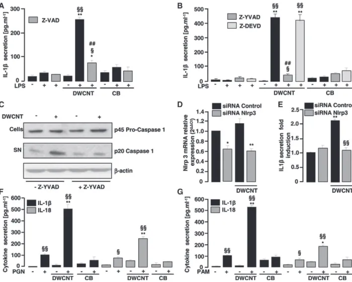
Caspase-1 is a key protease activated by the Nlrp3 inflamma-some complex involved in the maturation of IL-1β protein. To determine unequivocally whether caspase-1 is involved in the DWCNT-induced IL-1β secretion by LPS-primed PHMs, we evaluated IL-1β secretion in the presence of specific caspase inhibitors. The use of z-vad, an inhibitor of proteases that belongs to the caspase family, revealed that IL-1β secretion by PHMs after DWCNT challenge was dependent on caspase activation because the z-vad inhibits DWCNT-induced IL-1β secretion by LPS-primed PHMs (Figure 2, A). Moreover, the treatment of PHMs with the specific caspase-1 inhibitor z-yvad significantly decreased DWCNT-induced IL-1β secretion, whereas the specific caspase-3 inhibitor z-devd did not change this secretion (Figure 2, B). This data showed that the IL-1β secretion induced by DWCNTs was exclusively linked to caspase-1 activation (Figure 2, B).
Caspase-1 activation involves autocatalytic processing of the 45 kDa pro-caspase-1 to generate two subunits, p20 and p10.8,20 To further explore whether caspase-1 was activated in DWCNT-stimulated LPS-primed PHMs, we determined the appearance of the p20 cleavage product by western blot (Figure 2, C). We observed an increased p20 subunit level in response to DWCNTs, which is significantly decreased in the presence of 1 inhibitor, confirming that DWCNT-mediated caspase-1 activation to secrete IL-caspase-1β (Figure 2, C). Collectively, these data suggest involvement of the Nlrp3 inflammasome in the DWCNT-induced IL-1β response.
As Nlrp3 is involved in a specific caspase-1 activation pathway for the processing of pro-inflammatory cytokines by particles, the expression of Nlrp3 receptor was silenced in PHMs using Nlrp3 siRNA (Figure 2, D). Figure 2, E shows that the siRNA-mediated knockdown of Nlrp3 decreased IL-1β secretion in PHMs treated with DWCNTs. Thus, we demonstrate here for the first time the role of Nlrp3 receptor as the DWCNT responsive mediator of inflammasome assembly in human monocytes.
We next analyzed the requirement of TLR signalling for Nlrp3 inflammasome activation by DWCNTs (Figure 2, F and G). To determine whether DWCNT-induced IL-1β secretion was specif-ically TLR4 dependent or not, DWCNTs were associated with peptidoglycan, a TLR2 ligand, or with PAM3CSK4, a TLR1/2
activator21 (Figure 2, F and G). Importantly, the ability of DWCNTs to promote IL-1β and IL-18 secretion was not limited to LPS, as these NPs also significantly enhanced IL-1β and IL-18 secretion in response to a range of TLR agonists (Figure 2, F and G). These data demonstrated that IL-1β secretion in response to DWCNT treatment requires a first signal dependent on TLRs. DWCNTs–induced IL-1β release in human monocytes requires a potassium efflux, phagocytosis, and lysosomal acidification, but not ROS production
Despite the identification and characterization of numerous sterile and microbial activators of the Nlrp3 inflammasome, the precise mechanism mediating inflammasome activation remains
unclear.8,22-25Several pathways leading to its activation have been described, such as efflux of cellular potassium, cathepsin B and vacuolar acidification, and/or the generation of ROS.7,8,13,20,22To characterize the precise mechanism mediating inflammasome activation by DWCNTs, we evaluated the involvement of the potassium efflux and the phagocytosis process. Preventing the potassium efflux by increasing extracellular potassium abolished DWCNT-induced IL-1β secretion (Figure 3, A). Moreover, p20 subunit level strongly decreased in the presence of high concentrations of potassium, establishing that caspase-1 cleavage induced by DWCNTs was dependent on potassium efflux (Figure 3, B). In addition, the disruption of actin-mediated phagocytosis by Latrunculin A suppressed DWCNT-induced IL-1β secretion in
PHMs (Figure 3, C), indicating that DWCNT phagocytosis was required for inflammasome activation. Moreover, using TEM, we demonstrated that some DWCNTs were located in phagosomes, strongly suggesting that PHMs were able to phagocytose these particles (Figure 3, D). These results suggest that the phagocytosis of DWCNTs is required for inflammasome activation.
ROS are potent activators of the Nlrp3 inflammasome. We investigated whether DWCNT phagocytosis was linked to Nlrp3 inflammasome-dependent ROS activation. Although PHMs produced ROS in response to TPA, we demonstrated that DWCNTs did not induce ROS production (Figure 4, A). Moreover, the use of Trolox, a chemical ROS scavenger, did not suppress DWCNT-induced IL-1β secretion (Figure 4, B).
Figure 2. DWCNT–induced IL-1β release in human monocytes results from Nlrp3-dependent caspase-1 activation. (A,B) IL-1β release in culture supernatant of untreated or LPS-primed human monocytes stimulated with DWCNTs or CB in the presence of z-vad caspase inhibitor, z-yvad caspase-1 inhibitor or z-devd-FMK caspase-3 inhibitor. *P ≤ 0.05, **P ≤ 0.01 in comparison with the LPS-primed human monocytes.§P ≤ 0.05,§§P ≤ 0.01 in comparison with the respective unprimed human monocytes. ##P ≤ 0.01 in comparison with LPS-primed human monocytes stimulated with DWCNTs or CB. (C) Representative immunoblot of mature p20 form and p45 pro-form respectively in supernatant and lysate of LPS-primed human monocytes stimulated or not with DWCNTs in presence or not of z-yvad. (D) Nlrp3 mRNA level in LPS-primed human monocytes transfected with either a control siRNA or a Nlrp3 siRNA and stimulated or not with DWCNTs. *P ≤ 0.05 compared to human monocytes transfected with control siRNA. (E) IL-1β release in culture supernatant of LPS-primed human monocytes transfected with either a control siRNA or a Nlrp3 siRNA stimulated or not with DWCNTs. **P ≤ 0.01 compared to human monocytes transfected with control siRNA.§§P ≤ 0.01 compared to human monocytes transfected with control siRNA and stimulated with DWCNTs. (F) IL-1β and IL18 release in culture supernatant of untreated or peptidoglycan (PGN) TLR2 activator-primed human monocytes stimulated with DWCNTs or CB. (G) IL-1β and IL18 release in culture supernatant of untreated or PAM3CSK4 TLR1/2 activator-primed human
monocytes stimulated with DWCNTs or CB. *P ≤ 0.05, **P ≤ 0.01 compared to the PGN- or PAM3CSK4-primed human monocytes. §
P ≤ 0.05,§§P ≤ 0.01 compared to the respective unprimed human monocytes. Data are means ± SEM. The data are representative of three independent experiments.
Together, these data show that ROS production did not play a key role in the mechanism by which phagocytosis is coupled to DWCNT-induced inflammasome activation.
It has been proposed that Nlrp3 inflammasome may be activated by lysosomal damage and the subsequent release of cathepsin-B into the cytoplasm of cells.24,26 Inhibition of the activation of all cathepsins by nonspecific inhibitor significantly reduced DWCNT-induced IL-1β secretion (Figure 4, C). it is interesting to note that the use of specific cathepsin-B inhibitor highlighted cathepsin-B as the main protease involved in IL-1β release by PHMs in response to DWCNTs (Figure 4, C). In addition, treating PHMs with Bafylomycin A1, an inhibitor of the H+ ATPase system required for lysosomal acidification, significantly reduced the IL-1β response induced by DWCNTs (Figure 4, D). All together, these data suggest that DWCNT treatment of PHMs resulted in intracellular changes, including lysosomal acidification. The release of cathepsin-B lysosomal product into the cytosol may promote the generation of Nlrp3 inflammasome activation signals.
Discussion
Carbon nanotubes have a wide range of applications in various sectors and their use is foreseen to increase in the future. Thus, public concern about their adverse effects on human health
has also risen. Recent studies clearly demonstrated that CNTs induce a strong pro-inflammatory reaction through the secretion of IL-1β and IL-6 cytokines in mice exposed to these particles.4,5 Brown et al showed that MWCNTs induced a strong secretion of IL-1β only in PMA-primed THP1 monocytic cell line.27 Although the pro-inflammatory potential of CNTs has been suggested, the signaling pathways leading to the increase of pro-inflammatory cytokine secretion remain unknown.
In this study, we investigated the capacity of double-walled CNTs (DWCNTs) to stimulate the release of pro-inflammatory cytokines in PHMs. The cytokines of the IL-1 family (IL-1α, IL-1β, IL-18) are particularly potent pro-inflammatory mediators strongly involved in the development of autoimmune disorders, such as silicosis or asbestosis.7,8,20 The particularity of these cytokines is that their secretion involves a signaling pathway distinct from that of TNFα and IL-6 inflammatory cytokines.7,20 Indeed, after induction of their mRNAs by pro-inflammatory mediators (TLR ligands) TNFα and IL-6 proteins are directly secreted, whereas the production of IL-1 family cytokines involves a complex regulation that requires two different signals.20 First, the induction of IL-1β mRNA through the stimulation of pattern recognition receptors, such as TLRs, is required for synthesis of pro–IL-1β protein in cells. Then, a second stimulus is necessary for inflammasome activation leading to caspase-1–dependent cleavage and release of the biologically active and mature IL-1 family cytokines.20Several
Figure 3. DWCNTs–induced IL-1β release in human monocytes requires potassium efflux and phagocytosis. (A) IL-1β release in culture supernatant of untreated or LPS-primed human monocytes stimulated with DWCNTs or ATP in the presence of elevated KCl or NaCl concentrations. **P ≤ 0.01 in comparison with to LPS-primed human monocytes in the presence of NaCl.§
P ≤ 0.05,§§
P ≤ 0.01 in comparison with LPS-primed human monocytes stimulated with DWCNTs or ATP in the presence of NaCl. (B) Representative immunoblot of mature p20 form and p45 pro-form in lysate of LPS-primed human monocytes stimulated with DWCNTs in the presence or not of elevated KCl or NaCl concentrations. (C) IL-1β release in culture supernatant of untreated or LPS-primed human monocytes stimulated with DWCNTs or ATP in the presence of Latrunculin A. **P ≤ 0.01 in comparison with the LPS-primed human monocytes.§§
P ≤ 0.01 in comparison with the respective unprimed human monocytes.##
P ≤ 0.01 in comparison with to LPS-primed human monocytes stimulated with DWCNTs or ATP. (D) Representative transmission electron microscopy image of primary human monocytes stimulated with DWCNTs. Data are means ± SEM. The data are representative of three independent experiments.
particles (asbestos) and NPs (nanosilica) have been found to specifically induce IL-1β secretion through the activation of inflammasome post-transcriptional mechanisms.7,8 In this con-text, we investigated the impact of DWCNTs on the different steps of the production of IL-1β by PHMs. We demonstrate in this study that DWCNTs induced a strong secretion of IL-1β and IL-18 in LPS-primed PHMs but not in unprimed PHMs, supporting the idea that TLR agonists are indispensable to enhance DWCNT-induced IL-1β and IL-18 release. Consistently, previous studies have shown that only PMA-primed THP1 cells and LPS-primed murine monocytes secrete IL-1β when they are challenged with MWCNTs.27 An interesting aspect is that we established that DWCNTs induced only IL-1β and IL-18 release in LPS-primed PHMs. Indeed, the treatment of LPS-primed PHMs with DWCNTs did not amplify TNFα or IL-6 secretion. These data corroborate the findings of previous studies highlighting the ability of silica and asbestos to specifically increase IL-1β and IL-18 secretion, but not TNFα or IL-6 in LPS-primed macrophages.7,8In this study, we also demonstrated that
DWCNTs did not induce IL-1β mRNA gene expression, but they activated the protease 1. This DWCNT-induced caspase-1 activation promoted the maturation of IL-caspase-1β, strongly suggesting involvement of the inflammasome in this process. Although DWCNTs alone did not alter the IL-1β mRNA levels in PHMs, these particles seem to activate an inflammasome-dependent signaling pathway required for their secretion. Among the numerous inflammasomes identified, the Nlrp3 inflammasome is the best characterized.20Many particles, such as silica and asbestos, have been shown to specifically trigger activation of the Nlrp3 inflammasome, leading to caspase-1 activation-induced IL-1β and IL-18 secretion.7,8,20 In this study, we show that the silencing of the Nlrp3 gene strongly reduced DWCNT-induced IL-1β secretion, demonstrating that the Nlrp3 inflammasome senses DWCNT-associated danger signals and contributes to triggering IL-1β and IL-18 release in PHMs.
Activation of the Nlrp3 inflammasome is subject to several events, such as the efflux of cellular potassium, the phagocytosis of particles, the generation of ROS, cathepsin B activation and/or
Figure 4. DWCNT–induced IL-1β release in human monocytes requires lysosomal acidification but not ROS production. (A) ROS production in human monocytes untreated (control) or treated with DWCNTs or TPA. DWCNTs–induced respiratory burst of human monocytes was measured by chemioluminescence. Total chemioluminescence emission (area under the curve expressed in counts × seconds) was observed continuously for 60 minutes in the presence or not of DWCNTs or TPA. **P ≤ 0.01 in comparison with untreated human monocytes (control). (B) IL-1β release in culture supernatant of untreated or LPS-primed human monocytes stimulated with DWCNTs in the presence of the ROS scavenger Trolox. **P ≤ 0.01 in comparison with the LPS-primed human monocytes.§§
P ≤ 0.01 in comparison with the respective unprimed human monocytes. (C) IL-1β release in culture supernatant of untreated or LPS-primed human monocytes stimulated with DWCNTs or ATP in the presence of cathepsin inhibitor (CA-074) or cathepsin B inhibitor (CA-074Me). **P ≤ 0.01 in comparison with the LPS-primed human monocytes.§P ≤ 0.05,§§P ≤ 0.01 in comparison with the respective unprimed human monocytes.
##
P ≤ 0.01 in comparison with LPS-primed human monocytes stimulated with DWCNTs or ATP. (D) IL-1β release in culture supernatant of untreated or primed human monocytes stimulated with DWCNTs or ATP in the presence of Bafilomicyn A1. *P ≤ 0.05, **P ≤ 0.01 in comparison with the LPS-primed human monocytes.§
P ≤ 0.05,§§
P ≤ 0.01 in comparison with the respective unprimed human monocytes.##
P ≤ 0.01 in compared with LPS-primed human monocytes stimulated with DWCNTs. (E) IL-1β mRNA level in untreated or LPS-primed human monocytes stimulated or not with DWCNTs in the presence of Bafilomicyn A1 or CA-074Me. *P ≤ 0.05, **P ≤ 0.01 in comparison with the LPS-primed human monocytes. Data are means ± SEM. The data are representative of three independent experiments.
the vacuolar acidification.7,8,13,20,22 The efflux of cellular potassium appears to be a common step shared by various activators of the Nlrp3 inflammasome.7,8,20,22Thus, it has been suggested that the inflammasome works as a sensor of cellular membrane disruption characterized by a loss of intracellular potassium.22We showed that prevention of the potassium efflux by increasing extracellular potassium abrogated caspase-1 activation and IL-1β secretion, revealing that DWCNT-induced potassium efflux triggers Nlrp3 inflammasome activation. These data demonstrate that DWCNT–induced potassium efflux was necessary for Nlrp3 inflammasome activation, suggesting that these particles may affect cell membrane integrity. Moreover, we showed that some DWCNTs are located in vesicles, demon-strating that some DWCNTs are completely phagocytosed. However, due to their great length heterogeneity (0.1 to 100 μm) and their high potential for aggregation, certain large particles were not completely phagocytosed by monocytes. In these conditions, Brown et al previously showed the existence of frustrated phagocytosis of CNTs by mononuclear cells.28 Recently, it has also been demonstrated that large particles, such as alum, asbestos, and silica can induce the so-called frustrated phagocytosis at the surface of the cells, provoking the formation of actin cytoskeletal filaments.8,20 In addition, the pharmacological inhibition of actin cytoskeleton-dependent phagocytosis with Latrunculin A inhibited DWCNT-induced IL-1β secretion, suggesting that both phagocytosis and frustrated phagocytosis are involved in Nlrp3 inflammasome activation by DWCNTs. Dostert et al has previously shown that the inhibition of cytoskeletal filament generation with cytochalasin D or colchicine disrupts the ability of asbestos and silica particles to trigger IL-1β secretion, demonstrating that the frustrated phagocytosis is involved in the NLRP3 inflammasome activation,8 and this is consistent with our findings. It has recently been shown that there is an alternative route for particle penetration into cells that allows the activation of the inflamma-some. Indeed, the entry into macrophages and keratinocytes of TiO2NPs by passive diffusion induces inflammasome activation
independently of actin-cytoskeleton rearrangements.13,29,30 In this study, we showed that cytoskeletal filament rearrangement is essential in the activation of the inflammasome by DWCNTs. The difference between CNTs and TiO2 could be attributed to
their physicochemical discrepancies. Indeed, the shorter length of NPs of TiO2may allow them to penetrate cells more easily by
diffusion.13,29,30An interesting aspect is that although TiO2and
nano-ZnO used the same route to enter the cells, nano-ZnO did not activate the Nlrp3 inflammasome.13 In view of these differences, we suggest that independently of the way in which they penetrate cells, both the NP structure and its chemical properties are essential to trigger Nlrp3 inflammasome activa-tion. In this study, the modification of the physicochemical structure of the DWCNT surface by oxidation with nitric acid did not affect their ability to induce IL-1 β release by PHMs, thus demonstrating that their hydrophobic properties are not involved in the DWCNT-induced IL-1β secretion.
Recent reports have suggested that ROS produced during phagocytosis may be an important signal for inflammasome activation by several stimuli, such as asbestos, silica, and peptidoglycan particles.7,8,31 Here, we demonstrated that ROS
production was not involved in Nlrp3 inflammasome activation by DWCNTs. Consistent with our results, it has been described that CNT-induced ROS production was length dependent in the THP1 monocytic cell line.27 In their study, Brown et al demonstrated that short CNTs failed to promote ROS production, but they were able to stimulate IL-1β release in PMA-primed THP1.27,30 In line with this, amorphous silica-NP–mediated Nlrp3 inflammasome activation in murine dendritic cells was triggered independently of ROS production,32 suggesting that ROS are not indispensable for Nlrp3 activation by NPs.
Our data also indicated that both cathepsin B activity and lysosomal acidification contributed to DWCNT-mediated acti-vation of the Nlrp3 inflammasome. Indeed, although neither cathepsin B or H+ATPase inhibitors altered the mRNA level of pro IL-1β, DWCNT-induced IL-1β secretion was significantly decreased by the presence of these two inhibitors. These data strongly suggested that phagosomal maturation is involved in the DWCNT-induced Nlrp3 inflammasome activation. Phagosomal rupture and cathepsin B activation have previously been shown to be important in Nlrp3 inflammasome activation by silica crystals and aluminum.23 The release of lysosomal products, such as cathepsin B, into the cytosol might promote the generation of danger signals sensed by the Nlrp3 inflammasome, resulting in inflammasome assembly.
We demonstrate here, for the first time, that DWCNTs induce IL-1β and IL-18 pro-inflammatory cytokine secretion by PHMs. Importantly, we identify the Nlrp3 inflammasome as the DWCNT-responsive element in human monocytes. Moreover, the potassium efflux and phagocytosis processes are key molecular pathways involved in the DWCNT-induced Nlrp3 inflammasome activation (see Graphical Abstract). Given the critical role of the Nlrp3 inflammasome in the development of severe chronic inflammation and cancer formation7,8 and the impact of the DWCNTs observed on its activation, future decisions will be necessary to prevent the possible health hazards that these particles may represent.
Acknowledgments
We thank Isabelle Fourquaux, from Centre de Microscopie Electronique Appliquée à la Biologie (CMEAB), for TEM assistance.
Appendix A. Supplementary data
Supplementary data to this article can be found online at
doi:10.1016/j.nano.2011.11.004. References
1. Poland CA, Duffin R, Kinloch I, Maynard A, Wallace WA, Seaton A, et al. Carbon nanotubes introduced into the abdominal cavity of mice show asbestos-like pathogenicity in a pilot study. Nat Nanotechnol 2008;3:423-8.
2. Ryman-Rasmussen JP, Cesta MF, Brody AR, Shipley-Phillips JK, Everitt JI, Tewksbury EW, et al. Inhaled carbon nanotubes reach the subpleural tissue in mice. Nat Nanotechnol 2009;4:747-51.
3. Shvedova AA, Kisin E, Murray AR, Johnson VJ, Gorelik O, Arepalli S, et al. Inhalation vs. aspiration of single-walled carbon nanotubes in
C57BL/6 mice: inflammation, fibrosis, oxidative stress, and mutagen-esis. Am J Physiol Lung Cell Mol Physiol 2008;295:L552-65. 4. Crouzier D, Follot S, Gentilhomme E, Flahaut E, Arnaud R, Dabouis V,
et al. Carbon nanotubes induce inflammation but decrease the production of reactive oxygen species in lung. Toxicology 2010;272:39-45. 5. Inoue K, Takano H, Koike E, Yanagisawa R, Sakurai M, Tasaka S, et al.
Effects of pulmonary exposure to carbon nanotubes on lung and systemic inflammation with coagulatory disturbance induced by lipopolysaccharide in mice. Exp Biol Med (Maywood) 2008;233: 1583-90.
6. Heister E, Lamprecht C, Neves V, Tilmaciu C, Datas L, Flahaut E, et al. Higher dispersion efficacy of functionalized carbon nanotubes in chemical and biological environments. ACS Nano 2010;4:2615-26. 7. Cassel SL, Eisenbarth SC, Iyer SS, Sadler JJ, Colegio OR, Tephly LA,
et al. The Nalp3 inflammasome is essential for the development of silicosis. Proc Natl Acad Sci U S A 2008;105:9035-40.
8. Dostert C, Petrilli V, Van Bruggen R, Steele C, Mossman BT, Tschopp J. Innate immune activation through Nalp3 inflammasome sensing of asbestos and silica. Science 2008;320:674-7.
9. Burns K, Martinon F, Tschopp J. New insights into the mechanism of IL-1beta maturation. Curr Opin Immunol 2003;15:26-30.
10. Martinon F, Tschopp J. Inflammatory caspases: linking an intracellular innate immune system to autoinflammatory diseases. Cell 2004;117: 561-74.
11. Gurcel L, Abrami L, Girardin S, Tschopp J, van der Goot FG. Caspase-1 activation of lipid metabolic pathways in response to bacterial pore-forming toxins promotes cell survival. Cell 2006;126:1135-45. 12. Mariathasan S, Weiss DS, Newton K, McBride J, O'Rourke K,
Roose-Girma M, et al. Cryopyrin activates the inflammasome in response to toxins and ATP. Nature 2006;440:228-32.
13. Yazdi AS, Guarda G, Riteau N, Drexler SK, Tardivel A, Couillin I, et al. Nanoparticles activate the NLR pyrin domain containing 3 (Nlrp3) inflammasome and cause pulmonary inflammation through release of IL-1alpha and IL-1beta. Proc Natl Acad Sci U S A 2010; 107:19449-54.
14. Martin-Blondel G, Gales A, Bernad J, Cuzin L, Delobel P, Barange K, et al. Low interleukin-10 production by monocytes of patients with a self-limiting hepatitis C virus infection. J Viral Hepat 2009;16:485-91. 15. Flahaut E, Bacsa R, Peigney A, Laurent C. Gram-scale CCVD synthesis
of double-walled carbon nanotubes. Chem Commun (Camb) 2003: 1442-3.
16. Brozena AH, Moskowitz J, Shao B, Deng S, Liao H, Gaskell KJ, et al. Outer wall selectively oxidized, water-soluble double-walled carbon nanotubes. J Am Chem Soc 2010;132:3932-8.
17. Gales A, Conduche A, Bernad J, Lefevre L, Olagnier D, Beraud M, et al. PPARgamma controls dectin-1 expression required for host antifungal defense against Candida albicans. PLoS Pathog 2010;6: e1000714.
18. Lefevre L, Gales A, Olagnier D, Bernad J, Perez L, Burcelin R, et al. PPARgamma ligands switched high fat diet-induced macrophage M2b
polarization toward M2a thereby improving intestinal Candida elimina-tion. PLoS One 2010:5:e12828.
19. Netea MG, Nold-Petry CA, Nold MF, Joosten LA, Opitz B, van der Meer JH, et al. Differential requirement for the activation of the inflammasome for processing and release of IL-1beta in monocytes and macrophages. Blood 2009;113:2324-35.
20. Martinon F, Mayor A, Tschopp J. The inflammasomes: guardians of the body. Annu Rev Immunol 2009;27:229-65.
21. Ozinsky A, Underhill DM, Fontenot JD, Hajjar AM, Smith KD, Wilson CB, et al. The repertoire for pattern recognition of pathogens by the innate immune system is defined by cooperation between toll-like receptors. Proc Natl Acad Sci U S A 2000;97:13766-71.
22. Eisenbarth SC, Colegio OR, O'Connor W, Sutterwala FS, Flavell RA. Crucial role for the Nalp3 inflammasome in the immunostimulatory properties of aluminium adjuvants. Nature 2008;453:1122-6.
23. Hornung V, Bauernfeind F, Halle A, Samstad EO, Kono H, Rock KL, et al. Silica crystals and aluminum salts activate the NALP3 inflamma-some through phagosomal destabilization. Nat Immunol 2008;9:847-56. 24. Rajamaki K, Lappalainen J, Oorni K, Valimaki E, Matikainen S, Kovanen PT, et al. Cholesterol crystals activate the NLRP3 inflamma-some in human macrophages: a novel link between cholesterol metabolism and inflammation. PLoS One 2010;5:e11765.
25. Zhou R, Yazdi AS, Menu P, Tschopp J. A role for mitochondria in NLRP3 inflammasome activation. Nature 2011;469:221-5.
26. McNeela EA, Burke A, Neill DR, Baxter C, Fernandes VE, Ferreira D, et al. Pneumolysin activates the NLRP3 inflammasome and promotes proinflammatory cytokines independently of TLR4. PLoS Pathog 2010;6:e1001191.
27. Brown DM, Donaldson K, Stone V. Nuclear translocation of Nrf2 and expression of antioxidant defence genes in THP-1 cells exposed to carbon nanotubes. J Biomed Nanotechnol 2010;6:224-33.
28. Brown D, Kinloch I, Bangert U, Windle A, Walter D, Walker J, et al. An in vitro study of the potential of carbon nanotubes and nanofibres to induce inflammatory mediators and frustrated phagocytosis. Carbon 2007;45:1743-56.
29. Donaldson K, Murphy F, Schinwald A, Duffin R, Poland CA. Identifying the pulmonary hazard of high aspect ratio nanoparticles to enable their safety-by-design. Nanomedicine (London. England) 2011;6:143-56.
30. Donaldson K, Murphy FA, Duffin R, Poland CA. Asbestos, carbon nanotubes and the pleural mesothelium: a review of the hypothesis regarding the role of long fibre retention in the parietal pleura, inflammation and mesothelioma. Part Fibre Toxicol 2010;7:5. 31. Shimada T, Park BG, Wolf AJ, Brikos C, Goodridge HS, Becker CA,
et al. Staphylococcus aureus evades lysozyme-based peptidoglycan digestion that links phagocytosis, inflammasome activation, and IL-1beta secretion. Cell Host Microbe 2011;7:38-49.
32. Winter M, Beer HD, Hornung V, Kramer U, Schins RP, Forster I. Activation of the inflammasome by amorphous silica and TiO(2) nanoparticles in murine dendritic cells. Nanotoxicology 2011;5:326-40.