Université de Montréal
Cost-effectiveness of Transcatheter Mitral Valve Leaflet
Repair for the Treatment of Mitral Regurgitation
in Heart Failure
par
Anita W. Asgar, MD
Sciences Biomédicales Faculté de médicine
Mémoire présenté à la Faculté Médicine
en vue de l’obtention du grade de Maitrise en Sciences (MSc) En Sciences biomédicales
Option Recherche Clinique
Décembre, 2015
Résumé
Contexte: La régurgitation mitrale (RM) est une maladie valvulaire nécessitant une intervention dans les cas les plus grave. Une réparation percutanée de la valve mitrale avec le dispositif MitraClip est un traitement sécuritaire et efficace pour les patients à haut risque chirurgical. Nous voulons évaluer les résultats cliniques et l'impact économique de cette thérapie par rapport à la gestion médicale des patients en insuffisance cardiaque avec insuffisance mitrale symptomatique.
Méthodes: L'étude a été composée de deux phases; une étude d'observation de patients souffrant d'insuffisance cardiaque et de régurgitation mitrale traitée avec une thérapie médicale ou le MitraClip, et un modèle économique. Les résultats de l'étude observationnelle ont été utilisés pour estimer les paramètres du modèle de décision, qui a estimé les coûts et les avantages d'une cohorte hypothétique de patients atteints d'insuffisance cardiaque et insuffisance mitrale sévère traitée avec soit un traitement médical standard ou MitraClip.
Résultats: La cohorte de patients traités avec le système MitraClip était appariée par score de propension à une population de patients atteints d'insuffisance cardiaque, et leurs résultats ont été comparés. Avec un suivi moyen de 22 mois, la mortalité était de 21% dans la cohorte MitraClip et de 42% dans la cohorte de gestion médicale (p = 0,007). Le modèle de décision a démontré que MitraClip augmente l'espérance de vie de 1,87 à 3,60 années et des années de vie pondérées par la qualité (QALY) de 1,13 à 2,76 ans. Le coût marginal était 52.500 $ dollars canadiens, correspondant à un rapport coût-efficacité différentiel (RCED) de 32,300.00 $ par QALY gagné. Les résultats étaient sensibles à l'avantage de survie.
Conclusion: Dans cette cohorte de patients atteints d'insuffisance cardiaque symptomatique et d insuffisance mitrale significative, la thérapie avec le MitraClip est associée à une survie supérieure et est rentable par rapport au traitement médical.
Mots-clés : insuffisance mitrale, insuffisance cardiaque, analyse de couts, réparation de la valve mitrale
Abstract
Background: Mitral regurgitation (MR) is a common valvular heart disorder requiring intervention once it becomes severe. Transcatheter mitral valve leaflet repair with the MitraClip device is a safe and effective therapy for selected patients denied surgery. We sought to evaluate the clinical outcomes and economic impact of this therapy compared to medical management in heart failure patients with symptomatic MR.
Methods: The study was comprised of two phases; an observational study of patients with heart failure and MR treated with either medical therapy or the MitraClip, and an economic model. Results of the observational study were used to estimate parameters for the decision model, which estimated costs, and benefits in a hypothetical cohort of patients with heart failure and moderate to severe MR treated with either standard medical therapy or MitraClip.
Results: The cohort of patients treated with the MitraClip was propensity matched to a population of heart failure patients, and their outcomes compared. At a mean follow up of 22 months, all-cause mortality was 21% in the MitraClip cohort and 42% in the medical management cohort (p=0.007). The decision model demonstrated that MitraClip increased life expectancy from 1.87 to 3.60 years and quality-adjusted life years (QALY) from 1.13 to 2.76 years. The incremental cost was $52,500 Canadian dollars, corresponding to an incremental cost-effectiveness ratio (ICER) of $32,300.00 per QALY gained. Results were sensitive to the survival benefit.
Conclusion: In this cohort of heart failure patients with symptomatic moderate-severe MR, therapy with the MitraClip was associated with superior survival and is cost-effective compared to medical therapy.
Keywords: mitral regurgitation, heart failure, transcatheter mitral repair, cost-effectiveness
Table of Contents
Résumé ... i
Abstract ... ii
Table of Contents ... iv
List of tables ... vi
List of figures ... vii
List of abbreviations ... viii
Acknowledgements ... xi
Introduction ... 1
Background ... 3
Mitral Regurgitation ... 3
Heart Failure ... 5
Transcatheter Mitral Valve Leaflet Repair ... 6
MitraClip Device ... 7
Economic Impact of Heart Failure ... 13
Methods ... 14 Study Objectives ... 14 Study Hypotheses ... 14 Study Design ... 14 Observational Study ... 15 Economic Model ... 17 Results ... 24
Observational Study of MitraClip ... 24
MitraClip Procedure ... 24
Medical Management Cohort ... 25
Cost-Effectiveness of MitraClip vs. Medical Therapy ... 30
Sensitivity Analyses ... 31
Discussion ... 35
Summary ... 35
Hospitalizations for heart failure with mitral regurgitation ... 35
Survival benefit of treatment of mitral regurgitation ... 36
Cost-effectiveness of MitraClip compared to medical management ... 37
Study Limitations ... 38
External validity ... 40
Conclusion ... 42
List of tables
Table 1. International Registry data of MitraClip in predominantly FMR ... 10 Table 2. Health Utilities and Event Rates with Ranges used in Base Case (Mean Value) and Probabilistic Sensitivity Analyses ... 21 Table 3. Health Care Resource Costs ... 22 Table 4. Baseline Characteristics of MitraClip and Medical Management Cohorts ... 26 Table 5. Comparison of Outcomes in MitraClip and Medical therapy Cohorts at 30 days and 1 year ... 27 Table 6. Economic Outcomes for MitraClip and Medical Therapy ... 30 Table 7. Comparison of Outcomes in MitraClip Cohort with published Registry Data ... 41
List of figures
Figure 1. Anatomy of the mitral valve complex ... 3 Figure 2. Schematic and echo representation of ischemic MR (top panel), and non-ischemic MR (bottom panel) ... 4 Figure 3. Schematic drawing of surgical Alfieri double orifice repair and MitraClip ... 7 Left panel : artist rendering of a surgical Alfieri repair with sutures ... 7 Right panel : artist rendering of a MitraClip device in place in the mitral valve creating a double orifice mitral valve (Courtesy of Abbott Vascular, Menlo Park, CA) ... 8 Figure 4. Transcatheter mitral valve repair with the MitraClip device (from top left to bottom right: MitraClip device; close-up of MitraClip device; MitraClip device in place in jet of mitral regurgitation; double orifice mitral valve; MitraClip in place following device release) (Courtesy of Abbott Vascular, Menlo Park, CA) ... 8 Figure 5. Decision Model for Cost-effectiveness Analysis ... 19 Figure 6. Kaplan-‐Meier survival curves for patients treated with MitraClip and medical management ... 28 Figure 7. Kaplan-Meier curves for MitraClip and Medical Management overlaid with Weibull extrapolations to ten years ... 29 Figure 8. Tornado diagram of one-way sensitivity analysis ... 32 Figure 9. Probabilistic sensitivity analysis (scatter plot) of cost-effectiveness of MitraClip compared to medical management ... 33 Figure 10. Cost-effectiveness acceptability curve for MitraClip therapy compared to medical therapy in heart failure patients with significant mitral regurgitation ... 34
List of abbreviations
ACC American College of Cardiology AHA American Heart Association CE Certification European
CEAC Cost-effectiveness acceptability curve CHF Congestive Heart Failure
CRT Cardiac resynchronization therapy DMR Degenerative mitral regurgitation
EACTS European Association of Cardio-Thoracic Surgeons
ER Emergency room
ESC European Society of Cardiology FDA Food and Drug Administration FMR Functional mitral regurgitation GDMT Guideline-directed medical therapy HF Heart failure
ICD Implantable cardioverter defibrillator ICER Incremental cost-effectiveness ratio ICU Intensive care unit
LA Left atrium
LOS Length of Stay (hospital) LV Left ventricle
LVOT Left ventricular outflow tract MA Mitral annulus
MC MitraClip
MM Medical management MR Mitral regurgitation MV Mitral valve
MVA Mitral valve annuloplasty NYHA New York Heart Association
PSA Probabilistic sensitivity analysis QALY Quality-adjusted life year STS Society of Thoracic Surgeons TEE Trans-esophageal echocardiography
This work is dedicated to the heart failure patients that underwent mitral
leaflet repair in the hopes of improving their condition and quality of life.
Your openness to undergo a new procedure has made this work possible
and hopefully one day this procedure will be more available to the
Acknowledgements
I would like to extend my sincerest appreciation and gratitude to my supervisor and mentor, Dr. Paul Khairy for his support in this project and for allowing me to spread my wings and follow my desire to develop a skill in health economics and understand the economic impact of new technology in structural heart disease intervention.
Sincere thanks to Lisa Bernard and Heather Cameron for their support in this project and for taking the time to teach a novice the basics of decision modelling, how to build a model in Excel, and for your assistance in quality assurance of this model, all seven versions.
Finally, my deepest gratitude to my husband Marc-Aurele. Your unwavering support of my relentless desire to learn and belief in my abilities has made me a better person and given me the strength to finish this project.
Introduction
Mitral regurgitation (MR) is one of the most common valvular heart disorders, with an estimated prevalence in the US of ~1.7%, increasing with age to ~9.3% in those >75 years in a population study performed in New York State(1). In the 2001 EuroHeart Survey, MR was second in frequency only to aortic stenosis with a prevalence of 24.8% in patients with valvular heart disease(2). The standard of care for severe symptomatic MR is surgical mitral valve repair or replacement according to published guidelines(3, 4). Nevertheless, a significant number of patients do not receive intervention due to severe comorbidities and high surgical risk and are treated medically, particularly those with left ventricular dysfunction, symptoms of heart failure (HF) and secondary or functional MR (FMR)(5). In such patients, the presence of significant MR has been shown to independently predict mortality and hospitalizations for HF(6). HF is costly for the healthcare system; exceeding $40 billion dollars in 2012 in the US(7) therefore effective therapies may provide significant clinical and economic benefits.
Transcatheter mitral valve repair using the MitraClip (Abbott Vascular, Menlo Park, CA) has been commercially available in Europe since 2008 and in Canada since 2010. Such therapy involves the transcatheter placement of a metal clip on the leaflets of the mitral valve at the site of valvular regurgitation thereby reducing MR and resulting in a double-orifice mitral valve(8). Evaluation of this technology in surgical candidates has established superior safety, albeit with less efficacy when compared with surgical repair or replacement(9).
Current clinical experience with the MitraClip has focused on high-risk patients rather than surgical candidates, and in particular those with HF and FMR. In the post-approval ACCESS-EU registry, the MitraClip was implanted in 567 patients with a clip implant rate of 99.6% and MR reduction in 91% of patients, with no procedural mortality(10). Through 12-month follow-up, NYHA class and 6-minute walk distance were substantially improved. Numerous other centers in Europe have published their clinical experience of MitraClip in FMR but as yet there has not been a comparison to patients treated medically(11).
Large-scale randomized controlled trials are currently underway in HF patients to evaluate the efficacy of this intervention compared to medical therapy. We sought to compare a cohort of patients with HF and FMR treated with MitraClip to a cohort of medically
managed patients at our institution. The economic burden of HF on the healthcare system, lack of effective interventional options for many patients, and the substantial up-front costs of such technology, are the basis of this evaluation of the cost-effectiveness of the MitraClip based on data from patients treated with this device at our institution.
Background
Mitral Regurgitation
Mitral regurgitation (MR) is one of the most common valvular heart disorders, with an estimated prevalence in the US of ~1.7%, increasing with age to ~9.3% in those over >75 years(1). In the 2001 EuroHeart Survey, MR was second only to aortic stenosis with a prevalence of 24.8% of all patients with valvular heart disease(2).
MR is classified as primary (or degenerative) when the regurgitation is principally due to a structural abnormality of the mitral valve, whether the leaflets, chordae tendinae, papillary muscles or mitral annulus as shown in Figure 1. Secondary (or functional) MR refers to the presence of MR without intrinsic MV disease, usually in patients with left ventricular dysfunction. FMR is more common than degenerative MR (DMR)(12), and is associated with a worse prognosis (compounded by the underlying cardiomyopathy and other comorbidities).
FMR can be further classified as either ischemic or non-ischemic in nature (Figure 2). Ischemic MR is the more common etiology, and occurs in patients with coronary artery disease with regional wall motion abnormalities due to prior myocardial infarction, typically resulting in apical and lateral displacement of the posteromedial PM causing tethering of the posterior leaflet. Secondary chords on the anterior leaflet may also cause tethering resulting in pseudo-prolapse of the anterior leaflet as it slides above the posterior leaflet, producing a posteriorly directed MR jet(13). In contrast to ischemic MR, non-ischemic MR (which is most commonly due to idiopathic dilated cardiomyopathy, but can be due to dilated cardiomyopathy of any etiology) is characterized by global LV dilatation with increased sphericity. In this condition the LV loses its normal “football” shape, becoming more rounded, or “basketball”-like. Displacement of both PMs and apical tethering of the chordae tendinae and MV leaflets results typically in a centrally directed regurgitant jet(13). LV dilation and remodelling in non-ischemic cardiomyopathy results in symmetric MA dilatation greatest in the septal-lateral direction that correlates with the severity of ventricular dysfunction(14).
Figure 2. Schematic and echo representation of ischemic MR (top panel), and non-ischemic MR (bottom panel)
The top panel demonstrates tethering of the posterior mitral valve leaflet due to a regional wall motion abnormality, as seen on the mid-esophageal long-axis view of a TEE, with an eccentric jet of MR. The bottom panel illustrates global dilatation of the left ventricle resulting in displacement of the papillary muscles and a wide, central jet of MR in the mid-esophagel two chamber view.
Heart Failure
Heart failure (HF) is an epidemic and major public health concern with over 500,000 new cases diagnosed annually worldwide and a prevalence that is expected to increase by 25% by 2030(15, 16). Current management for HF involves pharmacologic therapy with beta-blockers, angiotensin converting enzyme inhibitors or angiotensin receptor blockers and aldosterone antagonists, as described in the recent ACC/AHA Heart Failure Guidelines(17).
The prevalence of secondary MR in HF patients is high. In a study of 1256 patients with dilated cardiomyopathy of either ischemic or non-ischemic etiology, the prevalence of severe MR was 24%(6). A study of 2057 HF patients with an ejection fraction less than 40% from the Duke Cardiovascular Databank noted the presence of moderate to severe or severe MR in 29.8%(18). There is a strong association between secondary MR and all-cause mortality and hospitalizations for HF: In the previously mentioned study of dilated cardiomyopathy, severe secondary MR was an independent predictor of death or HF hospitalization at median 2.5-year follow-up (adjusted HR: 1.5 [95% CI: 1.2-1.9]), independent of left ventricular function(6). In fact, secondary MR is a powerful predictor of death or transplant, even with less severe HF(19).
Guideline directed medical therapy (GDMT) is the first line of treatment for FMR, and consists of aggressive management of HF as per recent guidelines(17). Unfortunately, morbidity and mortality of patients with LV dysfunction and FMR remain high despite GDMT. In a study of 404 patients with at least mild FMR due to ischemic or non-ischemic cardiomyopathy treated with GDMT, cardiac mortality at mean follow-up of 4 years occurred in 43% and 45% of patients with moderate and severe MR respectively, compared to only 6% with mild MR (P=0.003)(20). The presence of moderate or severe MR was also an
independent predictor of new onset HF in those patients with a history of ischemic systolic dysfunction (relative risk [95%CI] = 3.2 [1.9–5.2], P=0.0001).
Transcatheter Mitral Valve Leaflet Repair
Despite the poor prognosis with GDMT, most HF patients with FMR not requiring CABG are not referred to MV surgery due to high surgical risk in the setting of multiple comorbidities, and the lack of a proven survival benefit(5). In fact, in a review of patients with MR treated at the Cleveland Clinic, FMR was more likely to be treated medically rather than with surgery, with 47.5% of patients managed medically compared to surgical intervention in 26.8%. In those patients managed medically 5-year mortality was 50%(21). As a result of the under-utilization of surgical therapy due to increased patient risk, transcatheter strategies have emerged as potential treatment options for such patients.
The question remains whether treatment of mitral regurgitation in addition to GDMT in HF is beneficial. Recent evidence from the NIH randomized trial in patients with moderate ischemic MR randomized to revascularization or revascularization and mitral valve repair did not demonstrate a statistically significant difference in the primary endpoint of LV remodelling as measured by left ventricular end-systolic volume index. Patients treated with mitral valve repair did have a higher incidence of neurologic complications but there were no observed differences in mortality or quality of life although the trial was not powered for these endpoints(22). It is unclear whether the failure of the trial to show a clinical benefit was related to surgical morbidity or sample size.
Currently, the COAPT Trial of MitraClip vs. Medical therapy in functional mitral regurgitation is underway and will attempt to answer this question. The primary endpoint of this randomized trial is rehospitalization for HF at one year, and secondary endpoints include mortality, reduction in mitral regurgitation and improvement in quality of life.
MitraClip Device
Transcatheter mitral leaflet repair is a percutaneous technique based on the surgical edge-to-edge mitral leaflet repair described by Alfieri (Figure 3) performed using the MitraClip(23). The MitraClip (Abbott Vascular, Menlo Park, CA) is a polyester-covered cobalt-chromium clip that is inserted via the femoral vein and advanced under trans-esophageal echocardiographic guidance into the LA following trans-septal puncture (Figure 4). The clip is opened, positioned above the regurgitant jet and advanced into the LV. It is then retracted to grasp the free edges of the mitral leaflets, the grippers are dropped and the clip is closed and released. Multiple clips may be safely placed if necessary, with no reported cases to date of mitral stenosis. The MitraClip has received CE mark and Health Canada approval, and limited FDA approval in the US for treatment of patients with DMR who are at prohibitive risk for surgery on the basis of data from the EVEREST II trial(24) and High risk registry(25) which were performed in predominantly although not exclusively DMR patients. Due to the ongoing randomized clinical trial of MitraClip in FMR, MitraClip is not currently FDA approved for this indication.
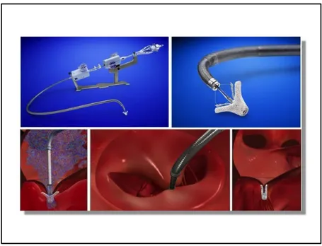
Figure 3. Schematic drawing of surgical Alfieri double orifice repair and MitraClip Left panel : artist rendering of a surgical Alfieri repair with sutures
Right panel : artist rendering of a MitraClip device in place in the mitral valve creating a double orifice mitral valve (Courtesy of Abbott Vascular, Menlo Park, CA)
Figure 4. Transcatheter mitral valve repair with the MitraClip device (from top left to bottom right: MitraClip device; close-up of MitraClip device; MitraClip device in place in jet of mitral regurgitation; double orifice mitral valve; MitraClip in place following device release) (Courtesy of Abbott Vascular, Menlo Park, CA)
Clinical Results
The initial safety and feasibility of the MitraClip was confirmed in the EVEREST I pilot study of 27 patients. A clip was successfully placed in 24 patients with no procedural complications. MR was successfully reduced in 52% of patients with a result that was maintained at 6-month follow up.(8) Following the EVEREST I pilot experience, the MitraClip was compared to surgical MV repair in the 278 patient randomized controlled EVEREST II trial in relatively low risk patients with 3+-4+ MR. Compared to MV surgery, the MitraClip procedure was substantially safer, but not as effective in reducing MR and LV
remodelling(24). Moreover, reflecting the early learning curve with this device, acute procedural success (MR ≤2+ at discharge) was achieved in only 77% of patients, and 21% of patients’ required MV surgery. Nonetheless, with follow-up now to 4 years, NYHA class and overall survival were similar in the 2 groups (26). Of note, however, 73% of the patents in this trial had DMR, and 27% had FMR. A significant interaction was present between the randomized therapy and the primary composite endpoint of death, MV surgery, and 3+-4+ MR at both 1 and 4 years according to MR etiology; patients with DMR had significantly improved outcomes with MV repair, whereas outcomes were at least as good with the MitraClip in patients with FMR(24, 26).
Since device commercialization in Europe, the MitraClip has been used extensively in patients at high risk for MV surgery, more frequently in FMR than DMR(27, 28). In the post-approval ACCESS-EU registry, the MitraClip was implanted in 567 patients at 14 sites between April 2009 and April 2011. The mean logistic EuroSCORE was 23, and 77% of the patients had 3+-4+ FMR. The clip implant rate was 99.6%, with multiple clips used in 40% of patients. MR was reduced to ≤2+ in 91% of patients, and there were 0 procedural deaths. Through 12 month follow-up NYHA class and 6-minute walk distance have substantially improved. The MitraClip has also been used with success in HF patients who are non-responders to CRT (an especially high-risk group), with resultant improvements in MR grade, functional capacity, and evidence of left ventricular remodelling(29).
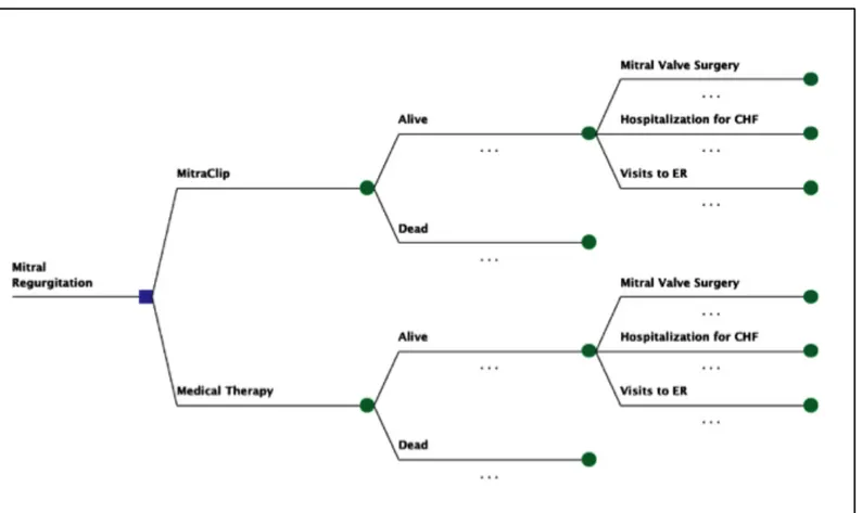
At present, there are several randomized clinical trials underway addressing the question of the use of MitraClip in FMR. Until such data become available there is registry data from numerous registries of FMR patients that have demonstrated high rates of procedural success and favorable short-term outcomes in patients treated with MitraClip. The largest published registries of FMR are summarized in Table 1(10, 30-42).
Table 1. International Registry data of MitraClip in predominantly FMR
The largest published registry to date is the Transcatheter Mitral Valve Interventions (TRAMI) Registry(30). Among 1,064 patients treated with the MitraClip at 20 German centers, the median age was 75 years; 87% had NYHA III/IV HF symptoms; 69% had LVEF <50%; FMR was present in 71% of patients; and the median STS mortality score was 10. Procedural success was achieved in 95% of patients, with no procedural deaths. At ~3 months of follow-up, 12% of patients had died and 12% had been hospitalized for HF, although 66% remained in NYHA class I/II.
Similarly, in the 25-center, 8-country 2011–2012 European Sentinel Pilot Registry, 72% of 628 MitraClip-treated patients had FMR, 86% had NYHA class III/IV symptoms, and the mean EuroSCORE was 20.4(31). Acute procedural success was high (95.4%), with multiple clips used in 39% of patients. In-hospital mortality (2.9%) and 1-year mortality (15.3%) were similar in patients with FMR and DMR, although rehospitalization for HF was more common in the FMR group (25.8% vs. 12.0%, p=0.009). At 1 year severe MR was present in only 6% of patents. Pooled data from the EVEREST II High-risk Registry and US REALISM registry
Table 1. Large-scale published registries of the MitraClip: baseline characteristics and acute procedural success
Registry N Mean age
(years) Male Mean or median risk NYHA Class III/IV Mean LVEF FMR etiology ≤2+ MR post Multiple clips Procedural success¶ TRAMI 1064 75 62% 10%* 87% † 71% 96% 1.5 mean 95% ACCESS-EU 567 78 64% 23%** 85% †† 77% 91% 40% 99.6% European Sentinel 628 74 63% 20%** 86% 43% 72% 98% 37% 95% EVEREST and REALISM 351 76 61% 11%* 85% 48% 70% 86% 39% - GRASP 171 71 62% 7%* 81% 37% 78% 93% 41% 99% MARS 142 71 64% 17%** 68% 47% 54% 77% 47% 94% Taramasso et al 109 69 84% 22%** 82% 28% 100% 87% 65% 99% Mitra-Swiss 100 77 67% 17%** 82% 48% 62% 85% 40% 85% French multicenter 62 73 72% 19%** 81% 40% 74% 88% 17% 95% Treede et al 202 75 63% 44%** 98% 44% 65% 92% 35% 92% Bozdag-Turan et al 121 77 69% 11%* 96% 42% 59% 99% 28% 97% Rudolph et al 104 74 62% 36%** 100% 43% 66% 92% 38% 92% Braun et al 119 71 67% 28%**‡ 86% 35%‡ 35%‡ - - 86% Neuss et al 157 74 67% 22%** 100% 41% 73% 100% 16% 98%
* By the Society for Thoracic Surgery score; ** By the logistic EuroScore; †LVEF ≤50% in 69% of patients; ††LVEF ≤40% in 53% of patients; ‡In patients with FMR; ¶According to the registry protocol definition, which varied per study.
have been recently published in which the MitraClip was used in 351 patients with an STS score or surgeon-predicted operative mortality of ≥12% (70% of whom had FMR)(32). By paired echocardiographic core lab analysis MR was ≤2+ in 89.7% of patients at discharge and in 83.4% of patients at 1 year. Mortality was 4.8% at 30 days and 22.8% at 1 year. LV end-diastolic and end-systolic dimensions decreased through 1 year follow-up, the physical and mental components of the SF-36 quality-of-life score improved, and the proportion of patients with NYHA class III/IV symptoms was reduced from 82.1% at baseline 17.1% at 1 year. The rate of hospitalizations for HF was significantly reduced in the year after compared to the year before the MitraClip (median per patient 0.41 vs. 0.79, p<0.0001). All outcomes were directionally consistent in patients with FMR and DMR. The MitraClip has also been used with success in HF patients who are non-responders to cardiac resynchronization therapy (CRT), an especially high-risk group, with resultant improvements in MR grade, functional capacity, and LV remodelling(29). In this study, patients remained in NYHA class III-IV despite CRT and were treated with MitraClip to address significant mitral regurgitation. Following the MitraClip procedure there was progressive improvement in NYHA Class and LV remodelling at both 6 and 12 months.
The role of MitraClip in the treatment of patients with FMR has recently been addressed by societal guidelines. The 2012 ESC/EACTS valve guidelines provide a class IIb (level of evidence C) recommendation to consider use of the MitraClip in patients with symptomatic severe FMR despite GDMT and CRT who are inoperable or at high surgical risk with life expectancy >1 year(4). The 2012 ESC HF guidelines similarly note that percutaneous edge-to-edge repair may be considered in order to improve symptoms in patients with an indication for valve repair that are judged inoperable or at unacceptably high surgical risk(43). Finally, the 2013 ACC/AHA heart failure guidelines provide a class IIb (level of evidence B) recommendation to consider use of the MitraClip in patients with symptomatic severe FMR despite GDMT after “careful candidate selection”(7).
Given the enthusiasm for such new technology in the field of cardiology, an understanding of the clinical impact and the economic ramifications of percutaneous treatment of secondary MR in these patients is critical to ensuring appropriate use of what remains limited resources.
Economic Impact of Heart Failure
In addition to the impact on mortality and morbidity, HF places a major strain on health care resources, accounting for 2–5% of the total health-care budget in most developed countries(44). In 2012, the economic burden of HF was estimated to be $3.9 billion in Canada(45). The total cost of HF management consists of several components, including hospital management for acute decompensation, physician and outpatient visits, and medical therapy. However, device-based treatments, such as implantable defibrillators, biventricular cardiac pacing devices for CRT, and ventricular mechanical circulatory support, have now emerged as a central and costly part of HF treatment. Health economic analysis or cost-effectiveness analysis has been increasingly used in countries such as the United Kingdom, Australia and Canada to understand the impact of health technologies prior to their widespread adoption. Such analysis employs thresholds for decision of cost-effectiveness, which in Canada is between $20,000-$100,000 per quality adjusted life year (QALY) gained(46).
Expensive technology is not new to the field of heart failure and devices such as CRT have previously undergone similar assessments of effectiveness analyses. A cost-effectiveness study based on data from the randomized COMPANION trial compared the costs of optimal medical therapy with those of CRT with pacing only (CRT-P) and CRT with defibrillator (D). Their analyses demonstrated an ICER of $19,600 per QALY for CRT-P and a ICER of $43,000 for CRT-D, both of which were felt to be in the range of reasonable costs for a new therapeutic intervention(47).
Methods
The goal of this study was to evaluate the outcomes of HF patients with significant FMR treated with the MitraClip at our institution, compare these outcomes with a cohort of medically treated patients and finally to evaluate the cost-effectiveness of this therapy.
Study Objectives
1. Prospectively evaluate the outcomes of a cohort of patients with significant functional regurgitation and congestive HF treated with the MitraClip at our institution
2. Compare the outcomes of the MitraClip cohort with a historical cohort of patients with significant MR treated with medical therapy
3. Estimate the cost-effectiveness of MitraClip therapy compared to medical therapy in patient with significant MR and HF
Study Hypotheses
1. HF patients with significant MR treated with MitraClip will have lower rates of recurrent hospitalizations for HF compared to those treated with medical therapy. 2. Therapy with the MitraClip will be cost-effective compared to medical therapy in
patients with HF and significant MR.
Study Design
The study was comprised of two phases: a comparison of propensity matched populations from an observational study of patients with HF and MR that were treated with either medical management or the MitraClip; and an economic model. Results of the observational study were used to estimate parameters for the economic model. The local Ethics Committee approved the study (Project #12-1403).
Observational Study
MitraClip Cohort
This prospective cohort was comprised of patients treated with the MitraClip (Abbott Vascular, Menlo Park, CA) at the Montreal Heart Institute from 2010 - 2013. Indication for the procedure was determined by local institutional practice and following consultation with the treating physician, cardiologist and cardiac surgeon. Eligible patients had symptomatic or asymptomatic moderate-to-severe (3+) or severe (4+) MR and were considered high risk for surgical intervention following multidisciplinary team discussion. Patients underwent trans-thoracic and trans-esophageal echocardiography to evaluate anatomical suitability. Exclusion criteria for the procedure included the following: mitral valve area <4 cm2
by planimetry, significant valvular or annular calcification, or visible thrombus in the left atrium. Procedures were performed as previously described(9, 48), and all patients signed informed consent and were approved under the Health Canada Special Access program to undergo the intervention. Data was collected on the following demographic variables: age, gender, left ventricular ejection fraction, history of ischemic heart disease, atrial fibrillation, hypertension, previous coronary artery bypass graft surgery, previous percutaneous coronary intervention, implantable cardioverter-defibrillator, cardiac resynchronization therapy, diabetes, and therapy with beta-blockers, ACE inhibitors, diuretics, angiotensin receptor blockers, and spironolactone; as well as mortality, rehospitalization for CHF and visits to the emergency room.
Medical Management Cohort
This retrospective comparator group consisted of medically managed patients with moderate to severe and severe (3-4+) MR followed at the Heart Failure Clinic at the Montreal Heart Institute from 2008-2010. The HF Clinic maintains a database of patients with HF and captures demographic data, medical therapy, diagnostic tests, interventions, rehospitalizations, hospital and ER visits, and mortality. Patients’ entry into the cohort was considered to be the date that significant MR was diagnosed by echocardiography.
The medical cohort was less symptomatic with the majority of patients reporting NYHA class II symptoms, compared to the predominantly class IV symptoms in the MitraClip group. Given the significant differences in baseline functional class between the MitraClip and medical management cohorts, it was not possible to include NYHA class in the propensity matching.
Clinical Outcomes
Clinical outcomes of interest in the matched cohort included the following; emergency room (ER) visits, rehospitalizations for HF, mitral valve surgery and mortality. For the MitraClip cohort, this data was obtained from patient interviews and chart review. For the medical management cohort, this data was obtained directly from the HF Clinic database. Data for each outcome was tabulated for the individual cohorts over the follow up period to calculate a rate and then probability of the outcome per patient. For example, a total of 100 readmissions for HF over a one-year period in the MitraClip cohort of 50 patients would be interpreted as a rate of 2 admissions per patient per year.
Statistical Analysis
To create a matched cohort, medical management patients were matched to those treated with the MitraClip using a propensity score. A multivariate logistic regression model with presence of MitraClip as the dependent variable included the following independent variables: age, gender, left ventricular ejection fraction, history of ischemic heart disease, atrial fibrillation, hypertension, previous coronary artery bypass graft surgery, previous percutaneous coronary intervention, implantable cardioverter-defibrillator, cardiac resynchronization therapy, diabetes, and therapy with beta-blockers, ACE inhibitors, diuretics, angiotensin receptor blockers, and spironolactone. Propensity scores (predicted probability of having MitraClip) were obtained for each subject with MitraClip (MC) and medical therapy (MM). Absolute differences between propensity scores were computed for each pair of MC and MM subjects. Each MC subject was matched with the MM subject that yielded the smallest absolute difference in a 1:5 greedy matching scheme, matching was performed with replacement.
Continuous variables were presented as means and standard deviations. Categorical variables were presented as frequencies and percentages. Survival in each cohort was evaluated using Kaplan-Meier curves and differences in survival were compared using a log-rank test.
Economic Model
An economic model was developed in Excel (Microsoft Corporation, Redmond, WA, USA) to estimate the costs, life-years and quality-adjusted life years (QALY) for the studied patients. This data was then used to calculate the incremental cost per QALY gained and per life-year gained.
The model followed a hypothetical cohort of HF patients with significant MR in one-month time increments from age 75 years until death or age 85. Patients were treated with either standard medical therapy (including cardiac resynchronization therapy as indicated) or the MitraClip device. We programmed the model inputs such that the estimates of survival, emergency room (ER) visits, hospitalizations and rates of mitral valve surgery were the same as the results obtained in the observational study. Outcomes of interest were life expectancy (measured in years), QALYs, and costs (reported in 2013 Canadian dollars) and the incremental cost-effectiveness ratio. The model was analyzed from the perspective of the Canadian publicly funded health care system. All health outcomes and costs were discounted at 5% per year as per the recommendations of the Canadian Agency for Drugs and Technologies in Health(49). Discounting is performed to standardize flows of costs and benefits that occur at different points in time. For external model validation, we compared outcomes of the modeled cohort over time with outcomes in independent registries(6, 10).
Model Overview: Data and Assumptions
We constructed a decision model of symptomatic severe MR to simulate disease progression and added MitraClip as a treatment option (see Figure 5). In the first month of the model, at time zero, patients in the MitraClip group underwent the procedure and may have survived or died. Additionally, in that first month they may have also experienced a complication related to the procedure, mitral valve surgery, re-intervention with the MitraClip, hospitalization for congestive heart failure (CHF) and/or visits to the ER. In every subsequent monthly cycle, MitraClip patients may have died, undergone mitral valve surgery, had re-intervention, or have had ER visits or hospitalizations for HF. For the medical therapy group,
in any one-month cycle, patients may have undergone mitral valve surgery, been hospitalized for heart failure, visited the ER or died.
We estimated mortality and peri-procedural complication rates after MitraClip using data from the observational study. The probability of death, hospitalization for heart failure, ER visits, and mitral valve surgery in the medical cohort were likewise obtained from the observational study. Mortality was extrapolated using parametric survival models for a time horizon of ten years. An exponential, log-normal and weibull extrapolation were performed, the weibull was chosen as it was a better fit for the data according to the Akaike information criterion (AIC)(50).
During each cycle of the model, cohort specific probabilities calculated from actual event rates, for heart failure hospitalizations and ER visits were applied such that all patients alive remained at risk for these outcomes. The cohort-specific probability for mitral valve surgery was applied to both groups during the first twelve months only. Model parameters are detailed in Table 2. The time horizon for the model was ten years. Model assumptions for the base case analysis included; baseline NYHA for the medical therapy cohort was equivalent to that of MitraClip patients prior to intervention, NYHA class was increased in both cohorts by one class every two years, and the probability of mitral valve surgery applied only in the first year for both cohorts.
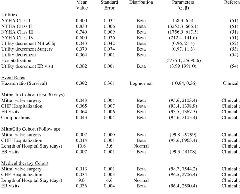
Table 2. Health Utilities and Event Rates with Ranges used in Base Case (Mean Value) and Probabilistic Sensitivity Analyses
Mean Value Standard Error Distribution Parameters (α, β) Reference Utilities
NYHA Class I 0.900 0.037 Beta (58.3, 6.5) (51)
NYHA Class II 0.830 0.006 Beta (3252.3, 666.1) (51)
NYHA Class III 0.740 0.009 Beta (1756.9, 617.3) (51)
NYHA Class IV 0.600 0.026 Beta (212.4, 141.6) (51)
Utility decrement MitraClip 0.043 0.042 Beta (0.96, 21.4) (52)
Utility decrement Surgery 0.079 0.074 Beta (0.97, 11.3) (53)
Utility decrement Hospitalization
0.064 0.001 Beta
(3776.1, 55690.6)
(54)
Utility decrement ER visit 0.002 0.001 Beta (3.99,1991.0) (54)
Event Rates
Hazard ratio (Survival) 0.392 0.361 Log normal (-0.94, 0.36) Clinical data
MitraClip Cohort (first 30 days)
Mitral valve surgery 0.043 0.004 Beta (95.6, 2103.4) Clinical data
CHF Hospitalization 0.065 0.007 Beta (93.4, 1338.9) Clinical data
ER visits 0.064 0.006 Beta (93.5, 1367.3) Clinical data
Complications 0.043 0.004 Beta (95.6, 2103.4) Clinical data
MitraClip Cohort (Follow up)
Mitral valve surgery 0.002 0.000 Beta (99.8, 49799) Clinical data
CHF Hospitalization 0.014 0.001 Beta (98.6, 6965.4) Clinical data
Length of Hospital Stay (days) 10.6 5.6 Normal Clinical data
ER visits 0.007 0.001 Beta (99.3, 14108) Clinical data
Medical therapy Cohort
Mitral valve surgery 0.013 0.001 Beta (98.7, 7544.2) Clinical data
CHF Hospitalization 0.034 0.003 Beta (96.5, 2706.4) Clinical data
Length of Hospital Stay (days) 9.0 6.6 Normal Clinical data
ER visits 0.036 0.004 Beta (96.4, 2590.4) Clinical data
Costs
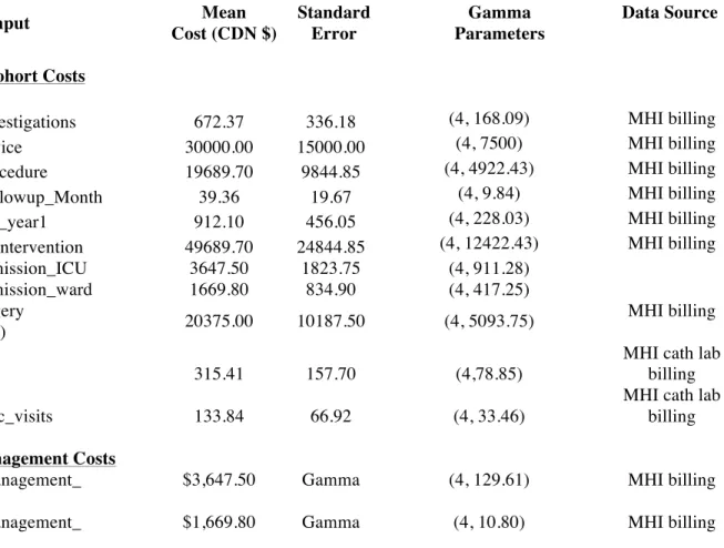
Detailed resource utilization and costs were collected for the MitraClip and medical therapy cohorts, as outlined in Table 3. Costs were calculated using the most important cost drivers from clinical data including diagnostic evaluation costs directly incurred as a result of the MitraClip procedure, procedural costs, and inpatient treatment costs at a large tertiary care hospital in Montreal (Montreal Heart Institute). The cost of MitraClip is per procedure (irrespective of the number of clips used). Follow-up costs included protocol driven visits and tests. Costs of hospitalizations and emergency room visits were obtained from the Montreal Heart Institute. Data on costs for the medical therapy cohort were obtained by reviewing outpatient hospital clinic visits, emergency room visits and hospitalizations recorded in the Heart Failure Clinic Database. Costs are summarized in Table 3. Hospitalization costs at other centers were assumed to be equal to those incurred at our tertiary care center in 2013.
Table 3. Health Care Resource Costs
Cost Input Mean
Cost (CDN $) Standard Error Gamma Parameters Data Source
MitraClip Cohort Costs
cost_MC_investigations 672.37 336.18 (4, 168.09) MHI billing
cost_MC_device 30000.00 15000.00 (4, 7500) MHI billing
cost_MC_procedure 19689.70 9844.85 (4, 4922.43) MHI billing
cost_MC_Followup_Month 39.36 19.67 (4, 9.84) MHI billing
cost_MC_FU_year1 912.10 456.05 (4, 228.03) MHI billing
cost_MC_Reintervention 49689.70 24844.85 (4, 12422.43) MHI billing
cost_CHFadmission_ICU 3647.50 1823.75 (4, 911.28) cost_CHFadmission_ward 1669.80 834.90 (4, 417.25) cost_MVSurgery (Replacement) 20375.00 10187.50 (4, 5093.75) MHI billing cost_ERvisits 315.41 157.70 (4,78.85)
MHI cath lab billing
cost_HFClinic_visits 133.84 66.92 (4, 33.46)
MHI cath lab billing
Medical Management Costs
cost_MM_management_ annual
$3,647.50 Gamma (4, 129.61) MHI billing
cost_MM_management_ month
Utilities
Quality adjusted life years (QALY) (life expectancy adjusted for quality of life of the health state experienced) were calculated for each patient in the alive state using published health utilities, which measure quality of life from a 0 (dead) to 1 (perfect health) scale, for heart failure according to NYHA Class(51). We assumed that patients in the medical therapy cohort remained in NYHA Class III-IV for the duration of the model. The NYHA Class assigned to patients treated with MitraClip during the first year of follow-up was based on actual data. For projected time intervals beyond the clinical study data, an assumption was made that patients would deteriorate by one NYHA Class every two years.
Short-term utility decrements (i.e. disutility) for the MitraClip procedure were approximated using published decrements for percutaneous coronary intervention(55) and were applied in the first cycle of the model. A utility decrement for mitral valve surgery obtained from the literature(53) was applied to both groups for the first year of the model only. Utility decrements were also applied for heart failure hospitalizations(54) and emergency room visits(56) according to the proportion of patients alive and at risk.
Analysis
We performed extensive deterministic sensitivity analyses to explore the impact of uncertainty in key parameters on the analysis results. A probabilistic sensitivity analysis to further characterize uncertainty in model parameters was performed using 10000 simulations. A beta distribution was applied to all probabilities and utilities, gamma distributions to all costs, and a log normal distribution for all hazard ratios (see Table 2). Results are represented in the form of a scatter plot and cost-effectiveness acceptability curve (CEAC) representing the probability of the MitraClip being cost-effective over a range of different willingness to pay thresholds
Results
Observational Study of MitraClip
A total of 50 consecutive patients underwent the MitraClip procedure from December 2010 until March 2013 and their baseline characteristics are described in Table 4. The average age was 75.4±9.1 years and, 74% were male. The majority of patients (78%) had a previous history of ischemic heart disease, with 52% (n=26) having had previous CABG and 40% (n=20) previous coronary intervention (PCI). Atrial fibrillation was present in over half the cohort (n=29) and device therapy was used in 54% (n=27) of patients. Patients had symptomatic heart failure, 98% were NYHA class III or IV. MR severity was assessed as 3+or 4+ in all patients and the underlying etiology was functional in 90% of cases. A small subset of patients had high-risk degenerative MR (n=5). The mean ejection fraction was 38.3 ± 15.8%.
MitraClip Procedure
The MitraClip procedure was performed under general anesthetic using trans-esophageal guidance as previously described (9). MitraClip device placement was successful in 96% of patients (n=48). Failure to place a device occurred in two patients in whom there was severe restriction of a shortened posterior leaflet that precluded grasping. MR severity was reduced to ≤ 2+ in 94% (n=47) of the initial cohort. Two clips were used in 71% (n=34) on patients, with one clip in the remaining 29% (n=14).
30-day Major Adverse Events
Four patients (8%) died within 30 days of the MitraClip procedure. All were considered procedure-related but none occurred intra-procedurally. The two patients with unsuccessful MitraClip procedures (i.e., no clip implanted) died due to progressive heart failure and low cardiac output. A third patient had single-leaflet device attachment 48
post-operative complications. The fourth patient died 48 hours post-intervention due to an acute intra-cerebral haemorrhage on warfarin for chronic atrial fibrillation. An additional patient required surgical mitral valve replacement two days post-procedure due to persistent severe MR in the setting of a mitral valve cleft, with an uneventful recovery. No patients were lost to follow up however patients were entered into the cohort as the MitraClip procedure was performed from 2010-2013 therefore the individual patient follow up is variable depending on when they underwent the procedure.
Medical Management Cohort
The medical management cohort was comprised of 42 patients that were matched to the MitraClip group on the basis of comorbidities and medical therapy. The baseline characteristics of the medical management cohort are described in Table 3. In comparison to the MitraClip cohort, the patients were younger, with a mean age 68.2 ±15.5 years. Comorbidities were similar however there were lower rates of ischemic heart disease (71%), CABG (48%) and previous PCI (33%). Patients were less symptomatic, with the majority of patients being NYHA Class II or III and but this parameter was not used in the matching process. Echocardiographic assessment demonstrated MR severity of 3+ or 4+ in all patients however the mean ejection fraction of 31.8 ± 13.6% was lower than that measured in the MitraClip cohort. Accordingly, there were higher rates of device therapy in this cohort, 59% with pacemaker or implantable defibrillator.
Table 4. Baseline Characteristics of MitraClip and Medical Management Cohorts
MitraClip Cohort (n=50) Medical Management
Cohort (n=42)
Mean age (years) 75.4± 9.1 68.2 ±15.5
% Males 74% (37) 77% (33)
Mitral Regurgitation Severity
3+ 58% (29) 76% (32)
4+ 42% (21) 24% (10)
Ischemic Heart Disease 78% (39) 71% (30)
Atrial Fibrillation 58% (29) 64% (27) Hypertension 58% (29) 57% (24) Diabetes 42% (21) 31% (13) Previous CABG 52% (26) 48% (20) Previous PCI 40% (20) 33% (14) Pacemaker/ICD 34% (17) 59% (25)
Cardiac Resynchronization Therapy (CRT) 20% (10) 14%(6)
Left Ventricular Ejection Fraction (%) 38.3 ± 15.8 31.8 ± 13.6
NYHA Class at Baseline
II 2% (1) 74% (31) III 32% (16) 21.4% (9) IV 66% (33) 0% (0) Medical Therapy ACE Inhibitors 44% (22) 43% (18) Beta Blocker 86% (43) 83% (35) Diuretics 88% (44) 86% (36) Angiotensin-‐Receptor Blockers 28% (14) 26% (11) Aldosterone Antagonists 50% (25) 62% (26)
CABG denotes coronary artery bypass graft; PCI, percutaneous coronary intervention; ICD, implantable cardioverter-‐defibrillator; NYHA, New York Heart Association; ACE, angiotensin converting enzyme
Table 5. Comparison of Outcomes in MitraClip and Medical therapy Cohorts at 30 days and 1 year
MitraClip (n=50) Medical
management (n=42)
Outcome 30 days 12 months 12 months
All – cause Mortality 8% (4) 18% (9) 24% (10)
Mitral Valve surgery 4% (2) 6% (3) 21% (9)
# CHF Hospitalizations/patient 0.06 0.16 0.57 # ER visits/patient 0.06 0.08 0.60
CHF denotes congestive heart failure; ER, emergency room
Clinical Follow Up
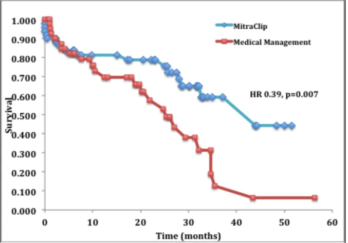
Clinical follow up of patients in both cohorts at 12 months are shown in Table 5. All-cause mortality was 18% and 24% in the MitraClip and medical management cohorts respectively. The number of hospitalizations for heart failure was 0.16 and 0.57 per patient in each group. Emergency room visits at 12 months were 0.08 and .60 per patient. Longer-term follow up was available in the MitraClip and medical management cohorts at a mean of 22±15 and 33±21 months respectively. At these time points, all-cause mortality was 21% in the MitraClip cohort and 42% in the medical management cohort, hazard ratio 0.39, 95% confidence interval [CI]: 0.19 to 0.79, p=0.007. Kaplan-Meier survival curves are plotted in Figure 6, with Weibull extrapolations for a time horizon of ten years overlaid in Figure 7.
Figure 6. Kaplan-‐Meier survival curves for patients treated with MitraClip and medical management
Figure 7. Kaplan-Meier curves for MitraClip and Medical Management overlaid with Weibull extrapolations to ten years
Cost-Effectiveness of MitraClip vs. Medical Therapy
Costs of the MitraClip device and procedure (approximately $80,000 CDN) were partially offset ($30,000) by lower hospitalization, ER visits and mitral valve surgical costs when compared to medical therapy.
Table 6 summarizes the discounted results of the cost-effectiveness analysis. Under the above assumptions, the discounted cost of a MitraClip per patient was $88,200.00 compared to $35,600.00 for medical therapy over a ten-year time horizon. The discounted life years gained was 3.60 in the MitraClip cohort and 1.87 in the medical therapy cohort. Given an incremental difference in QALYs of 1.63 this results in an ICER of $32,300.00 per QALY gained.
Table 6. Economic Outcomes for MitraClip and Medical Therapy
Medical
Management MitraClip Therapy Incremental Difference
Costs $35,600.00 $88,200.00 $52,600.00
Life Years (LY) 1.87 3.60 1.74
Quality-‐adjusted life years (QALY) 1.13 2.76 1.63
Incremental cost-‐effectiveness ratio of MitraClip to medical management $/LY gained $/QALY gained $30,300.00 $32,300.00
Sensitivity Analyses
The model was robust for the majority of variables on one-way sensitivity analyses, as shown in the tornado diagram, Figure 8. The model was sensitive to changes in the hazard ratio for survival, and time horizon with the MitraClip remaining cost-effective (assuming a threshold of $100,000 per QALY gained(57)) at the upper 95% CI of the hazard ratio, 0.795 however with an increase in the ICER to $66,300.00. At the time horizon of two years the ICER increased to approximately $89,000.00 suggesting that overall life expectancy has an impact on the cost-effectiveness. In the absence of an improvement of quality of life in those treated with the MitraClip the ICER also increased but remained below the threshold of $100,000.00 as displayed in the tornado diagram. The major incremental cost drivers in the model were implant costs (+$50,000.00 CDN) and disease management costs ($11,500.00 CDN) over the time horizon of the model. One-way sensitivity analyses showed that shorter length of hospital stay (2 vs. 4 days) for the procedure and place of hospitalization (critical care unit vs. regular ward) had a minor impact on the incremental costs.
Figure 8. Tornado diagram of one-way sensitivity analysis
The tornado diagram above illustrates the results of the one-way sensitivity analysis of the model. The midpoint of the bar graphs represents the ICER of the base case analysis, those values in blue represent an ICER less than the base case while those in red, an ICER higher than the base case analysis.
A probabilistic sensitivity analysis (PSA) to further characterize uncertainty in model parameters was performed using 10000 simulations. The PSA demonstrated that treatment with the MitraClip compared with medical therapy was cost-effective in 67% of simulations using a willingness-to-pay threshold of $50,000 and in 95% of simulations using a willingness-to-pay threshold of $100,000 (Figure 9 and 10).
Figure 9. Probabilistic sensitivity analysis (scatter plot) of cost-effectiveness of MitraClip compared to medical management
Figure 10. Cost-effectiveness acceptability curve for MitraClip therapy compared to medical therapy in heart failure patients with significant mitral regurgitation
The cost-effectiveness acceptability curve above demonstrates the likelihood of MitraClip being cost-effective, based on the data generated by the probabilistic sensitivity analysis, and depending on the willingness to pay of the healthcare system. In the setting of a willingness to pay of $50,000 for a new treatment, therapy with MitraClip is cost-effective in 67% of cases of patients treated. In a scenario where the healthcare system is willing to pay $100,000 for a new therapy, MitraClip would be cost-effective in 95% of cases.
Discussion
Summary
This cost-effectiveness modelling study was based on data from heart failure patients with significant MR treated in a clinical setting with either medical management or MitraClip therapy. In this study, therapy with MitraClip was found to be cost-effective with an ICER of $32,300.00 per QALY. This result was driven primarily by an improvement in mortality, however patients treated with MitraClip also had lower rates of re-hospitalization for heart failure and fewer visits to the emergency room.
Hospitalizations for heart failure with mitral regurgitation
The presence of FMR in patients with left ventricular dysfunction is an independent risk factor for re-hospitalization for heart failure and mortality, and this risk is further increased in those with moderate or severe MR(6). GDMT is the treatment of choice for such patients however even in the presence of optimal medical therapy; MR is associated with only a 50% survival at 4 years for those with moderate or severe MR. Furthermore, the presence of moderate or severe MR is an independent predictor of recurrent heart failure (RR 3.2, 95% CI 1.9–5.2, P<0.0001)(20).
MitraClip has been previously used in high-risk patients and those with FMR. The EVEREST II High Risk Study was performed in those patients felt to be at high surgical risk as estimated by a STS (Society of Thoracic Surgeons) Score >12%. In this study, 59% of patients had FMR and the mean LVEF was 54%. Patients were treated with the MitraClip to reduce MR with procedural success achieved in 83%. At 1 year, MR reduction was sustained in 79% of patients with FMR. The number of patients with CHF hospitalizations also decreased significantly from 42% (33 of 78) in the 12 months before the MitraClip procedure to 16% (12 of 75) (p<0.02) in the 12 months after discharge after the MitraClip procedure, a 45% reduction(58). We found similar results in our matched cohort, with 0.57 admissions per patient/year in the medical management group compared to 0.16 admissions per patient/year in the MitraClip group.
Survival benefit of treatment of mitral regurgitation
The presence of MR is known to negatively impact survival in patients with heart failure with little improvement with medical therapy alone. Despite this fact, it is unclear whether surgical or transcatheter valve repair can actually improve survival. Initial results with surgical undersized mitral valve annuloplasty (MVA) were promising with symptomatic improvement in the majority of patients(59). This enthusiasm was somewhat dampened however when follow up data on patients treated with annuloplasty demonstrated that there was no mortality benefit from surgery in patients with left ventricular dysfunction and MR(60). This retrospective study compared outcomes of patients undergoing MVA to propensity-matched patients treated with medical therapy with the goal of identifying predictors of mortality or use of mechanical left ventricular support. The presence of coronary artery disease was found to be a risk factor for death, and the use of medical therapy for heart failure was associated with a reduced risk of mortality. Mitral valve annuloplasty had no impact on reducing mortality(60).
More recently, a retrospective study evaluated the impact of transcatheter mitral repair with MitraClip, surgical treatment or conservative medical management on survival in high-risk patients with predominantly FMR. This study compared 139 consecutive patients with high-risk MR treated with MitraClip to a surgical comparator group (n=53) and patients management medically (n=59). Patients were propensity matched for comorbidities and surgical risk although despite this, the surgical risk was highest in those treated with MitraClip. At a follow up of one year, survival was similar in the MitraClip and surgical treatment groups (85.8% and 85.2%, respectively) but significantly lower in those treated conservatively (67.7% survival at one year); resulting in a hazard ratio of 0.41, 95% confidence interval [CI]: 0.22 to 0.78, p < 0.006 for MitraClip therapy (61). These results are very similar to those in our study, which demonstrated a hazard ratio of 0.39 95% CI: 0.19 to 0.79, p=0.007.
To further clarify the hypothesis that therapy with MitraClip is associated with improved survival compared to conservative medical management, randomized trials are currently underway in the United States, and Europe with the results anticipated in 2017.
Cost-effectiveness of MitraClip compared to medical management
This cost-effectiveness analysis was performed to examine the cost utility of the MitraClip in high-risk patients with predominantly FMR. This analysis demonstrates that as much as 38% of MitraClip procedure and device costs may be offset by reductions in hospitalizations, ER visits and mitral valve surgeries compared to medical therapy. Given the Canadian societal willingness to pay threshold of $20,000 - $40,000(46), MitraClip is cost-effective with a deterministic ICER of $32,300 per QALY gained and median probabilistic ICER of just under $50,000 CDN per QALY gained. Our analysis is unique in that it utilizes a propensity-matched cohort of patients with heart failure and FMR in order to minimize differences in patient populations undergoing each treatment. The model was most influenced by the hazard ratio for survival, demonstrating that the therapy is not cost-effective in the absence of a survival benefit.
Our results are similar to an analysis of the MitraClip published from the perspective of the National Health Service in the United Kingdom. This evaluation utilized data from the high-risk registry of EVEREST to create a decision model. Our results are consistent with this analysis, which demonstrated that treatment with the MitraClip was cost-effective in high-risk patients. The UK model was found to be most sensitive to the time horizon chosen rather than device or procedure cost(62).
A recently published cost-effectiveness study of MitraClip and medical treated patients found similar results to our analyses. This propensity matched cohort analysis also compared patients treated with MitraClip to those on medical treatment alone and demonstrated that treatment with MitraClip was cost-effective with an ICER of 5000-8000 euros/QALY(63).
Significance of Study Results
Heart failure now has an increasing number of treatment options ranging from GDMT to advanced heart failure therapies such as defibrillators, CRT, left ventricular assist devices, and transcatheter mitral valve repair for those with significant MR. Thus far, evidence-based medical therapy with beta-blockers, angiotensin-converting enzyme (ACE) inhibitors and angiotensin receptor blockers have been demonstrated to be cost-effective in most cases, with an ICER between $1000-$10,000 per QALY, and in some settings cost-saving(64). Device therapy with defibrillators and CRT has an increased cost of $43,000 to $60,000 per QALY(44, 65) but are thought to be cost-effective given the associated reductions in hospitalizations and mortality. However, consideration must be given to the maintenance costs of such devices, particularly in the setting of primary prevention. Mechanical circulatory support with left ventricular assist devices have been increasingly considered for patients as a bridge to transplant or as destination therapy but are extremely costly, with an ICER of over $300,000 per QALY for the Heart Mate II device(66). Treatment with the MitraClip device has been shown to improve QoL in heart failure patients following optimal medical therapy and in CRT non-responders(29, 67). Nevertheless, cost-effectiveness in this population has yet to be evaluated in a randomized trial. Given the prognosis of patients with advanced heart failure and secondary MR, the question of whether the addition of MitraClip to standard therapy can provide meaningful health benefits to the population at an acceptable cost is particularly germane.
Our study has evaluated the cost-effectiveness of MitraClip in an actual cohort of patients with heart failure and FMR and compared these results to a cohort of patients managed medically. In this setting, MitraClip has been shown to be a cost-effective therapy with an impact on both mortality and hospitalizations for recurrent heart failure. This study provides valuable information to clinicians and hospital administrators responsible for care of patients in whom transcatheter mitral valve repair with the MitraClip may be considered.