HAL Id: dumas-01518315
https://dumas.ccsd.cnrs.fr/dumas-01518315
Submitted on 4 May 2017
HAL is a multi-disciplinary open access
archive for the deposit and dissemination of sci-entific research documents, whether they are pub-lished or not. The documents may come from teaching and research institutions in France or abroad, or from public or private research centers.
L’archive ouverte pluridisciplinaire HAL, est destinée au dépôt et à la diffusion de documents scientifiques de niveau recherche, publiés ou non, émanant des établissements d’enseignement et de recherche français ou étrangers, des laboratoires publics ou privés.
MiR-483-5p and miR-139-5p promote invasion and
tumorigenicity of adrenocortical carcinoma cells
Claire Agosta
To cite this version:
Claire Agosta. MiR-483-5p and miR-139-5p promote invasion and tumorigenicity of adrenocortical carcinoma cells. Human health and pathology. 2015. �dumas-01518315�
AVERTISSEMENT
Ce document est le fruit d'un long travail approuvé par le
jury de soutenance et mis à disposition de l'ensemble de la
communauté universitaire élargie.
Il n’a pas été réévalué depuis la date de soutenance.
Il est soumis à la propriété intellectuelle de l'auteur. Ceci
implique une obligation de citation et de référencement
lors de l’utilisation de ce document.
D’autre part, toute contrefaçon, plagiat, reproduction illicite
encourt une poursuite pénale.
Contact au SID de Grenoble :
bump-theses@univ-grenoble-alpes.fr
LIENS
LIENS
Code de la Propriété Intellectuelle. articles L 122. 4
Code de la Propriété Intellectuelle. articles L 335.2- L 335.10
http://www.cfcopies.com/juridique/droit-auteur
UNIVERSITE JOSEPH FOURIER
FACULTE DE MEDECINE DE GRENOBLE
Année : 2015 N°
T H È S E - A R T I C L E
PRESENTÉE POUR L’OBTENTION DU DOCTORAT EN MÉDECINE
DIPLÔME D’ÉTAT
D.E.S d’ENDOCRINOLOGIE, DIABÈTE, MALADIES MÉTABOLIQUES
Claire AGOSTA
Thèse soutenue publiquement à la faculté de médecine de Grenoble*
Le 13 novembre 2015
DEVANT LE JURY COMPOSÉ DE :
Président du jury : M. le Professeur Olivier CHABRE
Directeur de thèse : Me. le Docteur Nadia CHERRADI
Membres du jury :
M. le Professeur Philippe CHAFFANJON
Me. le Professeur Pascale HOFFMANN
Me. le Docteur Nathalie STURM
*La Faculté de Médecine de Grenoble n’entend donner aucune approbation ni improbation aux opinions émises dans les thèses ; ces opinions sont considérées comme propres à leurs auteurs.
MiR-483-5p et miR-139-5p
contribuent à l’invasivité et la tumorigénicité
du carcinome corticosurrénalien
2
REMERCIEMENTS
Tout d’abord, je remercie sincèrement les membres du jury d’avoir jugé mon travail de thèse, A Monsieur le Professeur Olivier Chabre,
Merci de m’avoir formée et guidée pendant ces 5 et bientôt 6 années internat. Vous savez nous transmettre l’amour de la médecine et surtout la passion de l’endocrinologie ! Après un externat à Marseille, je suis très heureuse d’avoir choisi la montagne à Grenoble pour mon internat et de vous avoir eu comme maître. Vous être un modèle d’enseignement et vous m’avez donné des bases solides pour l’exercice de cette belle spécialité. Je vous remercie pour votre écoute et vos encouragements pendant mes études, ainsi que votre soutien pour allier vie personnelle et professionnelle.
A Madame le Docteur Nadia Cherradi,
Grâce à toi j’ai passé une merveilleuse année de recherche dans le laboratoire de biologie du cancer et de l’infection au CEA de Grenoble. J’y ai été si bien accueillie par toute l’équipe que je remercie chaleureusement (Jean Jacques Feige, Nadia A., Agnès, Fred, Mickael, Aude, Christine, Josiane, Sophie, Khadija, Waël, Féli, Vanessa…). Merci à Jonathan que je n’ai pas eu l’occasion de connaître mais qui a beaucoup travaillé sur les suites de ce projet et qui l’a enrichi. J’ai particulièrement apprécié travailler avec toi Nadia pour ta rigueur, ton encadrement, et ton enthousiasme au travail. Merci pour ce que tu m'as apporté sur le plan scientifique, grâce à toi la recherche fondamentale n'est (presque) plus un secret pour moi et je dois dire que j'y ai pris goût ! Merci aussi pour ton amitié qui je l’espère perdurera.
A Monsieur le Professeur Pierre-Yves Benhamou,
Comme je vous l'ai dit je regrette votre absence aujourd'hui comme membre du jury, mais vous savez mon estime pour vous. Je vous remercie pour vos qualités humaines que nous avons pu admirer en visite auprès des patients, et aussi auprès de nous les internes (qui n’a pas versé une larme à vos côtés ?). Votre expertise en diabétologie est pour moi une référence, et vos qualités de chercheur toujours connecté à la nouvelle technologie me fascine.
3 A Madame le Docteur (et Professeur) Nathalie Sturm,
Merci pour mon semestre en anatomopathologie qui a failli faire changer mon choix de spécialité. Tu m’as fait découvrir les lames de thyroïde, hypophyse et surrénale, décrites de façon précise par ton grand savoir faire. Nous sommes fiers de t’avoir comme référente de l’endocrino-pathologie. Merci pour ton aide pour la mise en place de la collection de tumeurs surrénaliennes du CRB qui n’existerait pas sans ta rigueur dans la conservation des échantillons tumoraux depuis des années. Les couloirs du DAPC me sont devenus familiers et je remercie les correspondants du CRB qui m’ont permis d’avancer dans la banque : monsieur Philippe Lorimier et Gabriel. J’en profite pour dire mon amitié à Diane et Julie, internes et futures assistantes d’anapath, avec qui nous avons passé de belles années. A Monsieur le Professeur Philippe Chaffanjon,
Vous me faites l’honneur de juger mon travail de recherche sur la carcinogénèse corticosurrénalienne, domaine qui vous est cher, je vous en remercie. La collaboration entre la chirurgie, l’endocrinologie clinique, l’anatomo-pathologie et la recherche fondamentale est primordiale pour la prise en charge des patients.
A Madame le Professeur Pascale Hoffmann,
Tout d’abord merci pour votre accueil dans votre service de PMA, j’y ai été très bien formée et ce semestre a confirmé mon choix d’exercice professionnel en alliant la médecine de la reproduction et l’endocrinologie. J’apprécie la place que vous donnez aux endocrinologues dans votre service et notamment aux internes en formation. Je vous remercie de juger mon travail aujourd’hui, car en plus de votre qualité de gynécologue-obstétricienne, j’ai pu connaître votre ouverture d’esprit pour bien d’autres domaines cliniques et de recherche fondamentale. En effet, vous faites partie de la même équipe de recherche sur l’Angiogenèse physiologique et tumorale dirigée par Jean-Jacques Feige au CEA de Grenoble, dans laquelle a été réalisé ce travail.
Je remercie aussi l’équipe de PMA adorable avec moi ces 6 derniers mois juste avant la thèse… En particulier Nicole, Agnès, Sylvie, Monique, Brigitte, Eliane, Odile, Aurore, Laure et Alexia. Vous avez été d’un grand soutien et m’avez permis d’affronter un épisode difficile dont on se souviendra de très loin… Je me suis sentie dans une grande famille. Je remercie tout particulièrement Nicole qui m’a beaucoup appris de cette belle spécialité et auprès de qui on apprécie tous travailler de par son
4 expérience médicale et ses grandes qualités humaines ! Bravo pour cette belle équipe de PMA du CHU de Grenoble (biologistes, médecins, sages femmes, secrétaires…), pour votre rôle majeur auprès des couples et pour avoir renforcé mon goût pour la médecine de la reproduction.
Merci à toute l’équipe d’endocrinologie et de diabétologie du CHU de Grenoble. J’ai passé des belles et longues années d’internat dans le pavillon des ECRINS dont je connais les recoins mais qui ne cesse d’évoluer.
Merci aux médecins de l’équipe. J’apprécie les relations bienveillantes et les compétences de chacun, complémentaires et riches. Merci à Nelly, Marie, Anne-Laure B., Sandrine L., Isabelle, Sandrine C. et aussi à mes merveilleuses assistantes Marine C., Marine B. et Marie Aude !
Un message nostalgique pour les infirmières, aides-soignantes et secrétaires du service d’endocrinologie avec qui j’ai passé 1 an en tant qu’interne au deuxième étage… Ces deux semestres ont été riches en émotions et j’ai vraiment apprécié y travailler pour un intérêt commun ! Hélène (la doyenne), Coco, Blandine, Dominique, Lydia, Marie-Agnès, Marie-Odile, Béa, Ratiba, Marie-O, Gaëlle, Murielle, Marie-Antoinette, Sylvie…
Last but not least, merci à la dream-team des co-internes d’endoc-diabéto, toujours très soudée (c’est une chance !) et avec qui on passe des moments inoubliables dans le service et ailleurs (je pense aux mojitos géants du bar du cousin à Sandra aux JNDES annuelles à Paris…on s’en souvient !!). Je suis persuadée que notre amitié perdurera en plus de notre lien en tant que collègues.
Maurine, avec qui j’ai passé un premier semestre aux ECRINS mémorable accompagnées des zouzous Benoit ,Jojo et Georgounet ! et ça continue… Bonne suite pour toi à Chambé avec Cyril et futur baby… que du bonheur.
Sandra et Marie, on va finir l’internat quasi en même temps, et je m’en réjouie. J’apprécie l’intimité et la confiance dans nos relations, et qui ne se perdent pas !.
Marie-Aude, Anne Laure C. et Claudine, quelle joie de vous connaître et de partager l’amour de la « marmite » !.
5 Enfin un message pour l’équipe de diabétologie de la Martinique, le Dr Fagour notamment qui m’a accueillie dans son service. J’ai passé un semestre rêvé d’inter-CHU très enrichissant.
Il me reste à remercier mes proches sans qui la vie professionnelle n’a aucun sens !
Claire, mon amie de toujours et qui partage la même passion de l’endocrinologie. Je te remercie pour ton soutien, ton écoute et tes grandes qualités. Nos chemins sont liés !, tout comme avec Milie (un rayon de soleil) vous comptez beaucoup pour moi !
Mica, Rej, Quentin, Marianne… les années folles du lycée Thiers, puis chacun a pris un horizon différent… mais pour toujours mieux se retrouver !
Mes copains d’enfance de Martinique, bien entendu Olivia et Michou, et les Beauvais, Anton, Oliviers… la belle époque qui continue malgré la distance !! je vous porte dans mon cœur.
A mon parrain et ma marraine, tous mes oncles et tantes, mes cousins si nombreux avec qui je garde des liens intenses. Clin d’œil à Caro la pionnière des Grenoblois !
A mes grands parents chéris, mémé de Carry et mon pépé, mamie et bon papa : je vous aime ! A ma belle famille avec qui je vis de bons moments et où je me sens bien.
Cécile, Cyril et Lucie, mes frérots adorés, toujours soudés… vous êtes tout pour moi ! Et longue vie à la marmaille actuelle (Gwénaël, Alizé, Timéo) et future… Thomas et Cécile merci pour votre aide grâce à votre côté geek bien pratique quand il y a alerte rouge (ou un choix de pomme à faire) !. A mes parents adorés, vous m’avez toujours guidée et soutenue, votre présence m’est indispensable. Ma vocation de médecin n’a pas été simple tous les jours parce qu’elle « consomme » beaucoup de temps… Heureusement, à vos côtés j’ai appris que l’épanouissement professionnel est une chose, mais que la richesse de la vie personnelle est plus important que tout (vive les vacances en famille). Enfin, à mes deux (bientôt trois et pourquoi pas quatre) amours de ma vie…. Mon doudou, Maël et mon choubidou, Timéo. Quel boulot cette fin d’année 2015 avec nos deux thèses ! ouf… un peu (ou beaucoup) de répit nous attend !. Merci pour tous les bons moments, et la traversée des plus difficiles… ce qui montre bien que notre projet de vie est plus fort !! Love.
6
TABLE DES MATIÈRES
! Remerciements! 2! Table!des!matières! 6! Résumé! 8! Abstract! 9! MiR948395p!and!miR913995p!promote!invasion!and!tumorigenicity!of!adrenocortical!carcinoma! cells! 10! INTRODUCTION) 11! METHODS) 14! Patient!cohort! 14! Cell!culture! 14! NCI!H295R!cells!transfection! 15! Total!RNA!extraction!and!quantification!of!microRNAs!using!RTAqPCR! 15! Prediction!of!miRNA!target!genes! 16! Quantification!of!target!mRNA!levels!by!real!time!RTAqPCR! 17! Analysis!of!protein!expression!by!Western!blot! 18! Luciferase!reporter!gene!assay! 19! Cell!proliferation!analysis! 20! Cell!cycle!and!apoptosis!analysis! 20! Transwell!migration!and!invasion!assay! 21! AnchorageAindependent!colony!formation!assay!in!soft!agar! 22! Statistical!analyses! 22! RESULTS) 23! miRNAs!expression!profile!in!tumor!tissues!and!identification!of!putative!target!genes! 23! Inverse!correlation!between!NDRG2/NDRG4!expression!and!miRA483A5p/miRA139A5p!expression! levels!in!tumor!tissues! 24! Effect!of!miRA483A5p!and!miRA139A5p!inhibitors!on!NDRG!expression!in!NCI!H295R!cells! 24!7 MiRA483A5p!and!miRA139A5p!functionally!interact!with!the!3’Auntranslated!region!of!NDRG2!and! NDRG4,!respectively! 25! MiRA483A5p!and!miRA139A5p!did!not!affect!NCI!H295R!cell!proliferation!or!apoptosis.! 26! MiRA483A5p!and!miRA139A5p!promote!migration!and!invasion!of!NCI!H295R!cells! 27! MiRA483A5p!and!miRA139A5p!are!involved!in!anchorageAindependent!colony!formation!of!NCI! H295R!cells! 27! DISCUSSION) 29! Références! 35! Table!and!Figures! 41! Table!1.!Putative!miRNA/mRNA!target!interaction!in!ACC.! 41! Figure!1.!!Schematic!representations!of!miRA483A5p!and!miRA139A5p!interaction!with!NDRG2!and! NDRG4!respectively,!according!to!prediction!softwares.! 41! Figure!2.!Inverse!correlation!between!microRNAs!expression!and!putative!target!gene!expression! in!a!cohort!of!human!adrenocortical!tissues.! 42! Figure!3.!Effect!of!miRA483A5p!and!miRA139A5p!inhibitors!on!NDRG!expression!in!NCI!H295R!cells. ! 42! Figure!4.!MiRA483A5p!and!miRA139A5p!functionally!interact!with!NDRG2!and!NDRG4!3’UTR.! 43! Figure!5.!Effect!of!miRA483A5p!and!miRA139A5p!inhibition,!or!miRA195!and!miRA335! overexpression,!on!cell!cycle!and!cell!proliferation!in!NCIAH295R!cell!line.! 44! Figure!6.!Effect!of!miRA483A5p!and!miRA139A5p!inhibition,!or!miRA195!and!miRA335! overexpression,!on!NCIAH295R!cell!apoptosis.! 44! Figure!7.!Effect!of!miRA483A5p!and!miRA139A5p!inhibition,!or!NDRG!target!gene!overexpression!on! migration!and!invasion!capacities!of!NCIAH295R!cells.! 45! Figure!8.!Effect!of!miRA483A5p!and!miRA139A5p!inhibition!on!anchorageAindependent!growth!of! NCIAH295R!cells.! 45! Figure!9.!The!multiple!functions!of!the!NAMyc!DownstreamARegulated!gene!(NDRG)!family.! 46! CONCLUSION) 57! Intégration!de!la!collection!de!tumeurs!surrénaliennes!dans!le!centre!de!ressources!biologiques! (CRB)!du!CHU!de!Grenoble! 59! Liste!des!enseignants!à!l’UFR!de!médecine! 63! Serment!d’hippocrate! 67!
8
RÉSUMÉ
Contexte. Les microARNs ont émergé comme bio-marqueurs potentiels pour le diagnostic et le pronostic des tumeurs corticosurrénaliennes (TCS). Notre équipe a montré que 483-5p et miR-139-5p sont sur-exprimés dans les carcinomes corticosurrénaliens (CCS) (Chabre et al. 2013). Toutefois, le rôle de ces miRNAs dans la pathogenèse des CCS est encore inconnu.
Objectif. L'objectif était d'identifier les gènes cibles de miR-483-5p et miR-139-5p dans le CCS et d'étudier leur contribution dans la tumorigenèse corticosurrénalienne.
Méthodes. La prédiction des cibles a été réalisée en utilisant les bases de données miRWalk et TargetScan, combinées avec le profil d'expression génique des tumeurs corticosurrénaliennes, publié précédemment (de Reyniès et al. 2009). Le niveau d’expression tumoral de miR-483-5p et miR-139-5p a été mesuré par RT-qPCR dans une cohorte de 28 patients issus de la banque du réseau national COMETE (COrtico- MEdullo Tumeurs Endocrines), et corrélé au niveau d’expression des gènes cibles hypothétiques. La région 3’-non traduite (3’UTR) des gènes cibles a été clonée en aval d’un gène rapporteur Luciferase pour évaluer l’interaction microARN-ARN messager. Le rôle des deux microARNs dans le phénotype malin de la lignée cellulaire NCI- H295R a été étudié par transfection d’inhibiteurs de microARNs.
Résultats. Nous avons identifié NDRG2 (N-Myc Downstream-Regulated Gene 2) et NDRG4 comme cibles de miR-483-5p et miR-139-5p, respectivement. L'expression de NDRG2 et NDRG4 dans les CCS était inversement corrélée à l’expression de miR-483-5p et miR-139-5p, respectivement. L’inhibition de miR-483-5p et miR-139-5p dans les cellules NCI-H295R entraîne une augmentation du niveau d’ARNm de NDRG2 et NDRG4. De plus, les transcrits de NDRG2 et NDRG4 sont régulés négativement via un site d’interaction spécifique avec les microARNs. Enfin, l’inhibition de miR-483-5p et de miR-139-5p n'a pas affecté la prolifération ou l'apoptose des cellules NCI- H295R, mais a inhibé leur caractère invasif et leur croissance en gel d’agar sans ancrage cellulaire.
Conclusion. Nos données suggèrent que miR-483-5p et miR-139-5p contribuent à l’agressivité des CCS en réprimant l’expression de deux gènes suppresseurs de tumeurs, NDRG2 et NDRG4.
Mots clefs. microARNs, carcinome corticosurrénalien, N-Myc Downstream-Regulated Genes, invasion, miR-483-5p, miR-139-5p
9
ABSTRACT
Background. MicroRNAs have emerged as potential biomarkers for diagnosis and prognosis of adrenocortical tumors. We have previously shown that miR-483-5p and miR-139-5p were up-regulated in adrenocortical carcinomas (ACC) (Chabre et al. 2013). However, the role of these microRNAs in the pathogenesis of ACC is still unknown.
Objective. Our aim was to identify target genes of miR-483-5p and miR-139-5p in ACC and to investigate their contribution to adrenocortical tumorigenesis.
Methods. Target prediction was performed using miRWalk and TargetScan databases combined with the previously published transcriptome of adrenocortical tumors (de Reyniès et al. 2009). Tumor miR-483-5p and miR-139-5p levels were measured by RT-qPCR in 28 patients from the French bio-bank COMETE and correlated with the expression of their putative targets. The 3’-untranslated regions of putative target genes were cloned into pMiR-Luciferase vectors to evaluate miRNA-mRNA interactions. The role of miR-483-5p or miR-139-5p in the malignant phenotype of NCI-H295R cells was investigated using miRNA inhibitors.
Results. We identified NDRG2 (N-Myc Downstream-Regulated Gene 2) and NDRG4 as targets of miR-483-5p and miR-139-5p, respectively. In ACC, expression of NDRG2 and NDRG4 was inversely correlated with miR-483-5p and miR-139-5p levels. Downregulation of miR-483-5p or miR-139-5p in NCI-H295R cells induced an increase of NDRG2 or NDRG4 mRNAs. Moreover, NDRG2 and NDRG4 transcripts were negatively regulated via specific miRNA target sites. Finally, silencing miR-483-5p or miR-139-5p did not affect proliferation or apoptosis of NCI-H295R cells but inhibited their anchorage-independent growth and invasion capacities.
Conclusion. Our data indicate for the first time that repression of NDRG2 and NDRG4 tumor suppressor genes by overexpressed miR-483-5p and miR-139-5p might contribute to ACC aggressiveness.
Keywords. microRNAs, adrenocortical carcinoma, N-Myc Downstream-Regulated Genes, invasion, miR-483-5p, miR-139-5p
10
MiR-483-5p and miR-139-5p promote invasion and tumorigenicity of
adrenocortical carcinoma cells
C. Agosta
1,2, O. Chabre
2,3,4,5, JJ. Feige
3,4,5and N. Cherradi
3,4,5 1Joseph Fourier University, UFR Pharmacie, F-38706 La Tronche, France2Hospital Albert Michallon, Departement of Endocrinology, F-38054 Grenoble, France
3Institut National de la Santé et de la Recherche Médicale, Unité 1036, F-38054 Grenoble, France 4Commissariat à l’Energie Atomique, Institute of Life Sciences Research and Technologies, Biology of Cancer and Infection, F-38054 Grenoble, France
5Université Grenoble Alpes, Unité Mixte de Recherche-S1036, 38000, Grenoble, France
Correspondence to:
nadia.cherradi@cea.fr
Short title: miR-483-5p and miR-139-5p in adrenocortical carcinoma
Word count: abstract 262 words, text 6570 words
Number of references: 66
Number of tables and figures: 1 Table, 9 Figures
List of abbreviations
aACC: aggressive AdrenoCortical Carcinoma naACC: non aggressive AdrenoCortical Carcinoma ACA: AdrenoCortical Adenoma
IGF: Insulin-like Growth Factor
NDRG: N-Myc Downstream-Regulated Genes PCR: Polymerase Chain Reaction
RNA: Ribonucleic Acid UTR: Untranslated Region
11
I N T R O D U C T I O N
Adrenocortical carcinoma (ACC) is a rare cancer with an incidence of 0.7–2 cases per million people per year. Nevertheless, ACC is a highly aggressive tumor with very poor prognosis. ACC can occur at any age, with a peak incidence between 40 and 50 years (1). The poor prognosis is attributable in part to the fact that many ACCs are not detected until they are at advanced stage: 36% to 46.6% of patients are stage IV at the time of diagnosis, as defined by the presence of distant metastasis according to the European Network for the Study of Adrenal Tumors (ENS@T) classification in two independent large cohorts of the German adrenocortical carcinoma registry and the U.S. National Cancer Institute SEER (Surveillance, Epidemiology and End Results) database, respectively (2,3). The 5-year disease-specific survival rate for stage IV patients in those cohorts was 13% and 6.9% respectively, and around 38.1% in the overall population (ACC stage I to IV). The only curative treatment for localized ACC (ENS@T stage I, II and III) is radical surgery, but postoperative recurrence is frequent. Unfortunately, treatment for aggressive form of ACC is limited and still ineffective (4–7). The tumor– node–metastasis (TNM) classification proposed by ENS@T in 2008 has shown a higher prognostic potential, and is now used as the reference staging system of ACC tumors (2). Other prognosis factors for recurrence used in clinical practice are resection status after surgery and proliferation markers including mitotic count and Ki-67 index in the primary tumor (8).
Therefore, there is a need for a better characterization of the genetic alterations and molecular mechanisms involved in sporadic tumor development in order to improve diagnosis and therapeutic medical care of those patients (5,9–12). Over the last decade, novel approaches have been used to identify cancer signatures by using high-throughput technologies for pan-genomic analysis of genetic and epigenetic alterations as well as gene expression profiling of ACC tumors. The integration of the 'omics' using tumor genomic (DNA mutations and chromosomal aberrations), transcriptomic (mRNA
12 and microRNA expression patterns) and epigenomic (DNA methylation profiles) data improve the diagnosis and prognosis classification of patients with ACC into subgroups with different clinical outcome, (13). The alteration mostly observed in sporadic ACCs is overexpression of IGF-2 growth factor, in almost 90% of ACC tumors. This alteration has been however observed also in rare cases of adrenocortical adenomas (ACAs) (14,15). Furthermore, other molecular pathways playing a major role in ACC pathogenesis are frequently altered by somatic gene mutations detected in two third of ACC tumors with poor outcome. The most frequent mutations are found in genes acting in the Wnt/Beta-catenin pathway and the TP53 signaling pathway or in cell cycle control (10,16–19). Nonetheless, these alterations do not concern all ACCs thus suggesting that other unidentified molecular defects are involved in the pathogenesis of ACC. Notably, alterations in microRNA expression profile is another interesting pathway, which is investigated more and more in adrenocortical tumors. Among others, miR-483-5p was found by different research groups to be the most significantly overexpressed microRNA in ACC tissue (20–27). More recently, we were first to show that miR-483-5p was increased in the serum of ACC patients (28). Further work confirmed our observations (29,30). Importantly, our data suggest that circulating miR-483-5p could be used as a biomarker for aggressive ACC (28).
MicroRNAs are small non-coding RNAs that regulate the expression of more than 50% coding genes at the post-transcriptional level through their binding to the 3’-untranslated region (UTR) of the target gene messenger RNA (mRNA). They are involved in a wide range of key biological mechanisms such as cell-cycle control, apoptosis, proliferation, differentiation, migration and invasion. MicroRNAs cause mRNA degradation or translational repression by perfect or imperfect pairing with the 3’UTR region of mRNAs, respectively (31). A large number of recent studies have shown that deregulation of microRNA expression is associated with a variety of tumors. These microRNAs play a tumor suppressor or oncogenic role depending of the cellular context (32). They exert their effect in
13 a tissue-specific manner depending on cancer type but also on tumor stage. In adrenocortical tumors, miR-195 and miR-335 were found down-regulated whereas miR-483-5p was upregulated in ACCs compared with ACAs. In addition, miR-139-5p was overexpressed only in aggressive ACC (recurring or metastatic tumors) but not in recurrence-free ACCs (28).
Nevertheless, up to now, the functional role of these microRNAs in ACCs has not been explored in depth. The pathways affected by differentially expressed microRNAs could represent potent targets for anti-cancer therapy and personalized medicine. Clinical applications of direct or indirect microRNA targeting for adrenal tumors need better characterization of the pathways altered by microRNAs and leading to ACC tumorigenesis.
In this study, using an aggressive human ACC cell line, we investigated the functional role of miR-483-5p and miR-139-5p at the molecular level by identifying their potential target genes and by analyzing their impact on basic cellular processes.
14
M E T H O D S
Patient cohort
The adrenocortical tumor samples were obtained from a validation cohort of 28 patients that has been described in a recent study (28). Ten adrenocortical adenomas (ACA) and 18 ACC were collected from the national tumor bank of the Cortico-Medulo Tumeurs Endocrines (COMETE) network (Cochin Hospital, Paris). Adrenocortical carcinoma (ACC) samples were divided into two groups: a first group consisting of aggressive tumors (aACC, n=9) and a second group consisting of non-aggressive tumors (naACC, n=9). Three normal adrenal cortex tissues (NA) were obtained from nephrectomized patients.
Cell culture
The human adrenocortical carcinoma NCI-H295R cell line was a gift from Dr William E. Rainey (Department of Molecular and Integrative Biology, University of Michigan). Cells were cultured as a monolayer on rat tail Collagen I-coated plates (0.07 mg/ml, BD Biosciences) in Dulbecco's Modified Eagle Medium: Nutrient Mixture F-12 (DMEM/F-12; GIBCO Life Technologies) supplemented with 1% (v/v) ITS Premix Universal Culture Supplement (containing insulin, transferrin, selenium, bovine serum albumin and linoleic acid, Corning Discovery Labware), 5% (v/v) Cosmic Calf Serum (HyClone, Thermo Scientific) and antibiotics (1:100 Penicillin-Streptomycin and 1:1000 Gentamycin, GIBCO Life Technologies). HEK293 cells, derived from human embryonic kidney, were purchased from ATCC and cultured in DMEM GlutaMAX High Glucose medium (Invitrogen) containing 10 % of fetal bovine serum (GE Healthcare) and antibiotics. Cells were cultured in a humidified incubator at 37°C in a 5% CO2-95% air atmosphere.
15 NCI H295R cells transfection
The NCI H295R cells were transiently transfected using commercially available mirVANA miRNA inhibitors (Ambion, Life Technologies) designed to inhibit the activity of endogenous mature miRNAs when introduced into cells. 50 nM of mirVANA miRNA inhibitors against hsa-miR-483-5p (Assay ID: MH11749) and hsa-miR-139-5p (Assay ID: MH12629) were transfected using 5 µl per well (6-well cell culture plate) or 2 µl per well (12-well plate) of Lipofectamine RNAiMAX (Invitrogen, Life Technologies) according to the manufacturer’s instructions. Briefly, the day before transfection cells were seeded in antibiotic-free medium until 50% confluency. Opti-MEM I Reduced Serum Medium (Invitrogen, Life Technologies) was used to dilute microRNAs and Lipofectamine. Alternatively, NCI H295R cells were transiently transfected with mirVANA miRNA mimics at a final concentration of 50 nM (Assay ID: hsa-miR-195: MC10827; hsa-miR-335: MC10063) using the same transfection reagent. For mock transfection conditions, Lipofectamine RNAiMAX was used in the absence of mimics or inhibitors. mirVANA miRNA Negative Control (Ambion, Life Technologies) was transfected as a non-specific random sequence miRNA validated to not produce measurable effects on known miRNA function. The efficiency of miRNA over-expression or miRNA knockdown was assessed by reverse transcription (RT) and quantitative PCR (RT-qPCR) 24h, 48h, and 72h after transfection.
Total RNA extraction and quantification of microRNAs using RT-qPCR
Total RNA as well as total protein extracts from NCI H295R cells were isolated simultaneously from the same sample using mirVANA PARIS Kit (Ambion, Life Technologies) according to the manufacturer’s instructions. MiRNA levels in cell lysates were measured by RT-qPCR using TaqMan miRNA assays (Life Technologies). Ten nanograms of cell total RNA were reverse-transcribed using the TaqMan miRNA Reverse Transcription kit and miRNA-specific stem-loop primers (Applied
16 Biosystems, Life Technologies) in a 15 µl-RT reaction (composed of 0.15 µl 100 mM dNTPs mix, 1.5 µl 10x RT buffer, 0.19 µl RNase inhibitor (20 units/µl), 4.16 µl H2O, 1 µl multiscribe reverse transcriptase, and 5 µl input RNA), using a TGradient Thermal Cycler (Biometra, Goettingen, Germany) at 16°C for 30 min, 42°C for 30 min, and 85°C for 5 min. Real time PCR was performed on the 5’-extended cDNA with Applied Biosystems TaqMan 2X Universal PCR Master Mix and the appropriate 5’-TaqMan MicroRNA Assay Mix for each miRNA of interest (Assay ID: hsa-miR-483-5p: 002338; hsa-miR-139-hsa-miR-483-5p: 002289; hsa-miR-195: 000494; hsa-miR-335: 000546 and RNU48: 001006). Briefly, 4.5 µl of 2.5-fold diluted RT product was combined with 5.5 µl of PCR assay reagents to generate a PCR volume of 10 µl. Real-time PCR was carried out on a C1000 Thermal cycler (CFX96 Real Time system, Bio-Rad) at 95°C for 10 min, followed by 40 cycles at 95°C for 15 s and 60°C for 1 min. Data were analyzed with CFX Manager Software version V1.5.534.0511 (Bio-Rad). All experiments were performed duplicate. The RNU48 RNA was used as an endogenous control for normalization in NCI H295R cells. Normalized expression was calculated using the comparative Ct method and fold changes were derived from the 2-ΔΔCt values for each miRNA.
Prediction of miRNA target genes
The target genes of the deregulated microRNAs were predicted using different algorithms based on complementary approaches (33): TargetScan which is based on sequence complementarity (http://www.targetscan.org), RNAHybrid which is based on thermodynamic stability (http://bibiserv.techfak.uni-bielefeld.de/rnahybrid) and miRWalk which is based on comparative analysis of several softwares (http://www.umm.uni-heidelberg.de/apps/zmf/mirwalk/index.html). We selected target genes with the lowest p value (p<0,05) which were provided by these target prediction softwares and that had an inversely correlated expression level with the corresponding microRNA
17 expression levels (analysis of the previously published transcriptomic study of adrenocortical tumors by de Reynies et al (34)).
Quantification of target mRNA levels by real time RT-qPCR
N-Myc Downstream-Regulated Gene 2 and 4 (NDRG2 and NDRG4) expression was assessed by real time RT-qPCR. A first step of reverse transcription was carried out with 1µg of total RNA extract from cell lysates or from tissue sample using iScript cDNA Synthesis Kit (Bio-Rad) according to manufacturer’s instructions. Quantitative PCR was performed using 2.5 µl of 2.5-fold diluted RT product and 7.5 µl of reaction mix (composed of 5 µl of GoTaq qPCR Master Mix (Promega), 2.15 µl of H2O, and 0.35 µl of forward and reverse PCR Primers mixed together at a final primer concentration of 0.35 µM) to generate a PCR volume of 10 µl. All primers were purchased from Sigma-Aldrich (human RPL13A: forward primer 5’-TTAATTCCTCATGCGTTGCCTGCC-3’ and reverse primer TTCCTTGCTCCCAGCTTCCTATGT-3’; human HPRT: forward primer 5’-ATGGACAGGACTGAACGTCTTGCT-3’ and reverse primer: 5’-TTGAGCACACAGAGGGCTACAATG-3’; human NDRG2: forward primer 5’-GCTCTGTCACTTTCACTGTCTAT-3’ and reverse primer AGTGGCTGGAAGCAAGATTTA-3’ ; human NDRG4: forward primer CCGGCCTAACTAGCACTTTAC-AGTGGCTGGAAGCAAGATTTA-3’ and reverse primer 5’-GTTCACCACGTTCCCAATCT -3’). Real-time PCR was carried out at 95°C for 2 min, followed by 40 cycles at 95°C for 15 s and 60°C for 1 min. The expression of NDRG2 and NDRG4 were normalized to RPL13A and HPRT as housekeeping genes. Normalized expression was calculated as described above. Correlation between microRNAs and putative target gene expression level in tumor samples was performed using raw RT-qPCR expression data normalized with housekeeping gene expression in each sample.
18 Analysis of protein expression by Western blot
Proteins were extracted from NCI H295R cells transfected with premiR-negative control, miR-483-5p or miR-139-5p inhibitors using Cell Disruption Buffer (Ambion, Life Technologies) and were quantitated with microBCA protein Assay kit (Thermo Scientific). SDS-polyacrylamide gel electrophoresis was performed according to Laemmli (35). Total proteins extracts were solubilized in sample buffer (60 mM Tris-HCl, pH 6.8, 2 % SDS, 5 % β-mercaptoethanol, 10% glycerol, 0.01% bromophenol blue), boiled for 5 min and loaded onto a 12% SDS-PAGE minigel. Electrophoresis was performed at 100 V for 30 min and afterwards at 150 V for 1h in a buffer containing 50 mM of Tris-HCl pH 8.6, 384 mM of Glycine and 0.1% (w/v) of SDS. SDS-PAGE-resolved proteins were electrophoretically transferred onto a polyvinylidene fluoride (PVDF) membrane as previously described (36). Following transfer, the membrane was incubated in a blocking buffer (Tris-buffer saline (TBS) containing 0.1 % Tween 20 and 5 % non-fat dry milk) for 1 h at room temperature. The blots were probed with monoclonal mouse anti-Actin (1:5000, Sigma-Aldrich), rabbit anti-NDRG2 or rabbit anti-NDRG4 (1:1000, Cell Signaling Technology) overnight at 4 °C in TBS/Tween. The membrane was thoroughly washed with the same buffer (3 x 10 min), then incubated for 1 hour with either horseradish peroxidase (HRP)-conjugated goat anti-mouse IgG (1:5000, Thermo Fisher) or HRP-conjugated goat anti-rabbit IgG (1:3000, Thermo Fisher). The PVDF sheet was washed as above and the antigen-antibody complex revealed by Enhanced Chemiluminescence, using the detection kit from BioRad and BioMax Kodak films (Sigma-Aldrich). Quantification of the signals was performed using ImageJ software. Protein levels were normalized to Actin to compensate for protein loading variations. Values are presented relative to protein content of control samples.
19 Luciferase reporter gene assay
The 3'UTR of NDRG2 and NDRG4 mRNA containing the predicted miR-483-5p and miR-139-5p target sequences, respectively, were amplified from cDNA extract of healthy human trophoblasts with high fidelity Advantage Genomic LA Polymerase (Sigma-Aldrich). Primers used for amplification were designed by adding a restriction enzyme site when necessary. The PCR products (708bp for NDRG2-3’UTR and 1158bp for NDRG4-3’UTR) were cloned into the pMIR-Report luciferase vector (Promega, Madison, WI) using the Multi-Cloning Site, downstream of the Firefly luciferase reporter gene and between Sac-I and Spe-I for 3’UTR or between Sac-I and Hind-III for NDRG2-3’UTR. All the plasmids were fully sequenced. Nucleotide-substitution mutations were carried out using directed mutagenesis at the 3’UTR regions of NDRG2 and NDRG4 (QuickChange XL site-directed mutagenesis kit, Agilent Technologies) with specific primers (pMiR-Luc-3’UTR-Mutant-NDRG2 : forward primer 5’-TGGAGGGTGCCCTGGGCAGCCCCACAACTTCTGC-3’ and reverse primer 5’-GCAGAAGTTGTGGGGCTGCCCAGGGCACCCTCCA-3’ ; pMiR-Luc-3’UTR-Mutant-NDRG4 : forward primer 5’- TTCATCCCTCTTGCGCCGCCTAACACTGCAAA-3’ and reverse primer 5’-TTTGCAGTGTTAGGCGGCGCAAGAGGGATGAA-3’) and the template corresponding to wild-type 3’UTRs (Figure 1). Post-transcriptional inhibition of the luciferase reporter gene by miR-483-5p and miR-139-5p was assayed in HEK 293 cells. Briefly, 1.5×10⁵ cells were seeded in duplicates into 12-well plates one day before the transfection and then co-transfected with either miR-483-5p mimics, miR-139-5p mimics or miRNA negative control at a final concentration of 50 nM, together with 1ng of pMIR-Luc Report construct containing NDRG2-3’UTR, NDRG4-3'UTR or Mutant-3’UTR, and with 50 ng of pRL-Tk-Renilla luciferase plasmid. Lipofectamine 2000 Transfection Reagent (Invitrogen, life technologies) was used according the manufacturer's instructions. Activities of Firefly and Renilla luciferase were measured sequentially 48 h post-transfection using the Dual-Luciferase reporter assay system (Promega) as recommended by
20 the manufacturer, on a LUMAT LB 9507 luminometer (EGG-Berthold). Results are presented as relative light units of Firefly luciferase activity over relative light units of Renilla luciferase activity to compensate for variations in transfection efficiency and results were plotted as a percentage of the activity measured in the appropriate control.
Cell proliferation analysis
NCI H295R cells were plated at a density of 4 x 10⁴ cells per well in 96-well microplates one day before transfection. Each condition was performed in triplicate using 0.3 µl Lipofectamine RNAiMAX per well and 50 nM of mirVANA miRNA inhibitors (for 483-5p and hsa-miR-139-5p) or miRNA mimics (for hsa-miR-195 and hsa-miR-335) or mirVANA miRNA negative control, diluted in complete medium without antibiotics up to a final volume of 100 µl per well. One day later, cells were serum-starved overnight and then stimulated with 2% serum. After 24h-, 48h-, or 72h- incubation, 10 µl of The Cell Proliferation reagent WST-1 (Roche Applied Science) was added into each well then cells were incubated at 37°C for 2 additional hours. Absorbance measurement of the formazan product was performed with a microplate reader (Thermo Scientific Multiskan EX) at 450 nm wavelength.
Cell cycle and apoptosis analysis
Cell cycle was analyzed using propidium iodide (Sigma-Aldrich) staining 24h after transfection of NCI H295R cells with 50 nM of microRNAs inhibitors, mimics, or miRNA negative control in 6-well plates. Cells were trypsinized, washed in PBS and centrifuged for 5 min at 1000 rpm then the pellet was placed on ice for 15 min. Cells were re-suspended in 400 µl of staining solution containing 10 µl of RNase 1mg.ml–1, 10 µl of propidium iodide, and 380 µl of 0.1% (w/v) Tri-sodium citrate dihydrate and 0.1% (v/v) Triton X-100 (Sigma-Aldrich), and incubated protected from light at 4°C overnight.
21 Flow cytometric measurement was performed on a FACSCalibur instrument (BD Biosciences) and analyses were performed for 20 000 events (cells) using CellQuest software. Apoptosis analysis was performed by staining cells in 100 µl of 1X Annexin V Binding Buffer containing 5 µl of FITC Annexin V and 5 µl of propidium iodide (FITC Annexin V Apoptosis Detection Kit I, BD Pharmingen) according to the manufacturer’s instructions. Cells were then analyzed within 1 hour using a flow cytometer as described above.
Transwell migration and invasion assay
In vitro cell migration assays were performed using translucent Transwell chambers (8 µm-pore size, BD Falcon Cell Culture Inserts). The lower face of the membrane was coated with rat tail Collagen I (BD Biosciences). NCI H295R cells were transfected in 12-well plates with 50 nM of miRNAs inhibitors, or miRNA negative control. 24h after transfection, the cells were trypsinized, resuspended in 0.5% serum containing medium and 15×103 cells (300µl) were added to the upper chamber placed in 24-well plates. Complete medium containing 15% of Cosmic Calf Serum was added to the bottom chambers (750 µl). After 24 hours of migration, cells that had not migrated to the lower surface of the membrane were removed from the upper face of the membrane using cotton swabs. Cells that had migrated were fixed for 20 min using 4% Paraformaldehyde solution (Sigma) and stained for 30 min using a Crystal Violet solution (0.005% in 10% methanol). Images of five random fields (×10) on each membrane were captured, and the number of migratory cells was counted; the mean values of three duplicate assays for each experimental condition were used for statistical analysis. Invasion assay was performed using inserts coated with 100µl of Matrigel Basement Membrane Matrix (BD Biosciences) diluted at 1:25 with DMEM/F-12. 3×10⁴ cells were seeded in 300µl of serum-free medium in the upper chambers whereas 15%-serum containing medium was added in the bottom chambers. Cells were fixed and stained 4-5 days later as described above.
22 Anchorage-independent colony formation assay in soft agar
Soft agar plates were prepared in 12-well plates using a bottom layer containing 0.6 % (w/v) of Noble Agar (Gibco, Becton Dickinson) dissolved in complete growth medium for NCI H295R (DMEM/F-12, 5% Cosmic Calf Serum, 1% ITS, antibiotics). NCI H295R cells were transfected with miRNA inhibitors and trypsinized, then 15 000 cells were suspended in 0.3 % (w/v) of soft agar in complete growth medium, and plated as a top layer. 250 µl of growth medium were added once a week. Colony formation was monitored over a period of 14 days. For colony counting, growth medium was replaced by 500 µl of 0.0001 % crystal violet in 10 % ethanol. After 2 h, the staining solution was removed and each well was carefully rinsed several times with tap water until the soft agar became nearly transparent. The total number of colonies above 6 µm was then counted using Image J Software.
Statistical analyses
Statistical analyses were performed with GraphPad Prism software version 4.03 (San Diego, California, USA) using one-way ANOVA or t tests where appropriate. Results are expressed as mean ± SE or mean ± SD. Statistical significance is indicated as * for p≤ 0.05; ** for p≤ 0.01; *** for p≤ 0.001.
23
R E S U L T S
miRNAs expression profile in tumor tissues and identification of putative target genes
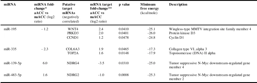
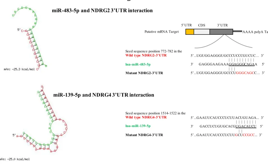
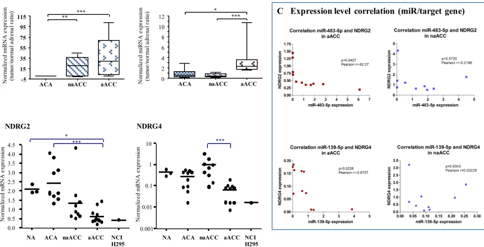
We focused on the four microRNAs that we have previously shown to be differentially expressed in ACCs compared with ACAs and that appeared as potential biomarkers for diagnosis and prognosis (28). This study was based on a microarray analysis followed by RT-qPCR validation, in two independent cohorts of 18 and 28 adrenocortical tumors. The level of miR-195 and miR-335 was found significantly decreased in both non-aggressive and aggressive ACC (naACCs and aACCs) as compared to ACA and normal adrenal cortex tissues, whereas miR-483-5p was markedly overexpressed in all ACCs. Interestingly, miR-139-5p expression level was reported to be up-regulated only in aACC tumors compared to naACC (28). To identify putative gene targets of the four differentially expressed miRNAs, we combined our data from an in silico analysis using different bioinformatics algorithms and data from gene expression analysis of adrenocortical tumors (34). Results are summarized in Table 1. We focused our further experiments on potential mRNA/miRNA interactions between miR-483-5p and miR-139-5p and two unidentified genes in the physiopathology of adrenocortical tumors: the N-myc Downstream Regulated Gene family member 2 (NDRG2) as a target of miR-483-5p and the family member 4 (NDRG4) as a target of miR-139-5p. These two genes were found down-regulated in the transcriptomic analysis of adrenocortical tumors (Table 1) whereas miR-483-5p and miR-139-5p expression levels were found up-regulated in the miRnome analysis (with mRNA or microRNA fold-change greater than 2-fold). Figure 1 shows the miRNA/mRNA target physical interaction and secondary structures predicted by RNAHybrid software, combined with the results from miRWalk and TargetScan prediction softwares.
24 Inverse correlation between NDRG2/NDRG4 expression and miR-483-5p/miR-139-5p expression levels in tumor tissues
To validate the data obtained with the microarray analyses, we measured the mRNA expression level of NDRG2 and NDRG4 by RT-qPCR in a cohort of 30 adrenocortical tissues including 18 carcinomas, 10 adenomas and 3 normal cortical tissues. We found a down-regulation of NDRG2 in aggressive ACCs compared with adenomas (p<0.001) and with normal tissues (p<0.05) whereas no significant difference was observed between non-aggressive ACCs and adenomas (Figure 2B). Importantly, NDRG4 mRNA was significantly down-regulated in aACC when compared with naACC (p<0,001; Figure 2B). As shown in Figure 2A, low NDRG2 expression was associated with high miR-483-5p expression in aACC whereas a high NDRG2 expression was associated with a low 483-5p expression in ACA. Similarly, low NDRG4 expression was associated with high miR-139-5p expression in aACC, as compared with naACC (Figure 2A). Interestingly, expression levels of NDRG2 and NDRG4 were dramatically low in the human NCI H295R adrenocortical carcinoma cell line and were comparable to those measured in aACC tumors (Figure 2B). Figure 2C represents the correlation between the expression of microRNAs and putative target genes in each aACC or naACC. Significant inverse correlations between expression levels of NDRG2 and miR-483-5p, and between expression levels of NDRG4 and miR-139-5p were found in aACC but not in naACC tumors.
Effect of miR-483-5p and miR-139-5p inhibitors on NDRG expression in NCI H295R cells
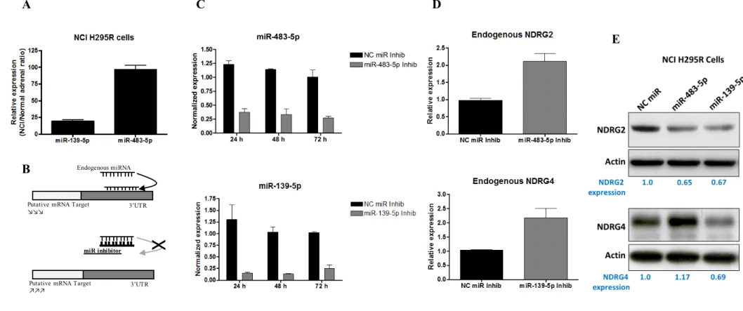
In light of the above results, we hypothesized that NDRG2 and NDRG4 might be target genes of miR-483-5p and miR-139-5p, respectively. We first confirmed by real-time PCR that miR-483-5p and miR-139-5p were overexpressed in NCI H295R: the normalized expressions of miR-483-5p and miR-139-5p were 90-fold and 18-fold higher, respectively, in NCI H295R cells compared with
25 normal adrenocortical tissues (Figure 3A). These results led us to examine whether inhibition of miR-483-5p and miR-139-5p could restore endogenous expression of NDRG2 and NDRG4 mRNAs, respectively, in NCI H295R cells (Figure 3B). To this end, miR-483-5p and miR-39-5p expression was down-regulated using specific inhibitors in NCI H295R cells. Figure 3C shows that the levels of miR-483-5p and miR-139-5p were efficiently decreased respectively by 3.3 and 8.6-fold within 24h as compared to Negative Control (NC) miRNA inhibitor and that inhibition was maintained at least up to 72h in NCI H295R cells. Quantitative PCR analysis revealed that miR-483-5p and miR-139-5p blockadge led to re-expression of NDRG2 and NDRG4 mRNA 72h post-transfection (Figure 3D). These results suggest that miR-483-5p and miR-139-5p regulate NDRG2 and NDRG4 expression, respectively, at the post-transcriptional level. Analysis of NDRG2 and NDRG4 at the protein level in the same experimental conditions did not show any significant up-regulation of both proteins (not shown). Therefore, NCI H295R cells were transiently transfected with miR-483-5p or miR-139-5p mimics to assess the effect on endogenous NDRG protein expression. As shown in Figure 3E, miR-139-5p overexpression decreased NDRG4 whereas miR-483-5p decreased NDRG2 protein levels. Unexpectedly, miR-139-5p also decreased NDRG2 protein expression. These results suggest that NDRG2 mRNA levels may be modulated by both miRNAs.
MiR-483-5p and miR-139-5p functionally interact with the 3’-untranslated region of NDRG2 and NDRG4, respectively
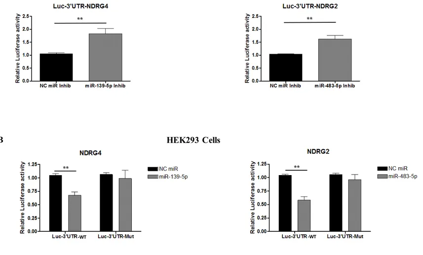
We then generated reporter constructs in which the wild-type 3’-untranslated region containing the predicted miRNA binding site was cloned downstream of the luciferase reporter gene (pMIR-Luc-3’UTR constructs). To investigate whether miR-483-5p and miR-139-5p target the (pMIR-Luc-3’UTR of their putative mRNA targets, miR-483-5p or miR-139-5p inhibitors or negative control inhibitors were co-transfected with these reporter plasmids into NCI H295R cells. Figure 4A shows that miR-483-5p
26 inhibitor significantly increased luciferase activity of pMIR-Luc-NDRG2 construct to 1.6-fold of luciferase activity measured in NC inhibitor-treated cells. On the other hand, miR-139-5p inhibitor increased the luciferase activity of pMIR-Luc-NDRG4 construct to 1.8-fold of the one measured in NC inhibitor-treated cells. Conversely, co-transfection experiments were performed with miR-483-5p and miR-139-5p mimics or miRNA negative control in the presence of reporter plasmids in HEK293 cells. Figure 4B shows that miR-483-5p significantly decreased the luciferase activity of pMIR-Luc-NDRG2 construct to 0.6-fold of luciferase activity in NC miR-treated cells and that miR-139-5p decreased the luciferase activity of pMIR-Luc-NDRG4 construct to 0.7-fold, suggesting that both miRNA target their predicted 3’UTR binding site. To determine whether these effects were due to a direct interaction between NDRG2 3’UTR and miR-483-5p (at the 787 bp position) and between NDRG4 3’UTR and miR-139-5p (at the 1522 bp position), we generated reporter constructs in which the 3’UTR region containing the predicted miRNA binding site was mutated (Figure 2B). Figure 4B shows that miR-483-5p and miR-139-5p transfection in HEK293 cells did not affect the luciferase activity of pMIR-Luc-NDRG2 and pMIR-luc-NDRG4 mutated constructs, respectively, suggesting that indeed miR-483-5p and miR-139-5p interact physically with their target 3’UTR.
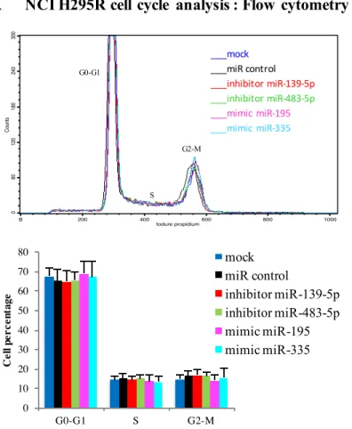
MiR-483-5p and miR-139-5p did not affect NCI H295R cell proliferation or apoptosis.
As our results indicated that miR-483-5p and miR-139-5p were overexpressed in ACC (oncogenic miRNAs) and that, conversely, miR-195 and miR-335 were under-expressed (tumor suppressor miRNAs), we next investigated the role of these miRNAs in adrenocortical cell tumorigenesis. NCI R295R cells were transfected with 483-5p and 139-5p inhibitors or with 195 and miR-335 mimics. WST1 proliferation assay and cell cycle analysis showed no statistically significant differences between NCI H295R cells transfected with miR-483-5p and miR-139-5p inhibitors or with miR-195 and miR-335 mimics and cells transfected with miRNA negative controls or
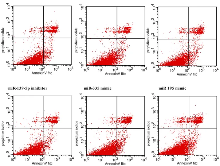
mock-27 transfected ones (Figure 5). Similarly, Annexin V-based cell apoptosis assays showed no significant differences between miR-483-5p or miR-139-5p inhibitor-transfected cells, miR-195 or miR-335 mimic-transfected cells and miRNA negative control-transfected cells (Figure 6). These results indicate that miR-483-5p and miR-139-5p are not involved in the regulation of NCI H295R proliferation or apoptosis in two-dimensional growth assays.
MiR-483-5p and miR-139-5p promote migration and invasion of NCI H295R cells
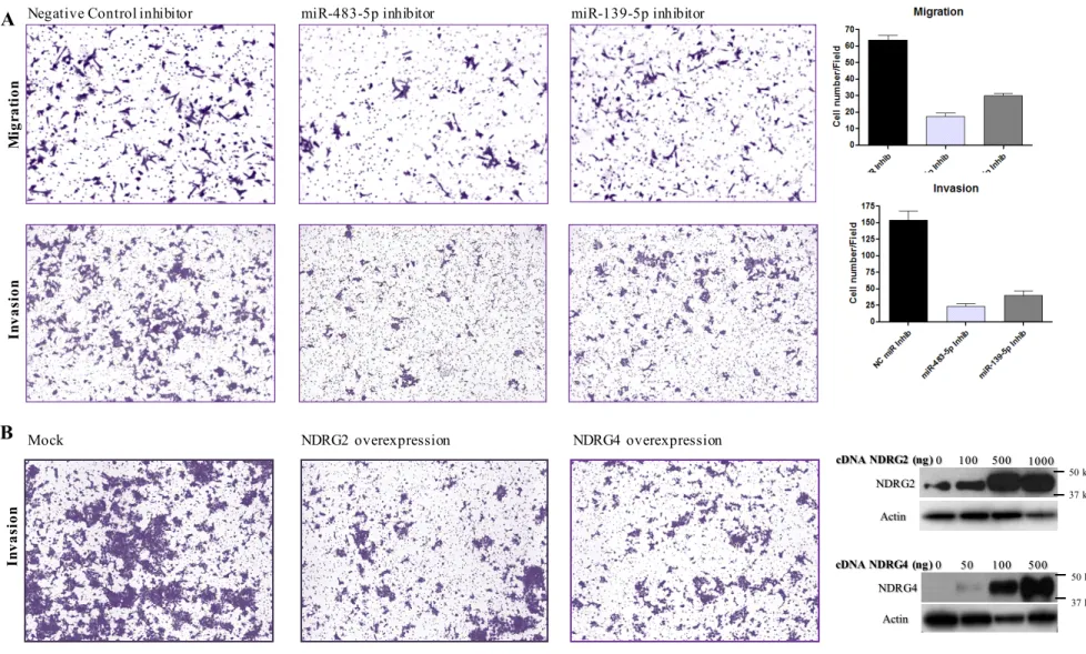
To evaluate the potential role of miR-483-5p and miR-139-5p in ACC aggressiveness, we analyzed the migration and invasion capacities of NCI H295R using Boyden chamber-based assay (Transwell Assay as described in Material and Methods). Figure 7A shows that the migration of NCI H295R cells transfected with miR-483-5p or miR-139-5p inhibitors through the insert membrane was significantly reduced. Furthermore, miR-483-5p or miR-139-5p inhibitors also induced a marked inhibition of NCI H295R cell invasion through the Matrigel, when compared to negative control miR inhibitor. As both miRNAs target NDRG2 and NDRG4, we evaluated whether restoration of NDRG2 and NDRG4 could affect invasion capacities of NCI H295R cells. Figure 7B shows that the invasion of NCI H295R cells transfected with NDRG2 or NDRG4 constructs was reduced as compared to control cells, suggesting that NCI invasiveness is mediated, at least in part, through down-regulation of NDRG2 and NDRG4 by miR-483-5p and miR-139-5p.
MiR-483-5p and miR-139-5p are involved in anchorage-independent colony formation of NCI H295R cells
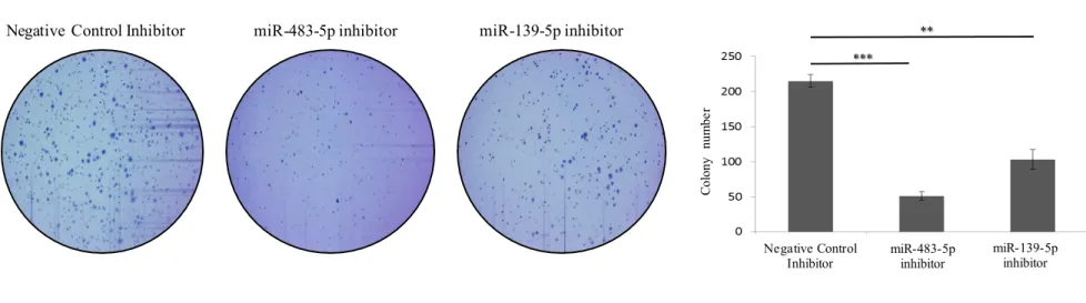
Anchorage-independent growth (AIG) is a common characteristic of cancer cells. To determine the effect of miR-483-5p or miR-139-5p silencing on AIG in NCI H295R cells, miRNA inhibitor-transfected cells were analyzed for their ability to growth in soft agar (Figure 8). Although
miR-28 483-5p or miR-139-5p inhibitors had no significant effect on NCI H295R two-dimensional monolayer growth (Figure 5), the number and size of colonies were decreased when the cells were grown in soft agar, suggesting that both miRNAs are involved in loss of contact inhibition in adrenocortical cancer cells. These results point a role of miR-483-5p or miR-139-5p overexpression in the aggressive phenotype of NCI H295R cells.
29
D I S C U S S I O N
ACC is rare and highly aggressive malignancy, associated with a pejorative prognosis due to distant metastasis in most cases. Unfortunately, current treatment fails in many patients with advanced ACC, including the first line of combined chemotherapy currently used in metastatic tumors (37). The underlying molecular mechanisms resulting in carcinogenesis and aggressiveness remain obscure and stand for the major limitation in finding novel molecular-targeted strategies (5). So far, therapies targeting VEGF and its receptors, or multi-tyrosine kinase inhibitors failed to demonstrate clinical efficacy in ACC patients (4,9,11,12). Hopes were pinned on drugs targeting the insulin growth factor 2 (IGF-2), the most up-regulated gene in ACC, and its receptor IGF-1R, but a recent phase 3 trial showed disappointing results (38). Thus, networking and progress in the molecular characterization of ACC are ongoing thanks to the ENS@T network and The Cancer Genome Atlas (TCGA) international consortium. Researchers are developing molecular tools to better identify potential targets for personalized treatment, potential prognostic markers and predictive factors of new treatments efficacy, in order to further introduce them into the clinics (13,17). Several studies are ongoing to improve the classification of adrenocortical tumors based on pan-genomic analysis of genetic mutations, chromosomal aberrations, DNA methylation (the methylome) and gene expression (the transcriptome). The integration of microRNA expression (the miRNome) is an important part of this project.
In our study, we found that miR-483-5p and miR-139-5p target potential tumor suppressors, NDRG2 and NDRG4, respectively. Furthermore, miR-483-5p and miR-139-5p inhibition could effectively suppress the invasive potential of adrenocortical carcinoma cells in vitro. Thus, 483-5p and miR-139-5p might act as oncomiRNAs in ACC pathogenesis and represent potential therapeutic targets for this highly aggressive and therapy-refractory tumor.
30 Genetic aberration patterns have been defined previously and even if there are considered as early or late events occurring in ACC tumorigenesis, they are not sufficient to explain the entire malignant transformation process and chronology (13). Numerous studies have shown an overexpression of IGF2, a constitutive activation of Wnt/beta-catenin pathway, and an inactivation of the p53 suppressor gene in most ACCs. Abnormalities in the imprinted 11p15 region (involving maternal 11p15 Loss of Heterozygosity (LOH) with duplication of the active IGF-2 paternal allele) result in strong overexpression of IGF-2 and occur in more than 85% of ACCs. The markedly elevated IGF2 in sporadic ACC is likely to involve additional mechanisms of transcriptional regulation (14,15). Interestingly, miR-483-5p which was previously found to be overexpressed in ACC tumors compared with ACAs, resides within the second intron of IGF-2 and both were found significantly co-expressed in ACC tumors (24,26,28,29).
We focused our analysis on the most deregulated miRNA in ACC, according to our previous study: 195 and 335, which are underexpressed in ACC tumors compared with ACA, and miR-483-5p and miR-139-5p which are overexpressed (28). Using several prediction softwares, we identified several potential gene targets. Integrating the transcriptome and the miRnome data of ACCs allowed us to select gene targets inversely correlated with miRNA expression profile in ACCs. Surprisingly, when we restore the expression of miR-195 and miR-335 in NCI H295R cells, we did not observe any significant effect on the putative gene target expression (data not shown). Therefore, molecular mechanisms involved in the dysregulation of these miRNAs in ACC and their molecular targets remain to be explored further. We observed an increase in NDRG2 and NDRG4 expression mRNA levels by suppressing miR-483-3p or miR-139-5p expression in the human ACC cell line NCI-H295R. In addition, the functional interaction between miR-483-5p and NDRG2, and between miR-139-5p and NDRG4, was confirmed by luciferase reporter assays, indicating that each miRNA post-transcriptionally regulate NDRG2 and NDRG4 expression via a binding site in their 3’UTR
31 region. Interestingly, the mutation of the specific miRNA binding region of NDRG2- or NDRG4-3’UTR showed no effect on luciferase activity, indicating a compromised interaction between both microRNAs and their target gene. Unexpectedly and by contrast to the results obtained for NDRG2/4 mRNA levels, we did not found any significant increases in NDRG2/4 protein expression in NCI H295R cells transfected with miRNA inhibitors (data not shown). However, it is worth mentioning that the basal endogenous miR-483-5p and miR-139-5p levels are very high in NCI H295R cells compared with normal adrenal cortex (100- and 20-fold increase in NCI H295R cells, respectively). Consequently, substantial amounts of endogenous miRNA remain available within the cells despite miRNA inhibition. We therefore decided to assess endogenous NDRG protein expression using miRNA mimics. We could observe a significant decrease in NDRG4 and NDRG2 protein expression following miR-139-5p and miR-483-5p overexpression. Unexpectedly, miR-139-5p also decreased NDRG2 protein levels. Using prediction softwares, we found that miR-139-5p was predicted to bind the 3’UTR region of NDRG2, although with a lower score than miR-483-5p. These observations confirm that multiple miRNAs can target the same gene and suggest that the combination of miR-139-5p and miR-483-5p activities might determine the expression of NDRG2 in NCI H295R cells.
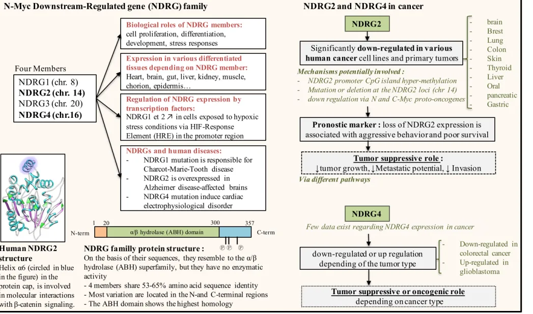
We demonstrated that NDRG2 expression in human adrenal tumors was significantly lower in both aggressive and non-aggressive ACC compared with ACA. Furthermore, low NDRG4 levels could discriminate between aggressive ACC and non-aggressive ACC. These results need to be confirmed in larger cohorts of patients. We showed that low NDRG2 and NDRG4 expression levels in ACC were inversely correlated with high expression levels of miR-483-5p and miR-139-5p, respectively. NDRG2 and NDRG4 belong to the N-myc downstream regulated gene family of proteins consisting of 4 members, NDRG1 to NDRG4, which share 53-65% amino acid identity. Figure 9 represents a literature overview on NDRG family functions. The NDRG genes are located on different chromosomes, and the distinct subcellular (nuclear versus cytoplasm) and tissue distributions of the
32 different members suggest that each of them may have different function. Although there is evidence that they play fundamental roles in physiological processes such as cell proliferation, differentiation, development, and stress responses, the exact function of these proteins needs to be explored further (39–41). The mRNA and protein level of NDRG2 has been shown to be significantly down-regulated in a variety of human malignant tumors such as breast cancer, colorectal adenocarcinoma, glioblastoma, hepatocellular carcinoma, thyroid cancer, pancreatic cancer and gastric cancer (42–46). Thus, several studies support a tumor suppressor role for NDRG2 in cancer. The downregulation of NDRG2 in malignant tumors was reported to be closely correlated with poor overall survival, metastasis, and highly invasive phenotype (47–50). Therefore, NDRG2 was considered as a potentially valuable prognostic marker in patients. There are less data available concerning NDRG4 expression in cancer, but it seems to have tumor-suppressive or oncogenic functions depending on the tissue type (51–53). The regulation of NDRG protein expression involves complex and various mechanisms including not only transcription repressors such as N-myc and C-myc proto-oncogenes, but also promoter hyper-methylation. Interestingly, NDRG2 promoter CpG island methylation and downregulation have been observed in many cancers including ACC (54–56). Our data highlight an additional mechanism for the regulation of NDRG2 and NDRG4 expression in ACCs, through miR-483-5p- and miR-139-5p-mediated post-transcriptional control. Similar mechanisms have been reported in recent studies showing that miR-122 in hepatocellular carcinoma, or miR-650 in colorectal carcinoma, repress the expression of NDRG3 and NDRG2, respectively (57,58).
Our data show that silencing miR-483-5p or miR-139-5p and overexpressing miR-195 or miR-335 in NCI-H295R cells did not affect cell proliferation or apoptosis. By contrast, a previous study reported that inhibition of miR-483-5p or overexpression of miR-195 in NCI-H295 cells resulted in a significant reduction of cell proliferation (23). Although we do not have explanation for these discrepancies, one should mention that in Ozata’s study, the NCI H295R cells were electroporated
33 with 20-times higher concentrations (1 µM) of miRNA inhibitors or miRNA mimics, which could lead to non-specific effects on gene expression.
We observed that miR-483-5p inhibitor significantly decreased NCI H295R cell migration and invasion. These results are in agreement with those reported very recently by Song and co-workers, who demonstrated that miR-483-5p is a critical prometastatic miRNA in lung adenocarcinoma (59). We found that miR-139-5p inhibition also disrupts NCI H295R cell invasion and migration. This is contrast to the data reported by Krishnan et al demonstrating that miR-139-5p overexpression plays an anti-invasive role in breast cancer (60). However, one should keep in mind that miRNAs can play tumor suppressive or oncogenic functions depending on the cell context. As we have shown in our study that NDRG2 and NDRG4 mRNA expression is dramatically decreased in ACC as compared to normal adrenal cortex and as both transcripts are targets of miR-483-5p and miR-139-5p, respectively, we re-expressed NDRG2 and NDRG4 in NCI H295R cells and could show that invasion capacities of NCI H295R cells were impaired.
Anchorage-independent growth in soft agar, which is considered as the most stringent assay for detecting malignant cell transformation, revealed that miR-483-5p or miR-139-5p inhibition induced a marked decrease in the number and the size of the colonies compared with negative controls. These results suggest that both microRNAs are involved in phenotypic changes of NCI H295R cells such as loss of contact inhibition and cell ability to grow in a viscous fluid or gel. This process is assumed to be closely related to the in vivo carcinogenesis. In vivo strategies based on exogenous miRNA delivery (mimic or inhibitor) in cancer models remain the key challenge for the establishment of this novel class of RNA therapeutics (32,61). Therefore, our results are promising in the prospect of testing the effect of miR-483-5p and miR-139-5p inhibitors as therapeutic tools in an ACC model in mice. There are several major difficulties to overcome before considering modulating endogenous microRNA expression in patients, such as technical issues (administration mode etc.) and also
34 potential off-target actions and side effects due to the tissue-specific nature of microRNA action. The pathways affected by microRNA dysregulation in ACC tumors could also serve as indirect therapeutic targets (22). In this study, we identified for the first time two target genes among the NDRG family, NDRG2 for miR-483-5P and NDRG4 for miR-139-5p, that play a tumor suppressor role in ACC tumors and participate to the invasive phenotype of NCI H295R. We hypothesize that the downstream pathways altered by both oncomiRs and their targets involves the Epithelial-Mesenchymal Transition (EMT) which is required for metastasis, and largely influenced by the WNT/Beta-catenin signaling (62,63). Recent studies have shown a correlation between loss of NDRG2 and invasive characteristics of different cancer cell models and suggested an important role for EMT as a molecular mechanism involved in these processes (64–66). Further experiments have to be performed to explore this hypothesis.
Moreover, the recent discovery of the overexpression of miR-483-5p and miR-139-5p in the serum of patients affected by ACC opens the way for the study of their possible role as intercellular communication molecules (28). MiR-139-5p and particularly miR-483-5p were found highly abundant in the culture medium of NCI H295R cells (data not shown), in line with what has been observed in the serum of ACC patients. Tumor-derived circulating miRNAs may be transferred to recipient cells such as endothelial cells or other cells of the tumor micro-environment and may remodel their gene expression. This could promote angiogenesis, and lead to vascular endothelial barrier destruction and metastasis of tumor cells. Our preliminary results open promising perspectives in this field.
35
RÉFÉRENCES
1. Kebebew E, Reiff E, Duh Q-Y, Clark OH, McMillan A. Extent of disease at presentation and outcome for adrenocortical carcinoma: have we made progress? World J Surg. mai 2006;30(5):872 8. 2. Fassnacht M, Johanssen S, Quinkler M, Bucsky P, Willenberg HS, Beuschlein F, et al. Limited prognostic value of the 2004 International Union Against Cancer staging classification for adrenocortical carcinoma: proposal for a Revised TNM Classification. Cancer. 15 janv 2009;115(2):243 50.
3. Lughezzani G, Sun M, Perrotte P, Jeldres C, Alasker A, Isbarn H, et al. The European Network for the Study of Adrenal Tumors staging system is prognostically superior to the international union against cancer-staging system: a North American validation. Eur J Cancer Oxf Engl 1990. mars 2010;46(4):713 9.
4. Else T, Kim AC, Sabolch A, Raymond VM, Kandathil A, Caoili EM, et al. Adrenocortical carcinoma. Endocr Rev. avr 2014;35(2):282 326.
5. Fassnacht M, Kroiss M, Allolio B. Update in adrenocortical carcinoma. J Clin Endocrinol Metab. déc 2013;98(12):4551 64.
6. Libé R. Adrenocortical carcinoma (ACC): diagnosis, prognosis, and treatment. Front Cell Dev Biol. 2015;3:45.
7. Mihai R. Diagnosis, treatment and outcome of adrenocortical cancer. Br J Surg. mars 2015;102(4):291 306.
8. Libé R, Borget I, Ronchi CL, Zaggia B, Kroiss M, Kerkhofs T, et al. Prognostic factors in stage III-IV adrenocortical carcinomas (ACC): an European Network for the Study of Adrenal Tumor (ENSAT) study. Ann Oncol Off J Eur Soc Med Oncol ESMO. oct 2015;26(10):2119 25.
9. Kirschner LS. The next generation of therapies for adrenocortical cancers. Trends Endocrinol Metab TEM. juill 2012;23(7):343 50.
10. Lehmann T, Wrzesinski T. The molecular basis of adrenocortical cancer. Cancer Genet. avr 2012;205(4):131 7.
11. Ragazzon B, Assié G, Bertherat J. Transcriptome analysis of adrenocortical cancers: from molecular classification to the identification of new treatments. Endocr Relat Cancer. avr 2011;18(2):R15 27.