C
HEMICAL
C
ONSTITUENTS
OF
THE
L
EAVES
OF
C
ALONCOBA
WELWITSCHII
G
ILG
Pascal D. Douanlaa,b, Marguerite Hortence K. Tchuendema,*, Alembert T. Tchindab, Turibio
Kuiate Tabopdaa, Denis Zofouc, Ewa Cieckiewiczd, Michel Frédérichd, Augustin E.
Nkengfacka
a Department of Organic Chemistry, Faculty of Science, The University of Yaoundé-1, P.O.
Box 812, Yaounde, Cameroon
b Laboratory of Phytochemistry, Institute of Medical Research and Medicinal Plant Studies,
(IMPM), P.O. Box 6163, Yaounde, Cameroon
c Biotechnology Unit, University of Buea, P.O. Box 63, Buea, South West Region, Cameroon d Université de Liège, Laboratoire de Pharmacognosie, Centre Interfacultaire de
Recherche sur le médicament (CIRM), Département de Pharmacie, Université de Liège, B36, B-4000, Liège, Belgium
KEYWORDS: Flacourtiaceae Caloncoba welwitschii Flavonol glucosides Friedelanes
Antiplasmodial activities ABSTRACT
Two new compounds namely acetylcaloncobaside (1) and friedocaloncobic acid (2) were isolated from the leaves of Caloncoba welwitschii together with six known compounds. Their structures were determined by standard spectroscopic methods including one- and two-dimensional NMR, EI-MS, and HRESIMS. The new compounds were established as kaempferol 4',7-dimethoxy-3-0-(3",4",6"-0-triacetyl)-β-NULL-glucopyranoside (1) and 15β-hydroxy-3-oxo-28-friedelanoic acid (2). The isolated compounds were evaluated for their ability to inhibit the 3D7 strain of Plasmodium falciparum, using the semi-automated in vitro model with parasite lactate dehydrogenase assay.
1. Introduction
The genus Caloncoba comprises 20 species of trees and shrubs, and constitutes one of the smallest genera of the Flacourtiaceae family represented in tropical Africa (Hutchinson and Dalziel, 1954; Ziegler et al., 2002). Caloncoba welwitschii (Oliv.) Gilg. is a tree common in the rain forests of western and central Cameroon (Giner-Pons et al., 1992; Hutchinson and Dalziel, 1954). The leaf-sap is used to treat headaches, and the plant itself is prescribed as a mean of killing body-lice (Burkill, 1994). The decoction of seeds is used in african folk medicine to treat leprosis (Raponder-Walker and Sillans, 1961). Previous phytochemical investigation of some Caloncoba species afforded cycloartanes (Mpetga et al., 2012a,b, 2014), cyclopentanoid amino acids (Clausen et al., 2002), friedelane, dammarane and malabaricane triterpenes (Giner-Pons et al., 1993, 1992;
Tchuendem et al., 1996; Ziegler et al., 2002). To the best of our knowledge, there is no prior report on the chemical constituents of C. welwitschii. As part of a program to search for antiplasmodial natural products, we report herein the isolation and structure elucidation of a new acetylated glucoside flavonol and a new friedelane triterpene, along with six known compounds.
2. Results and discussion
Repeated column chromatography over silica gel and Sephadex LH-20 of CH2Cl2/MeOH
(1:1) extract of the leaves of C. welwitschii led to the isolation of a new flavonol acetylated glucoside (1) (Fig. 1) and a new friedelane triterpene acid (2) (Fig. 2), along with one known friedelane (3), a mixture of β-sitosterol and stigmasterol (4a and 4b), a mixture of β-sitosterol and stigmasterol glucoside (5a and 5b), and a known flavonoid, luteolin 7-0-β-NULL-glucopyranoside (6). The known friedelane underwent chemical
transformations and yielded two other known friedelanes (3a and 3b) which were also elucidated.
Compound 1 was obtained as yellow crystals, mp 204-206 °C. The positive HR-ESIMS showed a quasi-molecular ion peak at m/z 603.16830 [M + H]+, (caled 603.16829)
consistent with the molecular formula C29H30O14, accounting for fifteen degrees of
insaturation. Its IR spectrum showed absorptions for hydroxyl (υmax = 3328.9 cm-1) and
carbonyl (υmax = 1749.3 cm-1) groups. The 1H NMR showed a low field singlet appearing at
SH 12.00 characteristic of chelated hydroxyl group with the carbonyl function of a
flavonoid moiety (Harborne et al., 1975; Mabry et al., 1970). In addition, this spectrum displayed the characteristic pair of A2X2 aromatic system at δH 8.13 (dd, J = 8.8, 2.2 Hz,
H-2', 6') and 6.96 (dd, J = 8.8, 2.2 Hz, H-3', 5'), suggesting a 1,4-disubstituted aromatic nucleus; two doublet signals at δH 6.38 (d, J = 2.2 Hz) and 6.48 (d, J = 2.2 Hz), assigned to
two meta protons of an aromatic ring. These informations were supported by 1H-1H COSY
correlations observed between these aromatic protons. The 1H NMR of 1 also showed
resonances for two methoxy group linked to the aromatic nucleus [δH/δC 3.88/55.4
(7-OCH3), 3.89/56.0 (4'-OCH3)]. The above described spectral data suggested a kaempferol
skeleton bearing methoxy groups at C-7 and C-4' (Harborne et al., 1975). The 13C NMR
spectrum also corroborated the kaempferol skeleton (see Table 1). The remaining signals observed on the 1H- and 13C NMR spectra, were characteristic of a triacetylated glucose
residue attached to the flavonol framework. In fact, these spectra exhibited resonance for four oxygenated methines [δH/δC 3.85/72.2 (CH-2"), 5.18/74.9 (CH-3"), 4.99/ 68.2 (CH-4"),
3.57/72.1 (CH-5")], one oxygenated methylene [δH/δC 4.02 and 3.84/62.1 (CH2-6")], one
anomeric methine [δH/δC 4.90/ 105.7 (CH-1")] and three methyls of acyl grups [δH/δC
2.11/20.9 (6"-OAc), 2.01/20.7 (4"-OAc), 1.92/20.5 (3"-OAc)]. HMBC correlations observed between the acetyl carbon atoms at δc 170.5 (6"-COCH3), 169.8 (4"-COCH3), and 170.4
(3"-COCH3) and deshielded sugar protons at δH 4.02, 3.84 (Ha-6", Hb-6"), 4.99 (H-4"), and 5.18 (H-3"), respectively, strongly supported the location of the three acetyl group. A long range connectivity was also observed between the anomeric proton at δH 4.90 (H-l") with the aglycone carbone at δc 135.3 (C-3) suggesting the position of the glucose moiety at C-3. This is consistent with the downfield value of C-3.Acid hydrolysis of (1) afforded a flavonoid and a free sugar. The sugar was identified by TLC comparison with authentic sample and his absolute configurations were determined by the measurement of optical rotation after separation by PTLC to NULL-glucose. On the basis of the above spectral data and by their comparison with literature values (Fathy et al., 2002; Mustafa et al., 2000), the structure of compound 1 was established as kaempferol 4',7-dimethoxy-3-0-(3",4",6"-0-triacetyl)-β-NULL-glucopyranoside, which is a new compound to which the trivial name acetylcaloncobaside was assigned.
Fig. 1. Chemical structure of compounds 1 and 2.
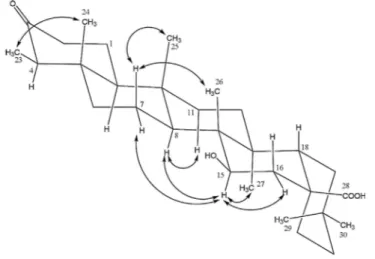
Fig. 2. NOESY correlations of compound 2.
Table 1.
1H and
13C NMR data of compound 1 recorded in CDC1
3.
Positions δc δH Positions δc δH
2 158.4 (s) _ Glucose
4 178.0 (s) - 2" 72.2 3.85 (dd, 9.6, 8.0) 4a 105.4 (s) - 3" 74.9 5.18 (t, 9.6) 5 161.7 (s) - 4" 68.2 4.99 (t, 10.0) 6 98.4 (d) 6.38 (d, 2.2) 5" 72.1 3.57 (ddd, 10.0, 5.4, 2.9) 7 166.1 (s) - 6" 62.1 4.02 (dd, 12.2, 5.4) 3.84 (dd, 12.2, 2.4) 8 92.6 (s) 6.48 (d, 2.2) 3"-COCH3 170.4 -8a 156.9 (s) - 3"-COCH3 20.5 1.92 (s) 1' 122.0 (s) - 4"-COCH3 169.8 -2' 131.2 (d) 8.13 (dd, 8.8, 2.2) 4"-COCH3 20.7 2.01 (s) 3' 113.6 (d) 6.96 (dd, 8.8, 2.2) 6"-COCH3 170.5 -4' 162.2 (s) - 6"-COCH3 20.9 2.11 (s) 5' 113.6 (d) 6.96 (dd, 8.8, 2.2) 6' 131.2 (d) 8.13 (dd, 8.8, 2.2) 7-OCH3 55.4 3.88 (s) 4'-OCH3 56.0 3.89 (s) 5-OH - 12.00 (s)
Compound 2 was isolated as an amorphous powder. The positive HR-ESIMS showed a pseudo-molecular ion peak at m/z 495.35147 [M + Na]+, (calcd 495.35146 for C30H48O4Na) suggesting the molecular formula C30H48O4, accounting for seven degrees of insaturation. Its IR spectrum indicated the presence of hydroxyl (υmax = 3434 cm-1), carboxylic acid (υmax = 1735 cm-1), and carbonyl (umax = 1715 cm-1) groups. The ID NMR (1H and APT) spectra showed thirty carbon signals with seven methyls, ten methylenes, five methines, and eight quaternary groups. These data indicated that compound 2 is a pentacyclic triterpene bearing a carboxylic acid signal at δc 182.2, a carbinol methine signal at δc 73.5, and a ketone carbonyl signal at δc 214.5. The friedelane triterpene skeleton of compound 2 was confirmed by the presence of characteristic signals in its 1H and 13C NMR spectra (Table 2) respectively at δH 0.87 (H3-23, d, J = 6.7 Hz) and δc 6.6 (C-23) (Giner-Pons et al., 1992; Mahato and Kundu, 1994). The attachment of acid group at C-17 position was supported by the downfield carbon resonances of C-17 (δc 44.0) (Hang et al., 2006) compared to that of friedelin at C-17 (δc 30.0) (Akihisa et al., 1991). This was confirmed by the HMBC correlation of H-18 (SH 2.33) with C-17 (δc 44.0). The position of the 15-hydroxy group was deduced from the HMBC correlation of H15 (δH 3.60) with C26 (δc 13.1), C14 (δc 39.7) and C8 (δc 53.3). The β -orientation of the hydroxyl group at C-15 was determined using the NOESY spectrum, which showed key correlation peaks between H-15 (δ„ 3.60) and H3-27 (δ„ 1.02), H-8 (δ„ 1.47), H-7 (δH 1.90), and H-16 (δH 2.33). The full structure was deduced from the analysis of its 1D-and 2D-NMR spectral data (COSY, HMQC, NOESY, and HMBC) and by their comparison with literature values (Hang et al., 2006). Its name was established as
15β-hydroxy-3-oxo-28-friedelanoic acid, a new compound to which the trivial name friedo-caloncobic acid was assigned.
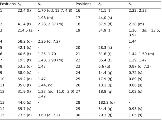
Table 2. 1H and 13C NMR data of compounds 2 in CDCI
3-CD3OD. Positions δc δH Positions δc δH 1 22.4 (t) 1.70 (dd, 12.7, 4.8) 16 41.1 (t) 2.22, 2.33 1.98 (m) 17 44,0 (s) -2 41.4 (t) 2.28, 2.37 (m) 18 37.9 (d) 2.28 (m) 3 214.5 (s) - 19 34.9 (t) 1.16 (dd, 13.5, 3.9) 4 58.2 (d) 2.26 (q, 7.2) 1.44 5 42.1 (s) - 20 28.3 (s) -6 40.6 (t) 1.25, 1.70 21 31.6 (t) 1.44, 1.59 (m) 7 19.5 (t) 1.48, 1.90 (m) 22 35.4 (t) 1.29, 1.47 8 53.3 (d) 1.47 23 6.6 (q) 0.87 (d, 7.2) 9 38.0 (s) - 24 14.4 (q) 0.72 (s) 10 59.2 (d) 1.47 25 17.9 (q) 0.89 (s) 11 35.0 (t) 1.44, nd 26 13.1 (q) 0.86 (s) 12 31.9 (t) 1.23 (dd, 11.0, 3.0) 1.42 27 18.8 (q) 1.02 (s) 13 44.0 (s) - 28 182.2 (q) -14 39.7 (s) - 29 34.4 (q) 0.95 (s) 15 73.5 (d) 3.60 (d, 7.2) 30 29.3 (q) 1.05 (s) nd = not determined.
In addition to these two compounds, six other known compounds were also isolated and identified as 3-oxo-28-friedelanoic acid (3) (Hang et al., 2006), ^-sitosterol and stigmasterol (4a and 4b), β-sitosterol and stigmasterol glucosides (5a and 5b), and luteolin 7-0-β-NULL-glucopyranoside (6) (Zhang et al., 2006).
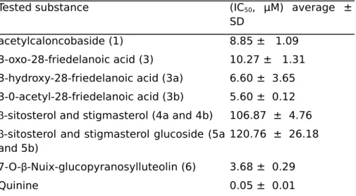
All the isolated compounds were evaluated in vitro against the 3D7 strain of Plasmodium falciparum. Acetylcaloncobaside (1), 3-oxo-28-friedelanoic acid (3), and 7-0-β-NULL-glucopyranosylluteolin (6) exhibited moderate to significant antiplasmodial in vitro activity with IC50 values of 8.85 ± 1.09, 10.27 ± 1.31, and 3.68 ± 0.29 µM, respectively.
Compound 2, which is only different from 3 by the hydroxyl group at C-15, was inactive, suggesting that the presence of OH group on the triterpene skeleton may reduce the antiplasmodial activity. In order to verify the effect of the OH group, we have reduced the carbonyl function of the 3-oxo-28-friedelanoic acid obtained in a good amount to 3β-hydroxy-28-friedelanoic acid (3a). Unexpectedly, the reduced product rather showed better activity. This result revealed that the presence of a hydroxyl group at position 3 increased the activity, although it is not observed with the hydroxyl group at position 15. Acetylation of the 3β-hydroxy-28-friedelanoic acid (3a) also leads to 3β-0-acetyl-28-friede-lanoic acid (3b) with increased activity. In conclusion, (3a) and (3b) are more active than product (3) on the 3D7 strain of Plasmodium falciparum, with inhibitory concentrations of (6.60 ± 3.65) and (5.67 ± 0.12) µM, respectively (Table 3).
Table 3. In vitro antiplasmodial activity of compounds from Cahncoba
welwitschii.
Tested substance (IC50, µM) average ±
SD
acetylcaloncobaside (1) 8.85 ± 1.09 3-oxo-28-friedelanoic acid (3) 10.27 ± 1.31 3-hydroxy-28-friedelanoic acid (3a) 6.60 ± 3.65 3-0-acetyl-28-friedelanoic acid (3b) 5.60 ± 0.12 β-sitosterol and stigmasterol (4a and 4b) 106.87 ± 4.76 β-sitosterol and stigmasterol glucoside (5a
and 5b)
120.76 ± 26.18
7-O-β-Nuix-glucopyranosylluteolin (6) 3.68 ± 0.29
Quinine 0.05 ± 0.01
3. Materials and methods
3.1. GENERAL EXPERIMENTAL PROCEDURES
Melting points were measured on an Electrothermal IA9100 Series, digital melting point apparatus (Bibby scientific, Great Britain) and are uncorrected. IR spectra were obtained with a FT-IR spectrum 2000 (Perkin-Elmer) spectrophotometer equipped with a BTGS detector. The 1H- and 13C NMR spectra were recorded on Bruker Avance II 500 MHz spectrometers, resonating at 500 MHz and 125 MHz respectively. Chemical shifts (5) were expressed in ppm with reference to TMS and coupling constants (J) were given in Hz. HR-ESIMS analyses were performed on a LTQ-Orbitrap XL hybrid mass spectrometer (Thermo Fisher Scientific, Bremen, Germany). Data were acquired in positive ion mode using full-scan MS with a mass range of 100-2000 m/z. The or-bitrap operated at 30000 resolutions (FWHM definition). All experimental data were acquired using daily external calibration prior to data acquisition. The electrospray inlet conditions were applied: spray voltage, 5 kV; sheath gas (N2) flow rate, 20 a.u.; auxiliary gas (N2) flow rate, 10 a.u.; capillary temperature, 275 °C; capillary voltage, 45 V; tube lens, 80 V. Methanol was purchased from Biosolve (LC-MS grade). Column chromatography was performed on normasil silica gel (63-200 µm) and Sephadex LH-20 (40-70 µm). TLC was carried out on precoated silica gel 60 F254 aluminum plates and detection accomplished by dipping into a 10% H2S04 in ethanol followed by heating. Solvents were distilled prior to use.
3.2. PLANT MATERIAL
The leaves of C. welwitschii were collected in Mount Kala near Yaounde, central Region of Cameroon, in July 2012. The plant was authenticated by Mr Victor Nana, a botanist of the National Herbarium of Cameroon (Yaounde), where a voucher specimen (45385/HNC) was deposited.
3.3. EXTRACTION AND ISOLATION
The leaves of C. welwitschii were dried, powdered and finally macerated in a percolator apparatus with the mixture methanol-methylene chloride (1:1) during 48 h at room
temperature and then concentrated using a rotavapor to yield a crude extract (140 g). Part of the extract (120 g) was subjected to vacuum chromatography over a silica gel column. The column was first eluted with 100% hexane and then with mixtures of hexane and increasing amounts of ethyl acetate and finally with methanol. The elution process was monitored by TLC and the fractions of similar TLC behavior were combined. All fractions were eluted in Sephadex LH-20 with the system Hex/CH2Cl2/MeOH (7: 4: 0.5) to eliminate the chlorophyll. The chlorophyll-free fractions were separated and purified on silica gel column chromatography to give 8 compounds. The fraction F2 (Hex/EtOAc 20%) gave 3-oxo-28-friede-lanoic acid (105 mg). From the fraction F4 (Hex/EtOAc 30%), two white crystalline substances were obtained and identified as a mixture β-sitosterol (6 mg) and stigmasterol (16 mg), and 3-oxo-15β-hydroxy-28-friedelanoic acid (9 mg) 2. Fraction F7 (Hex/EtOAc 50%) yielded kaempferol 4',7-dimethoxy-3-0-(3",4",6"-0-triacetyl)-β
-NULL-glucopyr-anoside (8 mg) 1, and a mixture of β-sitosterol and stigmasterol glucosides (13 mg). Fraction F10 (EtOAc/MeOH 10%) gave luteolin 7-O-β-NULL-glucopyranoside (10 mg).
3.4. ACID HYDROLYSIS
A soln. of 1 (4 mg) in 0.2 M HC1 (dioxane/H20 1:1, 3 mL) was heated at 95 °C for 30 min under Air. After cooling, the mixture was neutralized by passage through an Amberlite-IRA-93ZU (Organo, Tokyo, Japan) column and chromatographed (Diaion HP-20, 40% MeOH followed byMe2CO/EtOH 1:1) to give aglycone fractions (2.0 mg) and a sugar fraction (1.3 mg). The sugar fraction was then passed through an Sep-Pak-C18 cartridge (Waters, Milford, MA, USA; with 40% MeOH) and Toyopak-IC-SP-M-cartridge (Tosoh; with 40% MeOH), it was analyzed by HPLC (MeCN/H20 17: 3, flow rate, 0.9mLmin_1; detection, RI and OR): NULL-glucopyranoside (rt 18.6, pos. OR) was detected in 1.
3.5. PHYSICAL AND SPECTRAL PROPERTIES OF ISOLATES
Compound (1): Yellow powder; mp 204-206 °C; [α]g6 -37.5 (c 0.1, CHCl3-MeOH, 1:1); IR (KBr) vmax 3328, 1749 cm-1; 1H (500 MHz, CDCl3) and 13C (125 MHz, CDCI3) NMR data, see Table 1; positive ESI-HRMS: m/z 603.16830 [M + H]+ (caled for C29H3JOJ4, 603.16829).
Compound (2): White amorphous powder; [α]26D +20.5 (c 0.1, CHCl3-MeOH, 1:1); IR (KBr)
fmax 3434, 1735, 1715 cm-1; αH (500 MHz, CDCI
3) and 13C (125 MHz, CDCI3) NMR data,
see Table 2; positive ESI-HRMS: m/z 495.35147 [M + Na] + (calcd 495.35146 for C30H48O4Na).
3.6. REDUCTION OF 3-OXO-28-FRIEDELANOIC ACID (3)
A sample of 3 (25 mg) was heated gently with a few grains of boiling stones and 25 mL of 95° ethanol in a 250 mL round bottom flask perfectly dry surmounted with a condenser. To this solution was added 0.4 g of sodium borohydride (NaBH4) and stirred at room temperature for 10 min. To this reaction mixture was added 30 mL of distilled water and refluxed for 5 min. The total conversion of the starting material was observed by TLC. After cooling, 60 mL of cold distilled water was added in the flask; the contents of the ball was poured into a perfectly clean beaker; crystallization was waited in a bath: water + crushed ice; the cold solution filtered on a Büchner funnel and the crystals rinsing with a small amount of cold water, and then wring. This operation led to obtain 3β-hydroxy-28-friedelanoic acid (3a). White powder; mp 283-285 °C; τH (500 MHz, CDCI3) and 13C (125 MHz, CDCI3) NMR data, see Table 2.
3.7. ACETYLATION OF 3A
A sample of 3a (10 mg) was treated for 4 h at 70 °C on a water bath, with Ac20 (2 mL) in dry pyridine (1 mL), in the presence of a catalytic amount of DMAP. The reaction mixture was poured in crushed ice-water and extracted with EtOAc. The organic layer was washed respectively with 2 M HC1 and 1 M NaHC03, and dried. Gel permeation over Sephadex LH-20 (CHCl3-MeOH, 1:1) gave the acetylated derivative 3β -O-acetyl-28-friedelanoic acid (3b). White powder; mp 276-278 °C; 1H (500 MHz, CDCI3) and 13C (125 MHz, CDCI3) NMR data, see Table 2.
3.8. IN VITRO ANTIPLASMODIAL ASSAYS
The 3D7 (MRA-102) was kindly donated by BEI-Resources (MR4, Manassas, VA, USA), and maintained in continuous culture, with back up stored in liquid nitrogen, using the method of Trager and Jensen with some modifications (Trager and Jensen 1976; Zofou et al., 2011, 2014). All the chemicals except Albumax II (Gibco; Invitrogen, USA), were ordered from Sigma-Aldrich Inc (Germany). The cultures were monitored and parasitemia assessed using both fluorescence (acridine orange) and normal light (Giemsa stain) microscopes. The anti-plasmodial screen was carried out in 96-well microtitration plates as described by Desjardins et al. with some modifications (Desjardins et al., 1978; Zofou et al., 2014). The parasitaemia was measured using the parasite lactate dehydrogenase (pLDH) assay as previously described (Zofou et al., 2011).
Appendix A. Supplementary data
Supplementary data associated with this article can be found, in the online version, at
https://doi.org/10.1016/j.phytol.2017.10.019
References
Akihisa, T., Kokke, W.C.M.C, Tamura, T., Matsumoto, T., 1991. Sterols of Kalanchoepinnata: first report of the isolation of both C-24 epimers of 24-alkyl-Δ25- sterols from a higher
plant. Lipids 26, 660-665.
Burkill, H.M. 1994. The Useful Plants of West Tropical Africa, Vol 2. Royal Botanic Gardens, Kew.
Chaturvedula, V. S. P., Prakash, I., 2012. Isolation of stigmasterol and β-sitosterol from the dichloromethane extract of Rubus suavissimus. Int. Curr. Pharm.J. 1, 239-242.
Clausen, V., Frydenvang, K., Koopmann, R., Jorgersen, L.B., Abbiw, D.K., Ekpe, P., Jaroszweski, J.W., 2002. Plant analysis by butterflies: occurrence of cyclopente-nylglycines in Passifloraceae, Flacourtiaceae and Turneraceae and discovery of the novel nonproteinogenic amino acid 2'-(3'-cyclopentenyl) glycine in Rinorea. J. Nat. Prod. 65, 542-547.
Desjardins, R.E.T., Canfield, C.J., Haynes, J.D., Chulay, J.D., 1978. Quantitative assessment of antimalarial activity in vitro by a semi-automated microdilution technique. Antimicrob. Agents Chemother. 16, 710-718.
Fathy, M.S., Afaf, H.S., Amal, E.K., Shahera, M.E., 2002. An Acylated Kaempferol Glycoside from Flowers of Foeniculum vulgare and F. Dulce. Molecules 7, 245-251.
Giner-Pons, R.M., Gray, A.I., Lavaud, C, Massiot, G., Gibbons, S., Waterman, P.G., 1992. 30-Norfriedelane triterpenes from the stem bark of CaLoncoba glauca. Phytochemistry 31, 223-225.
Giner-Pons, R.M., Gray, A.I., Gibbons, S., Waterman, P.G., 1993. Friedelane triterpenes from the stem bark of Caloncoba glauca. Phytochemistry 33, 237-239.
Hang, N.T.M., Chien, N.Q., Hung, N.V., 2006. Triterpenes from the leaves of the Vietnamese plant Cak>phyUum inophyUum L. J. Chem. 44, 115-118.
Harborne, J.B., Mabry, T.J., Mabry, H., 1975. The Flavonoids I. Academic Press, New York, pp. 192.
Hutchinson, J., Dalziel, J.M., 1954. Flora of West Tropical Africa 1 H.M.S.O, London (revised by Keay, R.W.J.) p. 189.
Mabry, T.J., Markham, K.R., Thomas, M.B., 1970. The Systemic Identification of Flavonoids. Springer-Verlag, New York Heidelberg Berlin.p.187.
Mahato, S.B., Kundu, A.P., 1994. 13C NMR spectra of pentacyclic triterpenoids-a
compilation and some salient features. Phytochemistry 37, 1517-1575.
Mpetga, J.D.S., Tene, M., Wabo, H.K., Li, S.-F., Kong, L.-M., He, H.-P., Hao, X.-J., Tane, P., 2012a. Cytotoxic cycloartanes from the fruits of Caloncoba glauca. Phytochem. Lett. 5, 183-187.
Mpetga, J.D.S., Shen, Y., Tane, P., Li, S.-F., He, H.-P., Wabo, H.K., Tene, M., Leng, Y., Hao, X.-J., 2012b. Cycloartane and friedelane triterpenoids from the leaves of Caloncoba glauca and their evaluation for inhibition of 1 Lθ-hydroxysteroid dehydrogenases. J.Nat. Prod. 75, 599-604.
Mpetga, J.D.S., He, H.-P., Hao, X.-J., Leng, Y., Tane, P., 2014. Further cycloartane and friedelane triterpenoids from the leaves of Caloncoba glauca. Phytochem. Lett. 7, 52-56. Mustafa, K., Nurettin, Y., Hasan, B.S., Hasan, G., 2000. Flavonol glycosides from Consolida armeniaca. Turk. J. Chem. 24, 191-197.
Raponder-Walker, A., Sillans, R., 1961. In: Le Chevalier, Paul (Ed.), Plantes Utiles Du Gabon, Paris VI, p. 181.
Tchuendem, M.H.K., Ayafor, J.F., Connolly, J.D., 1996. Lophocarpin and 2Lθ-hydro-xylophocarpin, two new 30-norfriedelane triterpenes from Caloncoba lophocarpa. Nat. Prod. Lett. 9, 27-32.
Trager, W., Jensen, J., 1976. Human malaria parasites in continuous culture. Science 190, 673-675.
Zhang, J., Ding, A.W., Li, Y.B., Qian, D.W., Duan, J.A., Yin, Z.Q., 2006. Two new flavonoid glycosides from Chrysanthemum morifolium. Chin. Chem. Lett. 17, 1051-1053.
Ziegler, H.L., Staerk, D., Christiensen, J., Olsen, CE., Sittie, A.A., Jarosweski, J.W., 2002. New dammarane and malabaricane triterpenes from Caloncoba echinata. J. Nat. Prod. 65, 1764-1765.
Zofou, D., Kengne, A.B.O., Tene, M., Ngemenya, M.N., Tane, P., Titanji, V.P.K., 2011. Invitro antiplasmodial activity and cytotoxicity of crude extracts and compounds from the stem bark of Kigelia africana (Lam.) Benth (Bignoniaceae). Parasitol. Res. 108,1383-1390. Zofou, D., Nyasa, R.B., Nsagha, D.S., Ntie-Kang, F., Meriki, H.D., Assob, J.C., Kuete, V., 2014. Control of malaria and other vector-borne protozoan diseases in the tropics: enduring challenges despite considerable progress and achievements. Infect. Dis.Poverty 3, 1.