AVIS
Ce document a été numérisé par la Division de la gestion des documents et des archives de l’Université de Montréal.
L’auteur a autorisé l’Université de Montréal à reproduire et diffuser, en totalité ou en partie, par quelque moyen que ce soit et sur quelque support que ce soit, et exclusivement à des fins non lucratives d’enseignement et de recherche, des copies de ce mémoire ou de cette thèse.
L’auteur et les coauteurs le cas échéant conservent la propriété du droit d’auteur et des droits moraux qui protègent ce document. Ni la thèse ou le mémoire, ni des extraits substantiels de ce document, ne doivent être imprimés ou autrement reproduits sans l’autorisation de l’auteur.
Afin de se conformer à la Loi canadienne sur la protection des renseignements personnels, quelques formulaires secondaires, coordonnées ou signatures intégrées au texte ont pu être enlevés de ce document. Bien que cela ait pu affecter la pagination, il n’y a aucun contenu manquant.
NOTICE
This document was digitized by the Records Management & Archives Division of Université de Montréal.
The author of this thesis or dissertation has granted a nonexclusive license allowing Université de Montréal to reproduce and publish the document, in part or in whole, and in any format, solely for noncommercial educational and research purposes.
The author and co-authors if applicable retain copyright ownership and moral rights in this document. Neither the whole thesis or dissertation, nor substantial extracts from it, may be printed or otherwise reproduced without the author’s permission.
In compliance with the Canadian Privacy Act some supporting forms, contact information or signatures may have been removed from the document. While this may affect the document page count, it does not represent any loss of content from the document.
Histone HiA exogène induit la différenciation et la sénescence des cellules cancéreuses
par
Annamaria Hadnagy
Département de Pathologie et Biologie cellulaire Faculté de Médecine
Mémoire présenté
à
la Faculté des études supérieuresen vue de l'obtention du grade de Maître ès sciences (M. Sc.)
en Pathologie et Biologie cellulaire option Biologie du cancer
Mai, 2008
Université de Montréal Faculté des études supérieures
Ce mémoire intitulé :
Histone H2A exogène induit la différenciation et la sénescence des cellules cancéreuses
Présenté par Annamaria Hadnagy
a été évalué par un jury composé des personnes suivantes:
IY . Edward Bradley Président -rapporteur Dré . Danuta Balicki Directrice de recherche Dr . Louis Gaboury Co-directeur de recherche Dr . Raymond Beaulieu Membre du jury Il
.RÉSUMÉ
Malgré les progrès récents dans le traitement contre le cancer, il existe encore beaucoup de défis à relever dans ce domaine. La présente étude visait d'abord à évaluer le potentiel de l'histone exogène H2A du thymus de veau à inhiber la prolifération des cellules cancéreuses, puis, à en disséquer le mécanisme d'action. Bien que les histones participent principalement à l'organisation de la chromatine, dans le compartiment intranucléaire, ces protéines exercent aussi d'importantes fonctions hors du noyau. Dans cette étude, nous démontrons que l'histone H2A inhibe la croissance des cellules cancéreuses, réduit la capacité des cellules à croître dans un milieu semi-solide d'agarose (<< anchorage-independent growth ») et provoque l'arrêt du cycle cellulaire. Nous démontrons également que l'histone IDA induit la différenciation et la sénescence des cellules du cancer du sein. Le mécanisme d'action de l'histone H2A implique l'augmentation de l'expression de p21, un inhibiteur du cycle cellulaire ou de p53, un gène suppresseur de tumeurs,
dépendamment de la lignée cellulaire analysée. Les modifications
post-traductionnelles de l'histone H2A conditionnent ses effets anti-prolifératifs. Tandis que l'histone H2A du thymus de veau contient autant la forme acétylée que non-acétylée de l'histone H2A, l'histone humaine recombinante H2A exprimée dans les bactéries ne contient que la forme non-acétylée et n'exerce aucun effet sur la prolifération des cellules MCF-7. Somme toute, ces résultats suggèrent que l'histone H2A pourrait devenir un agent anti-prolifératif ayant des applications dans la thérapie anti-tumorale.
Mots clés : histone H2A, cancer du sein, sénescence, différenciation, épigénétique, inhibiteurs de l'histone déacétylase (HDACi), p21, pS3.
ABSTRACT
Despite the recent progress made in cancer therapy, there remain out standing challenges in cancer therapeutics. The present study was designed to evaluate whether exogenous calf thymus histone H2A inhibits the proliferation of cancer ceUs, and to dissect out its mechanism of action. While the physiological compartment of histones is within the nucleus, where they participate in chromatin organization, extranuclear functions are also being characterized. In this study we show that exogenous calf thymus histone H2A arrests ceU growth, reduces anchorage-independent growth capacity and induces ceU cycle arrest in the MCF-7 human breast adenocarcinoma cellline. Moreover, we show for the first time that exogenous calf thymus histone H2A induces cellular differentiation and senescence in breast cancer cells. The mechanism underlying histone H2A effects involved cell cycle inhibitor p2I and tumor suppressor gene p53 in a cell line-dependent manner. While calf thymus histone
H2A,
which contains both acetylated and unacetylated histones, inhibits the proliferation of MCF-7 cells, recombinant human histone H2A expressed in bacteria is ineffective under the same conditions. Thus, we suggest that the anti-proiiferative etIects of histone H2A require the presence of post-translational modifications. In conclusion, these results suggest that histone H2A may be useful as an anti-proliferative agent in cancer therapy.Keywords: histone H2A, breast cancer, senescence, ditIerentiation, epigenetics, histone deacetylases inhibitors (HDACi), p2I, p53.
TA BLE DES MATIÈRES
RÉSUMÉ ... ID
ABSTRACT ... V
TABLE DES MATIÈRES ... VI
LISTE DES FIGURES ... XI
LISTE DES ABRÉVIATIONS ... XIV
REMERCIEMENTS ... XVI
1. INTRODUCTION ... 1
2. ARTICLE #1 : HISTONE TAIL MODIFICATIONS AND NON-CANONICAL FUNCTIONS OF HISTONES: PERSPECTIVES IN CANCER EPIGENETICS ... 3
2. i. CONTRIBUTION PERSONNELLE: ... 4 2.2. ABSTRACT: ... 6 2.3. HISTONES ... 7 2.4. EPIGENETICS IN CANCER ... 7 2.4.]. Histone acetylation .. ... , ... 9 2.4.2. DNA methylation ... 10 2.5. EPIGENETICDRUGS ... il 2.5.1. Histone deacetylase inhibitors (HDACi) ... 12
2.5.2. DNA methyltransferase inhibitors (DNMTi) ... 13
2.5.3. Dif.lërentiation therapy ... ... 17
2.5.4. Induction of cellular senescence ... ... 18
2.6. "NON-CANONICAL" FUNCTIONS OF HISTONES: PERSPECTIVES IN CANCER THERAPEUTICS ... 19
2.6.2. Histone H1.2 ... 21
2.6.3. Histone H2A ... ... 23
2.6.4. Histone H2AX ... ... 23
2.7. CONCLUSION AND FUTURE GOALS ... 24
2.7.1. Exogenous pro teins enter the nucleus ... 25
2.7.2. HDAC competition model ... ... 26
2.8. ACKNOWLEDGEMENTS ... 28
2.9. REFERENCE LIST ... 28
3. ARTICLE #2: EXOGENOUS HISTONE H2A INDUCES P21-DEPENDENT GROWTH ARREST AND SENESCENCE IN HUMAN BREAST CANCER CELLS ... 38
3.1. CONTRIBUTION PERSONNELLE: ... 39
3.2. ABSTRACT ... 42
3.3. INTRODUCTION ... 43
3.4. MATERIALS AND METHODS ... 45
3.4.1. Cell culture and histone ... 45
3.4.2. Cellular proliferation assay .... ... 46
3.4.3. Anchorage-independent growth in soft agar ... 47
3.4.4. Flow cytometric analysis of DNA content ... ... 47
3.4.5. Senescence-associated
P
-galactosidase staining ... 473.4.6. lmmunoblot analysis ... ... 48
3.4. 7. RT-PCR ... ... 49
3.4.8. ROS analysis ... ... 49
3.5. RESULTS ... 49
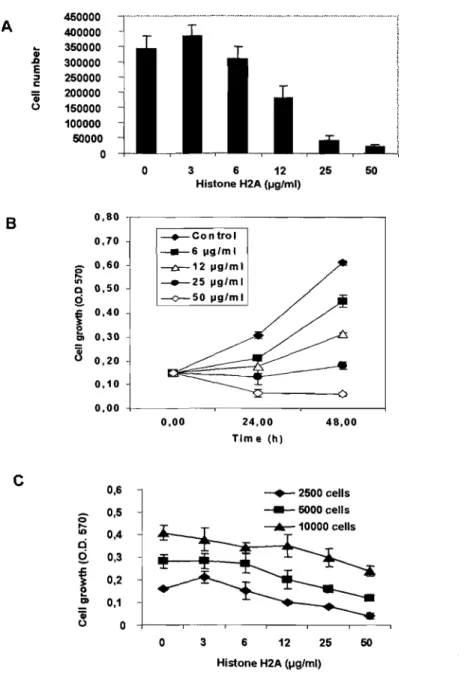
3.5.1. Exogenous histone H2A inhibits ce Il proliferation ... 49
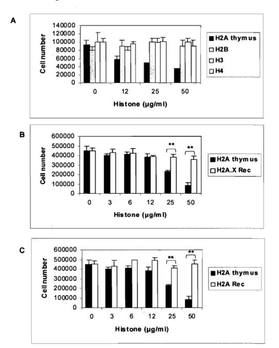
3.5.2. Histone-induced cell growth arrest is specifie to histone H2A ... 53
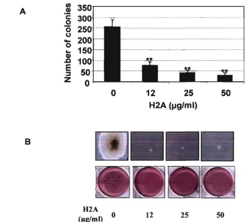
3.5.3. Histone H2A inhibits anchorage-independence ofMCF-7 cells ... 55
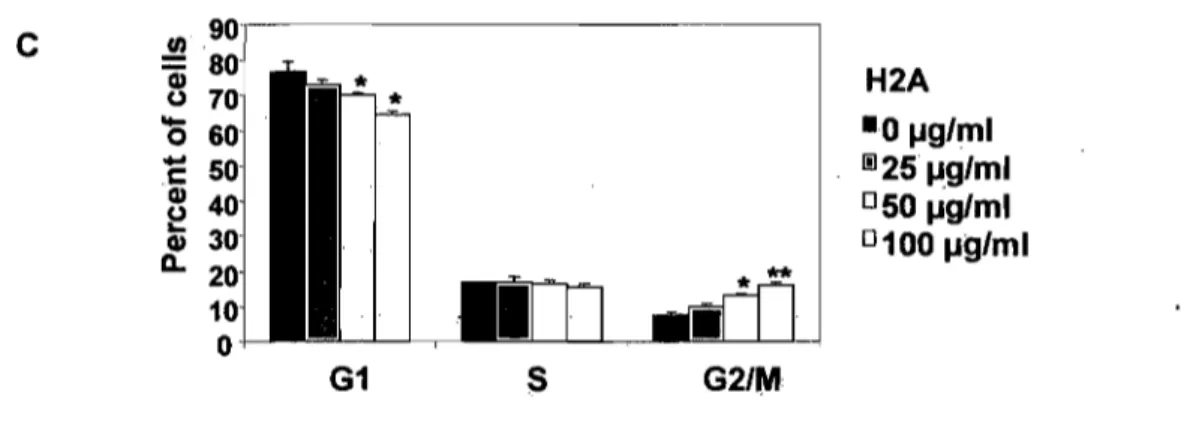
3.5.4. Histone H2A blocks the cell cycle ... 57
3.5.6. p2I and p53 expression in H2A-treated MCF-7 ... ... 60
3.6. DISCUSSION ... 64
3.7. ACKNOWLEDGEMENTS ... 70
3.8. REFERENCE LIST ... 70
4. METHODES EXPÉRIMENTALES~ ...•...•...•...•..••...•..•••...•...•.. 79
4.1. CULTURE CELLULAIRE ET HISTONES ... 79
4.2. INCORPORATION DE LA THyMIDINE ... 79
4.3. ACCUMULATION DES LIPIDES ... 80
4.4. DOSAGE DE L'ACTIVITÉ DE L' ACÉTYLCHOLINESTÉRASE (TEST ELLMAN) ... 80
4.5. WESTERN BLOT ... 82
4.6. RT-PCR ... 83
4.7. TEST D' APOPTOSE PAR MARQU AGE AVEC ANNEXIN V -FITCIPI ... 84
4.8. EXTRACTION ACIDE DES HISTONES ... 85
4.9. COLORATION DES GELS SDS-PAGE AU BLEU DE COOMASSIE ... 85
4.10. ÉTUDES IN VIVO ... 86
4.11. TEST POUR LA DÉTECTION ET LE TRAITEMENT DES MYCOPLASMES ... 89
5. RÉSULTATS ... 90
5.1. HISTONE R1.2 RECOMBINANTE PROVOQUE UNE DIMINUTION DU NOMBRE DE CELLULES MÉTABOLIQUEMENT ACTIVES ... : ... 90
5.2. L'HISTONE H1.2 RECOMBINANTE N'INDUIT PAS DE CHANGEMENTS MORPHOLOGIQUES DANS LES CELLULES MCF -7 ... 91
5.3. HISTONE Rl.2 RECOMBINANTE INDUIT L'APOPTOSE DANS LES CELLULES HELA ... 92
5.4. L'IMPACT DE LA PRÉSENCE DES MYCOPLASMES DANS LES CELLULES SUR LES EFFETS DE L'HISTONE H2A ... 93
5.5. HISTONE H2A INDUIT DES CHANGEMENTS MORPHOLOGIQUES ET INHIBE LA PROLIFÉRATION DES CELLULES PRIMAIRES D'ADÉNOCARCINOME MAMMAIRE HUMAIN ... 94
5.6. HISTONE IDA INHIBE LA PROLIFÉRATION CELLULAIRE (MÉTHODE D'INCORPORATION DE
THYMIDINE TRITIÉE) ... 95
5.7. HISTONE H2A INDUIT DES CHANGEMENTS MORPHOLOGIQUES DANS LES CELLULES MDA-MB-231 ... 97
5.8. HISTONE IDA INDUIT L'EXPRESSION DE LA B -CASÉINE ... 98
5.9. HISTONE IDA INDUIT L'ACCUMULATION DES LIPIDES ... 99
5.10. HISTONE H2A AUGMENTE L'ACTIVITÉ DE L'ACÉTYLCHOLIESTÉRASE (AcHE) DANS LES CELLULES SH-SY5Y ... 101
5.11. EXPRESSION DE p21 ET p53 DANS LES CELLULES MDA-MB-231 APRÈs LE TRAITEMENT AVEC L'HISTONE IDA ... 102
5.12. EFFET DE L'HISTONE H2A SUR LE NIVEAU D' ACÉTYLATION DES HISTONES H3 ET H4 ... 103
5.13. HISTONE H2A DIMINUE LA TAILLE DES TUMEURS IN VIVO SANS AFFECTER LE POIDS DES SOURIS ... 105
5.14. HISTONE IDA INHIBE L'EXPRESSION DE LA PROTÉINE C-MYC ... 108
5.15. HISTONE H2A INHIBE L'EXPRESSION DE LA TÉLOMÉRASE ... 109
6. DISCUSSION ... 111
6.1. MISE EN CONTEXTE ... III 6.2. EFFETS ANTI-PROLIFÉRATIFS ... 113 6.3. MÉCANISME D' ACTION ... 115 6.3.1. Sénescence et différenciation ... 115 6.3.2. La voie de signalisation p53/p21 ... : ... ... 120 6.3.3. Mécanisme de compétition ... ... 123 7. CONCLUSIONS ET PERSPECTIVES ...•... 127 8. REFERENCE LIST ... 129
9. ARTICLE #3 : SP ANAL YSIS MAY BE USED TO IDENTIFY CANCER STEM CELL POPULATIONS ... 140
9.1. CONTRIBUTION PERSONNELLE : ... 141
9.2. ABSTRACT ... 144
9.3. NORMAL AND CANCER STEM CELLS ... 145
9.4. ABC TRANSPORTERS ... 147
9.5. SIDE POPULATIONS (SP) ... 149
9.5.1. SP in ceillines ... 151
9.5.2. SP in normal tissues ... 154
9.5.3. SP in tumors ... ... 156
9.6. CONCLUSION AND FUTURE GOALS ... 160
9.7. ACKNOWLEDGEMENTS ... 164
LlSTE DES FlGU"Q.ES
Figure 1 (introduction): HDAC competition model. ... 27
Figure 1. Effects of histone H2A on MCF-7 cell proliferation ... 51
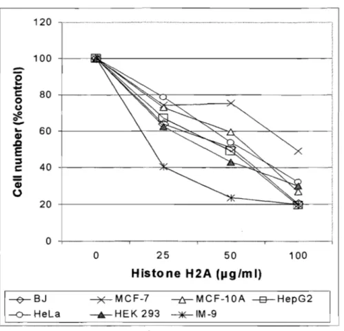
Figure 2. Histone H2A inhibits the growth of several cell types ... 52 Figure 3. (A) Effect of the core histones H2A, H2B, H3 and H4 extracted from calf thymus on MCF-7 cell proliferation ... 54
Figure 3. (B) The effect of calf thymus histone H2A was compared with human
recombinant histone H2A.X on MCF-7 cell proliferation ... 55 Figure 3. (C) Effect of calf thymus histone H2A was compared with human recombinant histone H2A on MCF-7 cell proliferation ... 55
Figure
4.
(A,B) Histone H2A reduces the anchorage-independent growth ofM CF -7 cells •••••••••.•••.•••.••••.••••••••.••••••••..•••••••••••..••...•••••••••••••••••••••••••...•••••••••••••.••• 56 Figure 4. (C) Histone H2A induces cell cycle arrest ... 57 Figure 5. (A,B) Histone H2A-treated MCF-7 cells exhibited enlarged and flattened cell morphology ... 59 Figure 5. (C,D) Histone H2A treatment resulted in the appearance of SA-(:S-galactosidase-positive cells ...••....•••...••....••.•..•..•••••••••••.•.••••••••••••.•.••••••••••. 59 Figure 6. lmmunoblot analysis of p21 and p53 proteins in MCF-7 cells exposed to histone H2A ...•...•...•.•.•.•... 61 Figure 7. Treatment wÎth histone H2A did not change p53 RNA expression ... 62 Figure 8. (A) Acetylated histones H2A are present in calf thymus histone H2A preparation ...•...•...•...•...•.•...•... 63
Figure 8. (B) Reactive oxygen species (ROS) generation is not involved in growth arrest effect of histone H2A ... 63 Figure 9: L'apparence macroscopique d'une tumeur formée chez les souris après l'injection des cellules cancéreuse MDA-MB-231. ... 88 Figure 10: Effet de l'histone H1.2 recombinante sur les cellules cancéreuses •.. 90 Figure 11: Changements morphologiques des cellules MCF-1 suite au traitement avec l'histone H1.2 recombinante et H2A ... 91 Figure 12: Induction de l'apoptose dans les cellules Hela après le traitement avec l' histone Hl.2 recombinante ... 93 Figure 13 : L'impact de la contamination par les mycoplasmes sur les effets de l' histone H2A ... 94 Figure 14: Effet de l'histone H2A sur les cellules primaÎres d;adénocarcinome mammaire humain ... 95 Figure 15 : Inhibition de la prolifération des cellules MCF-1 et MDA-MB-231 par l'histone H2A ... 96 Figure 16: Changements morphologiques des cellules MDA-MB-231 suite au traitemen t avec
l'
histoneH2A ... 98
Figure 17 : Effet de l'histone H2A sur l'expression de la p-caséine ... 99 Figure 18 : Accumulation des lipides dans les cellules MCF-7 et Hs578T suite au traitement avec l'histone H2A ... 100 Figure 19: Changements dans l'activité de l'AchE dans des cellules de neuroblastome suite au traitement avec l'histone H2A ... I01Figure 20 : L'expression de p21 et p53 dans les cellules MDA-MB-231 après le traitement avec l' histone H2A ... 102 Figure 21 : Acétylation des histones H3 et H4 après le traitement avec l'histone H2A ... 104 Figure 22 : Effet de l'histone H2A in vivo ... 106 Figure 23 : Métastases au niveau des poumons ... 107 Figure 24 : L'expression de c-myc après le traitement avec l'histone H2A ...•.• I08 Figure 25 : Inhibition de l'expression de la hTERT par l'histone H2A ... I09 Figure 1 (article #3): Importance of keeping a constant number of stem cells in normal physiology versus malignant transformation ... 163
,LISTE UES ABRÉVIATIONS 5-Aza-CR : 5-azacytidine
5-Aza-CdR: 5-aza-2-deoXycytidine
ABC transporters : ATP-binding
cassette transporters AchE : acétylcholinestérase
AL T : alternative lengthening of
telomeres
APL :
acute promyelocytic leukemia ATM : ataxia telangiectasia mutated ATR: ATM and Rad3-related ATRA: all-trans-retinoic acidBCRP : breast cancer resistance
protein
tIP
A : comite institutionnel deprotection des animaux du CHUM Chk2 : checkpoint kinase 2
CPP : cell-penetratlng proteins
CRCHUM: Centre hospitalier de
l'Université de Montréal CT A : cancerltestis antigenes CTCL : cutaneous T -celllymphoma DNA-PK: DNA-dependent protein kinase
DNMT : DNA methyltransferase
DNMTi : DNA methyltransferase
inhibitors
DTNB : dithiobisnitrobenzoate ER : estrogen receptor
FDA: food and drug administration HDAC : histone deacetylases
HDACi : histone deacetylases
inhibitors
hTERT: human telomerase reverse transcriptase MDRl: multidrug resistance transporter 1
MDS :
myelodysplastic syndrome MTT : 3-(4,5-dimethylthiazol-2-yl)-2,5-diphenyltetrazolium bromide NAC : N-acetyl-L-cysteine P-gp : p-glycoprotein PI : propidium iodide PI3-K : phosphatidyl-inositol-3-kinase PTD : protein transduction domain RAR : retinoic acid receptor ROS : reactive oxygen speciesSAHA: suberoxylanilide hydroxamic acid SA-~-gal: senescence-associated ~~ galactosidase Smc l' structural maintenance of chromosomes SP : side population
xv
TSA : trichostatin A X-gal: 5-bromo-4-chloro-3-indoyl ~ -galactosideRENIERCIEMENTS
Cette étude a été rendue possible grâce aux fonds des Instituts de recherche en santé du Canada (IRSC) et aux Fonds de la recherche en santé du Québec (FRSQ) du Dre. Danuta Balicki. Annamaria Hadnagy a reçu une bourse d'excellence offerte par le Département de Pathologie et Biologie cellulaire et détient actuellement une bourse d'études supérieures du Canada (BESC) offerte par le Conseil de recherche en sciences naturelles et en génie du Canada (CRSNG).
Je remercie la Dre. Danuta Balicki d'avoir accepté de diriger ma maîtrise et d'avoir subventionné cette recherche. Je remercie le Dr. Raymond Beaulieu pour le support scientifique offert, le Dr. Louis Gaboury d'avoir accepté de co-diriger ma maîtrise et le Dr. Edward Bradley d'avoir accepté de faire partie de mon comité de parrainage, de présider le jury de mon mémoire de maîtrise et de nous avoir fourni deux des lignées cellulaires que nous avons utilisées dans cette étude. Je remercie le Dr Mohammadi Kaouass pour son aide dans l'exécution des manipulations expérimentales. Les Drs Balicki et Beaulieu sont remerciés pour leurs contributions dans tous les articles de ce mémoire et le Dr Kaouass et M Mansour pour leurs contributions dans Particle 2. Je remercie egalement la Dre. Julie Douville pour son aide pour l'encadrement scientifique, l'exécution des manipulations et la rédaction de
ce mémoire de maîtrise. Mes remerciements vont également à M. Saad Mansour pour
les anaiyses de cytométrie en flux. Mme Rhyna Salinas et M Ovid Da Silva sont remerciés pour leur contribution aux articles de ce mémoire.
Le cancer est la première cause de décès au Québec, selon la Société canadienne du cancer (http://www.cancer.caJccs/intemet/niw_splash/O%2C%2C3172%2COO.html).La mise au point d'un traitement efficace contre le cancer qui ne comporterait que très
peu d'effets secondaires demeure un objectif important pour la communauté
scientifique.
L'épigénétique a pour objet l'étude des changements transmissibles de l'expression des gènes indépendants des changements de la séquence d'ADN (Goldberg et al. 2007;Yoo and Jones 2006).
Le cancer est une maladie associée à des altérations à la fois génétiques et epigénétiques. Les altérations genétiques, telles que les modifications dans la séquence des gènes, sont difficiles à renverser, en revanche les altérations épigénétiques, comme l'acétylation des histones et la méthylation de l'ADN, sont beaucoup plus accessibles à la réparation. C'est pourquoi des médicaments épigénétiques ont été développés afin de corriger les altérations épigénétiques qui caractérisent le développement et la progression du cancer.
Récemment, plusieurs études ont rapporté un rôle des histones dans la croissance des cellules cancéreuses. Nos propres résultats suggèrent que l'histone H2A exogène purifiée à partir de thymus de veau inhibe la prolifération des cellules cancéreuses.
La revue de la littérature de ce mémoire de maîtrise a été présentée sous forme d'un article de revue. Cet article expose les fonctions classiques des histones ainsi que de nouvelles fonctions qui commencent à être décrites. Cette revue présente
\
aussi un aperçu de la thérapie épigénétique. Enfin, un nouveau modèle pour la thérapie épigénétique y est décrit.
2. ARTICLE #1: HISTONE TAn, MODIFICATIONS AND
NON-CANONICAL FUNCTIONS OF HISTONES: PERSPECTIVES IN CANCER EPIGENETICS
Annamaria Hadnagy B.Sc.§t Raymond Beaulieu M.D., M.Sc.§t, and Danuta Balicki M.D., Ph.D§t
Research Centre and Department ofMedicine§, HÔtel-Dieu du Centre hospitalier de l'Université de Montréal (CHUM) and Department ofMedicinet and Biology and
Cellular Pathologyt, Université de Montréal, Montréal, Québec, Canada
ARTICLE PUBLIÉ DANS
2.1. CONTRIBUTION PERSONNELLE:
Histone tail modifications and non-canonical functions of histones: perspectives in cancer epigenetics
Annamaria Hadnagy §t, Raymond Beaulieu §t, and Danuta Balicki M.D.§t 1
Research Centre and Department of Medicine§, Hôtel-Dieu du Centre hospitalier de l'Université de Montréal (CHUM) and Department of Medicinet and Biology and Cellular Pathologyt, Université de Montréal, Montréal, Québec, Canada
Running Title: Histones and cancer therapy
Keywords: epigenetics, histones, HDAC inhibitors, methylation, acetylation
Abbreviation List: HDAC (histone deacetylases), HDACi (histone deacetylase inhibitors), DNMT (DNA methyltransferases), DNMTi (DNA methyltransferase inhibitors).
1 D.B. is a scholar of Fonds de la recherche en santé du Québec (FRSQ). A.H. is a
recipient of a Natural Sciences and Engineering Research Council of Canada (NSERC) graduate scholarship. This study was also made possible through a Canadian Institutes of Health Research (CIHR) operating grant to D.B..
Requests for reprints to: Danuta Balicki M.D., Ph.D. corresponding author Hôtel-Dieu du CHUM, Pavillon Masson
3850, rue Saint-Urbain, Montréal, Québec, H2W 1 T7 Phone: (514) 890-8000 #15518
FAX: (514) 412-7204 E-mail:
2.2. ABSTRACT:
Over the past few years histone deacetylase (HDAC) inhibitors have come to occupy an important place in the effort to develop novel, but less toxic anti-cancer therapy. HDAC inhibitors block HDAC which are the enzymes responsible for histone deacetylation and therefore, they modulate gene expression. The cellular effects of HDAC inhibitors include growth arrest and the induction of differentiation. Early successes in cancer therapeutics obtained using the se drugs alone or in combination with other anti-cancer drugs emphasize the important place of post-translational modifications of histones in cancer therapy. Histone tail modifications along with DNA methylation are the most studied epigenetic events related to cancer progression. Moreover, extranuclear functions of histones have also been described. Since HDAC inhibitors block HDACs and thereby increase histone acetylation, we propose a model wherein exogenous acetylated histones or other related acetylated proteins that are introduced into the nucleus become HDAC substrates, and thereby compete with endogenous histones for HDACs. This competition may lead to the increased acetylation of the endogenous histones, as in the case of HDAC inhibitor therapy. Moreover, other mechanisms of action, such as binding to chromatin and
2.3. HISTONES
Histones are the most abundant proteins bound to DNA in eukaryotic cells and amongst the most evolutionary conserved proteins known (1). They are small basic proteins with a molecular weight between Il and 20 kilodalton (Kda) and they contain a high percentage of positively charged amino acids (approximately 20%), such as lysine and arginine. Eukaryotic cells contain mainly five types of histones: histone linker Hl and core histones H2A, H2B, H3 and H4. AlI four core histones, i.e. histone H2A, H2B, H3 and H4, share a similar structure, with a central "fold domain" and terminal "tails", N-terminal and C-terminal (2). The fold domain contributes to octamer histone assembly and terminal tails are crucial for the normal functioning of cellular processes, including replication and transcription (3) as they
are targets for post-translational modifications: acetylation, methylation,
phosphorylation and ubiquitination (4). Linker histone Hl binds nucleosomes together and thus participates in a higher order compaction of chromatin (5).
Beside these canonical histones, several histone variants have been described such as histone H2A.X implicated in DNA repair and genomic stability (6) and histone H1.2, a pro-apoptotic protein that is translocated trom the nucleus to mitochondria (7) .
2.4. EPIGENETICS IN CANCER
The term "epigenetics" was introduced by Conrad Waddington (Waddington, 1942 cited by (8)). Epigenetics is the study of heritable changes in gene expression that are not related to changes in DNA sequence (4;8).
In eukaryotic ceIls, DNA is wrapped around histones and thus, is limited in its accessibility in biological processes such as replication, transcription, and DNA repair. Therefore, cellular mechanisms of chromatin modulation were identified in cells, including recruitment of nucleosome remodeling factors, such as SWI/SNF (9) and modulation of the contact between DNA and histones through post-translational modifications of histones (3).
Epigenetics changes include DNA methylation, histone modifications, chromatîn remodeling and expression of microRNAs (10). These processes may be deregulated in several diseases, including cancer, neurological and cardiovascular disease.
Cancer is characterized by a deregulation in normal cell proliferation caused by genetic or epigenetic alterations. Genetic alterations of various genes implicated in normal cell proliferation such as tumor suppression genes lead to abnormal protein expression encoded by these genes and thus to 10ss of normal functioning of these
proteins. Histone tail modifications, such as acetylation, methylation,
phosphorylation and ubiquitination along with DNA methylation are the most studied epigenetic events related to cancer progression (4). Epigenetic events modulate gene expression without modification of primary gene sequence. For example, hypermethylation of DN A promoter regions of genes that control normal cell proliferation, such as tumor suppression genes, is associated with gene silencing and
thus, with tumor progression (11). It was suggested that the DNA hypermethylome
can be associated with tumor aggressiveness and it may be used as a clinical marker in cancer cell characterization. Fiegl et al. (12) suggested that HER-2/neu positive
aggressive breast cancer cells are characterized by a specific DNA methylation profile. They identified 3 genes: PGR (co ding for the progesterone receptor), HSD 17B4 (coding for type 4 17 -beta-hydroxysteroid dehydrogenase, an enzyme involved in estrogen metabolism), and CDH13 (coding for H-cadherin) whose DNA methylation correlates with Her2/neu status and, subsequently they suggested that this methylation profile may explain the aggressiveness and reduced responsiveness to antiestrogen treatment.
Thus, epigenetic events play an important role in cancer development and progression and an understanding of these events has recent led to advances 10
epigenetic therapy. Moreover, epigenetic modifications may also be implicated 10
prognosis and drug response of the patients. Thus, Esteller et al. (13) have
demonstrated that methylation of the DNA-repair enzyme 06 -methyl-guanine-DNA
methyltransferase (MGMT) promoter, an enzyme involved in resistance to alkylating agents, can be used as a predictor of responsiveness to treatment with these drugs. Moreover, they suggested that MGMT methylation may be a better prognostic factor than those used classically, such as tumor grade and patient age.
2.4.1. HISTONE ACETYLATION
Histone acetylation and deacetylation play an important role in chromatin remodeling and, thus, in gene expression. There is a fine balance between acetylation and deacetylation of histones in normal cells, and the enzymes catalyzing these modifications are histone acetyltransferases (HAT) and histone deacetylases (HDAC) respectively (14). While histone acetylation is associated with an open chromatin and
enhanced transcription, histone deacetylation is associated with closed chromatin and transcriptional repression. For example, acetylation of N-terminal. core histones facilitate the recruitment of transcription factors, such as TFIIIA (15) and histone H3 and H4 acetylation is associated with an open (H3-H4)2 tetrameric particle allowing access oftranscriptional machinery to DNA (3).
Along with transcriptional modulation, histone acetylation plays an important role in other biological processes such as replication and DNA repair, as histone acetylation facilitates the movement of the replication machinery along the DNA strand (16) and creates a favorable environment for DNA repair (17).
2.4.2. DNA METHYLATION
Methylation of CpG islands (DNA regions characterized by a high incidence of the nucleotide pair C followed by G) is an epigenetic event characterized by the transfer of a methyl group to the C-S position of cytosine. This process is catalyzed by DNA methyltransferases (DNMT) (18).
Changes in DNA methylation can result in either DNA hypermethylation or DNA hypomethylation. Both modifications have been identified in cancer cells. Gene promoter hypermethylation and global gene hypomethylation play important roles in tumorigenesis (4). Gene promoter hypermethylation was associated with the inhibition of cancer-related genes such as tumor suppressor genes and DNA mÏsmatch repair genes (4; Il). DNA methylation is associated with chromatin compaction as the loss of DNA methylation alters the binding of the linker histone Hl (19). DNA hypomethylation, the first epigenetic event identified in cancer cells
(11), can lead to gene activation, such as oncogenes (11) and to genomic instability (4).
Following the completion of the human genome project which was designed to identify aIl human genes, a human epigenome project has been launched to identify DNA methylation sites in human genes in major tissues (10). This project acknowledges the role of epigenetic modifications in human diseases such as cancer. Epigenetic modifications play an integral role in cancer. For example, Espada et al. (18) have demonstrated that the loss of DNA methylation in cells lacking DNMT1 is associated with an increase of the acetylation and decrease of the methylation of histone H3. These results are not surprising since a specific interaction between DNMT1 and HDAC has also been reported (20). Moreover, DNMT1 itself is associated with deacetylase activity (20).
2.5. EPIGENETIC DRUGS
The implications of epigenetic events in gene expression and DNA repair, and therefore in the tumorigenesis process, make them a valuable target for cancer therapy. Tumorigenesis is associated with genetic and epigenetic alterations. While genetic alterations such as gene deletions cannot be reversed, certain epigenetic alterations cano Thus, the rationale for epigenetic therapy is reactivation of the expression of several genes silenced by epigenetic events during tumorigenesis. As we mentioned previously, abnormal histone acetylation and DNA methylation were identified in cancer cells. Thus, drugs that target these epigenetic alterations are being studied, and are termed epigenetic drugs. There are two classes of epigenetic
drugs that are currently being investigated: histone deacetylase inhibitors (HDACi) and DNA methyltransferase inhibitors (DNMTi).
2.5.1. HISTONE DEACETYLASE INHffiITORS (HDACI)
Alterations of histone acetylation are reported in cancers (21). As mentioned previously, two types of enzymes, HDAC and HAT, modulate histone acetylation. The deregulation of HDAC functions· have been associated with hematological cancers, e.g. HDACs are recruited by the acute promyelocytic leukernia fusion protein (pML-RARa.) (22). They have also been associated with solid tumors, e.g. the breast and ovarian cancer susceptibility gene BRCAl is associated with HDACs, as BRCAl interacts with components of the HDAC complex (23).
The mechanism of action of HDACi is not entirely understood. The rationale behind the development of HDACis was that these inhibitors lead to an increased acetylation of histones and thus, rnight reactivate genes, such as the cell cycle inhibitor p2l, that are silenced during carcinogenesis (24). However, recently other mechanisms of action of HDACi have been identified such as the generation of oxidative stress (25) and induction of premature sister chromatid separation that renders the rnitotic spindle assembly checkpoint ineffective (26).
Several HDACis are currently being investigated, including suberoylanilide hydroxamic acid (SARA) (27), suberoyl-3-aminopyridinearnide hydroxarnic acid (pyroxarnide) (28) and the benzarnide derivate MS-275 (29). SARA obtained U.S. Food and Drug Administration (FDA) approval for clinical use for the treatment of
cutaneous T-cell lymphoma (CTCLi and, currently, it is in a phase III clinical trial for the treatment of advanced mesothelioma3.
2.5.2. DNA METHYLTRANSFERASE INHmITORS (DNMTI)
Although the global low level of gene methylation is associated with cancer, hypermethylation was observed in the promoter region of several genes implicated in carcinogenesis, which correlates with gene silencing (4). DNMTi, nucleoside and non-nucleoside analogues, are epigenetic drugs that target DNA hypermethylation. While nucleoside analogues require DNA incorporation for DNMT inhibition, non-nucleoside analogues can block DNMT directly without DNA incorporation (4). Inhibition of enzymes responsible for DNA methylation during the pro cess of tumorigenesis results in reactivation of previously silenced cancer related genes, such as tumor suppression genes, DNA mismatch repair genes and cell cycle related genes (30).
Recently, it has been suggested that epigenetic modifications, such as methylation can regulate the expression of microRNAs (31). They are small RNA
molecules encoded in the genome and they control expression of several genes by translational repression. For example, Saito et al. (31), have demonstrated that DNMTi treatment increases the expression of microRNA-127, a member of the microRNA family. The target of this microRNA is a proto-oncogene and therefore, the authors suggested that up-regulation of microRNA, by epigenetic therapy may be a novel strategy in cancer treatment.
There are several DNMTi under preclinical and c1inical investigation, two of them, members of nucleoside analogues farnily, 5-azacytidine (5-Aza-CR) and 5-aza-2-deoxycytidine (5-Aza-CdR) obtained Food and Drug Adrninistation (FDA) approval for c1inical use in the treatment of myelodysplastic syndrome (MDS)l.
Zebularine (l-[P-D-ribofuranosil]-1,2-dihydropyrimidin-2-1) tS a 5-Aza-CR
derivative that has been recently described as a novel DNMTi which is more stable and less toxic compared with 5-Aza-CR and 5-Aza-CdR (32). As the se drugs are incorporated into DNA, theyare associated with cytotoxieity (33).
Nonnucleoside DNMTi are a1so under investigation. For example, MG98, a phosphorothioate antisense oligodeoxynucleotide that is a specifie inhibitor of rnRNA for human DNA methyltransferase 1 (DNMTl) is currently being tested in clinical trials (34). Fini et al. (35) have evaluated the anticancer effect of Annurca polyphenol extract (APE) in sporadic eolorectal cancers (CRC). They demonstrated that APE functions as DNMTi with comparable effects to those obtained with CdR, but with no side effects, including myelossupression as reported for
5-Aza-CdR
Although epigenetic drugs can be used as monotherapy, their effects can be optirnized by combination therapies, such as combinations of demethylating agents and HDACi (30;36). Mongan et al. (36) have demonstrated that the combination of valproic acid, a short-chain fatty acid structurally related to the butyrate c1ass of HDACi with 5-Aza-CdR, a DNMTi and with retinoic acid lead to reactivation of the tumor suppression gene RARp2 that is epigenetically silenced in breast cancer cells.
Epigenetic drugs can be used in combination with each other, but also with
chemotherapeutic agents. For example, it has been demonstrated in a phase l trial
that the administration of valproic acid, an HDACi, with epirubicin, an anthracyc1ine antitumor antibiotic, can improve the patients' response to epirubicin. Moreover, an antitumor response was obtained in patients with anthracycline-resistant tumors (37). A promising application of combination therapy is the reactivation of the estrogen receptor (ER) in breast cancer, followed by antiestrogen treatment (38). In general, ER+ tumors are characterized by a better prognosis and treatment outcome compared to ER- tumors. Loss of ER expression in breast cancer is caused by genetic events, such as DNA sequence mutation (30), but also by epigenetic events, such as hypermethylation of ER promoter (38). Sharma et al. (38) have demonstrated that treatment of the MDA-MB-231 ER- breast cancer cell Hne with a combination of trichostatin, an HDACi and 5-aza-CdR, a DNMTi can restore ER expression, and thus became sensitive to hormonal therapy, such as tamoxifen, an antiestrogen agent.
An interesting combination therapy includes epigenetic drugs and cancer
immunotherapy. Development of tumor-associated antigen-directed vaccines is one strategy involved in cancer immunotherapy but, tumor-associated antigens are characterized by a limited and heterogeneous expression in tumors. Sigalotti et al. (39) have demonstrated that the expression of cancer antigens is related to methylation status of their promoter. Therefore, they have shown that DNMTi treatment induces expression of cancer/testis antigenes (CTA) and thus, it may improve CTA-directed immunotherapy. Thus, it is hoped that combination therapy
including epigenetic drugs will overcorne sorne of the resistanee to therapy that we currently face.
Most conventional cancer therapies, such as chemotherapy and radiation therapy are highly toxic and non-specifie. Advances made in our understanding of the rnolecular basis of cancer facilitated the developrnent of novel drugs, directed more specifically to the cancer cell. This is the case of imatinib mesylate, an ABL kinase inhibitor effective against chronic rnyeloid leukemia and trastuzumab, a monoclonal antibody against HER-2 receptor, which is effective against breast cancer cells that overexpress HER-2 receptor. It has been suggested that cancer epigenetic therapy rnay aiso be specific to cancer cells. Thus, Ungerstedt et al. (40) have shown that HDACi induce cell death in transformed cells, while normal cells are relatively resistant. One possible explanation for this resistance is the increased expression of thioredoxin, a protein irnplicated in cell protection against oxidative stress. Thus, increased expression of thireodoxin in normal cells, but not in cancer cells, may protect cells against the cytotoxic effects ofHDACi.
The induction of DNA damage is the mechanism underlying cancer cell death following chemotherapy or radiation therapy, which will activate cellular DNA damage response and trigger apoptosis (7). Besides apoptosis, other strategies may be used to induce cancer cell growth arrest, including cellular differentiation and senescence. This review will focus on differentiation and senescence, as these two mechanisms were often reported to be associated with epigenetic drugs.
2.5.3. DIFFERENTIATION THERAPY
The absence of the cellular differentiation, or anaplasia, is a hallmark of malignant tumors and it is associated wÎth morphological and functÎonal changes. The morphological changes of anaplastic cells include alterations in the size and cellular morphology (pleomorphism) and a higher nucleus-cytoplasmic ratio as compared with normal cells. A highly transformed cell is undifferentiated, and loses the functional characteristics of the normal cell of origin. In contrast, differentiated cells preserve the functional properties of normal cells.
As it is weIl accepted that undifferentiated tumors are associated with a poor prognosis, and hence the induction of differentiation is a promising strategy in cancer therapy. The rationale behind differentiation therapy lies in the finding that undifferentiated cancer cells are characterized by an unlimited potential to proliferate and, thus, the induction of differentiation will halt their proliferative capacity. The underlying molecular mechanisms include the induction of specific gene expression, e.g. p21 a cell cycle inhibitor (29).
One of the most successful differentiation agents tested so far is all-trans-retinoic acid (ATRA) used in the treatment of acute promyelocytic leukemia (APL) (22). Under normal physiological conditions retinoic acid binds to the retinoic acid receptor (RARa) and releases the histone deacetylase complex leading to transcriptional activation and hematopoietic cell maturation. While this pathway is disrupted in APL, high levels of retinoic acid can restore this pathway and allow the maturation arrest (22). However, after promising initial remissions obtained in the patients treated with retinoic acid, many of these patients acquire retinoid resistance. Lin et al. (41) made the assumption that this resistance is the result of the constitutive
activation of histone deacetylases and subsequently, they demonstrate that the association of Thichostatin A (TSA), an HDACi with retinoic acid enhances the differentiating effect of retinoic acid and may overcome retinoic acid resistance.
Induction of differentiation by HDACi is not limited to hematopoietic tumors, but can also be used in the treatment of solid tumors. For example, Munster et al. (27) have shown that SARA induces growth arrest and differentiation in human breast cancer cells.
2.5.4. INDUCTION OF CELLULAR SENESCENCE
Cellular senescence is a state of irreversible growth arrest associated with morphological and functional changes. Senescent cells are characterized by the
presence of the senescence-associated ~-galactosidase marker (SA-~-gal). This
enzyme is a lysosomal hydrolase active at pH 4 in normal cells, but also active at pH 6 in senescent cells. The increase in SA-~-gal activity at pH 6 is a result of the increased lysosomal content in senescent cells (42). Hayf1ick et al. (43) made the initial suggestion that cells can divide in culture a finite number of times, and beyond that they will stop dividing. This phenomenon is called replicative senescence. Two signaIs can trigger senescence: telomeres shortening which is associated with replicative senescence and cellular stress exposure associated with stress or aberrant signaling-induced senescence (STASIS) (44).
One important step in the carcinogenesis process IS overcommg normal
cellular senescence and acquiring limitless replicative potential. Maintenance of telomere length is mandatory for a cell to acquire the immortal phenotype. Two
mechanisms are implicated in this process. The first one is the reactivation of telomerase, the enzyme responsible for the maintenance of telomere length in most cancer cells and the second is the alternative lengthening of telomeres (ALT) by intra-telomeric recombination mechanism in cancer cells that do not express the telomerase. Epigenetic mechanisms appear to be involved in both processes. While, telomerase reactivation is associated with histone H3 and H4 hyperacetylation, lack of telomerase expression in AL T cells is associated with histone H3 and H4 hypoacetylation (45).
It has been suggested that since cellular senescence is associated with growth
arrest, the induction of senescence can be applied to cancer therapeutics (46). It has
been demonstrated that chemotherapeutic drugs, such as etoposide (47) and differentiation agents, such as retinoic acid (48) can trigger cellular senescence. The
induction of cellular senescence has been also reported for epigenetic drugs, 1. e.
HOACi (49) and DNMTi (50).
2.6. "NON-CANONICAL" FUNCTIONS OF HISTONES: PERSPECTIVES IN CANCER THERAPEUTICS
While most of the focus on histones is classically involved in chromatin organization in the nucleus, the extracellular localization of histones has also been identified. For example, extracellular histones associated with DNA, i.e. nucleosomes, are found in the circulation under various pathological conditions, su ch as autoimmune disease (51). Patients with malignant diseases undergoing chemotherapy also have higher levels of circulating nucleosomes, which correlates with a higher rate of cellular death caused by chemotherapy (52).
Extracellular histones have been also located on the surface of the human T HPB-ALL cell line and phytohemaglutinin (PHA)-activated human peripheral blood lymphocytes (53). Rose et al. (54) have shown that ileal epithelial cells release histone Hl, while undergoing apoptosis, and that histone Hl possesses antimicrobial activity.
Histone participation in chromatin organization, gene expression and DNA reprur is weil documented. For example, histone H2B participates in post-replicational DNA repair (PRR) (55) and in the cellular response to double-stranded
DNA breaks (DSB) (56). It was also demonstrated that histone H2B is
phosphorylated in apoptotic cells and, thus, it may be used as a hallmark of apoptotic cells (57). The increased acetylation of histones H3 and H4 has been systematically reported after treatment with HDACi (24;28) and it was associated with transcriptional activation of several genes implicated in tumor growth suppression (24).
By "non-canonical" functions of histones we understand those functions that are not related to chromatin organization. It has been demonstrated that linker histone Hl, as weIl as core histones H2A, H2B, H3 and H4, can be used as transfection agents to de1iver DNA into various cells (reviewed in (58). In addition to their functions in DNA metabolism, histones Hl and H2A and histone variants HI.2 and H2A.X display additional activities that may have important repercussions in cancer therapy.
2.6.1. HISTONE Hl
Besides its participation In chromatin organization, linker histone Hl
possesses other functions. Widlak et al. (59) have demonstrated that the C-terminal tail of histone Hl (CTD) activates the apoptotic nuc1ease DNA fragmentation factor
DFF40/CAD via protein-protein interactions. It has also been demonstrated that
histone Hl suppresses tumor cell growth in vitro in the Burkitt's lymphoma Daudi cel1line and the IM-9 lymphoblastic leukemia cellline, as well as in vivo in a mouse xenograft model (60). Histone Hl inhibits cellular proliferation and induces apoptosis in the human breast adenocarcinoma cell lines MCF-7 and MDA-MB-231 (61). Class et al. (60) suggested that the mechanism underlying histone Hl cytotoxicity is the presence of histone Hl-binding proteins on the cell surface that will trigger cellular responses such as apoptosis. Moreover, the entire histone Hl molecule may be required to obtain the observed cellular response since different peptides derived from histone Hl have no inhibitory effect (60).
2.6.2. HISTONE H1.2
Histone H1.2 is a member of the histone Hl family (5). Pohlmeyer et al. (62) have shown that histone Hl derived from calf thymus possesses cytotoxic effects on leukemia cells. The authors identified histone H1.2 as a major component of the histone Hl preparation and thus, they conc1uded that histone HI.2 is responsible for the cytotoxic effects observed. Moreover, exogenous recombinant histone H1.2 triggers apoptosis in human HeLa cervical carcinoma cells4.
Recently, it has been demonstrated that histone H1.2 is involved in X-ray-induced apoptosis (7). Among aIl histones, histone H1.2 along with histone H1.l bind with the lowest affinity to chromatin and thus, it was suggested that histone Hl.2 may be a very sensitive DNA double break sensor (5). Indeed, Konishi et al. (7) have shown that after X-ray irradiation, histone Hl.2 translocates from the nucleus to the cytoplasm. The mechanism underlying this histone H1.2 translocation and the activation of apoptosis is not completely u~derstood. However, they suggest that histone Hl.2 triggers apoptosis by the release of cytochrome c from mitochondria following activation of Bak, a member of Bcl-2 family. Thus, histone Hl.2 becomes a pro-apoptotic protein once it reaches the cytoplasmic compartment and translocates to mitochondria.
Based on the se reports, Gine et al. (63) analyzed the cytosolic release of histone Hl. 2 in primary tumoral chronic lymphocytic leukemia cells after treatment with genotoxic and non-genotoxic agents. They have shown that resistance to treatment with genotoxic agents is associated with the lack of histone Hl.2 release. Therefore, the release of histone Hl.2 may indicate the treatment outcome.
Another interesting function of histone Hl.2 is its antimicrobial activity. Jacobsen et al. (64) have demonstrated in vitro and in vivo the efficacy of histone Hl.2 against bum wound infection pathogens. Moreover, because of its low hemolytic effect, it has been suggested that histone Hl.2 may be used as a systemic antimicrobial agent (64).
2.6.3. HISTONE H2A
Histone H2A contains a histone fold domain and a N and C-terminal tail, just like other core histones. In contrast to other core histones, histone H2A has the largest family of variants (65). It contains a cluster of DNA binding sites localized near the N-terminal tail (66) and it possesses the largest consensus C-terminal tail (67).
Exogenous histone H2A inhibits cellular proliferation in several cancer cell lines, including the human MCF-7 non-invasive adenocarcinoma cell li ne and the human MDA-MB-231 invasive adenocarcinoma cell line. Histone IDA blocks cell cycle progression and induces differentiation and cellular senescence in these cells. The mechanism of action underlying these effects is the increase of the cell cycle inhibitor p21 5,6.
2.6.4. HISTONE H2A.X
Histone IDA.X represents 2-25 % of the histone IDA family (68) and its C-terminal motif SQ(DIE)(IILN) distinguishes it from other IDA variants (68). The serine (S) in this motif is serine 139, and is the site of a y-phosphorylation. Recently, Celeste et al. (6) have demonstrated that the loss of one IDA.X allele compromises genomic integrity and increases cancer incidence in the absence of the tumor suppresor gene p53.
5 Kaouass M., Hadnagy A., Mansour S., Beaulieu R, Balicki D. " Post-translational modifications of
histone H2A are pivotaI in its inhibition of human breast cancer cell proliferation via senescence. Poster presentation at San Antonio Breast Cancer Symposium, 2006
Phosphorylation of histone H2AX is catalyzed by members of the phosphatidyl-inositol-3-kinase (PI3-K) family, including ATM (ataxia telangiectasia mutated), ATR (ATM and Rad3-related) and DNA-PK (DNA-dependent protein kinase) (69). However, ATM may be the major kinase that contributes to H2AX phosphorylation in response to DNA double-strand breaks (69). This phosphorylation occurs rapidly in response to DNA double strand breaks induced by ionizing radiation (IR) (68). IR is used in radiotherapy of cancer and it has been demonstrated that it induces histone H2AX phosphorylation (70). Taneja et al. (70) have demonstrated that the level of the phosphorylated forro of histone H2AX ('YH2AX) after IR exposure may predict tumor response to radiotherapy. Moreover, they suggested that histone 'YH2AX might represent a biological target in therapy of radioresistant tumors, since blocking histone H2AX phosphorylation increases IR-induce apoptosis in cancer cells.
Liu et al. (71) have demonstrated that imatinib mesylate treatment of gastrointestinal stromal tumor cells (GIST) indu ces an increase in histone H2AX level. Moreover, this up-regulation is critical for imatinib mesylate efficiency and thus, novel therapeutic strategies designed to increase histone H2AX levels, such as proteasome inhibition, might prevent imatinib resistance of GIST c~l1s.
2.7. CONCLUSION AND FUTURE GOALS
Epigenetic drugs represent a promising strategy In cancer therapy, as
monotherapy as weIl as combination therapies. The intense focus on HDACi was rewarded by the recent introduction of SAHA into clinical settings. While the
classical substrates of HDAC are acetylated histones, it has been suggested that not only histone proteins, but acetylated non-histone proteins may aiso be HDAC substrates (72).
2.7.1. EXOGENOUS PRO TEINS ENTER THE NUCLEUS
It has been demonstrated that exogenous proteins may be taken up by
Physarum cells, an eukaryotic organism implicated in cellular metabolism. This is the case of exogenous H2NH2B dimers. Thus, Thiriet et al. (73) have demonstrated that these exogenous di mers are assembled into nucleosomes. Moreover, it has been
shown that the amino~ternùnal taiis of H2NH2B dimers are not required for their
nuclear import, but that they are pivotaI for efficient chromatin assembly.
Balicki et al. have demonstrated that (66) exogenous histone H2A enters the cytoplasm and nucleus within 24 hours. Subsequently, Hariton-Gazal et al. (74) demonstrated that core histones translocate directly across mammalian cell membranes and that this translocation is temperature- and energy- independent and uninhibited by endocytosis inhibitors, such as colchicine, nocodazole, cytochalasin
D, brefeldin A, chlorochine and nystatin. Rosenbluh et al. (75) have demonstrated
that core histones penetrate lipid bilayers and Mycoplasma membranes. As several pro teins have the ability to penetrate cellular membranes, this property was related to the presence of a specific domain rich in arginine and lysine residues termed the protein transduction domain (PTD). Proteins containing this domain were termed
considered CPPs (75). Thus, aU these studies suggested that exogenous histone H2A penetrates the nuclear compartment of the cell.
2.7.2. HDAC COMPETITION MODEL
We propose a model (Figure 1) where once exogenous acetylated histone H2A enters the nuclear compartment, it becomes a substrate for HDAC and thereby competes with endogenous nuclear histones for HDAC. The consequence of this competition will be an increase in the acetylation status of endogenous histones, analogous to treatment with HDACi. Our unpublished data regarding the effects of exogenous histone H2A on cancer cells support this model, as we have observed similarities between HDACi and histone H2A effects. The inhibition of cellular proliferation, induction of cell cycle arrest, increase of p21 expression, initiation of cellular differentiation and senescence are the common effects of HDACi (24;28;49) and exogenous histone H2A that we have observed, and which support this modef. However, we do not exclude the possibility that exogenously introduced histones may bind directly to chromatin and modulate gene expression.
In conclusion, the acetylation status of histones plays an important role in cancer progression and treatment. Thereby, epigenetic drugs, such as HDACi, have been studied and they are being introduced into clinical cancer therapy, e.g. SAHA. We propose a model whereby the histone acetylation status can be modulated not on1y by HDAC inhibition, but also by competition for HDAC. As HDAC substrates may potentially be histone and non-histone proteins, the development of acetylated
substrates of HDACs capable of reaching the nuclear compartment holds promise as an alternative strategy in epigenetic therapy.
A B
Figure 1 (introduction): HDAC competition model.
Figure lA demonstrates normal homeostasis where deacetylation of endogenous histones is accompli shed by HDAC and inhibited by HDACi. Figure lB demonstrates that exogenous histone H2A translocates into the nucleus and disrupts the normal activities of HDAC and HDACi by serving as an additional substrate for HDAC. As a result of this competition for HDAC, less HDAC is available to deacetylate histones H3 and H4, leading to an accumulation of the acetylated forms of these histones, as is also described with HDACi. The increased acetylation of histones ID and H4 has been associated with the transcriptional activation of several genes involved in the suppression of tumor growth.
2.8. ACKNOWLEDGEMENTS
The expert assistance of Rhyna Salinas in the preparation of this manuscript lS
gratefully acknowledged.
2.9. REFERENCE LIST
1. Komberg RD, Lorch Y. Twenty-five years of the nucleosome,
fundamental particle ofthe eukaryote chromosome. Cell 1999;98:285-94.
2. Luger K, Mader AW, Richmond RK, Sargent DF, Richmond Tl
Crystal structure of the nucleosome core particle at 2.8 A resolution. Nature 1997;389:251-60.
3. Morales V, Richard-Foy H. Role of histone N-termÏnal tails and their
acetylation in nucleosome dynamics. Mol Cell Biol 2000;20:7230-7.
4. Yoo CB, Jones PA. Epigenetic therapy of cancer: past, present and
future. Nat Rev Drug Discov 2006;5:37-50.
5. Th'ng JP, Sung R, Ye M, Hendzel Ml Hl family histones in the
nucleus. Control of binding and localization by the C-termÏnal domain. J Biol Chem 2005;280:27809-14.
6. Celeste A, Difilippantonio S, Difilippantonio MJ et al. H2AX
haploinsufficiency modifies genomic stability and tumor susceptibility. Cell 2003;114:371-83.
7. Konishi A, Shimizu S, Hirota J et al. Involvement of histone H1.2 in apoptosis induced by DNA double-strand breaks. Ce1l2003;114:673-88.
8. Goldberg AD, Altis CD, Bernstein E. Epigenetics: a landscape takes
shape. CeIl2007;128:635-8.
9. Logie C, Tse C, Hansen JC, Peterson CL. The core histone N-terminal
domains are required for multiple rounds of catalytic chromatin remodeling by the SWIlSNF and RSC complexes. Biochemistry 1999;38:2514-22.
10. Esteller M. The necessity of a human eplgenome project.
Carcinogenesis 2006;27: 1121-5.
11. Feinberg AP, Tycko B. The history of cancer epigenetics. Nat Rev
Cancer 2004;4:143-53.
12. Fiegl H, Millinger S, Goebel G et al. Breast cancer DNA methylation profiles in cancer cells and tumor stroma: association with HER-2/neu status in primary breast cancer. Cancer Res 2006;66:29-33.
13. Esteller M, Garcia-Foncillas J, Andion E et al. Inactivation of the DNA-repair gene MGMT and the clinical response of gliomas to alkylating agents. N
Engl J Med 2000;343:1350-4.
14. Davie JR, Spencer VA. Control of histone modifications. J Cell
Biochem 1999;SuppI32-33:141-8.
15. Lee DY, Hayes JI, Pruss D, Wolffe AP. A positive role for histone acetylation in transcription factor access to nucleosomal DNA. Cell1993;72:73-84.
16. Alexiadis V, Halmer L, Gruss C. Influence of core histone acetylation
/
17. Masumoto H, Hawke D, Kobayashi R, Verreault A. A role for cell-cycle-regulated histone H3 lysine 56 acetylation in the DNA damage response. Nature 2005;436:294-8.
18. Espada J, Ballestar E, Fraga MF et al. Human DNA methyltransferase
1 is required for maintenance of the histone H3 modification pattern. J Biol Chem 2004;279:37175-84.
19. Gilbert N, Thomson l, Boyle S, Allan J, Ramsahoye B, Bickmore
W A. DNA methylation affects nuclear organization, histone modifications, and linker histone binding but not chromatin compaction. J Cell Biol 2007)77:401-11.
20. Fuks F, Burgers WA, Brehm A, Hughes-Davies L, Kouzarides T.
DNA methyltransferase Dnmtl associates with histone deacetylase activity. Nat Genet 2000;24:88-91.
21. Gilbert J, Gore SD, Herman JG, Carducci MA. The clinical
application of targeting cancer through histone acetylation and hypomethylation. Clin Cancer Res 2004;1 0:4589-96.
22. Grignani F, De MS, Nervi C et al. Fusion proteins of the retinoic acid receptor-alpha recruit histone deacetylase in promyelocytic leukaemia. Nature 1998;391 :815-8.
23. Yard en RI, Brody Le. BRCA1 interacts with components of the
histone deacetylase complex. Proc Natl Acad Sci USA 1999;96:4983-8.
24. Gui CY, Ngo L, Xu WS, Richon VM, Marks PA. Histone deacetylase
(HOAC) inhibitor activation of p21 W AF 1 involves changes in promoter-associated proteins, including HOACI. Proc Nad Acad Sci USA 2004;101:1241-6.
25. Louis M, Rosato RR, Brault L et al. The histone deacetylase inhibitor sodium butyrate induces breast cancer cell apoptosis through diverse cytotoxic actions inc1uding glutathione depletion and oxidative stress. Int J Oncol 2004;25: 1701-11.
26. Magnaghi-Jaulin L, Eot-Houllier G, Fulcrand G, Jaulin C. Histone
deacetylase inhibitors induce premature sister chromatid separation and override the mitotic spindle assembly checkpoint. Cancer Res 2007;67:6360-7.
27. Munster PN, Troso-Sandoval T, Rosen N, Rifkind R, Marks PA,
Richon VM. The histone deacetylase inhibitor suberoylanilide hydroxamic acid induces differentiation of human breast cancer cells. Cancer Res 2001;61 ; 8492-7.
28. Butler LM, Webb Y, Agus DB et al. Inhibition of transformed cell
growth and induction of cellular differentiation by pyroxamide, an inhibitor of histone deacetylase. Clin Cancer Res 2001;7:962-70.
29. Gojo 1, Jiemjit A, Trepel JB et al. Phase 1 and pharmacologic study of MS-275, a histone deacetylase inhibitor, in adults with refractory and relapsed acute leukemias. Blood 2007;109:2781-90.
30. Yan L, Yang X, Davidson NE. Role ofDNA methylation and histone
acetylation in steroid receptor expression in breast cancer. J Mammary Gland Biol Neoplasia 2001;6: 183-92.
31. Saito Y, Liang G, Egger G et al. Specific activation of microRNA-127
with downregulation of the proto-oncogene BCL6 by chromatin-modifYing drugs in human cancer cells. Cancer Ce1l2006;9:435-43.
32. Y 00 CB, Cheng JC, Jones PA. Zebularine: a new drug for epigenetic
therapy. Biochem Soc Trans 2004;32:910-2.
33. Lyko F, Brown R DNA methyltransferase inhibitors and the
development of epigenetic cancer therapies. J Nad Cancer Inst 2005;97: 1498-506.
34. Stewart DJ, Donehower RC, Eisenhauer EA et al. A phase 1
pharmacokinetic and pharmacodynamic study of the DNA methyltransferase 1
inhibitor MG98 administered twice weekly.
Ann
OncoI2003;14:766-74.35. Fini L, Selgrad M, Fogliano V et al. Annurca apple polyphenols have
potent demethylating activity and can reactivate silenced tumor suppressor genes in colorectal cancer cells. J Nutr 2007;137:2622-8.
36. Mongan NP, Gudas LI. Valproic acid, in combination with all-trans
retinoic acid and 5-aza-2'-deoxycytidine, restores expression of silenced RARbeta2 in breast cancer cells. Mol Cancer Ther 2005;4:477-86.
37. Munster P, Marcrnon D, Bicaku E et al. Phase 1 trial of histone
deacetylase inhibition by valproic acid followed by the topoisomerase Il inhibitor epirubicin in advanced solid tumors: a clinical and translational study. J Clin Onco} 2007;25: 1979-85.
38. Sharma D, Saxena NK, Davidson NE, Vertino PM. Restoration of
tamoxifen sensitivity in estrogen receptor-negative breast cancer cells: tamoxifen-bound reactivated ER recruits distinctive corepressor complexes. Cancer Res 2006;66:6370-8.
39. Sigalotti L, Fratta E, Coral S et al. Intratumor heterogeneity of
methy\ation-regulated and functionally reverted by 5-aza-2'-deoxycytidine. Cancer Res 2004;64:9167-71.
40. Ungerstedt JS, Sowa Y, Xu WS et al. Rote of thioredoxin in the
response of normal and transformed cells to histone deacetylase inhibitors. Proc Natl Acad Sci USA 2005;102:673-8.
41. Lin RJ, Nagy L, Inoue S, Shao W, Miller WH, Jr., Evans RM. Role of
the histone deacetylase complex in acute promyelocytic leukaernia. Nature 1998;391 :811-4.
42. Dirnri GP, Lee X, Basile G et al. A biomarker that identifies senescent
human cells in culture and in aging skin in vivo. Proc Nat! Acad Sei USA 1995;92:9363-7.
43. Hayflick L, Moorhead PS. The serial cuItivation of human diploid cell
strains. Exp Cell Res 1961;25:585-621.
44. Gire V. [Senescence: a telomeric limit to immortality or a cellular
response to physiologic stresses?]. Med Sci (paris) 2005 ;21: 491-7.
45. Atkinson SP, Hoare SF, Glasspool RM, Keith WN. Lack of
telomerase gene expression in alternative lengthening of telomere cells is associated with chromatin remodeling of the hTR and hTERT gene promoters. Cancer Res 2005;65:7585-90.
46. Dimri GP. What has senescence got to do with cancer? Cancer Cell