Université de Montréal
Role of EFNBs and EphB4 in T cell development and
functions
par Wei Jin
Département de Sciences Biomédicales Faculté de Médecine
Thèse présentée à la faculté des études supérieures en vue de l’obtention du grade de
Philosophiae Doctor (Ph.D.) en sciences biomédicales
August.2013
Résumé
Kinases Eph est la plus grande famille de tyrosines kinases récepteurs Éphrines (EFN) est un ligand de Ephs. Eph et EFN sont toutes les molécules de surface cellulaire. L’interaction entre Ephs et EFNs permet de transmettre des signaux dans les deux directions (c.-à-d. partir de Ephs à EFNs, et de EFNs à Ephs.)
Eph et EFNs sont largement impliqués dans divers processus développementaux, physiologiques et physiopathologiques. Notre groupe et d'autres groupes ont rapporté les rôles de Ephs / EFNs dans le système immunitaire. Pour approfondir la fonction de EphBs / EFNBs dans le développement des lymphocytes T et des réponses immunitaires, nous avons généré des souris EFNB1, EFNB2, et EphB4 knock-out conditionnel (KO) et des souris EFNB1 / 2 doubles KO.
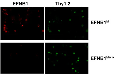
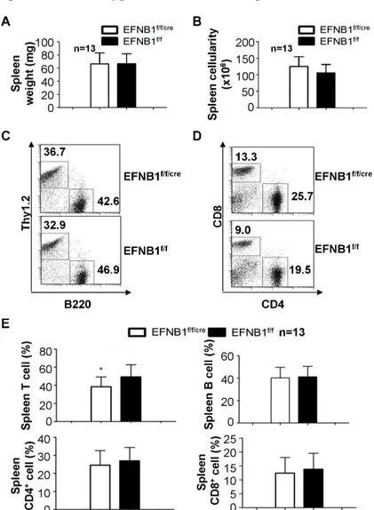
Dans les projets qui utilisent EFNB1 et EFNB2 comme souris knock-out, nous avons spécifiquement supprimé EFNB1 ou EFNB2 dans les cellules T. Les souris présentaient une taille normale, la cellularité du thymus et de la rate, ainsi que des sous-populations de cellules T étaient normales dans ces organes. Les progéniteurs de la moelle osseuse de souris KO et les souris WT ont repeuplé les organes lymphoïdes de l’hôte à des degrés similaires. L'activation et la prolifération des cellules KO T étaient comparables à celles des souris témoins. Les cellules CD4 naïves KO différenciées en Th1, Th2, Th17 et Treg étaient similaires aux cellules CD4 naïves de souris contrôle. Chez les souris KO EFNB2, nous avons observé une augmentation relative importante des thymocytes CD4CD8 : les double négatifs dans le thymus. L'analyse par cytométrie en flux a révélé qu'il y avait une augmentation modérée de la sous-population DN3 dans le thymus. Les résultats suggèrent qu’EFNB2 est impliqué dans le développement des thymocytes. Nos résultats indiquent que les fonctions de EFNB1 et EFNB2 dans le compartiment des cellules T pourraient être compensées entre eux ou par d'autres EFNB. La redondance des fonctions suggèrent le contrôle critique d’EFNB1 et EFNB2 dans le développement des cellules T.
Dans le projet, en utilisant EFNB1/B2 (modèle double KO) (dKO), nous avons observé une fonction de régulation de EFNB1 et EFNB2. dans la stabilisation de l’expression l'IL-7R α , à la surface des cellules T, IL-7 joue un rôle important dans le développement des thymocytes, l'homéostasie des lymphocytes T , et leur survie. IL-7R α subit une internalisation
ii
contraignante de IL-7. Chez les souris DKO, nous avons observé une perte d’expression de l’ IL-7Rα dans les thymocytes et les cellules T. En outre, l’ internalisation IL-7Rα a été accélérée dans les cellules CD4 dKO, suite à la stimulation IL-7. Dans la lignée cellulaire de lymphome T, EL4, la surexpression de EFNB1 ou EFNB2 retarde l'internalisation de l'IL-7Rα. Nous avons aussi démontré les signalisations compromises de l’ IL-7 et de la prolifération homéostatique des cellules T dKO. Les études du méchanisme qui utilisent la fluorescence de transfert d'énergie par résonance et immunoprécipitation ont montré que l'interaction physique de EFNB1 et EFNB2 avec IL-7R était probablement responsable du retard de l’ internalisation IL-7Rα.
Dans le dernier projet, nous avons étudié le développement des cellules T et la fonction des cellules épithéliales médullaires du thymus (mTEC), chez les souris knock-out EphB4. Les souris KO EphB4 ont démontré un poids et une cellularité qui sont normaux. La fonction et le développement de cellules T ne sont pas influencés par la suppression de l’ EphB4. Enfin, les souris KO ont développé une hypersensibilité de type retardée normale.
Dans l'ensemble, nos résultats suggèrent que l'interaction globale de croisement entre Eph et les membres de la famille EFN pourrir compenser la fonction d'un membre supprimé. Seule la suppression simultanée de plusieurs EFNBs va révéler leur vraie fonction dans le système immunitaire. En fait, une telle redondance montre les rôles vitaux d’Ephs et EFNS dans le système immunitaire.
Mots-clés : Eph; EFNB; KO, la délétion génique conditionnelle; le développementdes cellules T, la fonction des cellules T; IL-7Rα; cellules épithéliales Thymique médullaire (mTEC).
Abstract
Eph kinases are the largest family of cell surface receptor tyrosine kinases. The ligands of Ephs, ephrins (EFNs), are also cell surface molecules. Ephs interact with EFNs and the receptors and ligands transmit signals in both directions, i.e., from Ephs to EFNs and from EFNs to Ephs.
Ephs and EFNs are widely involved in various developmental, physiological pathophysiological processes. Our group and others have reported the roles of Ephs/EFNs in the immune system. To further investigate the function of EphBs/EFNBs in T cell development and responses, we generated EFNB1, EFNB2, EphB4 conditional gene knockout (KO) mice and EFNB1/2 double KO mice.
In the projects using EFNB1 and EFNB2 knockout mice, we specifically deleted EFNB1 or EFNB2 in T cells. The mice had normal size and cellularity of the thymus and spleen as well as normal T cell subpopulations in these organs. The bone marrow progenitors from KO mice and WT mice repopulated the host lymphoid organs to similar extents. The activation and proliferation of KO T cells was comparable to that of control mice. Naïve KO CD4 cells differentiated into Th1, Th2, Th17 and Treg cells similar to naïve control CD4 cells. In EFNB2 KO mice, we observed a significant relative increase of CD4CD8 double negative thymocytes in the thymus. Flowcytometry analysis revealed that there was a moderate increase in the DN3 subpopulation in the thymus. This suggests that EFNB2 is involved in thymocyte development. Our results indicate that the functions of EFNB1 and EFNB2 in the T cell compartment could be compensated by each other or by other members of the EFN family, and that such redundancy safeguards the pivotal roles of EFNB1 and EFNB2 in T cell development and function.
In the project using EFNB1/B2 double knockout (dKO) model, we revealed a novel regulatory function of EFNb1 and EFNb2 in stabilizing IL-7Rα expression on the T cell surface. IL-7 plays important roles in thymocyte development, T cell homeostasis and survival. IL-7Rα undergoes internalization upon IL-7 binding. In the dKO mice, we observed reduced IL-7Rα expression in thymocytes and T cells. Moreover, the IL-7Rα internalization was accelerated in dKO CD4 cells upon IL-7 stimulation. In T cell lymphoma cell line, EL4, over-expression of either EFNB1 or EFNB2 retarded the internalization of IL-7Rα. We further demonstrated
iv
compromised IL-7 signaling and homeostatic proliferation of dKO T cells. Mechanism study using fluorescence resonance energy transfer and immunoprecipitation demonstrated that physical interaction of EFNB1 and EFNB2 with IL-7Rα was likely responsible for the retarded IL-7Rα internalization.
In the last project, using medullary thymic epithelial cell (mTEC)-specific EphB4 knockout mice, we investigated T cell development and function after EphB4 deletion in mTEC. EphB4 KO mice demonstrated normal thymic weight and cellularity. T cell development and function were not influenced by the EphB4 deletion. Lastly, the KO mice developed normal delayed type hypersensitivity.
Overall, our results suggest that comprehensive cross interaction between Eph and EFN family members could compensate function of a given deleted member in the T cell development, and only simultaneous deletion of multiple EFNBs will reveal their true function in the immune system. In fact, such redundancy signifies vital roles of Ephs and EFNs in the immune system.
Keywords : Eph; EFNB; conditional gene knockout; T cell development; T cell function; IL-7Rα; Thymic epithelial cells
INDEX
Résumé ... i
Abstract ... iii
List of figures ... vi
List of abbreviations ... .x
Dedication ... .xiii
I. Introduction...1
I.1 Eph and Ephrin...2
I.1.1 Classification and structure of Eph and EFN...2
I.1.2 Signaling of Eph and EFN...3
I.1.3 Physiological role Eph/EFN signaling...7
I.1.3.1 Role of Eph/EFN in neuron development and injury repair...7
I.1.3.2 Role of Eph/EFN in bone development...9
I.1.3.3. Role of Eph/EFN signaling in angiogenesis...10
I.1.3.4 Eph and EFN in cancer...11
I.2 Roles of Eph/EFN in immune system...13
I.3 T cell development in the thymus...15
I.3.1 T cell development and lineage selection...15
I.3.2 Thymic epithelial cells guide the development of thymocytes...18
I.3.3 The effecter T cells...21
I.3.3.1 Th1 in the immune system...22
I.3.3.2 Th2 in the immune system...23
I.3.3.3 Th17 in immune responses...25
I.3.3.4 Regulatory T cells in the immune system...28
I.4 Working hypothesis of the project...30
II. ARTICLES...31
II.1 Article 1...32
II.1.1 BACKGROUND...34
II.1.2 MATERIALS AND METHODS...34
vi
II.1.4 DISCUSSION...39
II.1.5 CONCLUSIONS...40
II.1.6 ACKNOWLEDGEMENTS AND FUNDINGS...40
II.1.7 REFERENCES...41
II.1.8 FIGURES AND LEGENDS...42
II.2 Article 2...47
II.2.1 BACKGROUND...50
II.2.2 MATERIALS AND METHODS...51
II.2.3 RESULTS...54
II.2.4 DISCUSSION...57
II.2.5 CONCLUSIONS...58
II.2.6 AUTHORS' CONTRIBUTIONS...58
II.2.7 ACKNOWLEDGEMENTS AND FUNDINGS...58
II.2.8 REFERENCES...58
II.2.9 FIGURES AND LEGENDS...61
II.3 Article 3...70
II.3.1 INTRODUCTION...73
II.3.2 MATERIALS AND METHODS...74
II.3.3 RESULTS...77
II.3.4 DISCUSSION...80
II.3.5 ACKNOWLEDGEMENTS ...82
II.3.6 FIGURES AND LEGENDS...86
II.3.7 REFERENCES...97
II.4 Article 4...100
II.4.1 INTRODUCTION...102
II.4.2 MATERIALS AND METHODS...104
II.4.3 RESULTS...108
II.4.4 DISCUSSION...113
II.4.5 ACKNOWLEDGEMENTS...117
II.4.6 REFERENCES...117
II.4.8 SUPPLEMENTARY FIGURE LEGENDS...131
III. Discussion...135
III.1 Discussion...156
III.2 Summary and future directions...139
viii
Liste des figures et tableau
INTRODUCTIONFigure 1.1 Binding interactions and sequence relationships of Eph receptors and ephrins ... .3
Figure 1.2 Structure and bi-directional signaling of Ephs and EFNs...5
Figure 1.3 T cell migration during development in thymus...19
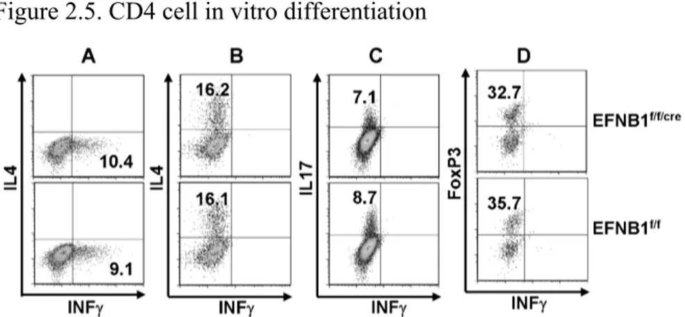
Table 1.1 The IL-17 family...26
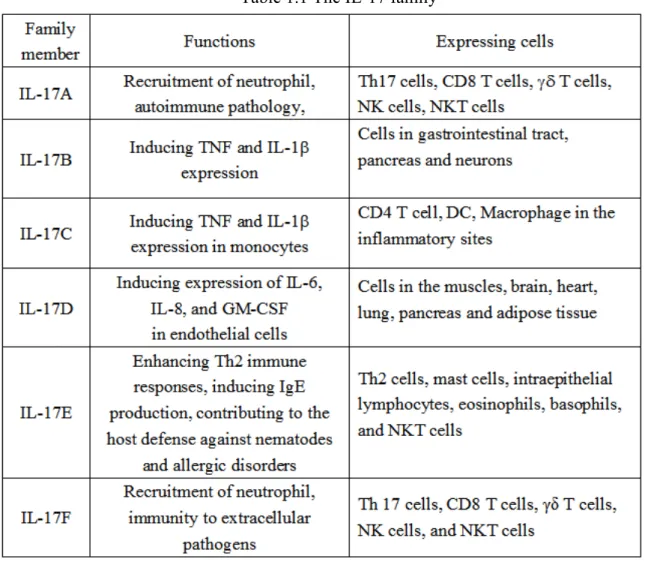
ARTICLE 1 Figure 2.1 T cell-specific deletion of EFNB1 in Lck-EFNB1f/f mice according to immunofluorescent microscopy...42
Figure 2.2 Phenotype of Lck-EFNB1f/f thymus...43
Figure 2.3 Phenotype of Lck-EFNB1f/f spleen...44
Figure 2.4 Normal activation and proliferation of Lck-EFNB1f/f T cells ...45
Figure 2.5 CD4 T cell in vitro differentiation...46
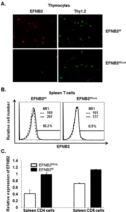
ARTICLE 2 Figure 2.6 T cell-specific deletion of EFNB2 according to immunofluorescent microscopy, flow thytometry and RT/qPCR ...61
Figure 2.7 Phenotype of EFNB2 KO thymuses ... ...63
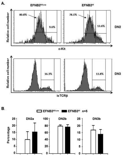
Figure 2.8 DN2 and DN3 subpopulations. ... 64
Figure 2.9 Phenotype of EFNB2 KO spleens ... 65
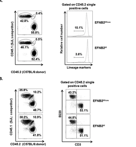
Figure 2.10 EFNB2 KO progenitors reconstitute the thymus and spleen in mixed chimeras. .. 66
Figure 2.11 Normal activation and proliferation of EFNB2 KO T cells...67
Figure 2.12 Normal differentiation of EFNB2 KO CD4 cell in vitro. ... 68
ARTICLE 3 Figure 2.13 mRNA expression of Ephb4 and related molecules in thymocytes and thymic stroma cells...86
Figure 2.14 TEC-specific deletion of Ephb4 in KO mice according to RT-qPCR...87
Figure 2.15 Phenotype of EphB4 KO thymi ... .89
Figure 2.17 Normal activation and proliferation of KO T cells ... 93 Figure 2.18 Naïve KO T cells differentiated normally into Th1, Th17 and Treg cells ... 95 Figure 2.19 KO mice mounted normal DTH responses...96
ARTICLE 4
Figure 2.20 Reduced IL-7Rα expression in dKO thymocytes and T cells ... 121 Figure 2.21 IL-7Rα internalization in EL-4 cells and spleen CD4 T cells ... 123 Figure 2.22 EFNb1 and EFNb2 expression modulates IL-7Ra expression on the cell surface124 Figure 2.23 7Rα co-localizes with EFNb1 and EFNb2 after 7Rα/EFNb1 or
IL-7Rα/EFNb2 cross-linking.. ... 126 Figure 2.24 EFNb1/EFNb2 associate with IL-7Rα.according to AB FRET. ... 127 Figure 2.25 Interaction between EFNb1/EFNb2 and IL-7Rα according to SE FRET and immunoprecipitation ... 129
Supplementary Figure 1 Stable EFNb1 and EFNb2 expression in EL4 and CD4 cells upon anti-EFNb Ab or IL-7 stimulation. ... 131 Supplementary Figure 2 Stable Myc-tagged EFNb1 and EFNb2 expression and transient
HA-tagged IL-7Rα expression in CHO cells according to flow cytometry. ... 132 Supplementary Figure 3 Lack of effect of EFNb1 and EFNb2 on the expression of TCR activation-induced IL-7Rα regulation and IL-6-induced IL-6Rα down-regulation...133 Supplementary Figure 4 EFNb1 and EFNb2 engagement does not affect IL-7Rα down-regulation. ... 134
DISCUSSION
x
List of abbreviations
Abi-1 Abl-interacting protein-1 AHR airway hyperreactivity AIRE autoimmune regulator APC antigen-presenting cell
APECED autoimmune polyendocrinopathy-candidiasis-ectodermaldystrophy BAL bronchoalveolar lavage
CAP Cbl-associated protein CIA collagen-induced arthritis
CMJ cortico–medullary junction CNS central nerve system
CRD cysteine-rich region
cTEC cortex thymic epithelial cells DC dendritic cell
DIX Dishevelled-Axin DN double negative DP double positive Dsh Dishevelled
DTH delayed type of hypersensitivity
EAE experimental autoimmune encephalomyelitis EC endothelial cell
Efn Ephrins
Eph Erythropoietin-producing hepatocyte kinase ephexin1 Eph-interacting exchange protein 1 ETP early thymic progenitor
FGFR fibroblast growth factor receptor FNIII fibronectin type III
FRET Fluorescence resonance energy transfer
Frz Frizzled
GEF guanine nucleotide exchange factor GPI glycosylphosphatidylinositol;
GRIP1 Glutamate receptor interacting protein HCV hepatitis C virus
HIF-1 hypoxia-inducible factor-1 HIV Human immunodeficiency virus HMG high mobility group
HSC hematopoietic stem cells IBD inflammatory bowel diseases IL interleukin
Ip Immunoprecipitation
IPEX Immunodysregulation polyendocrinopathy enteropathy X-linked syndrome iTreg induced regulatory T cells
JAK Janus Kinase JM juxtamembrane JNK Jun kinase K5 Keratin-5
LBD ligand binding globular domain MAP mitogen-activated protein;
MAPK mitogen-activated protein kinase MBD2 methyl-CpG-binding domain protein 2 MHC major histocompatibility complex MS multiple sclerosis
mTEC medullar thymic epithelial cells nTreg natrual regulatory T cell
NURD nucleosome remodelling and histone deacetylase PAK p2l-activated kinase
PDZ Psd-95, Dlg and ZO1 PH pleckstrin homology
xii
PTB phosphotyrosine-binding
PTH parathyroid hormone
PTHrP parathyroid hormone-related protein PTP-BL protein tyrosine phosphatase BAS-like Pyk2 proline-rich tyrosine kinase 2
RA Rheumatoid arthritis RBD receptor-binding domain
RORgt retinoid orphan nuclear receptor RTK receptor tyrosine kinase;
SAM sterile alpha motif;
SDF-1 stromal-cell-derived factor 1 SH2 Src homology 2
SOS son of sevenless SP single positive
STAT Signal transducer and activator of transcription
T-bet T-box expressed in T cell TM transmembrane
TNF tumour-necrosis factor
Hereby, I would like to express my sincere gratitude to my supervisors Dr. Jiangping Wu and Dr. Hongyu Luo for their instructions on my research and career development through all these years. Moreover, I would like to thank my family for their great support during my study. Their understanding and encouragement provided me great power in ups and downs. Finally, I would like to thank all my colleagues and friends for their kind cooperation and support
I.1 Eph and Ephrin
Erythropoietin-producing hepatocyte kinases (Ephs) are the largest family of receptor tyrosine kinases (RTK). Upon interaction with their ligands the Ephrins (EFN), downstream signaling cascades of both receptor and ligand are initiated which is referred as bidirectional signaling [1]. Eph and EFN play important roles in a wide range of biological processes from neuron axon guidance, angiogenesis, skeletal development, tissue patterning to immune responses [2, 3].
I.1.1 Classification and structure of Eph and EFN
The Eph family includes fourteen members categorized into A and B families with nine
EphAs (EphA1–8 and EphA10) and five EphBs (EphB1–4 and EphB6)[4]. The ligands of Ephs, EFNs, are also cell surface molecules with eight members[2]. EFNs can also be divided into A and B family depending on the way they anchor to the cell surface and their affinity to
Ephs. There are five EFNAs (EFNsA1–5), and 3 EFNBs (EFNB1-3)[5] .(fig.1.1)
The extracellular domain of Eph is composed of a ligand binding globular domain (LBD), a cysteine-rich region (CRD) and two fibronectin type III (FNIII) repeats, which are followed by a transmembrane (TM) helix[6]. The cytoplasmic domain of Eph is composed of four functional unites with the juxtamembrane (JM) domain that contains two conserved tyrosine (Y) residues, a classical protein tyrosine kinase domain, a sterile α-motif (SAM) and a Psd-95, Dlg and ZO1 domain (PDZ)-binding motif. The PDZ-binding motif is located in the very last 4-5 amino acid residues (XYXV) at the C-terminus followed by a hydrophobic residue in the end [2].
The structures of EFNs vary between A and B families[7]. The ectodomain of both EFNA and
EFNB contains a conserved extracellular receptor-binding domain (RBD)[8]. However, EFNBs
differ from the membrane-bond EFNAs with transmembrane helix and intracellular domains which contain several conserved tyrosine residues including a PDZ-binding motif. Tyrosine residues of the EFNB intracellular domains can also be phosphorylated upon engagement with Eph receptors. EFNAs, on the other hand, are anchored onto the plasma membrane via glycosylphosphatidylinositol (GPI) linkage [6, 9, 10].
3
fig1.1 Binding interactions and sequence relationships of Eph receptors and EFNs. Mesh and blank indicate high binding affinity within A and B family, respectively. Half mesh and half blank represent the weak cross reactivity to members of the other family.
I.1.2 Signaling of Eph and EFN
One distinctive feature of Ephs family is that their ligands, EFNs, are also cell surface proteins[2]. Interaction between Eph and EFN relies on direct contact between adjacent cells.
Both Eph and EFN are able to transduce downstream signaling to their host cells upon binding to each other. The signal transduced by Eph is called forward signaling, while the signal transduced by EFN is called reverse signaling[6].(fig.1.2)
Eph forward signaling involves EFN-induced clustering, auto-phosphorylation and association with adaptor proteins through intracellular domains [2, 11]. Before EFN binding, Ephs are
evenly distributed across the cell membrane and demonstrate minimal kinase activity. Upon binding with the LBD of Ephs, EFNs insert the extended loop into a deep channel of the Eph LBD, while lower affinity interfaces join two Eph-EFN dimers with two adjacent Ephs, assembling into hetero-tetramers and developing into higher-order clusters, which induce auto-phosphorylation of Eph intracellular domain [12]. The extent of EFN aggregation critically influences Eph signaling. EFNs need to cluster to form dimers to oligomers to stimulate tyrosine phosphorylation of Eph intracellular domains and initiate downstream responses [13]. The tyrosine kinase activity of Eph are regulated by the configuration of its JM domain [14]. Two conserved tyrosine residues in the JM domain have been identified as major auto-phosphorylation sites [15]. In a resting state, unphosphorylated JM forms a closed, auto-inhibited conformation. Once being phosphorylated on the JM tyrosine residues, the JM domain releases the kinase domain, allowing it to convert into an active form. Meanwhile, tyrosine phosphorylation creates docking sites for binding domains of adaptor proteins such as Src homology 2 (SH2) or phosphotyrosine-binding (PTB) domains [16, 17].
The activated Eph tyrosine kinase domain in turn activates intracellular effector proteins, which modulate cytoskeletal dynamics by regulating small Rho family GTPase activity such as Rac1, Cdc42 and RhoA[18]. The activity of Rho GTPases is regulated by guanine nucleotide exchange factors (GEFs) and GTPase-activating proteins (GAPs). The Rho GEF ephexin1 (interacting exchange protein 1) binds to Eph constitutively. Prior to EFN binding, Eph-bound dephosphorylated ephexin1 activates RhoA, Rac1 and Cdc42, balancing GTPase activity in the cell [19]. Upon activation of forward signaling, tyrosine phosphorylation of ephexin1 shifts the balance specifically towards RhoA activation.[19] Another Rho GEF Vav2 only binds to phosphorylated tyrosine residue of Ephs when it is activated. Phosphorylated Vav2 promotes local Rac1-dependent endocytosis of the EFN–Eph complex, thereby terminating both forward and reverse signaling [19].
5
Fig.1.2 Bi-directional signaling of Ephs and EFNs. Ephs and EFNBs are all transmembrane proteins, while EFNA bound to cell membrane via GPI anchor. When cells expressing Eph receptors contact EFN expressing cells, the Eph receptors transduce forward signals into the host cell through their tyrosine kinase activity. Meanwhile, EFNBs transduce reverse signals through auto-phosphorylation and recruitment of adaptor proteins.
RasGAP forms a complex with p-RhoGAP, which negatively regulates the small GTPase Rho.
The Ras-Rho complex binds to Eph phosphotyrosine residues by its SH2 domain [15] . In the
case of EphB2, activation of EphB2 induces tyrosine phosphorylation of p62dok, a pleckstrin homology (PH) domain-containing protein that recruits RasGAP and the SH2 adapter protein Nck [20]. Nck interacts with serine/threonine kinases of the p2l-activated kinase (PAK) family, and SOS (son of sevenless), an activator of the small GTPases Ras [21].Upon being recruited to the membrane by Nck, PAK is able to self-active and regulate Jun kinase (JNK)/p38 mitogen-activated protein kinase (MAPK) signaling [22, 23].
Although serving as ligands, transmembrane EFNBs are also able to transduce signal into host cells through their intracellular domains upon binding to Ephs [23]. The intracellular domains of
EFNB1/B2/B3 show high sequence conservation including an almost identical 33 amino acids at the c-terminus which contains three tyrosine residues enabling the recruitment of SH2/SH3
adaptor proteins, a PDZ binding domain, and a D-domain for interaction with Erk/MAPK [24].
The D-domain is located at leucine 293 (L293) in the JM of EFNB3. It has been reported that, Erk2 binds much stronger to D-domain of EFNB3 than Erk1, though both Erk1/2 are able to bind to EFNB3. In neurons, interaction between EFN and Erk has been linked to the regulation of synapse density and the formation of dendritic spines. EFNB3/Erk2 binding retains Erk in the dendrites, thus negatively regulating Erk signaling [25].
Several proteins have been identified that bind via PDZ domain directly such as Glutamate receptor interacting protein (GRIP1), protein tyrosine phosphatase BAS-like (PTP-BL) and PDZ-RGS3 [26-28].PDZ-RGS3 contains a PDZ domain and a RGS domain [28]. PDZ-RGS3 binds to EFNB c-terminal PDZ binding motif via its PDZ domain constitutively. Meanwhile, its RGS domains act as GAPs for Gα subunits of G proteins to promote the hydrolysis of GTP, thus negatively regulating Gαi, Gαq, and Gα12/13 coupled signaling pathways [29]. However, clustering of EFNBs after activation is pivotal to the regulatory function of PDZ-RGS3 [28]. For example, stromal-cell-derived factor 1 (SDF-1) has been shown to induce cerebellar granule cell migration via CXCR4, a G-proteincoupled chemokine receptor. Only
when binding to Ephs could EFNB1 induce inhibition of this chemotaxis via PDZ-RGS3 [28].
PTP-BL contains five PDZ domains and a PTP domain, which interacts with EFNB1 with its fourth PDZ domain [27]. It has been reported that PTP-BL negatively regulates EFNB1 reverse signaling by dephosphorylating tyrosine residues of EFNB1 intracellular domain, which is the target of Src kinase during activation [30, 31] .
Stimulation of EFNB1 leads to the formation of large sphingo-lipid/cholesterol-enriched raft patches [26]. During this process, GRIP1 are recruited into such rafts through binding to the PDZ-binding motif of EFNB1. It has been reported that GRIP1 acts as a multi-PDZ scaffold protein which docks EFN and a serine/threonine kinase. Therefore, the recruitment of signaling molecules by GRIP1 to EFNB complex links EFNB reverse signaling to kinase cascades, propagating signal transduction or remodeling cytoskeletal organization[26].
Upon binding and clustering with Eph receptors, EFNBs also recruit and activate Src family kinases which phosphorylate specific tyrosine residues of the EFNB intracellular domain [32].
7
Phosphorylated tyrosine residues provide docking site for SH2-containing adaptor proteins, such as Grb4. Through its SH3 domains, Grb4 associates with a set of partners which regulate cytoskeleton dynamics including the Cbl-associated protein (CAP), Ab1, the Abl-interacting protein-1 (Abi-1), and PAK1 [33] .
Dishevelled (Dsh) contains three conserved protein domains: DIX (Dishevelled-Axin), PDZ and DEP [34]. Dsh might binds to the intracellular domain of EFNB1 through interaction with Grb4. Dsh is known to be a downstream molecule of Frizzled (Frz). It has been reported that Xenopus Dsh mediates EFNB1 reverse signaling through the planar cell polarity pathway [35]. Moreover, Dsh has also been implicated in mediating RhoA and Rho kinase activation downstream of EFNB1. Although Dsh binds to EFN-B1 constitutively, such effect only occurs in response to EFNB signaling [34].
Although Eph/EFN system transduces bidirectional signals, Ephs and EFNs may also play independent roles in concert with other cell-surface receptors [3, 36-38]. For example, EFNBs could be phosphorylated on tyrosine in response to activation of growth factor receptor, another family of RTK. Activated fibroblast growth factor receptors (FGFR) inhibit EFNB1 activities by bounding directly to EFNB1 in cis and phosphorylating its tyrosine residues [38]. Accumulating evidences have revealed more crosstalk partners of Eph and EFNs, such as Ryk, adhesion molecules as integrins and cadherins, as well as ion channels like NMDA receptor [3]. Recent research from our lab has identified new crosstalk between EFNBs and cytokine receptors IL6R and IL7Rα. Our data demonstrated that EFNB1 and EFNB2 interact with IL7Rα, preventing its internalization upon IL7 treatment, and hence, maintaining IL7R signaling. Similarly, EFNB1/B2 are also critical for transducing IL6R downstream signaling
[39, 40].
I.1.3 Physiological role Eph/EFN signaling
I.1.3.1 Role of Eph/EFN in neuron development and injury repair
The Ephs and EFNs were initially identified and mostly studied in central nerve system as axon guidance molecules. Ephs and EFNs are highly expressed in the developing nervous system, where they play important roles in guiding axons and synaptic formation[41].
EphBs selectively promote formation of the spinal synapses and play a critical role in spine maturation, which may involve regulation of cytoskeleton through several GEF for Rho GTPases such as Kalirin, Intersectin, and Tiam1[42]. It has been reported that ectodomain of
EphB2 associates with NMDA neurotransmitter receptors promoting clustering at synapses upon EFNB ligation [43]. Moreover, EphB2 also triggers presynaptic differentiation by
regulating AMPA neurotransmitter receptor localization through its PDZ binding domain interactions [44]. Knockdown of EphB2 in neuron cultures results in decreased functional synaptic inputs, spines, and presynaptic specializations. On the other hand, expressing EphB2 in non-neuronal cells can drive the formation of presynaptic structures in co-cultured neurons, indicating the influence of activated axonal Eph signaling [44] .
EFNB expression is also identified on synapsis, especially post-synaptically [45].In cultured neurons, excitatory synapses induced by EFNB3 overexpression are found located on the dendritic shaft. Accordingly, in EFNB3 knockout mice reduced shaft synapses in hippocampal area CA1 have been identified.Moreover, activation of EFNB reverse signaling in cultured hippocampal neurons with EphB2-Fc promotes synapse formation and dendritic spine maturation. Such effect may require recruitment of GIT1 through Grb4 [46].
Ephs and EFNs are also involved in neuron injury reparation[47]. Upregulation of multiple Ephs and EFNs has been detected at sites of nervous system injury. However, their roles during the injury reparation vary. On one hand, EphBs promote the regeneration of injured axons. Interaction of EphB3 expressed in the infiltrated macrophages and EFNB3 on retinal axons promotes axon sprouting in the injured mouse optic nerve. In addition, meningeal fibroblasts that also invade in the injury site express EphB2. Interaction of EphB2 and EFNB2 expressed in astrocytes promotes the segregation of fibroblasts and astrocytes and formation of the glial scar and surrounding basal lamina [48]. On the other hand, EphA4 inhibits nerve regeneration. EphA4 accumulates in both damaged corticospinal axons and astrocytes in injured spinal cord. It has been shown that activating EphA4 forward signaling induces axon retraction and the formation of glial scar in astrocytes, which all inhibit axon regeneration [47,
9
I.1.3.2 Role of Eph/EFN in bone development
During bone formation, expression of Ephs and EFNs has been identified in chondrocytes, osteoclasts, osteoblasts and osteocytes [2]. EphA4 is expressed in mouse growth plate cartilage as well as in human chondrocytic cell lines [50]. In human articular cartilage cells, expressions of EFNB2 and EphB4 have been reported.In addition, EFNB1 and B2 are expressed on osteoclasts and may stimulate EphB on osteoblasts cells [51].
In humans, EFNB1 mutations are associated with the craniofrontonasal syndrome (CFNS) which manifests a series of developmental abnormalities such as cleft palate, hypertelorism, frontonasal dysplasia, agenesis of the corpus callosum, and hypoplasia of the maxilla [52, 53] . In mice, both forward signaling through EphB2/B3 and reverse signaling through EFNB1 are required for skeletal formation [54]. EFNB1 has been reported to be required for correct positioning of the palatal shelves during embryonic development. Deletion of EFNB1 in osteoblast leads to exencephaly due to reduced size of calvarial bones. Lacking of EFNB1 also leads to reduced size and bone mineral density of long bone. Mechanism study further shows that EFNB1 reverse signaling dephosphorylates TAZ within a protein complex, releasing TAZ from the complex to translocate into nucleus and to induce expression of osterix, osteoblastic differentiation and mineralization [55].
During bones remodeling in adults, EphB/EFNB bidirectional signaling coordinates behavior of osteoblasts and osteoclasts [56]. During osteoclastogenesis, cytokines produced by osteoblasts activate the transcription factors c-Fos and NFATc1 in osteoclast precursors, promoting differentiation of osteoclast and also EFNB2 expression. Meanwhile, Ephs expressed by osteoblasts stimulate EFNB2 reverse signaling in osteoclasts, which suppresses osteoclast differentiation through a negative feedback loop that represses Fos and therefore
Nfatc1 transcription. The PDZ binding motif has been reported indispensable for such
suppression effect, probably through its binding with Dvl2 [56, 57]. On the other hand, osteoblasts also receive stimulations from osteoclasts via EphB4 forward signaling which promotes the differentiation of osteoblasts and new bone formation at sites of resorption by osteoclasts. Forward signaling through EphB4 into osteoblasts enhances osteoblast differentiation via the inhibition of RhoA [57] . Moreover, expression of EFNB2 is also upregulated in osteoblasts upon parathyroid hormone (PTH) or parathyroid hormone-related
protein (PTHrP) signaling, which is required, in coordinate with EphB4, for mineralization by osteoblasts [58]. Therefore, cell-cell contact communication between osteoclasts and
osteoblasts mediated by EFNB1/B2 and EphB4 switches bone resorption to bone formation by limiting osteoclast differentiation and enhances osteoblast differentiation.
I.1.3.3 Role of Eph/EFN signaling in angiogenesis
Ephs and EFNs are also expressed in the vasculature, where they play a critical role during angiogenesis, particularly in regulating cell sorting and segregation [59, 60]. Expression of
EFNB2 and EPHB4 has been identified in angiogenic endothelial cells (EC) in arterial and venus vasculature, respectively, and mediates arterial-venous vessel segregation and vascular remodeling [59, 61]. The expression pattern of EFNB2 and EphB4 contributes to the control of cell migration with distinct arterial-venous fates. It has been shown that EFNB2 reverse signaling induces endothelial cells migration in response to VEGF or EphB4 and constitute the dorsal aorta [62, 63]. Moreover, EFNB2 phosphorylation occurs exclusively in angiogenic
vessels of retina, healing wounds, and tumor [64]. On the other hand, ECs expressing EPHB4
preferentially form the cardinal vein [65].
Although the exact role of Eph and EFN in angiogenesis remains largely unclear, several lines of evidences have implicated their importance. First, EFNB2 deletion in vascular smooth muscle cells leads to spreading defects, focal adhesion lost, and excessive depolarized motility
[66]. Second, deletion of EphB4 or EFNB2 are embryo lethal marked with defects in the
primary vascular plexus, which suggests their critical role in early vascular development [67, 68]. Consistently, deletion of EFNB2 in the endothelium and endocardium of the developing vasculature and heart demonstrated a similar phenotype [69]. Further study showed that EFNB2
reverse signaling plays a key role in this process [70]. Third, during angiogenesis, recruitment of pericytes is required for proper organization of endothelial cells [66]. EFNB2 has been shown to be essential in maintaining proper vascular architecture through ensuring spatial organization of the pericytes covering microvessels. Deletion of EFNB2 in pericytes in vivo leads to extensive hemorrhage in multiple organs including the skin, the lung, the intestine, and kidney, which indicates vascular malformation [66].Further study using human umbilical vein endothelial cells (HUVECs) in vitro established the role of EFNB reverse signaling during cell–cell contacts between pericytes and extracellular matrix in vascular development.
11
It has been shown that Src-dependent phosphorylation of cytoplasmic domain of EFNBs and downsrtream STAT3-Jak2 signaling is required for extracellular matrix–mediated assembly of endothelial cells and pericytes [64].
In addition to developmental and normal physiological angiogenesis, Eph and EFN are also involved in tumor angiogenesis [71]. In tumor cells and the vasculature of tumors, expression of
Eph and EFN can also be detected, which promotes angiogenesis [59, 64, 72] . EFNB2 reverse signaling in tumor endothelial cells, pericytes and smooth muscle cells has been shown to be important for blood vessel assembly, enlargement and decreased permeability both in cell culture and in vivo [63, 73].It has been shown that EFNB2 signaling promotes the interaction between endothelial cells and vascular smooth muscle cells.[64] Accordingly, EphB4 expressed by tumor cells enhances blood vessel growth through interactions with endothelial EFNB2 [74]. Consistent with these findings, in several mouse tumor models, EFNB2 deletion inhibits tumor growth and angiogenesis [75]. Moreover, similar as normal physiological angiogenesis, intense EFNB2 phosphorylation has been identified at the tumor margins where angiogenesis is most robust [64].
In addition to EFNB2 reverse signaling, EphA2 forward signaling also plays an important role in tumor angiogenesis. EFNA1, the ligand of EphA2, is present in tumor endothelial cells as well as tumor cells. Interaction between EFNA1 and EphA2 leads to activation of PI3k, Vav, and Rac1 leading to regulation of endothelial cell shape and migration [76].
I.1.3.4 Eph and EFN in cancer
Since Eph and EFN function importantly during normal physiological processes in organizing temporally specific cell behaviours, dysregulation of Eph and EFN expression would contribute to cancer progression[77-79]. Eph and EFN expression levels have been correlated with cancer progression, metastatic spread and patient survival. For example, EphA2 is preferentially expressed in malignant breast and prostate cancers and its expression has been linked to increased malignancy and a poor clinical prognosis[76]. In addition, EphB4 is also widely expressed in various cancer cells. Upregulation of EphB4 has been correlated with cancer progression[72]. However, in malignant cancer cell lines and tumor specimens, down-regulation of some Eph and EFN has also been identified. Further studies in colorectal cancer
showed that, after initial up-regulation, expression of Eph is repressed epigenetically or transcriptionally in more advanced stages. However, expression regulation of Eph and EFN is not uniform in cancers. Differential transcriptional regulation has been reported for EphB2,
EphB4 and EFNB during colorectal cancer progression. Similarly, inversed expression pattern
of EphA2 compared with other EFNA in breast cancer cell lines has also been reported [80, 81]. In colorectal cancer, up-regulation of EphB is attributed to constitutive activation of the Wnt/β-catenin/Tcf pathway[2]. The observed down-regulation of EphB later in advanced colorectal cancers has been linked to hypoxia in the surrounding tissue [81]. Hypoxia induces hypoxia-inducible factor-1 (HIF-1) which competes with Tcf-4 for binding to β-catenin, resulting in suppression of EphB expression. EphB has been reported to be capable of suppressing tumor growth in colorectal cancer. In ApcMin/+ mouse model, mutation or losing expression of EphB promotes adenocarcinoma progression due to lack of E-cadherin-dependent spatial restriction by surrounding EFNB expressing epithelial cells [82].
In breast cancer, EphA2 and EphB4 are extensively studied. Both Ephs are widely expressed in human breast cancer cell lines with low level of activation [72, 83]. Overexpression of EphA2 in a human mammary epithelial cell line induces oncogenic transformation without stimulation of EFNs[83].On the other hand, knockdown of EphA2 or EphB4 has been shown to inhibit the tumorgenicity of several cancer cell lines. Several lines of evidences have suggested that low versus high level of activation of Ephs leads to converse outcome[11]. In the absence of forward signaling, aberrantly expressed Eph may crosstalk with oncogenic signaling pathways, such as EGF receptor family members, to enhance tumor cell proliferation and motility[37]. One the other hand, activation of EphA2 or EphB4 readily suppresses the survival and tumor formation of human breast cancer cells in xenograft model possibly due to inhibition of downstream Ras-Erk, PI3K-Akt and Abl-Crk pathways [83, 84].
In melanoma, the role of Ephs is contradictory [85, 86]. EphA forward signaling has been shown to promote tumor cell proliferation and be associated with formation of blood vessel-like structures [87]. In contrast, though EphA2 is upregulated in both mouse and human skin carcinomas, deletion of EphA2 in tumor cells leads to their elevated growth and invasion[86]. Similar to EphB/EFNB interaction in breast cancers, interaction between EphA2-expressing tumor cells and EFNA1-expressing surrounding tissue restricts expansion of the
EphA2-13
positive tumor cells by inhibiting Ras-dependent pathways [86]. On the other hand, activation of EphB4 by membrane-bond EFNB2 or soluble EFNB2-Fc yields different outcomes. While co-expressed EFNB2 promotes amoeboid via RhoA activation in EphB4-expressing cells, EFNB2-Fc inhibits proliferation, survival, migration, and invasion of human MDA-MB-435 cell line both in vitro and in a mouse xenograft model. Such effect may possibly involve the Abl and Crk pathways[72] .Moreover, EFNB2 reverse signaling has also been associated with β1-integrin signaling and promotes cell adhesion and migration [78].
I.2 Roles of Eph/EFN in immune system
A few Ephs and EFNs are also expressed in lymphoid organs and lymphocytes, which implicates their possible roles in immune system [88].Given the cell surface expression of Ephs/ EFNs, it is reasonable to predict that they may function in immunobiology where cell-cell direct contact is critical, such as thymocyte development in thymus, and T migration and differentiation in lymphoid organs, where T cells encounter antigen-presenting cells (APC). It has been reported that interfering Eph-EFN interactions with EphB2-Fc or EFNB1-Fc leads to hampered thymocyte development and elevated apoptosis in thymic organ culture [89]. Moreover, multiple researches on mice deficient in several EphA and EphB family members all reported dramatic decrease of thymocytes and peripheral T cells due to disorganized thymic architecture [90] . Previous work in our lab also demonstrated that, deletion of EFNB1/B2 in T cell compartment leads to not only diminished T cell populations but also compromised T cell functions[40] . All the evidences suggest that Eph/EFN interaction is critical for thymic structural organization and T cells development.
In addition to their roles in T cell development, Eph/EFN have also been shown to modulate T cell receptor (TCR)-mediated responses[88] . Several reports have shown that solid phase EFNB1/B2/B3 are capable of promoting TCR response via activation of EphBs. Activated EphB forms a clustering cap on T cells together with TCR in aggregated lipid rafts, and, therefore, lowers the activation threshold of T cells upon TCR ligation. Moreover, EphB ligation also promotes T cell proliferation, cytokine production, and cytotoxic T cell activity [91-93] .
EphB6 is one of the most extensively investigated members of EphB in the immune system [94-96]. It is highly expressed in human mature T cells. In mouse lymphocytes, EphB6 is mainly expressed in double positive (DP), a fraction of mature CD4+ T cells, and CD8+ T cells. Interestingly, expression of EphB6 is under a dynamic balance through rapid synthesis and shedding [96, 97] . The intracellular domain of EphB6 lacks kinase activity. However, stimulation of T cells with anti-EphB6 antibodies or EFNBs still leads to increased tyrosine phosphorylation and downstream signaling, which may be due to association of EphB6 with EphB1 and EphB4 co-expressed on the same cell [88-90] .It has been suggested that EphB6 is critical for enhancing TCR signaling. Our lab have previously reported that, T cells from EphB6-/- mice show impaired TCR signaling, proliferation, and cytokine secretion in vitro. Moreover, EphB6-/- mice show compromised immune responses although their T cell numbers are normal. Further study showed that, EphB6 ligation enhances suboptimal TCR signal and lead to drastic T cell proliferation, accompanied by enhanced production of several cytokines, such as interferon (IFN)-γ, interleukin (IL)-6, IL-10, TGF-β, tumour-necrosis factor (TNF)-α, and GM-CSF, but not IL-2 and IL-4 [95] .Such effect involves up-regulation of the p38 and p42/44 MAP kinases [91-93] .
On the other hand, excessive EphB signaling by EFNB1 and EFNB2 co-stimulation demonstrates inhibitory effect on TCR-mediated responses in T cells, which is most likely mediated by EphB4. It has been shown that EphB4 forward signaling inhibits T cell proliferation by targeting Lck [98, 99] . High concentration of EFNBs induces EphB4 phosphorylation which, in turn, recruits SHP1. SHP1 dephosphorylates protein tyrosine kinase Lck at Tyr-394, and hence, negatively regulates T-cell signaling [100, 101] .
Moreover, research in our lab has reported that, EFNBs can cluster with several cytokine receptors and this is critical for downstream signaling. We have shown that EFNB1 and EFNB2 co-cap with IL-6R and IL-7R ensuring their signaling upon cytokine engagement [39, 40] .
In addition to EphBs and EFNBs, EphAs and EFNAs expression are also detectable in thymocytes and T cells [88, 102] .Similar to their cousins, EphAs and EFNAs are involved in TCR signaling modulation. EFNA1-Fc inhibits IL-2 secretion and induces apoptosis in DP cells under strong TCR stimulation, which suggests their role in negative selection. In CD4+
15
cells, ligation of EFNA1 with antibodies has been reported to suppress TCR-mediated responses, suggesting that EFNA1 reverse signaling may play a role in this process. Furthermore, EphA/EFNA signaling regulates migration of thymocytes in the thymus and T cell trafficking in responses to chemokine stimulations, such as SDF1-α and integrin-dependent adhesion. Mechanistic study has revealed that upon EFNA1 ligation, phosphorylation of Lck in association with proline-rich tyrosine kinase 2 (Pyk2) and Vav1 is critically required. Moreover, involvement of the PI3K pathway and Rho GTPase downstream signaling are also implicated after EFNA1 ligation [88, 103, 104] .
I.3 T cell development in the thymus
T cells and other lymphocytes arise from the same origin, the hematopoietic stem cells (HSC) in the bone marrow. The committed precursors enter circulation and arrive in the thymus initiating the T cell development program [105, 106] . The thymus is the major organ for T cell development and maturation which guarantees the production of proper reactive T cells [107-109] .
I.3.1 T cell development and lineage selection
Development progress of T cells can be divided into several stages according the expression pattern of CD4 and CD8 co-receptors, which can be defined as CD4-CD8- double negative (DN), CD4+CD8+ double positive (DP) and CD4+/CD8+ single positive (SP). The DN stage can be further segmented by expression of CD25 and CD44: DN1, CD44+CD25-; DN2, CD44+CD25+; DN3, CD44-CD25+; and DN4, CD44-CD25- [110] . The whole procedure consists several important events, including the rearrangement and expression of TCR genes, population expansion, positive and negative selection, and acquisition of functional capabilities [111, 112] .Both αβ and γδ Τ cells are derived from the common precursors referred as early thymic progenitors (ETP). The ETP migrate to thymus cortex from bone marrow, and gradually commits to the T cells linage during DN stage [113] .
Lineage commitment is the first essential event during T cell development. It has been established that the lineage commitment requires the sustained repression of the expression of genes characteristic of the alternative lineage. In early stages of ETP until DN2, signalling of
Notch 1, upon interaction with its ligand Delta-like 4 (DL4) expressed on the thymic stroma, is required to guarantee the T lineage commitment [114-116] . Sustained Notch1 signalling promotes T lineage specific gene expression and cell survival. However, Notch1 alone is not sufficient for T lineage commitment. Other transcription factors, including Runx1, GATA-3 and E-box proteins, are required to cooperate with Notch1 to initiate T cell differentiation [117] .
Further development of T lineage committed DN thymocytes requires expression of RAG-1 and RAG-2 recombinase to initiate the rearrangements of three TCR gene loci–Tcrb, Tcrg and
Tcrd. Meanwhile, TCR-α is encoded by a non-rearranging locus. Among them, Tcrb starts
rearrangement first, which involve the deletion of intron and joining of segments of VβDβJβ genes as well as Cβ gene. The further transcription and translation procedures yield a rearranged TCRβ [118] . The successfully rearranged TCRβ pairs with invariant pre-Tα chain (pre-TCR), resulting in enhanced proliferation, followed by entry of the double positive (DP) stage. With the rearrangement of TCRβ, Tcrg and Tcrd gene expression are excluded. T cells bearing rearranged TCRb gene next go through the verification checkpoint known as β-selection, before entering DP stage. Survival in the β-selection requires the signalling through a TCR complex with a properly rearranged TCRβ chain, CD3γ, δ, ε, ζ chains and the pre-Tα. The pre-TCR complex is capable of transmitting signals through its intracellular intermediates without the requirement for extracellular ligands [117] .
On the other hand, successful rearrange the Tcrg and Tcrd loci leads to expression of the γδ−TCR and exclusion of Tcra and Tcrb. Similar to TCR αβ thymocytes, thymocytes adopted γδ direction also go through the check point for TCR rearrangement verification at DN3. However, unlike the αβ cells, there is no pre-TCRγδ. The DN3 checkpoint verifies signalling by mature TCRγδ complexes.Moreover, γδ lineage differentiation is independent on Notch signalling [119] .
It has been suggested that distinct strength of signals transmitted by pre-TCR or TCRγδ complexes could guide the αβ /γδ decision at DN3 stage. Strong signals transmitted by γδ TCR promote γδ lineage choice, whereas the weaker signal from pre-TCR guides the cells to αβ direction [120] .This notion is supported by the evidence that introducing signalling-defective CD3ζ chain into DN3 cells which disrupt TCRγδ signalling leaded to the generation
17
of TCRγδ- DP thymocytes [121] . Another study using a transgenic TCRγδ of defined antigen specificity have shown that thymocytes expressing a transgenic TCRγδ receptor are directed to γδ lineage in mice expressing the corresponding ligand, but they adopt an αβ fate when the ligand is absent, suggesting that the interaction between TCRγδ and its ligand is necessary to ensure the signal strength and, hence, direct the cells to γδ lineage [122] .
In addition to TCR signalling, environmental signals have also been proposed to play a role in directing γδ/αβ lineage decision before the DN3 checkpoint by affecting TCR gene rearrangement. The strong IL-7 signal is required for Tcrg but not for Tcrb gene rearrangement. DN2 cells can be subdivided based on the expression level of 7Rα, and IL-7Rαhi cells gave rise to higher proportion of γδ T cells than IL-7Rlow/-cells [123] . Whether this phenomenon is caused by pre-commitment or high frequency of Tcrd rearrangements in IL-7Rαhi cells remains to be elucidated.
At DN3, several transcriptional targets of TCR or pre-TCR signals play crucial role in facilitating cells to overcome this check point [124] . Further development of DN3 cells are halted by E-box binding proteins E2A and HEB [125] . It has been proposed that cells overcome this block by two complementary mechanisms. One is to reduce the expression E2A or HEB coding gene. The other is to increased expression of Id-family molecules, which inhibit the expression of E-protein [126] . Id3 is a member if the Id family, which is a transcriptional target of both pre-TCR and TCRγδ signals. It has been proposed that different signal strength mediated by pre-TCR or TCRγδ decides the extent of Id3 up regulation, which distinguishes the lineage decision [127] . High Id3 expression alone in γδ cells is sufficient to down regulate E-box binding protein level, hence, facilitating overcoming the E-protein blockage. However, cells expressing Id3 at lower level in response to pre-TCR signalling require the cooperation of Notch1 signalling in down regulating E-protein expression and compete with E-protein on
Tcra regulation region [122, 127-129] .
Another important effector is the Egr transcription factor family members (Egr1 and Egr3). Egrs are triggered by both pre-TCR and TCRγδ signals. Over expression of Egr1 interfered with αβ lineage development both in culture and in vivo[122]. It has been suggested that high expression of Ergs promotes γδ T cell development, possibly by facilitation of Id3 up regulation [127] .
In addition, it has also been proposed that, HMG transcription factor Sox13 is important for the development of some γδ T cells by inhibiting the generation of αβ T cells, possibly by antagonizing the function of TCF1. TCF1 functions to favour αβ gene rearrangement while inhibiting γδ gene rearrangement [130] . Therefore, by antagonizing TCF1, Sox13 contributes to γδ lineage decision.
Thymocytes, which successfully passed the β-selection, initiate CD4 and CD8 expression, becoming CD4+CD8+ DP thymocytes. Meanwhile, Tcra gene is rearranged resulting in the final expression of TCRαβ complexes on cell surface. Three effectors contribute to the generation of DP cells from β-selected cells. First, Runx1 is necessary for the proliferative burst that follows β-selection[131] . Second, RORγt, encoded by the Rorc gene, promotes cell survival by up regulating expression of the anti-apoptotic protein Bcl-xL [132] . Lastly, the high mobility group (HMG) protein, TCF1, cooperates with its partner β-catenin to suppress γδ rearrangement and prompt generation of DP cells[120, 133] . The next and final stage of αβ DP cells maturation in thymus is positive and negative selection. DP cells that express TCRαβ which recognizes peptide-self-MHC complexes on the cortex thymus epithelium are prompt to survive from apoptosis. The selected cells are able to recognize foreign antigens loaded on the same self-MHC of APCs in periphery. These cells continue to go through negative selection. This step ensures the elimination of self-reactive cells, as well as generation of CD4+ or CD8+ cells matching their MHC specificity. Thymocytes restricted to MHC class II adopt CD4 fate while MHC class I–restricted thymocytes become cytotoxic CD8 cells. This procedure leads to clonal deletion of thymocytes with receptors of the highest avidity for self-antigens, which provides central tolerance [134, 135].
I.3.2 Thymic epithelial cells guide the development of thymocytes
The thymus provides a microenvironment for thymocyte development. During the whole developmental process, thymocytes are at close proximity of thymic stroma and migrate within the thymus since the arrival of lymphoid progenitor cells in the thymus. The structure of the thymus is highly compartmentalized and composed mainly of two kinds of epithelial cells which form cortex and medullar scalfolds separately [136] . (Fig1.3)
19
Fig 1.3 T cell migration during development in thymus. Bone marrow-derived early lymphoid progenitors (ELP) enter the thymus through circulation at the corticomedullary junction (CMJ) and migrate to inner cortex while differentiating into DN1 and DN2 cells.
DN2 cells further differentiate into DN3 cells in the mid and outer cortex. DN3 cells accumulate in the subcapsular zone (SCZ) undergoing proliferation and differentiate to DN4 cells. The developing thymocytes migrate backwards to inner cortex layer and medullar while further developing into DP stage. DP cells undergo positive selection and differentiation into SP cells during migration through the cortex. The surviving SP cells enter the medulla, where they undergo negative selection and final functional maturation. Finally, mature T cells leave the thymus and enter theperiphery.
Early progenitors arrive at the thymus at cortico–medullary junction (CMJ) from circulation. Seeding of the progenitors requires interaction of P-selectin expressed by thymic endothelium and its ligand PSGL-1 expressed on the progenitors. Mice lack PSGL-1 demonstrated decreased intrathymic progenitors and increased empty niches for prothymocytes [137] . After successful seeding into thymus, it is critical for the progenitors to efficiently relocate from the CMJ to the outer cortex to initiate thymocyte development. Several chemokines secreted by
the cortex and medullar epithelial cells have been reported to guide this movement, which involves CXCL12, CCL19, and CCL21 [138-140] .
CXCL12 has been reported to be critical in guiding the migration of developing thymocytes in the cortex [141-143] . Expression of CXCL12 receptor, CXCR4, has been identified in all the DN subpopulations as well as a part of the DP cells, which implicates the requirement of CXCR4 in mediating the migration of thymocytes at the DN and DP stage. It has been demonstrated that CXCR4-deficient thymocytes fail to migrate from CMJ to deep cortex and unable to differentiate past the DN1 stage. Notably, expression of CXCL12 is largely homogenous in the cortex, indicating that CXCL12 signal alone may not be enough to polarize thymocyte migration all the way across the cortex to the capsule [138] . On the other hand, later studies have revealed that CXCR4 functions also as a co-stimulator in facilitating thymocytes differentiation during β-selection [144] . In an in vitro co-culture system, which uncouples the migration function of CXCR4 signalling, it has been shown that a loss of CXCR4 signalling results in partial blockage of thymocyte development at β-selection [145] .
CCL19 and CCL21 are predominantly expressed in the thymic medulla. However, expression of their receptor, CCR7, has been identified in both DN2 and DP stages [139] . In DN2 thymocytes, CCR7 has been shown to function with CXCR4 in guiding the exit of CMJ. A loss of CCR7 signalling leads to partial blockage of DN1-2 cells to exit CMJ [140].On the other hand, CCR7 signalling is also critical in mediating a reversed directional chemotaxis of the positively selected thymocytes from cortex to the medulla [146] .
Thymic stromal cells do no only secrete chemokines to guide thymocytes during their development. They also express proteins on their cell surface for the purpose of positive and negative selection as well. Cortex thymic epithelial cells (cTEC) express peptide-MHC complexes, which could bind to the TCR of developing thymocytes. DP thymocytes that bind to peptide-MHC at low-avidity are induced to receive survival signals and continue to differentiate into SP thymocytes [147]. In addition to positive selection, the peptide–MHC expressing cTECs contribute also to the generation of nTreg cells. Typically, expression of MHC classII by cTEC is critical for the nTreg generation in the thymus[148] .
Thymocytes survived from positive selection migrate into the medulla to undergo negative selection and further functional maturation under the guidance of mTECs [149] . mTECs
21
express a panel of tissue-restricted self-antigens randomly, which mimic the peripheral tissues, to ensure that no self-reactive thymocytes will be released into the circulation [150] .Expression of tissue-restricted self-antigens has been reported to be largely regulated by autoimmune regulator (AIRE), a deficiency of which causes autoimmune polyendocrinopathy-candidiasis-ectodermaldystrophy (APECED) in both human and mice due to a failure of central tolerance [151-153] .
Although thymic stromal cells provide support and guidance to developing thymocytes, the crosstalk between thymocytes and stromal cells also regulates stromal cell organization. It has been reported that the differentiation stage of thymocytes contributes to the regulation of cortical environment in the thymus. Mice deficient of thymocyte development at an early stage demonstrate TECs arrested at an immature stage, which express both keratin 5 and keratin 8[154] . However, normal cortex organization is presented in the thymus of mice with deficiency of thymocyte development beyond the DN3 stage [154] .In addition, overexpression of CD40L in thymocytes alters the balance of cortex and medulla [155].Meanwhile, knockout of CD40L or CD40 greatly reduces medullar area in the thymus [156] . Similarly, RANKL expressed by Vγ5 γδ T cells and post-selection αβ T cells in the thymus contributes to maturation of mTEC [157] . All the above evidences demonstrate that in the thymus the development of thymocytes and thymus epithelial cells are inter-dependant.
I.3.3 The T helper cells
T lymphocytes play important roles in host defence against foreign pathogens. CD4+ effector T cells differentiate into various subtypes depending on cytokine stimulations and coordinate different immune responses upon antigen engagements during activation [158] .The concept of different types of effector T lymphocytes arises from the observation of inversed relation between delayed type of hypersensitivity (DTH) and antibody levels upon different doses and forms of antigens [159] .Later research using propagating cloned lines of T cells and assays for cytokines leads to the identification of two subtypes of CD4+ effector T cells with distinct profile of cytokine secretion, namely Th1 and Th2 [160] .
I.3.3.1 Th1 in the immune system
Th1 is not a pre-committed lineage of CD4+ cells in nature. Rather, Th1 is derived from mature naive CD4+ T cells with stimulation of IL-12 via the activation of STAT4 [161] .IL-12 is secreted by activated macrophages and dendritic cells in response to intracellular pathogens [162] . Later research has identified a T-box protein expressed in T cells (T-bet) as the hallmark transcription factor that mediates INF-γ expression. Retrovirus induced expression of T-bet in a number of cell lines and primary T cells leads to the expression of INF-γ. Moreover, T-bet functions not only in controlling INF-γ expression, but also in switching the differentiation program of naive T cells as well. Overexpression of T-bet in naive T cells under a Th2 condition readily induces INF-γ expression and represses the expression of Il-4, Il-5 and Il-13, which are the hallmark cytokines of Th2 [163] .It has been reported that T-bet is required to sustain the Th1 lineage throughout the Th1 life cycle by two different mechanisms. During the initiation stage of naive CD4+ T cell differentiation, TCR signalling promotes the interaction between T-bet and GATA-3, the hallmark transcription factor defining the Th2 lineage, via Itk. Binding of T-bet with GATA3 prevents it from activating Th2 signature genes such as Il4 and Il5 [164] .After Th1 lineage is fully committed, T-bet is involved in suppressing the Socs1,
Socs3 and Tcf7 genes directly by recruiting the transcriptional repressor Bcl-6 to their
promoters [165] .
Thl participates in the elimination of intracellular microbes by producing IL-2, IFN- γ, and tumour-necrosis factor TNF- α [166] .IFN-γ is the principle effector cytokine of Th1, which mediates phagocyte-mediated defence against infections by promoting both phagocytosis and microbiocidal ability of macrophages. Moreover, IFN-γ stimulates the production of IgG which is involved in the opsonization and phagocytosis of particulate microbes. In addition, together with IL-2, IFN-γ also promotes CD8+ T cells differentiation into active cytotoxic cells. On the other hand, if Th1 is inappropriately activated, their proinflammatory cytokines released often cause inflammation and tissue injury [162].Th1 and related cytokines have been proven to be involved in the pathogenesis of granulomatous inflammation, arthritis and colitis [167] .For example, increased level of IL-12 has been associated with human Crohn`s disease as well as animal colitis models [168] . Moreover, neutralizing IL-12 largely suppresses the intestinal inflammation. [169] Further, T cells with deficient STAT4 or T-bet failed to induce
23
colitis in an adoptive transfer model [170, 171] .All the evidences clearly indicated the role of Th1 in this disease.
To minimize the aberrant side-effects, Th1 responses are under tight regulation, typically by IL-10. IL-10 was originally identified as a Th2-specific cytokine, which inhibits Th1-mediated responses [172] .Later researches have broaden the panel of IL-10-producing cell to Treg cells, macrophages, dendritic cells (DC), B cells, eosinophils, and mast cells [173] .IL-10 exerts its suppressive function by inhibiting the expression of IL-12, MHC-II, and co-stimulatory molecules by macrophages and DCs [174, 175] .Mice lacking Il10 autonomously develop colitis due to failed regulation of unwanted Th1 responses against the gut flora [176] .Conversely, the
Il10-/- mice demonstrate increased resistance to a panel of intracellular pathogens as well as accelerated virus clearance [177] .Interestingly, Th1 itself has also been reported to produce IL-10, which suggests an auto-regulation mechanism of Th1 in preventing tissue injury during inflammatory responses [178] .However, such mechanism was reported to be utilized by some pathogens to cope with Th1 attacks. As a matter of fact, elevated levels of IL-10-producing CD4+ and CD8+ T cells have been observed in the patients with persistent virus infections such as hepatitis C virus (HCV), human immunodeficiency virus (HIV), and Epstein-Barr virus [179-181] .The switch from a proinflammatory mode of Th1 to an inhibitory mode is believed to be due to anergy of the Th1 during a chronic antigenic challenge [182] .
I.3.3.2 Th2 in the immune system
Th2 is the other T helper cell lineage identified along with Th1, featuring the secretion of IL-4, IL-5, IL-13 as well as IL-10 in response to antigen stimulation [183] .Similar to Th1, Th2 is also differentiated from naive CD4+ T cells.
Th2 differentiation is initiated by IL-4R signalling. Binding of IL-4 to IL-4R on activated CD4+ T cells leads to dimerization of IL-4R subunits followed by phosphorylation of their cytoplasmic tails by the Janus Kinase (JAK) family of tyrosine kinases. Subsequently, the phosphorylated tails recruits and phosphorylates STAT6, which then translocates into nucleus and activates transcription of GATA-3 and in tandem, a variety of Th2 hall mark cytokines including IL-4, IL-5, as well as IL-13 [184] . STAT6 does not only initiate Th2 lineage polarization, but also contributes to the maintenance of Th2 lineage by repressing Th1-associated genes. It has been reported that STAT6 directly binds to some loci of Th1-featured