‘7/t’33q
?
Université de Montréal
Leukotriene B4 and platelet-activating factor: assessment of
biological significance in neutrophil trafficldng
par
Hanan Allia El Imam
faculté de pharmacie
Mémoire présenté à la Faculté des études supérieures
en vue de l’obtention du grade de
Maître ès sciences (M.Sc.)
en sciences pharmaceutiques
option pharmacologie
/4ét
Septembre 2003
(c?2o;4;;
_1 —
-iL
JUniversité
1b
de Montréal
Direction des bibliothèques
AVIS
L’auteur a autorisé l’Université de Montréal à reproduire et diffuser, en totalité ou en partie. par quelque moyen que ce soit et sur quelque support que ce soit, et exclusivement à des fins non lucratives d’enseignement et de
recherche, des copies de ce mémoire ou de cette thèse.
L’auteur et les coauteurs le cas échéant conservent la propriété du droit
d’auteur et des droits moraux qui protègent ce document. Ni la thèse ou le
mémoire, ni des extraits substantiels de ce document, ne doivent être imprimés ou autrement reproduits sans l’autorisation de l’auteur.
Afin de se conformer à la Loi canadienne sur la protection des renseignements personnels, quelques formulaires secondaires, coordonnées
ou signatures intégrées au texte ont pu être enlevés de ce document. Bien
que cela ait pu affecter la pagination, il n’y a aucun contenu manquant.
NOTICE
The author of this thesis or dissertation has granted a nonexclusive license allowing Université de Montréal to reproduce and publish the document, in part or in whole, and in any format, solely for noncommercial educational and
research purposes.
The author and co-authors if applicable retain copyright ownership and moral rights in this document. Neither the whole thesis or dissertation, nor
substantial extracts from it, may be printed or otherwise reproduced without
the authors permission.
In compliance with the Canadian Privacy Act some supporting forms, contact
information or signatures may have been removed from the document. While this may affect the document page count, it does not represent any loss of
II
Université de Montréal Faculté des études supérieures
Ce mémoire intitulé:
Leukotnene
34and platelet-activating factor: assessment of
biological sigmficance in neutrophil trafficking
présenté par:
Hanan Attia
ElImam
A été évalué par un jury composé des personnes suivantes:
Dr. Patrice Hiidgen Président-rapporteur Dr. Sylvie Marleau Directeur de recherche Dr. Piene B orgeat Co-directeur de recherche Dr. Patrick P. McDonald Membre externe du jury
III
SUMMARY
In order to delineate the role of the lipid mediators, platelet-activating factor (PAF) and leukotnene B4 (LTB4) in regulating polymorphonuclear neutrophil (PMN) and plasma extravasation at sites of acute inflammation, we used selective and potent PAF and LTB4 receptor antagonists in a rat dermal inflammation mode!. Briefly, rats were injected subcutaneously with fiigrastim (Neupogen) for 9-11 days before the experiment in order to raise the number of PIVIN circulating in the blood. Rats were then pretreated orally with UK-74,505 or SR-27417 (PAF receptor antagonists) and!or CP-105,696 (LTB4 receptor antagonist). Agonists under investigation, including LTB4 and PAF were injected intradermally at duplicate sites in each rat. Myeloperoxidase, an enzymatic marker contained in PMN azurophulic granules, was used to assess local PMN accumulation whiÏe the Evans blue dye was used to quantify plasma extravasation. In some experiments, local microcirculatory blood flow was assessed by laser-doppler blood flowmetry.
In a series of experiments, we studied the role of PAF and/or LTB4, as well as their potentially co-operative effect on PMN accumulation. b this end, rats were pretreated witli either a selective PAF or LTB4 receptor antagonist, or with both drugs, prior to intradermal injections of PAF and LTB4 to induce cutaneous inflammation. Interestingly, the resuits show an additive inhibitoiy effect of LTB4-and PAF-receptor antagonists on PAF-elicited PMN accumulation, suggesting a role for LTB4 in regulating, at least in part, PAF-induced PMN extravasation at the blood endothelium interface. In contrast, we did flot observe an additive effect of the dmgs when LTB4 was used as a stimulus to elicit PMN accumulation in rat skin. In this particular series of experiment, the LTB4 receptor antagonist CP-105,696 inhibited LTB4-stimulated PMN accumulation by 79% at the dose used, which may have precÏuded the observation of co-operative effect of agonists. Further studies are undergoing to elucidate this point.
Iv
In a second series of experiments, we investigated the role of PAF andlor LTB4 in the chemotactic effect of a number of chemically unrelated soluble mediators on PMN accumulation to dermal inflammatory sites. Our resuits support that PAF andJor LTB4 contribute, at least in part, to dermal inflammation elicited by TNF-cL and zymosan-activated plasma, the latter used as a source of C5iesarg. In contrast, the lipid mediators PAF and LTB4 do flot appear to contribute to IL-8-elicited PMN chemotaxis in vivo in rats. These resuits flirther support our working hypothesis that LTB4 and PAF biosynthesis at the bÏood-endothelium interface may act in an aurocrine or paracrine fashion to regulate events that are crucial to the PMN transmigration pfocess. Inasmuch as plasma extravasation is a phenomenon accompanying PIVEN diapedesis to the inflammatory sites, we have assessed the contribution of PAF and/or LTB4 to plasma exudation elicited by inflammatory mediators including PAF, LTD4 and substance P. Our results support that PAF and/or LTB4 contribute, at least in part, to plasma extravasation elicited by LTD4.
The results obtained in this work support a role for PAF and/or LTB4 in regulating PIVIN extravasation elicited by a number of inflammatoiy mediators at the blood-endothelial interface. Furthermore, PAF and LTB4 may co-operate to this aim, inasmuch as PAF and LTB4 receptor blockade had an additive effect in reducing PMN accumulation elicited by either agonist in vivo. Our observations open a new perspective to ffirther investigate the role of these lipid mediators in pathologic inflammatory settings.
Key words chemotaxis, dermal inflammation, laser-doppler, LTB4, myeloperoxidase, oedema, PAF, PMNs accumulation, rats.
V
RÉSUMÉ
Dans le but d’ étudier le rôle des médiateurs lipidiques, notamment le facteur d’activation plaquettaire (PAF) et le leucotriène B4 (LTB4) dans la régulation de la migration des neutrophile polymorphnucléaire (PMN) et de l’extravasation plasmatique aux sites d’inflammation aiguè, nous avons utilisé des antagonistes sélectifs des récepteurs du PAF et du LTB4 dans un modèle d’inflammation dermique chez le rat. Brièvement, les rats ont reçu des injections sous-cutanées de fiigrastim (Neupogen”) pendant une période de 9 à 11 jours avant de débuter le protocole expérimental afin d’augmenter le nombre des PMNs circulants dans le sang. Les rats ont été prétraités oralement avec des antagonistes sélectifs des récepteurs du PAF (UK-74,505 ou SR-27417) et/ou par un antagoniste sélectif des récepteurs du LIB4 (CP-105,696). Les agonistes pro-inflammatoires à l’étude, incluant le LTB4 et le PAF, ont été injectés par voie intradermique en duplicata sur la région dorsale de chaque rat. La myelopéroxidase, une enzyme contenue dans les granules azurophiles des PIVINs a été utilisée comme marqueur afin de déterminer la quantité de PMNs présents dans les biopsies, tandis que le bleu d’Evans a été utilisé pour quantifier l’oedème tissulaire. Dans quelques expériences, nous avons déterminé le flux sanguin microcirculatoire à l’aide d’un moniteur laser-doppler «laser-doppler bÏood flowmetry».
Dans une première série d’expérience, nous avons étudié le rôle du PAF et/ou du LTB4 et leur effet coopératif potentiel chez les rats prétraités avec un antagoniste sélectif du PAF et/ou du LTB4. Les résultats ont montré qu’il y a un effet inhibiteur additif des antagonistes du LTB4 et du PAF sur la migration extravasculaire des PMNs induite par le PAF, ce qui suggère une rôle régulateur pour le LTB4 dans la régulation de la migration des PIVINs à l’interface neutrophiles-cellules endothéliales au niveau des vaisseaux sanguins. Par contre, nous n’avons pas observé d’effet additif des antagonistes lorsque le LTB4 a été utilisé comme stimulus pour induire l’accumulation dermique des PIVItIs. Dans cette dernière série d’expériences, l’antagoniste CP- 105,696, sélectif pour les récepteurs du LTB4 administré seul , a
VI
qui a pu limiter l’observation d’une action coopérative entre les agonistes. Des études sont en cours présentement pour évaluer cette possibilité.
Dans une deuxième série d’expériences, nous avons étudié le rôle du PAF et du LTB4 dans l’effet chimiotactique de plusieurs médiateurs solubles de nature chimique différente. Nos résultats montrent que le PAF et/ou le LTB4 contribuent, du moins en partie, à l’effet chimiotactique induit par le TNF-Œ et le plasma activé par le zymosan (ZAP) utilisé comme source de C5a&sarg.
À
l’opposé, les médiateurs lipidiques PAF et LTB4 ne semblent pas contribuer à la chimiotaxie des PMNs induite par l’IL-8 in vivo chez le rat. Ces résultats supportent notre hypothèse de travail selon laquelle la biosynthèse du LTB4 et du PAF au niveau de l’interface leucocytes-cellules endothéliales, et leurs effets de nature autocrine ou paracrine, peuvent moduler certains événements dans le processus de transmigration des PMNs. Étant donné que l’extravasation du plasma est un phénomène qui accompagne la diapédèse des PMNs au site inflammatoire, nous avons étudié la contribution du PAF et/ou du LTB4 dans l’exsudation de plasma induite par des médiateurs inflammatoires tels que le PAF, le LTD4 et la substance P. Nos résultats montrent que le PAF et/ou LTB4 contribuent, au moins en partie, à l’extravasation plasmatique induite par le LTD4.Les résultats obtenus dans ce travail appuient un rôle du PAF et du LTB4 dans la régulation de l’extravasation des PMNs induite par plusieurs médiateurs inflammatoires au niveau de l’interface neutrophile-endothélium vasculaire. De plus, le PAF et le LTB4 peuvent coopérer ensemble à cette fin. Nos observations ouvrent de nouvelles perspectives pour l’étude du rôle des ces médiateurs lipidiques dans des situations pathologiques inflammatoires.
Mots clés: chimiotaxie, inflammation dermique, laser-doppler, LTB4, myelopéroxidase, oedème, PAF, accumulation des PMNs, rats.
VI’
TABLE 0F CONTENTS
$UMMÀRY ffl
RÉSUMÉ V
TABLE 0FCONTENTS III
LIST 0f TABLES X
LIST 0F FIGURES XI
LIST 0f ABBREVIATIONS
xiii
ACKNOWLEDGEMENTS XVI
CIIAPTERI: DTRODUCT1ON 1
1.1. Theinflammatoryresponse 2
1.1.1. 11e phases of inflammation 3
1.1.2. Theresponse toinjury andinfection 5
1.2. Ceils participating in acute inflammation 6
1.2.1. Mast celisandbasophils 6
1.2.2. Eosinophils 7 1.2.3. Polymorphonuclear neutrophils 7 1.2.3.1. PMNgranules $ 1.2.3.2. PMNsinhostdefence 10 1.2.3.3. PMNsandhosttissuedamage 13 1.2.3.4. PMNpnmm 14
1.2.3.5. Mechanisms ofPMNextravascular accumulation 14
1.2.3.5.1. Captureandrolling ofleukocytes 15
1.2.3.5.2. Integrin-mediated firm adhesion 15
1.2.3.5.3. PMNdiapedesis 19
1.2.3.5.4. PMNmigration intheextravascular space 19
1.3. Jnflammatoiymediators 20
1.3.1. HistamineandSerotonin 21
1.3.2. CvtokinesandChemokines 22
1.3.3. Produets offfie complement system 23
1.3.4. Lipidmediators 24
VIII
1.3.4.2. Prostaglandins (PGs) .26
1.3.4.2.1. Biosynthesis ofPGs in leukocytes 2$
1.3.4.2.2. Role ofPGs in inflammation 28
1.3.4.3. Leukotrienes 29
1.3.4.3.1. Leukotriene biosynthesis in leukocytes 30
1.3.4.3.2. Leukotriene receptors and signaling pathways 32
1.3.4.3.3. Role ofleukotrienes ininflammation 33
1.3.4.4. Platelet-activating factor 35
1.3.4.4.1. Platelet-activating factor synthesis in leukocytes 35
1.3.4.4.2. Platelet-activating factor receptors 37
1.3.4.4.3. RoleofPAFininflammation 39
1.4. Aims ofthe present research project 40
CHAPTER H: MATERIALS AND MEIHODS 42
2.1. Chemicals 43
2.2. AnimaIs 43
2.3. Isolationandpurification of rat pentoneal neutrophils 44
2.4. Preparation of zymosan-activated plasma (ZAP) 44
2.5. Expenmental protocol 44
2.5.1. Agonist prepamtion 45
2.5.2. Measurement ofPMNaccumulation in rat skin 45
2.5.2.1. MPOenzymaticassay 47
2.5.3. Measurement of local oedema formation in ratskin 47
2.5.3.1. Evansblueassay 48
2.5.4. Measurement ofthe microcirculatory blood flow in rat skin 48
2.6. Statistical analysis 49
CHAPTER ffi: RESULTS 51
3.1. Preliminaiyexperiments 52
3.1.1. Dose-dependent effect of aLTB4antagonist on LTB4-inducedPMN
accumulation in ratskin 52
3.1.2. Dose-dependent effect of selectivePAFantagonists, on PAF-induced
3.2. Effects of PAF andLTB4antagonists on PMN accumulation elicited by a vanety
of chemoattractant agomsts 55
3.2.1. Effect ofUK-74,505 andlor CP-105,696 on infiammatory mediator
induced PMN accumulation in rat skin 55
3.2.2. Effect of SR-27417 andlor CP-105,696 on infiammatory mediator-induced
PMN accumulation in ratskin 57
3.2.3. Effect ofUK-74,505 andlor CP-105,696 on PMN accumulation induced by chemically unrelated chemoattractants (TNF-a, IL-8 and ZAP) 62 3.3. Effects of PAF and LTB4antagonists on oedema formation elicited by different
agonists 65
3.3.1. Effect of SR-27417 andlor CP-105,696 on oedema formation elicited by
PAF 65
3.3.2. Effect of SR-27417 andlor CP-105,696 on oedema formation elicited by
LTD4 and substance P 65
3.4. Effect ofa PAF antagonist onthe microcirculatory blood flow in ratskin 69
CIIAPTER W: GE1NERAL DISCUSSION 72
4.1. Problem situation 73
4.2. Methodology considerations 73
4.3. Role ofLTB4 in PMN accumulation and oedema formation at cutaneous sites. 77 4.4. Role of PAF in PMN accumulation and oedema formation at cutaneous sites.. 79 4.5. Role ofLTB4 and PAF in PMN accumulation and oedema formation at
cutaneous sites 82
4.6. Perspectives 85
4.7. Conclusion 85
X
LIST 0F TABLES
Table 1.1. The content ofhuman neutrophil granulesand secretory vesicles 10 Table 1.2. Classes ofcell adhesion receptors and their ligands 18
xl
LIST 0F FIGURES
Figure 1.1. Cardinal signs of inflammation 4
Figure 1.2. Mechanism ofpolymorphonuclear neutrophil bacterial phagocytosis 12
Figure 1.3. Mechanism ofleukocyte transmigration 16
figure 1.4. Leukocyte and endothelial adhesion molecules 16 figure 1.5. Biosynthesis oflipid inftammatoiy mediators 25
Figure 1.6 . Metabolism ofarachidonic acid by 5-LO enzyme and biosynthesis of
leukotrienes 31
figure 1.7 Pathways of platelet-activating factor biosynthesis 36 Figure 1.8. Platelet-activating factor signaling pathways 32 Figure 2.1. Intradermal injections ofagonists in the rat dorsal skin 46
Figure 2.2. Punching ofthe skin biopsies 46
Figure 2.3. Laser-doppler apparatus 50
figure 3.1. Dose-dependent inhibition ofLTB4-elicited PMN accumulation by
pretreatment of rats with CP-105,696 53
Figure 3.2. Dose-dependent inhibition ofPAF-elicited PMN accumulation by
pretreatment of rats with UK-74,505 53
figure 3.3. Dose-dependent inhibition ofPAF-elicited PIv1N accumulation by
pretreatment of rats wïth SR-27417 54
figure 3.4. Effect ofUK-74,505 and/or CP-105,696 on PAF-elicited PMN
accumulation (A) & LTB4-elicited PMN accumulation (B) in rat dorsal
skin 56
Figure 3.5. Effect of SR-274 17 (1 mg!kg) and/or CP- 105,696 (10 mg!kg) on PAF elicited PMN accumulation (A) & LTB4-elicited PMN accumulation (B)
in rat dorsal skin 5$
figure 3.6. Effect of $R-27417 (0.3 mglkg) and/or CP-105,696 (10 mglkg) on PAF
elicited PMN accumulation (A) & LTB4-elicitedPIVINaccumulation (B)
in rat dorsal skin 60
Figure 3.7. Effect of SR-27417 (0.3 mgfkg) and/orCP-105,696 (10 mglkg) onZAP elicited PMN accumulation in rat dorsal skin 61
xli
figure 3.8. Effect ofUK-74,505 and/or CP-1 05,696 on TNF-Œ-elicited PMN
accumulation (A) & 1L8-e1icited PMN accumulation (B) in rat dorsal
skin 63
Figure 3.9. Effect ofUK74,505 and/or CP-105,696 on ZAP (3%)-elicited PMN
accumulation in rat dorsal skin 64
figure 3.10. Effect of SR-27417 (0.3 mg/kg) and/or CP-105,696 (10 mg/kg) on PAF
elicited oedema formation 66
Figure 3.11. Effect ofSR 27417 (0.3 mglkg) and/or CP-105,696 (10 mg/kg) on LTD4-elicited (A) & substance P-elicited (B) oedema formation 68 Figure 3.12. Representative tracing of the vasodilator responses to PAF and the
inhibitory effect of SR-274 17 70
Figure 3.13. Representative tracing of the vasodilator response to LTB4 and the effect
ofSR-27417 71
figure 4.1. Hypothetical scheme of events for the involvement of 5-LO products and
X”
LIST 0f ABBREVIATIONS
5-HPETE: 5-hydroperoxyeicosatetranoic acid
5-LO: 5-lipoxygenase
AA: arachidonic acid
ARDS: aduit respiratoiy distress syndrome
BAL: bronchoalveolar lavage
BLT: LTB4receptors
Ca2: calcium
CINCs: cytokine-induced neutrophil chemoatracttants
CMC carboxymethyl cellulose
COX: cyclo-oxygenase
cPLA2: cytosolic PLA2
CysLTs: cysteinyl leukotrienes
DNA: deoxyribonucleic acid
ECM: extracellular matrix
ECs: endothelial ceils
E-selectin: endothelial selectin
fAs: fattyacids
fLAP: 5-lipoxygenase activating protein
flvTLP: N-formyl-methionyl-leucyl-phenylalanine G-C SF: granulocyte-colony stimulating factor
GM-CSF: granulocyte-macrophage colony-stimulating factor GRK: G-protein-coupled receptor kinase-1
H202: hydrogen peroxide
HOC1: hypochiorous acid
IV.: intravenous
TIR: ischemia and reperifision
ICAM- 1: intercellular adhesion molecule- 1
IgE: immunoglobulin E
xlv IL-3: interleukin-3 IL-5: interleukin-5 IL-6: interleukin-6 R-8: interleukin-$ 1P3: inositol trisphosphate
LAD-1: leukocyte adhesion deficiency syndrome-1 LFA- 1: lymphocyte-associated ffinction antigen- 1
LO: lipoxygenase
LPS: lipopolysaccharide
L-selectin: leukocyte selectin
LIA4: leukotriene A4 LTB4: leukotriene B4 LTC4: leukotriene C4 LTD4: leukotriene D4 LIE4: leukotriene E4 LTs: leukotrienes
Lyso-PAF AcT acetyl-CoA: lyso-PAF acetyltransferase
Lyso-PAF: 1 -O-alkyl-2-lyso-sn-glycero-3-phosphocholine MAC-1: macrophage antigen- 1
MCP- 1: macrophage chemoattractant protein- 1
MDP: muramyl dipeptide
]\‘IIP-lu: macrophage inflammatory protein-1 alpha
MPO: myeloperoxidase
N0: nitric oxide
02: superoxide anion
00N02: peroxynitrite
PAF: platelet-activating factor PAF-AH: PAF acetylhydrolase
PAF-PCI: PAF-phosphocholine transferase
PAFR: PAF receptor
xv
PGE2: prostaglandin E2
PGH2: prostaglandin H2
PGHS: prostaglandin H synthase PGI2: prostaglandin‘2 (prostacyclin)
PKC: protein kinase C
PLA2: phospholipase A2
PLC: phospholipase C
PMNs: polymorphonuclear neutrophils
PPAR-Œ: peroxisome proliferator-activated receptor-Œ P-selectin: platelet selectin
RNS: reactive nitrogen species
ROS: reactive oxygen species
RPA: reversed passive Arthus
$.C: subcutaneous
SCR: short consensus repeat domain
SOD: superoxide dismutase
sPLA2: secretoiy PLA2
SRS-A: slow reacting substance of anaphylaxis TNF-ct: tumor necrosis factor- ct
TXA2: thromboxane A2
VCAM-l: vascular celi adhesion molecule-1 VLA-4: very late antigen-4
xv’
ACKNOWLEDGEMENTS
First and foremost, I would like to express my sincere gratitude and appreciation to my research supervisor, Dr. S. Marleau, for giving me the opportunity to work on a fascinating project. I thank her for suggesting the original unes of research presented in this thesis and for endless hours of fruitful discussion and constructive criticism. Throughout working on this research project, I was assisted in a thousand ways by her.
I especially would like to thank Dr. P. Borgeat, my research co-supervisor, for his assiduous avaiiability, and bis valuable scientific and judicious advice. I thank him as welÏ for bis helpful discussions and continued support.
Special appreciation is extended to Mis Eve-Marie Charbonneau for her excellent technicai assistance «Mercipour votre aide!». I should also record a debt of gratitude to my colleagues in the iaboratory and to ail members of the Faculty of Pharmacy whose enthusiasm gave me the courage to keep working on this area.
I would like to thank my friend Ana Sanchez for her careffil reading of my thesis. My gratitude is also extended to the following individuals who have contributed to this project: Simone Zriel and Lucie Racine.
I thank the members of the jury for accepting to review and evaluate this thesis.
I must also extend my deep sense of gratitude to the Canadian Institutes for Health Research (C11IR) for the financial support oftbis research.
I am especiaÏly indebted to my parents, brother and sisters, to whom I most reverent, as they have patiently endured ail the trials and difficuities encountered during this journey. Finaliy, I thank my husband, Dr. S. Moussa, who encouraged me to pursue my goals, and has supported me ever since in my scientific work.
XVII
Chapter 1: Introduction 2
Inflammation
1.1. The inflammatory response
Inflammation, a complex homeostatic reaction of the body, is a localized, protective response to trauma or microbial invasion that destroys, dilutes, or wails-off the injurious agents and the injured tissue. This response requires innate immunity and, in some cases, an adaptive immune response, which are the two main integral components of the host’s defence system. Innate immunity flot only acts as the first une of defence against noxious material, but afler recognition of an appropriate stimulus, it provides the necessary signais to instmct the adaptive immune system to mount a response. In turn, the adaptive response relies on the innate immune system to provide the necessary effectors, in the form of phagocytes and granulocytes, to deal with the initiating stimulus (Lawrence et al., 2002).
These responses involve cellular and molecular mechanisms and are orchestrated in such a manner as to protect the organism from ffirther insuit and to retum normal functions to the tissues (Gauldie, 1991). Early investigators considered inflammation as a primary host defence system. However, inflammation may also lead to debilitating diseases, such as arthritis and gout. Hence, the end point of an inflammatory reaction may be beneficial or harmful.
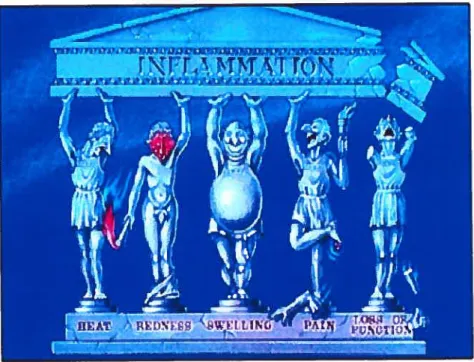
Macroscopically, inflammation is characterized in the acute form by five classic signs: ta) Heat (calor); increase in temperatufe is seen oniy in peripheral parts of the body, such as the skin. It is due to increased blood flow (hyperaemia) through the region, resulting in vascular dilatation and the delivery of warm blood to the area. (b) Redness (rubor); an acutely inflamed tissue appears red, due to dilatation of small blood vessels within the damaged area. (c) Swelling (tumor), swelling resuits from oedema, the accumulation offluid in the extravascular space. (d) Pain (dolor); results partly from the stretching and distortion of tissues due to inflammatoiy oedema and, in particular, from pus under pressure in an abscess cavity. (f) Loss of function
Chapter 1: Introduction 3
(functio laesa); movement of an inflamed area is consciously and reflexly inhibited
by pain (figure 1.1). These signs have been known since the ancient Greek and Roman era (Dennis et al., 1976; Gauldie, 1991; Walker and Fantone, 1994).
1.1.1. The phases of inflammation
Inflammation can be divided into several phases. The earliest, gross event of an inflammatoiy response is temporary vasoconstriction, e.g., narrowing of blood vessels caused by contraction of smooth muscle in the vesse! walls, which can be seen as blanching (whitening) of the skin. This is followed by several phases that occur over minutes, hours and days later, as following:
Acute vascutar response follows within seconds of the tissue injury and
last for some minutes. It resuits from vasodilation and increased capillary permeability due to alterations in the vascular endothelium, which leads to increased blood flow (hyperaernia) that causes redness (eiytherna) and the entry of fluid into the tissues (oedema).
Acute cellutar response takes place over the next few hours. This phase
occurs if there is sufficient damage to the tissues, or if infection has occurred. The appearance of granulocytes, particularly neutrophils, in the tissues is the specific sign of this phase. if the vessel is damaged, fibrinogen and fibronectin are deposited at the site of injury, platelets aggregate and become activated, and the red celis stack together to help stop bleeding and aid dot formation. The dead and dying celis contribute to pus formation.
Chronic cettular response may follow during the next few days. It is
characterized by the appearance of a mononuclear ceil infiltrate composed
of macrophages and lymphocytes. The macrophages are involved in
microbial killing and in clearing up cellular and tissue debris. Macrophages also seem to be very important in tissue remodeling.
Resotution, during which the normal tissue is restored as the blood dots
in-Chapter J: Introduction 4
Figure 1.1. Cardinal signs of inflammation
This cartoon depicts five Greeks representing the cardinal signs of
inflammation: heat, redness, swelling, pain and loss of function, which are as appropriate today as they were when first described by Celsus more than 2000 years ago. This figure was commissioned by D.A.W. and drawn by P. Culi for the Medical Illustration Department at St Bartholomew’s Medical College.
Chapter 1: Introduction 5
fihling with fibroblasts, collagen, and new endothelial celis (ECs). Generally, by this time, any infection will have been overcomed.
In acute inflammation, the hydrostatic pressure in postcapillary venules may overcome the osmotic pressure of plasma proteins. Therefore, fluid and low molecular substances have the tendency to penetrate into the surrounding area. The increased capillary permeability for plasma proteins is the key factor for the production of inflammatory exudate. In the interstitial area, high-molecular weight proteins may be spiit into smaller fragments that participate in the raising of the osmotic pressure of interstitial fluid.
Cellular exudate is formed during the second and the thfrd phase of inflammation, e.g., acute and chronic cellular responses. During the former, neutrophils are prevatent, whereas mononuclear celis (macrophages and lymphocytes) are predominant in later phases. Celi composition ofexudate differs not only depending on the phase of inflammation, but also on the type ofinflamed tissue and on the factors tfiggering the inflammatory process. Central effector and regulatory functions in acute inflammation rely on neutrophils. Eosinophils and basophils may also be involved. So, a number of different celi types are potentially
recwited into the area where damage has occurred, and these are responsible for
inactivation and removing of invading infectious agents and damaged tissues, as well as for inducing the formation of new tissue and reconstructing the damaged celi matrix, including basement membranes and connective tissue. A new blood supply to the area is also established during the repair process Martinez-Hemandez, 2001).
1.1.2. The response to injury and infection
The celis of the immune system are widely distributed throughout the body, but, if an infection or tissue damage occurs, it is necessary to concentrate them and their products at the site of injury. As discussed above, three major events occur during this response:
Chapter J: Introduction 6 • An increased blood supply to the tissue in danger” (vasodilatation). The
inflamed tissue looks like containing a greater number ofvessels.
• Retraction of the endothelial celis (EC5), allowing larger molecules than usual to escape from the capillaries, and thus, allowing the soluble mediators of immunity to reach the site of inflammation.
Migration of leukocytes out of the capillaries into the surrounding tissues (Walker and fantone, 1994).
Inflammatory responses must be well-ordered and controlled. Therefore, a wide variety of interconnected cellular and humoral (soluble) mechanisms are activated when tissue damage and infection occur. Celis and inflammatory mediators participating in these events are detailed in the following paragraphs.
1.2. Ceils parficipafing in acute inflammation
1.2.1. Mast celis and basophils
Mast ceils are localized within the connective tissue of the body, whereas basophils are present in low number in the circulation. They play a central role in a variety of inflammatory and allergic conditions; they are able to release potent inflammatory mediators such as histamine, proteases, chemotactic factors, cytokines and metabolites of arachidonic acid from the dense cytoplasmic granules into extracellular tissues by degranulation (Fantone and Ward, 2001). The degranulation could be induced by: (a) a physical destruction such as mechanical trauma; (b) chemicaÎ substances, such as toxins and proteases; (e) ceil mediators, peptidases and peroxidases such as cationic proteins derived from eosinophils and neutrophils; (d) immune mechanisms which may be immunoglobulin (1g) E-dependent or IgE independent. Botb, eosinophils and basophils contain high affinity receptors (FcRl) for IgE on their surface which can be triggered during the IgE-dependent mechanism.
C’hapter 1: Introduction 7
In the IgE-independent way, the anaphylatoxïns C5a, C3a and C4a, formed during activation of complement, trigger degranulation through C5a celi surface receptors.
1.2.2. Eosinophils
The eosinophil is a tenninally differentiated, end-stage leukocyte that resides predominantly in the submucosaÏ tissue and is recmited to sites of specific immune reactions, including allergic diseases. Its cytoplasm is fihled with large eosinophilic granules and its nucleus is almost aiways bilobed although trilobed forms are sometimes seen (Bainton, 1999). The granules are the principal identifying feature of eosinophils. They contain four distinct cationic proteins which exert a range of biological effects on host ceils and microbial targets: major basic protein, eosinophil cationic protein, eosinophil derived neurotoxin, and eosinophil peroxidase. In addition, histaminase and a variety of hydrolytic lysosomal enzymes are also present in the large specific granules (Peters et al., 1986). Recently, it has been recognized that eosinophils are capable of elaborating cytokines like granuÏocyte-macrophage colony-stimulating factor (GM-CSF), interleukin-3 (IL-3) and IL-5, (Kita et al., 1991; Desreumaux et al., 1993) as well as chemokines such as eotaxin (Ponath et aÏ., 1996), which play a roÏe in inflammatory responses. Eosinophils also synthesize lipid inflammatory mediators, in particular leukotriene C4 (LTC4) and platelet-activating factor (PAF). Both mediators contract airway smooth muscle, promote the secretion of mucus, alter vascular permeability and elicit eosinophil and neutrophil infiltration (Lewis and Austen, 1984; Lee et aL, 1984).
1.2.3. Polymorphonuclear neutrophits
Polymorphonuclear neutrophils (PMNs) represent 50 to 60% of the total cfrculating leukocytes in the normal adult human and constimte the t’first line of defence’1 against infectious agents or ‘Tnon-self’ substances that penetrate the body’s physical barriers. Their targets include bacteria, fungi, protozoa, viruses, and tumor ceils. The neutrophils are spent in three environments, including bone marrow, blood
Chapter 1: Introduction 8
and tissues. Bone marrow is the site of the important processes of the proliferation and terminal maturation of neutrophilic granulocytes before being released to the circulation (Bainton, 1999).
Neutrophils are terminally differentiated ceils rich in cytoplasmic granules and containing a lobulated chromatin-dense nucleus with no nucleolus; the nucleus of the mature neutrophil is segmented into three to five interconnected lobes. The bone marrow of a normal heahhy aduit produces more than 1011 neutrophils per day and more than 1012per day in settings of acute inflammation. Upon release from the bone marrow to the circulation, the ceils are in a non-activated state and have a haif-life of only 4 tolO h before marginating along the vessel walls and entering tissue poois, where they survive for 1 to 2 days. Celis of the circulating and marginated pools can exchange with each other. Senescent neutrophils are thought to undergo apoptosis (programmed ceil death) prior to removal by tissue macrophages (Saviil et al., 1993). The rate of neutrophil egress ftom the bone marrow is controlled by several factors, such as infection, stress and also by two colony-stimulating factors, granulocyte colony stimulating factor (G-C$f) and GM-CSf. Both factors direct the production and differentiation of bone marrow progenitor ceils. The rate of the neutrophil differentiation can increase as much as 1 0-fold during state of stress and infection (Cannistra and Griffin, 198$).
1.2.3.1. PMN granules
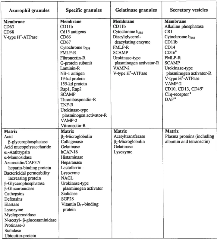
The initial classification into two major types of neutrophil granules was based on their content in myeloperoxidase. However, the granules can be subdivided into four distinct populations, azurophil, specific, gelatinase, and secretory, as summarized by Borregaard and Cowiand (1997). These populations (table 1.1) have been identified by cytochemical, immunocytochemical, and celi ftactionation procedures.
Chapter J: Introduction 9
Azurophil granules are known to contain myeloperoxidase (MPO), an antibacterial enzyme, and other numerous antimicrobial agents, lysozyme, and lysosomalenzymes (Bretz and Baggiolini, 1974). Defensins, azurophil-derived bacterial factors, and bactericidal permeability-increasing protein are bactericidal factors have been found in some azurophil granules (Rice et al., 1987; Gabay et al., 1986; Weiss and Olsson, 1987). In the azurophil granules ofhuman PMNs, cathepsin G, elastase, proteinase-3, and azurocidin are antimicrobial proteins which collectively could be called “serprocidins” and are closely related to serine proteases with microbicidal activity (Gabay et al., 1989). Specific granules, peroxidase-negative granules, which by definition do not contain peroxidase, contain lysozyme (Cramer and Breton-Gorius, 1987), lactoferrin (Bretz and Baggiolini, 1974), B12-binding proteins (Kane and Peters, 1975), and others as shown in table 1.1 (Bainton, 1999). The limiting membrane of specific granules and/or intracellular vesicles serves as a reservoir of receptors and other proteins involved in adherence, signal transduction, and functional activation ofmicrobicidal pathways (Bainton et al., 1987). Gelatinase granules are a subgroup of small, peroxidase-negative specific granules. They are defined by their high content of gelatinase (Borregaard and Cowiand, 1997). Secretory granules were originally discovered as highly mobilizable intracellular vesicles that contain alkaline phosphatase on their luminal surface (Borregaard et al., 1990). Mthough many studies have assumed that alkaline phosphatase was a plasma membrane marker, it seems instead to be in a cytoplasmic organelle, distinguishable from azurophil-, specific-, and gelatinase-containing granules, which is easily mobilized to the surface(Kobayashi and Robinson, 1991).
Among azurophul granule constituents, MPO is a critical enzyme converting hydrogen peroxide (H202) to hypochiorous acid. Together with hydrogen peroxide and a halide cofactor, MPO system forms the most effective microbicidal and cytotoxic mechanism of leukocytes. IVPO is responsible for the characteristic green color of pus (Klebanoff 1999).
Oiapter 1: Introduction 10
Azurophil granules Specific granules Gelatinase granules Secretory vesicles
Membrane Membrane Membrane Membrane
CD63 CD1 lb CD1 lb Mkaiine phosphatase
CD6X Cd15 antigens Cytochrome b558 CRi
V-type }{-ATPase CD66 Diacylglycerol- Cytochromeb558
CD67 deacylaling enzyme CD 1 lb
Cvtochrome b558 FMLP-R CD14
FMLP-R SCAMP CDl6a
Fibronectin-R Urokinase-type FMLP-R
G-protem subunit plasminogen activator-R SCAMP
Lamiiiin-R VAMP-2 Urokinase-type
NE-l antigen V-type W-ATPase plasminogen acfivator-R
1 9-kd protein V-type W-ATPase
155-kd protem VAMP-2
Rapl, Rap2 CD 10, CD13, CD45a
SCAMP Clq-receptor Thrombospondm-R DAF TNF-R Urokinase-type plasminogen aclivator-R VAMP-2 Vitronectm-R
Matrh Matnx Matrix Matnx
Acid [32-Microglobulm Acetyltransferase Plasma proteins (mcluding
f3-glycerophosphatase Collagenase f2-Microg1obuhn albumin and tetranectin) Acid mucopolysaccharide Ge1athase Gelatmase
ci1-Antitiypsin hCAP-18 Lysozyme
a-Mannosidase Histaminase
AzurocidinlCAP37l Heparanase hepann-binding protein Lactoferrin Bactericidal penneabfflty Lysozyme
increasmg protein NAGL
f3-Glycerophosphatase Urokinase-type f-Glucuromdase plasminogen aclivator
Cathepsms Sialidase
Defensins SGP2S
Elastase Vitawin B12-binding
Lysozyme protem Myeloperoxidase N-acetyl- -glucosaminidase Protinase-3 Sialidase Ubiquitin-protein
Table Li. The content ofhuman neutrophil granules ami secretoiy vesicles
From Bainton, DeveÏopmentalbiology ofneutrophils and eosinophils. In
Chapter J: Introduction 11
1.2.3.2. PMNs in host defence
The major role of neutrophils is to phagocytose and destroy infectious agents or celis, but they also limit the growth of some microbes, thereby buying time for adaptive (specific) immunological responses.
Phagocytosis is a complex process that involves several biochemical steps. Phagocytosis is triggered upon binding of opsonized or unopsonized microorganism through opsonin receptors (for complement fragments and antïbodies) or tbrough nonspecific glycosylated receptors that recognize certain lectins on target microorganism (Smith, 1997).
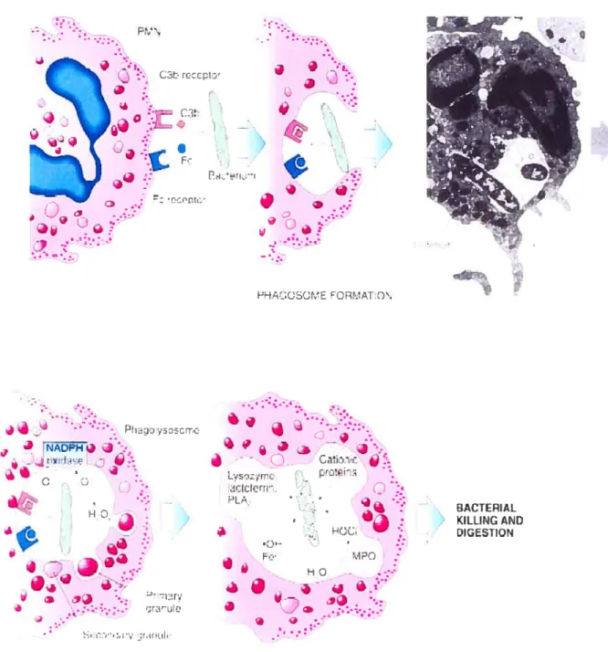
During phagocytosis, cytosolic granules (lysosomes) fuse with the invaginating plasma membrane (around the engulfing microorganism) to form a phagolysosome into which they release their contents, thereby creating a highly toxic microenvironment (figure 1.2). This normally prevents the release of the microbicidal components into the extracellular milieu. However, some targets may be too large to be phagocytosed, resulting in frustrated phagocytosis in which no phagosome is formed, and which may lead to the secretion of the cytosolic granules content into the eKtracellular milieu, where the targets may be killed (Kiebanoif, 1999).
The other microbicidal mechanism is the oxidative mechanism (oxidative burst), so called because ofthe 50-100 fold increase in 02 consumption which resuits in the production of the superoxide anion (02) and other cytotoxic reactive oxygen
species (ROS) and, potentially, reactive nitrogen species (RNS). 02 is formed, initially, by the reduction of molecular oxygen by a single electron that originates from NADPH; this process is catalyzed by the combined action of the plasma membrane NADPH oxidase and cytochrome b558. Mthough 02 may contribute to microbial killing, additional more potent ROS are generated from this precursor sucli
Chapter J: Introduction 12 PM\ reccpt:’ .J
9
7j t. _p PHAUSOME EORt.AT.Q\ Ph jj J - -J b’. à .• .Cii:’i r. — L,ntr_ t t_J Ctctrt H’ - PL i1 BACTEPIAL H KILLINCAND à!-
.7 -H iGESTIQN -HO :ilv ;r,rtih. •.‘•.DLO1AN..._ATtO\ AND rJA:P11Ox [tAS .r or
Figure 1.2. Mechanism ofpolymorphonuclear neutrophil bacterial phagocytosis From Fantone and Ward, Inflammation. In EssentiaÏ Fathotogy, 3’ ed, 2001.
t
C’hapter 1: Introduction 13
as H202 which is formed by the spontaneous dismutation of 02 and/or the catalytic
action of superoxide dismutase (SOD) (Smith, 1994). 11202react with the abundant
CF ions taken up from extracellular fluid to generate hypochiorous acid (HOC1), a reaction catalyzed by MPO contained in azurophil granules (Smith, 1997).
Nitric oxide (N0), which derived from the guanidino nitrogen in the conversion ofL-arginine to L-citmlline, may contribute to the microbicidal activity of neutrophils by reacting with ROS to form secondary cytotoxic species such as peroxynitrite (00N02) (McCall et ai, 1989).
1.2.3.3. PMNs and host tissue damage
While neutrophils are essential to host defence, they have also been implicated in many pathologic inflammatory conditions and in ischemia-reperfusion injury (Weiss, 1989; Ricevuti et al., 1991).
Host tissue damage may occur through several independent mechanisms. Ihese incÏude: premature activation during migration, extracellular release of toxic products during the killing of microbes or during removal of infected or damaged host ceils, and debris as a first step in tissue remodeling, or as a failure to terminate acute inflammatory responses (Smith, 1994; Smith, 1997).
for example, neutrophils have been implicated in the pathology of the aduit respiratory distress syndrome (ARDS) because of large influx of these ceils into the lung and the associated tissue damage caused by oxidants and hydrolytic enzymes released from activated neutrophils (Martin et al., 1991). Activation ofthe neutrophils by immune complexes in synovial fluid contributes to the pathology of rheumatoid arthritis (Robinson et al., 1992). Chronic activation of neutrophils may also initiate tumor development because some ROS generated by neutrophuls damage deoxyribonucleic acid (DNA) in vitro (Weitzman and Gordon, 1990).
Chapter 1: Introduction 14
1.2.3.4. PMN priming
Neutrophils may show three distinct states: dormant, primed, and activated. Priming means the neutrophiÏ’s functional responses (e.g., ROS production, chemotaxis) to a stimulus are amplified by previous exposure ofthe ceil to a priming agent. The priming agent, at low concentration, does flot normally cause a noticeable functional response (Guthrie et al., 1984; Bass et al., 1987; Swain et al., 2002). furthermore, there is al so evidence that primed neutrophuls can retum to a resting
state (Kitchen et al., 1996).
The priming agents include bacterial products like lipopolysaccharide (LPS), muramyl dipeptide (IVDP) and peptidoglycan, lipotechoic acid and N-fonnyl methionyl-leucyl-phenylalanine (fMLP); products from fungi like glucan and mannan; lipid mediators like leukotriene B4 (LTB4) and PAF; and cytokines like interleukin-8 (IL-8) and tumor necrosis factor-Œ(TNF-Œ) (Pabst, 1994).
Neutrophil priming is thought to play a key role in host defence process, and the regulation of priming may be essential for host survival. A number of studies have suggested that priming may be a good indicator of clinical disease activity. Enhanced neutrophil ROS production has been reported in patients with infection (Solberg et al., 1982). Primed neutrophuls have also been found in the blood of trauma patients (Zallen et al., 1999), as well as in patients with chronic inflammatory disease such as rheumatoid arthritis (Eggieton et al., 1995).
1.2.35. Mechanisms ofPMN extravascular accumulation
Circulating leukocytes can migrate from vessels into tissues under both normal and pathologic circumstances. The neutrophil migration from vasculature occurs by a multistep process (figure 1.3), dictated by sequential activation of adhesive proteins and thefr ligands on both leukocytes and ECs (von Andrian et al.,
Chapter J: Introduction 15
migrate to the endothelial surface. Initiation of the migration begins with the “capture” of PMNs from flowing blood by the vessel wall, and this is foflowed by their “rolling” along the vessel wall.
1.2.3.5.1. Capture and rolling ofleukocytes
Both the capture and rolling of leukocytes along the vessel wall is due to the reversible binding of transmembrane glycoprotein adhesive molecules caÏled selectins, which are found on both PMNs and ECs. Selectins have three major domains, of which the extracellular lectin domain is involved in ligand binding, an epidermal growth factor-like domain and a varying numb ers of short consensus repeat domain (SCR) that keep selectins away from the ceil surface (Crockett-Torabi and fantone, 1995). There are tbree types of selectins; the teukocyte setectin (L-selectin) appears to be critical for the rolling process. In addition, there are two endothelial selectins, the ptatetet setectin (P-selectin), which is rapidly and transiently expressed on ECs, and the endotheliat setectin (E-selectin), which appears a few hours later. The endothelial selectins are expressed only when appropriate inflammatory stimuli are present and interact with their PMN counterpart, which is a complex glycoprotein caïled P-selectin glycoprotein Iigand-1 (PSGL-1), belonging to the family of sialomucines (McEver and Cummings, 1997; Wagner and Roth, 2000).
1.2.3.5.2. Integrin-mediated firm adhesion
Cytoplasmic domains of bound L-selectin and PSGL-1 are linked to signal transduction pathways that lead to integrin activation in PIVINs (Crockett-Torabi and Fantone, 1995). Integrin activation is associated with the next step of the migration process, which is the firm adhesion ofthe leukocyte on ECs surface (figure 1.3). The
Chapter 1: Introduction 16 Ioodflow cets -_____ ___ _____ _____
PMN
.
7 9- ‘(C ESL1 ‘t, J--tntegrins PSGL-1 j’ PSetiflec,n ROLLING (Selectins) L FIRMADHESION Wi i2 integtins) J. TRANSMIGRATION (PCAM-1 etc.)Figure 1.3. Mechanism of leukocyte transmigration
From Fantone and Ward, JJ?flarnmation. In Essentiat Pathology. 3rn’
ed, 2001. /
.
.
/ /.
i\..
.i.
.t-ntegrins ‘t î L-selectinÏ
G:ycM 7M-Ï ICAM-ï g superarniIy Endothelial CeilFigure 1.4. Leukocyte and endothelial adhesion molecuies
From Fantone and Ward, Inftamn?ation. In Essentiat Pathology, 3rd
Chapter 1: Introduction 17
inregrins are a group of heterodimeric transmembrane glycoproteins found on PMNs and other hematopoietic ceils that mediate ceil-ceil or ceil-extracellular matrix (ECM) adhesion. There are 18 different integrins Œ-chains and 8 different f3-chains which pair together in specific pattem depending on the ceil type (Travis et al., 2003). PMN binding to activated endothelium is mediated mainly by two integrins: macrophage antigen-1 (MAC-1), also known as ŒMf32 or CD1 lb/CD1$, and lymphocyte associated fiinction antigen-1 (LFA-1), also known as ŒLf32 or CD1 ÎaJCD18 (figure 1.4). The importance of the integrin-mediated adhesion process is illustrated in patients with leukocyte adhesion deficiency syndrome (LAD-1), characterized by the absence of the f32 (CD 18) chain, which show severe recurrent, and life-threatening bacterïal infections secondary to an inability to locally recruit PIv[N (Anderson and
Springer, 1987).
The major counterligands ofintegrins belong to the immunoglobulin (7g)-liked superfamiÏy of adhesive rnotecules characterized by the presence of one or more immunoglobulin homology regions in their structure. IntercelluÏar adhesion molecule-I (ICAM-1), a member ofthis superfamily, is an important complementary endothelial ligand for MAC-1, exhibiting low constitutive presentation on EC membrane which is markedly induced by exposure ofECs to inflammatory cytokines (ligo et al., 1997). LFA-1 can also bind to ICAM-1, but it bas higher affinity to a related protein, ICAM-2, a ligand to which MAC-1 binds with low afflnity (Wagner and Roth, 2000). Vascular ceil adhesion molecule-1 (VCAM-1) is also an Ig-like molecule on ECs, but it binds selectively to f31-integrins such as cx4f3i integrin also called very late antigen-4 (VLA-4), which is critical for the migration of monocytes and eosinophils. However, VLA-4 has also been identified in both activated human and rat PMNs and may mediate VCAM-1-dependent adhesion of PMNs to endothelium in vitro (Davenpeck et al., 1998). A summary of integrin, selectin and Ig-like superfamlly ofadhesion molecules is found in table 2.1 (Horton, 1996).
Chapter 1: Introduction 18 family Homology region Examples CD no. Ligands Recognition motif in Extmcellular matrix in receptor (cluster of ligandlcounter-components with shared differentiation) receptor homology Integrin PEGG (ail f3 chains) I gpllbllla CD4 1/61 Blood proteins. Celi RGD. KQAGDV Collagen W, von Wilebrand domain (CD 1 1,al a2) LAF-1 CD 11/18 counter-receptors factor protein (Integrin I (e.g.ICAM) domain) ccvf33 CD5 1/61 Matrix, blood proteins RGD 2f31 CD49b/29 Collagen DGEA a4f31 CD49dJ29 Fibrioectin EILDV 1g superfamily 1g fold ICAMS, CD54 etc. Heterophylic interaction Multiple Perlecan (1g fold) VCAM Homophylic KYSFNYDGSE Fibronectin, tenascin, N-CAM LFA-3 counter-receptor thrombospondin (N-CAM CD2 type III repeat) Selectins C-type lectin L-, P-and E-CD62 Glycam-1, PSGL-1, Sialyl Le’ Aggrecan, versican (lectin), EGF repeat selectin CD34 etc. (CD 15) etc. Laminin, tenascin, Complernent regulatory CD62 thrombospondin, protein domain aggrecan versican (EFG repeat) Mucins Mucin side chain Leukosialin CD43 Selectins Muc-1 CD34 Platelet gpIV CD36 Thrombospondin, SVTCG (for Aggrecan, versican, lïnk CD36 Family collagen thrombospondin) protem CD44 Hyaluronidate-binding CD44 Hyaluromc acid, etc. Aggrecan, versican, linic site protein Cadhenns LDRE repeat E-, N-cadherin Homophylic HAV (IIO-AA module) Table 1.2. Classes ofcell adhesion receptors and their ligands Modified from Horton, Molecular bioÏogy oJceÏ/ adhesion rnoÏecuÏes, 1996.
C’hapter J: Introduction 19
1.2.3.5.3. PMN diapedesis
Subsequent to the firm adhesion, diapedesis of the PMN at endothelial celis junctions occur in a platelet endothelial ce!! adhesion molecule-1 (PECAM-1) or CD3 1-dependent manner. PECAM-1 is found on PMNs, platelets and ECs, serve as its own ligand and form homodimers with molecules on opposing celis. PECAM-1 is hypothesized to be a homing receptor to localize the transendothelial route for the migrating PMN. Treatment of the PMN or endothelial monolayers with an antibody to PECAM-1 blocks transmigration in vitro (Muller et ai, 1993).
1.2.3.5.4. PMN migration in the extravascular space
PMNs possess proteases capable of digesting collagen, laminin, and other extracellular components present in the vascular wall. Adhesion and migration are accompanied by the release of PMN-derived proteases (Hanlon et al., 1991). However, Mackeral et al. found that protease inhibitors are ineffective in stopping PMN migration through intact endothelial monolayers and basement membranes in
vitro. Thus, the requirement for protease release is uncertain (Mackarel et al., 1999).
Interaction of PMN f31—integrin with ECM proteins is important for transit of PMNs through the extravascular milieu. for example, PMN migration through the lung or synovial fibroblast barriers has been shown to require not only CD 1$ (132) but also VLA-4, VLA-5 (ci13i), VLA-6 (a3i) and VLA-9 (Œ9131), wbich are the most highly expressed f31—integrins in PMN, to bind the ECM components (Shang and Issekutz, 1997).
PECAM-1 is also involved in the extravascular transit of leukocytes. But unlike the homotypic interaction between PECAM molecules that mediate homing and diapedesis, migration through the subendothelial environment requfres
Chapter 1: Introduction 20
heterophilic binding of leukocytic PECAM-1 to an unidentified ligand (Wakelin et al., 1996).
1.3. Inflammatory mediators
When leukocytes reach the site of infection or inflammation, they release mediators which control leukocyte accumulation and activation ofneighboring ceils.
Inflammatory mediators are soluble molecules that act locally at the site of tissue damage and infection, and at more distant sites. These mediators may be low molecular weight proteins like cytokines and chemokines, hormones like neuroendocrine hormones, growth hormone and prolactin, or bioactive lipids like arachidonic acid and their metabolites (Smith, 1994).
Inflammatory mediators can be divided into exogenous and endogenous mediators. Exogenous mediators, such as bacterial products and toxins, evoke poweiful responses. For example, LPS can trigger complement activation, resuhing in the formation of the anaphylatoxins C3a and C5a which cause vasodilation and increase vascular permeability. LPS also activates the Hageman factor, leading to activation of the coagulation and fibrinolytic pathways as well as the kinin system. In addition, it elicits T celi proliferation, and has been described as a superantigen for T ceils. Endogenous mediators of inflammation are produced from the immune system (innate and adaptive) itself as well as from plasma. For example, they can be derived from molecules that are normally present in the plasma in an inactive form, such as peptide fragments of the complement, coagulation, and kinin systems. Mediators of inflammatoiy responses are also synthesized by a number of celi types that contain them as preformed molecules within storage granules, e.g. histamine, or which can be rapidly synthesized when they are required, for example, metabolites of arachidonic acid such as LTB4, LTC4 and leukotriene D4 (LTD4).
Chapter J: Introduction 21 Inflammatory mediators can also be classified into early and late phase mediators (White, 1999). Early phase mediators are important in acute inflammation and include histamine, serotonin and other vasoactive substances. They are produced by mast celis and platelets. In addition, lipid mediators, chemoatractants (e.g. C5a) and cytokines such as interleukin-1 (IL-1), interleukin-6 (IL-6), and TNF-Πbelong to the eafly phase mediators. Late phase mediators are found 6-12 hours afier initiation of inflammation. They are responsible for the regulation of vascular events. The later vascular events are mediated, at least in part, by products of arachidonic acid such as prostaglandin E2 (PGE2), LTD4 and LIC4.
Mediators which accumulate at local inflammatory sites such as skin blisters are somewhat different from those released following intravenous endotoxin. Mediators detected in blister fluid within 3 to 5 h of the inflammatory response include LTB4, C5a, IL-8 and IL-6. In contrast, IL-1f3, GM-CSF, and TNf-a are flot detected until afler $ hr in the blister. Afier i.v. endotoxemia, semm TNF reaches a peak level in 90-120 minutes and subsequently declines, to become undetectable within 4-6 h (Wang and Tracey, 1999).
1.3.1. ilistamine and Serotonin
They are the most important vasoactive mediators stored in mast ceil and basophil granules; both of them are also present in human platelets. Histamine is largely complexed to mucopolysaccharides such as heparin. Histamine has diverse biological ffinctions including local dilatation of small vessels, widespread arteriolar dilatation, local increased vascular permeability through ECs contraction, contraction
of nonvascular smooth muscle, chemotaxis for eosinophils, and blockade of T
lymphocyte function (Pearce, 1991). Serotonin is also capable of increasing vascular permeability, dilating capillaries and producing contraction of nonvascular smooth muscle.
Chapter 1: Introduction 22
1.3.2. Cytokines and Chemokines
Cytokines are soluble polypeptides that mediate and regulate various aspects of inflammation. The most important pro-adhesive cytokines that are present during most inflammatory responses are TNF-a and IL-1. The primary cellular sources of these mediators are macrophages and monocytes, and LPS is perhaps their most important inducer (Di Girolamo et al., 1997). Neutrophils also synthesize and secrete small amount of some cytokines including IL-1, IL-6, TNF-a, and GM-CSF, wbich may act in an autocrine or paracrine manner (Lloyd and Oppenheim, 1992). PMNs respond to TISF-o. by activating and expressing integrins, producing PAF and other mediators, and releasing granule contents. Likewise, ECs can respond to TNF-a exposure by mobilizing E-selectin, up-regulating ICAM-1 and activating procoagulant pathways (Burke-Gaffney and Helleweli, 1996). It was found that during inflammation and endotoxemia, PMNs release from their membrane a soluble TNF-a receptor (p55) that can bind and effectively inactivate circulating TNF-a (Wagner and Roth, 2000). IL-1 exerts similar physiological effects (Scholz et al., 1996). Thus, the early appearance of TNF-ct and IL-1 in plasma during inflammation is likely critical for the capture and firm adhesion ofPMNs to vascular endothelium.
Chemokines are a superfamily of low molecular weight cytokines with selective chemotactic properties. They have been subdivided into families on the basis of the relative position of their cysteine residues. The largest families are a chemokine (cysteine-X amino acid-cysteine or CXC) and f3-chemokine (cysteine cysteine or CC). IL-8, a member of the CXC superfamily, as well as macrophage inflammatory protein-1 alpha (MW-la), macrophage chemoattractant protein-1,-2, and -3 (MCP-1,-2,-3) and RANTES, which are members of the C-C superfamily, function primarily as chemoattractant molecules for macrophage and circulating leukocytes (Luster, 1998). Chemokines were first identified in vitro and initially thought to be produced by activated macrophages and monocytes. However under the proper conditions, their production and release has been elicited from neutrophils, endothelium, and a variety of parenchymal ceils in vitro. For example, activation of
Chapter 1: Introduction 23 neutrophils with particulate stimuli such as zymosan induces the generation of the CXC chemokine IL-8 (Au et al., 1998). further studies suggest that these ceils produce chemokines in animal modeis (Rovai et al., 199$). Chemokines are thought to provide the signais that convert the iow-affinity, seiectin-mediated interaction into the higher-affinity, integrin-mediated interaction that lead to extravasation of leukocytes (Luster, 1998).
Rodents do flot have an IL-8 analog and, instead, possess cytokine-ïnduced neutrophil chemoatracifants (CINCs). CNC-1, MLP-2, CLNC-2a and CINC-2f3 are released from LPS-stimulated rat macrophages in vitro and possess similar ability to elicit chemotaxis and degranulation of PMNs (Nakagawa et al., 1996). Rodents respond to human IL-8 and the i.v. administration of IL-8 inhibits PMN migration to extravascular sites of inflammation in rabbits (Ley et aI., 1993). Another in vivo study showed that eosinophil accumulation in response to IL-1f3 was significantly suppressed in rats treated with an anti-human IL-8 mAb DMJC7 (Sanz et ai., 1995).
Mthough LPS, TNF-Œ, and IL-Ï are flot chemoatactic for PMNs, their exposure to cultured ceils can induce the production of chemoattractants such as IL-8 and PAF (Burns et al., 1997).
1.3.3. Products ofthe complement system
The complement system is a potent mechanism for initiating and amplifying
inflammation. This is mediated through activated fragments of complement components. C3a, C4a and C5a are proteolytic products ofthe serine proteases ofthe compiement system. These anaphylatoxins are polypeptides containing approximately 75 amino acid residues. The C-terminal arginine in the molecule of C3a is offundamental importance for its biological activity, whereas removal ofthe C-terminal arginine ofC5a (CSacjesArg) only decreases its biological activity.
Chapter J. Introduction 24
The anaphylatoxins have powerful effects on blood vessel walls, causing contraction of smooth muscle and an increase in vascular permeability. C5a is extremely potent at stimulating neutrophil chemotaxis, adherence, respiratory burst generation and degranulation (Kohi and Bitter-Suermann, 1993). Ligation of the neutrophul C5a receptor at dermal skin sites is followed by mobilization of membrane arachidonic acid which is primarily metabolized to LTB4, another potent chemoattractant for neutrophils and monocytes, as obsewed in rabbits (Marleau et al., 1999).
1.3.4. Lipid mediators
Lipid mediators are a class of lipid molecules derived from the metabolism of membrane phospholipids. These include PAF and the vast array of eicosanoids, which are derived ftom the metabolism of arachidonic acid (figure 1.5).
1.3.4.1. Leukocyte phospholipases
During inflammation, cellular phospholipases, especially phospholipase A2 (PLA2) and C, are activated and degrade phospholipids to induce mobilization of fatty acids (FAs) from the membrane lipid pooi for the synthesis of lipid mediators at the site of cellular damage or inflammation (Heller et al., 199$). The fAs composing the normal inflammatory ceil membrane are saturated fAs, monounsaturated fAs, and polyunsaturated FAs, the latter composed of omega-3 and omega-6 FAs, which includes arachidonic acid (AA).
AA, the mother substance of pro-inflammatoiy eicosanoids, lias a short haif life and can be metabolized by two major pathways, the cyclo-oxygenase (COX) and lipoxygenase (LO) pathways. The COX pathway leads to the production of prostaglandin (PG) H2 which can be further metabolized to PGE2, PGI2 (prostacyclin) and thromboxane A2 (TXA2). These eicosanoids have potent local effects in the
Chapter J: Introduction 25
_____
Membrane phospholipids omega3fA
(omega-6fattyacids)
LTB4receptor antagonÏsts
Figure 1.5. Biosynthesis oflipid inflammatory mediators PLA2 iuhïbitors
—H
ostanes
I
ArachidonicacjdF—
PLA2 inhibitorsCOI inhibitors
H
TX syn inhibitors—{ TX synthase
TXA2inhibitors _______ ØLI
PAFI
T
PAF inhibitors 12 HET I5HETT
Chapter 1: Introduction 26
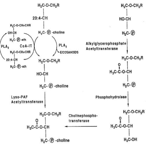
manifestation of inflammation. LO pathways lead to the production of leukotriene A4 (LTA4) which is further metabollzed into a number of leukotrienes: LTB4, LTC4, LTD4 and leukotriene E4 (LTE4), which have potent pro-inflammatory effects. Phospholipids can also be metabolized by induction of PLA2 activity to lyso-PAF which is then converted to PAF (Braquet et al., 1991; Bulger and Maier, 2000).
PLA2 is a key enzyme in the production of lipid mediators from membrane phospholipids as it catalyzes the hydrolysis of the sn-2 position of membrane phospholipids to release unsaturated fatty acids. PLA2 enzymes fali into four broad categories: the group W cytosolic PLA2 (cPLA2) calcium (Ca2)-dependent enzymes; the low molecular weight, secretory enzymes (sPLA2), including many groups like lB, HA, IIC, UD, 11E, 11F, ifi, V, X and XII; the Ca2tindependent, group VI enzymes; and the selective acetyl hydrolases of PAF, groups VII and VIII PLA2 (Dennis, 1997; Diaz and Arm, 2003). cPLA2 is present in most celis and tissues including PIvIN and monocytes, where it is critical to the production of PAF and AA, thereby playing an important role in both rapid and prolonged cellular response occurring during iiiflammatoiy processes. PLA2 activity is increased in response to norepinephrine, adenosine, bradykinin, PAF, TNF, and IL-1 stimulation (Anderson et al., 1994). In summary PLA2 groups and their properties are found in table 1.3 (Six and Dennis, 2000).
Phosphatidylinositol-bisphosphate is a major phospholipid in eukaryotic celis that can be hydrolyzed by PLC to diacylglycerol which is ffirther metabolized to AA by diacylglycerol lipase (Fantone and Ward, 2001).
1.3.4.2. Prostaglandins (PGs)
PGs have been detected in almost ail experimentai models of inflammation and clinical inflammatory conditions. For instance, the major PGs found in the synovial fluid of patients with arthritis are PGE2 and PGI2. In leukocytes, they are synthesized de nova ftom membrane-released AA when ceils are activated by
Chapter 1: Introduction 27
Group Initial/common Alternate Size Ca2 effects Characteristics sources names (kDa)
employed
W A Human U937 celis! cPLA2Π$5 <iM; membrane C2 domain, platelets RAW 264.7lrat transiocation aIf3-hydrolase
kidney regulatory
phosphoiylation B Human pancreasfllver cPLA2f3 114 <iM; membrane C2 domam,
heartlbmin transiocation a/f3-hydrolase
C Human hearUskeletal cPLA2y 61 None Prenylated,
muscle a/f3-hydrolase
VI A-1 P388D1 macrophages, IPLA2 or $4-$5 None Short splice,
CHO iPLA2-A $ ankyrin repeats
A-2 Human B-lymphocytes, IPLA2-B $8-90 None Long splice,
testis 7 ankyrin repeats
B Human heartlskeletal iPLA2«or 8$ None membrane-bound
muscle iPLA2-2
VII A Hmnan/mouse/porcine PAF-AH 45 None Secreted,
bovine plasma &3-hydrolase
Ser/His/Asp triad in VIIA and B
B Human/bovine PAF-AH (11) 40 None Intracdllular,
hverflddney myristoylated
VIII A Human brain PAF-AfflbŒ1 26 None Intracelhular,
(suburnt of G protein fold
trimer) SerfHis/Asp triad,
dimeric
B Human brain PAF-AHIb c 26 None Same as VIII A;
(subunit of active as
trimer) heireodimer or
homodimer
Table 1.3. Superfamily ofPLA2 enzymes
Chapter 1: Introduction 28
mechanical trauma or by specific cytokines, growth factors, or other stimuli, and act as autocrine and paracrine mediators.
1.3.4.2.1. Biosynthesis of PGs in leukocytes
At the endoplasmic reticulum and nuclear membrane, AA released by cPLA2 is presented to prostaglandin H synthase (PGHS), also known as COX for cyclo oxygenase, to form an intermediate prostaglandin, PGH2 (Funk, 2001). PGHS exists as two isoforms referred to as PGHS-1 (COX-1) and PGHS-2 (COX-2) (Smith et al., 2000). In simplistic terms, COX-1 is the enzyme responsible for basal, constitutive prostaglandin synthesis, whereas COX-2 is an inducible form of cyclooxygenase stimulated in the setting of acute inflammation in response to various cytoldnes, endotoxin and mitogens. Both enzymes catalyze the insertion of molecuÏar oxygen into arachidonic acid at Cil and Cl 5 resuit in formation of PGG2, and then, catalyze the reduction of PGG2 to PGH2 which is further metabolized by PG synthases into PGD2, PGE2, and PGF2 The cellular expression pattern of each synthase may profoundly influence the type of PG produced by particular celi. For example, platelets produce predominantly TXA2; ECs produce PGI2 and mast ceil produce PGD2 (Griffiths, 1999). In addition, monocytes produce mainly PGE2 through human peripheral blood mononuclear ceils produce a factor (MCF) (Robinson et al., 1979).
The effect of PGE2 resuits from its binding to PGE2 receptor subtypes EP2 and EP4, which stimulate production of the second messenger cAMP (Yoshikai, 2001).
1.3.4.2.2. Role ofPGs in inflammation
PGs generated by the PLA2/COX pathway play a dichotomous role in acute inflammation. PGE2 at physiological concentrations may be pro-ïnflammatory during the early phase of acute inflammation, while other PGs may regulate the resolution of
Chapter 1: Introduction 29
acute inflammation. PGE2 for instance, produced by macrophages (and ECs), contribute to the local vasodilation. PGE2 also downregulate macrophage response to cytokine stimulation (Fink, 1998). PGE2 concentration at inflammatory sites may determine whether its effect is pro- or anti-inflammatory; high concentrations of PGE2 may rather suppress the increased vascular permeability elicited by histamine, bradykinin, and C3a, thus preventing inflammatory reaction (Yoshikai, 2001).
In dermai inflammation, application of PGE2 or PGI2 alone increases blood flow, but does not promote oedema. However, co-injection of either agent with chemotactic mediators such as LTB4, IL-8, PAF-ci or bradykinin causes profound plasma protein leakage (Wedmore and Wiiliams, 1981). PGs also moduiate leukocyte function. For instance, PGE2, PGD2 and PGI2 ail inhibit PMN activation in vitro, as measured by chemotaxis and superoxide production (Wheeldon and Vardey, 1993). The inhibitory effect is associated with increase in cAMP formation. PGE2 is also a potent inhibitor of monocyte activation as it inhibits TNF production in vitro. In vivo, administration of COX-inhibitors enhances the release of TNF-ci in response to administration ofLPS (Pettipher and Wimberly, 1994).
In addition, PGs may reguiate lymphocyte function via modulation ofthe EP2 and EP4 receptor expression during acute inflammation (Griffiths, 1999). For example, PGE2 inhibits T-cell proliferation, cytokine production, and T-celi migration through a cAMP-mediated mechanism, invoiving EP2 or EP4 receptors.
1.3.4.3. Leukotrienes
The term “leukotriene” (Lis) refers to the ceilular source (leukocytes are one of the ma] or sources) as well as the con] ugated triene that characterizes their structure (Samueisson et al., 1979). Leukotrienes are formed from the catalytic oxygenation of 20-carbon unsaturated fatty acids, mainly arachidonic acid. Leukotrienes can be divided into two different classes, based upon their chemical structure and biological activity;
Chapter J: Introduction 30
Ihe cysteinyl leukotrienes (CysLTs), namely LTC4, LTD4, and LIE4, containing different amino acid residues
The dihydroxy-derivative leukotriene B4 (LIB4)
Briefly, CysLTs were originally described as the slow reacting substance of anaphylaxis (SRS-A) because of its slow and sustained smooth muscle contracting abilities. LTB4 (5 S, 1 2R-dihydroxy-6, 1 4-cis-8, 1 0-trans-eicosatetraenoic acid) was isolated and purified in 197$ from neutrophils upon activation by calcium ionophores such as A23 187 (Borgeat and Samueisson, 1979a), and characterized as a major AA metabolite in rabbit polymorphonuclear leukocytes (Borgeat and Samueisson,
Ï 979b).
1.3.4.3.1. Leukotriene biosynthesis in leukocytes
Ihe limiting step in the generation of Lis is the enzymatic release of AA from the ceil membrane phospholipids by PLA2. The first committed step in Lis formation from AA is through the enzymatic action of 5-lipoxygenase (5-LO) that requires ceil activation and influx of intracellular and extracellular calcium (Wong et al., 1991). Stimulation of neutrophils resuits in the transiocation of 5-LO, a cytosolic or nuclear soluble enzyme, to the nuclear envelope where it colocalizes with the 5-lipoxygenase activating protein (FLAP). FLAP is an integral membrane protein necessary for leukotriene synthesis, initially thought to act as a docking protein for 5-LO. Its role appears to be more complex, and may include efficient conversion ofAA to leukotrienes by handing the substrate to 5-LO (Abramovitz et al., 1993).
5-LO catalyzes the insertion of reactive oxygen into arachidonic acid at C-5 leading to formation of 5-hydroperoxyeicosatetranoic acid (5-HPETE). ihis unstable lipid hydroperoxide may be either reduced by peroxidase to the hydroxy metabolite (5-HETE) or can be stereospecifically dehydrated by a second 5-LO catalyzed step to eicosatetraenoic acid (LIA4) (figure 1.6) (Sirois and Borgeat, 1988). LiA4 undergoes transformation by one or more metabolic pathways depending on the cellular context; hydrolysis ofLiA4 by the LIA4 hydrolase occurs in the cytoplasm,