Formation de patron et modularité des canaux sensoriels de la
ligne latérale chez le poisson-castor, Amia calva
(Actinopterygii : Amiiformes)
Mémoire présenté
dans le cadre du programme de maîtrise en gestion de la faune et de ses habitats en vue de l'obtention du grade de maître ès sciences
PAR
© LEHOUX CAROLINE
UNIVERSITÉ DU QUÉBEC À RlMOUSKI Service de la bibliothèque
Avertissement
La diffusion de ce mémoire ou de cette thèse se fait dans le respect des droits de son auteur, qui a signé le formulaire
«
Autorisation de reproduire et de diffuser un rapport, un mémoire ou une thèse». En signant ce formulaire, l'auteur concède à l'Université du Québec à Rimouski une licence non exclusive d'utilisation et de publication de la totalité ou d'une partie importante de son travail de recherche pour des fins pédagogiques et non commerciales. Plus précisément, l'auteur autorise l'Université du Québec à Rimouski à reproduire, diffuser, prêter, distribuer ou vendre des copies de son travail de recherche à des fins non commerciales sur quelque support que ce soit, y compris l'Internet. Cette licence et cette autorisation n'entraînent pas une renonciation de la part de l'auteur à ses droits moraux ni à ses droits de propriété intellectuelle. Sauf entente contraire, l'auteur conserve la liberté de diffuser et de commercialiser ou non ce travail dont il possède un exemplaire.Composition du jury :
Martin-Hugues St-Laurent, président du jury, Université du Québec à Rimouski
Richard Cloutier, directeur de recherche, Université du Québec à Rimouski
Brian Hall, examinateur externe, Dalhousie University
REMERCIEMENTS
Je remercie les gens du Field Museum of Natural History, du Musée canadien de la nature, Huguette Massé et Mélissa Larochelle du Ministère du développement durable, de l'environnement, de la faune et des parcs de Longueuil qui ont gentiment prêté plusieurs spécimens.
Nous remercions le CRSNG pour le financement du projet de recherche (R.C.). Merci également au CRSNG et au FRQNT pour les bourses de recherche (C.L.). L'utilisation d'un micro-CT scan a été rendue possible grâce à l'agence de développement économique du Canada.
Je remercie tous mes collègues de laboratoire ; Marion Chevrinais, Cyrena Riley, Olivier Larouche, Élodie Libert, Catherine Morel, Daniel Potvin-Leduc, Michèle Leduc-Lapierre, Laurence Fischer-Rousseau, Isabelle Béchard, Vincent Roy et Thomas Grünbaum pour le soutien à toutes les étapes du projet et les nombreux, nombreux commentaires sur les nombreuses présentations. Merci aussi d'être mes amis et d'avoir fait que le passage à la maîtrise soit presque facile. Ce n'aurait pas été la même chose sans vous. Un merci tout particulier à Laurence pour l'aide apportée en géométrie morpho métrique. Merci aussi à Isabelle qui a réalisé la plupart des figures.
Merci à mon directeur de recherche, Richard Cloutier, pour m'avoir accueilli dans son laboratoire et avoir fait de son magasin de bonbons (des tiroirs remplis de fossiles!) mon lieu de travail pendant deux ans. Je remercie également les membres de mon comité : Brian Hall et Martin-Hugues St-Laurent.
Finalement, merci à ma famille pour avoir fourni un plein de motivation tout au long du projet. Merci à Brian B. d'avoir fait plusieurs corrections de ce mémoire et pour sa présence tout simplement. Un merci tout particulier à mon grand-père qui montre un intérêt remarquable dans tout ce que j'entreprends.
RÉSUMÉ
La modularité du crâne et l'intégration morphologique entre les systèmes anatomiques sont fondamentales en biologie évolutive du développement pour expliquer la diversité de plans d'organisation. Une étude récente au niveau des vaisseaux sanguins et des os du crâne a montré que la modularité est déterminée par la région anatomique et qu'elle n'est pas limitée à un système. L'interaction entre les os dermiques et les canaux de la ligne latérale est débattue depuis un siècle. Cependant, la modularité des canaux de la ligne latérale et leur intégration morphologique avec les os dermiques n'ont jamais été décrites. Une série ontogénétique de 97 spécimens de l'actinoptérygien basal Amia calva a permis de décrire l'ontogénie et la modularité des canaux de la ligne latérale ainsi que leur intégration avec les os crâniens. Les crânes ont été visualisés à l'aide d'un micro-CT scan. Le patteming des ramifications des groupes de pores du canal otique et de la commissure supratemporale a été décrit en codant la présence ou l'absence de chaque branche de manière à tester la présence de modules développementaux par la corrélation de Spearman dans l'ordre d'apparition des ramifications pour chaque groupe de pores. Des points de repères morpho métriques ont été posés sur 25 spécimens afin de tester des modules variationnels au niveau des canaux et l'intégration entre les canaux et les os en variation individuelle et en asymétrie aléatoire. Les ramifications suivent un patteming prédéterminé, supporté par la corrélation dans l'ordre d'apparition des bifurcations entre le côté gauche et droit. Des différences dans le patterning et le test de modularité basé sur la corrélation entre les groupes de points de repères suggèrent que le canal otique et la commissure supratemporale sont deux modules séparés. L'intégration entre les os et les canaux est insuffisante pour être détectée comme un module. Cependant, les deux blocs ne sont pas indépendants ajoutant du support à l'hypothèse de l'interaction développementale entre les os dermiques et les canaux sensoriels. Les groupes de pores ont tendance à disparaître sans affecter les autres groupes de pores suggérant qu'ils sont des unités quasi-indépendantes remplissant un critère pour la modularité. La modularité des canaux sensoriels suit une organisation hiérarchique qui s'explique par les étapes et les interactions développementales de la formation du système de la ligne latérale.
Mots clés: évo-dévo, modularité développementale, modularité variationnelle, canaux sensoriels, ligne latérale, os dermiques, Amia calva, patterning, morphométrie géométrique
ABSTRACT
Skull modularity and morphological integration among anatomical systems are fundamental in evolutionary developmental biology in order to explain the diversity of body plan. A recent study on cranial vasculature and the bony skull has shown that modularity is detem1ined by the anatomic region and thus not limited to a system. The interaction between dermal bones and lateral line canals has been debated for 100 years. However, modularity of lateral line canals and their morphological integration with dermal bones have never been described. An ontogenetic series of 97 specimens of the basal actinopterygian Amia calva was acquired to describe the ontogeny and modularity of lateral line canals and their integration with supporting cranial bones. Skulls were visualized with a micro-CT scan. Ramification patteming of groups of pores in the otic canal and the supratemporal commissure were described by coding the presence or absence of each branch in order to test for developmental modules by calculating Spearman rank correlations in order of appearance of ramifications between groups of pores. Landmarks were digitized on 25 specimens to test for variational modules at the level of canals and integration between canals and bones in individual variation and fluctuating asymmetry. Ramifications follow a particular patterning as supported by significant positive correlation in the order of appearance of bifurcation between sides. Differences in patterning and the test of modularity based on correlation between subset of landmarks suggest that the otic canal and the supratemporal commissure are two separate modules. The integration between bones and canals was insufficient to be detected as a module. However, the two blocks were not independent adding support to the hypothesis of developmental interaction between dermal bones and sensory canals. Groups of pores tended to disappear without affecting other groups of pores suggesting that they are quasi -independent units which fill a criterion for modularity. The hierarchichal organisation of modularity in sensory canals is explained by the developmental stages and interactions of the lateralline system formation.
Keywords :
evo-devo, developmental modularity, variational modularity, sens ory canals, lateral line, dermal bones, Amia calva, patterning, geometric morphometricsTABLE DES MATIÈRES
REMERCIEMENTS ... VII RÉSUMÉ ... IX ABSTRACT ... XI TABLE DES MATIÈRES ... XIII LISTE DES TABLEAUX ... XV LISTE DES FIGURES ... XVII LISTE DES ABRÉVIATIONS, DES SIGLES ET DES ACRONYMES ... XIX
INTRODUCTION GÉNÉRALE ... 1
CHAPITRE 1 LES BLOCS DE CONSTRUCTION DE LA TÊTE D'UN POISSON: MODULARITÉ DÉVELOPPEMENT ALE ET VARIATIONNELLE DANS UN SYSTÈME COMPLEXE ... 7
1.1 RESUME EN FRANÇAIS DE L'ARTICLE ... 7
1.2 BUILDING BLOCKS OF A FISH HEAD: DEVELOPMENTAL AND VARIATIONAL MODULARITY IN A COMPLEX SYSTEM ... 9
1.3 ABSTRACT ... 9
1.4 INTRODUCTION ... 10
1.5 MATERIAL AND METHODS ... 14
1.5.1 Specimens and imaging ... 14
1.5.2 Patterning of ramifications ... 15
1.5.3 Geometrie morphometrics ... 17
1.6 REsuLTs ... 19
1.6.2 Geometrie morphometries ... 21
1.7 DISCUSSION ... 21
1.7.1 Groups of pores patterning, variation and modularity ... 22
1.7.2 Modularity of sensory canals ... 23
1.7.3 Bone and canal integration ... 26
1.7.4 A hierarehieal model of modularity ... 27
1.7.5 Comparisons and future work ... 27
1.8
ACKNOWLEDGEMENTS ...28
1.9 TABLES ... 29 1.10 FIGURES ... 34 1.11 ApPENDIX 1 ... 40 1.12 ApPENDIX 2 ... 41 1.13 REFERENCES ... 43 CHAPITRE 2 CONCLUSION ... 49LISTE DES TABLEAUX
Table 1. Specimens used in the study listed by size ... 29
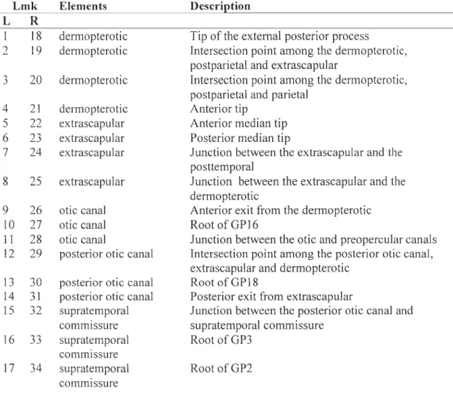
Table 2. Landmarks (lmk) position for each bone or canal for the left (L) and right (R) side ... 30 Table 3. Correlations in the order of appearance of bifurcation for GP 16-18 (otic canal) and GP2-3 (supratemporal commissure). Bold values indicate significant correlation. L: left side patteming, R: right side patteming ... 31
Table 4. Procrustes ANOVA for the complete dataset, including bones and canals (FA: fluctuating asymmetry) ... 32
Table 5. Results of modularity tests for three hypotheses for both symmetric and asymmetric components of variation using join or separate Procrustes fit. Bold numbers indicate the supported hypothesis. p: proportion of configurations with a lower RV coefficient than the hypothesis, P: p-value. / separates modules ... 33
LISTE DES FIGURES
Figure 1. Dorsal view of a posterior partial skull of Amia calva. Position of dermal bones, canals (dotted lines) and associated groups of pores. Digitized landmarks are shown by colored circles and numbered according to Table 2. Colors correspond to subsets presented as elements in Table 2. Red: Dermopterotic; Green: Extrascapular; Blue: Otic canal; Orange: Posterior part of the otic canal; Purple: Supratemporal commissure; Exsc: extrascapular; Dpt: dermopterotic; Dsp: dermosphenotic; Pa: parietal; P02: postorbital 2; Pp: postparietal; otc: otic canal, Pt: posttemporal; p.otc: posterior part of the otic canal; stc: supratemporal commissure; GP2, 3, 16, 18: Groups of pores 2, 3, 16 and 18 ... 34 Figure 2. Schematisation of a completely symmetrical group of pores with numbered bifurcations. Bifurcations are numbered clockwise, progressing towards extremities .
... 35 Figure 3. Modularity hypotheses tested by geometric morphometric analyses. Each shade corresponds to a module. A. Dermopterotic and extrascapular and respective canals as two separate modules. Black: dermopterotic and otic canal (landmarks 1-4, 9-11), White: extrascapular, posterior otic canal and supratemporal commissure (landmarks 5-8, 12-14, 15-17), B. Canals and bones as separate modules. Black: bones (landmarks 1-8), White: canals (landmarks 9-17), C. Otic canal and supratemporal commissure as separate modules. Black: dermopterotic (landmarks 1-4), White: otic canal (landmarks 8-11), Light grey: extrascapular (landmarks 5-8), Dark grey: Supratemporal commissure (landmarks 15-17) ... 36 Figure 4. Patteming comparison of group of pores 16 (GPI6), 18 (GPI8) and 3 (GP3) ... 37
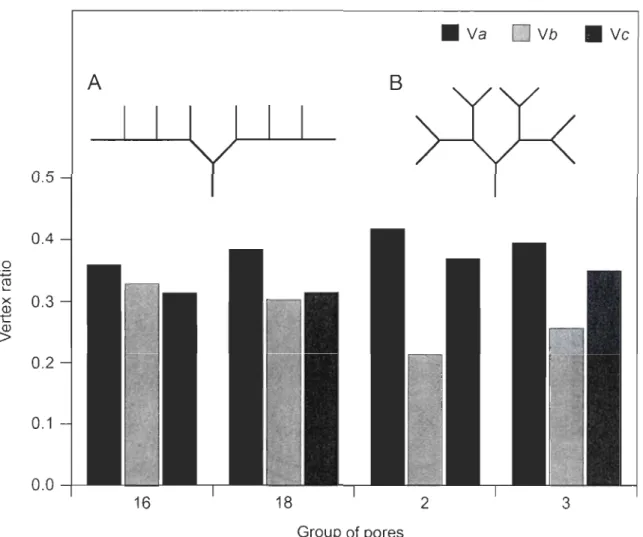
Figure 5. Comparison of ramification development for (A) the supratemporal commissure (GP2 and GP3) and (B) the otic canal (GP16 and GPI8). Ramifications of equivalent order develop simultaneously in the supratemporal commissure and in an anterio-posterior direction in the otic canal. Dotted segments represent developing ramifications and numbers show bifurcation identification ... 38 Figure 6. Comparison of spatial layout as indicated by vertex ratios for GP 16, 18, 2 and 3. GP16 and GP18 exhibit a growth with a higher probability of bifurcation for ramifications of higher order (A) which is consistent with a higher V b ratio. GP2 and GP3 grow symmetrically (B) which is consistent with a low V b ratio ... 39
LISTE DES ABRÉVIATIONS, DES SIGLES ET DES ACRONYMES CMN DFA dis2 dis2.5 dis3 Dpt Dsp ew Exsc FA FMNH GPI6 GPI8 GP2
Canadian Museum of Nature, Musée canadien de la nature, Gatineau (Qc)
Discriminant function analysis, Analyse discriminante
Distance entre le groupe de pores 2 et l'intersection entre le canal otique et la commissure supratemporale
Distance entre le groupe de pores seul et l'intersection entre le canal otique et la commissure supratemporale
Distance entre le groupe de pores 3 et l'intersection entre le canal otique et la commissure supratemporale
Dermoptérotique Dermosphénotique
Extrascapular width, largeur de l' extrascapulaire Extrascapulaire
Fluctuating asymmetry, asymétrie aléatoire
Field Museum ofNatural History, Chicago (IL, USA)
Groupe de pores 16 du canal otique Groupe de pores 18 du canal otique
GP3 L Imk
MDDEFP
MOD
ote p.otePa
PLS Po2 Pp PtQTL
R SL SLso steGroupe de pores 3 de la commissure supratemporale Left, côté gauche
landmarks
Ministère du développement durable, de l'envirollilement, de la faune et des parcs, Longueuil (QC)
Analyse de modularité canal otique
partie postérieure du canal otique Pariétal
Partial Least Square Postorbitaire 2 Postpariétal Posttemporal
Quantitative trait loci, Locus de caractères quantitatifs Right, côté droit
Coefficient de corrélation de Spearman Standard lenght, Longueur standard
Longueur standard à laquelle 50 % des individus ont développé la structure d'intérêt
Vertex de type a, lie deux vertex terminaux Vertex de type h, lie un vertex terminal Vertex de type c, ne lie aucun vertex terminal
INTRODUCTION GÉNÉRALE
La biologie évolutive du développement est une discipline centrale à la compréhension de l'évolution, puisque la variation disponible à la sélection naturelle est structurée par le développement en étant contrainte ou biaisée par des phénomènes tels que la modularité (Hendrikse et al., 2007). La modularité est la propriété des organismes à être composés de sous-unités quasi-indépendantes : les modules (Raff, 1996). Un module se définit comme une unité quasi-indépendante ayant des composantes fortement intégrées par rapport à un processus tel que le développement ou la fonction (Wagner el al., 2007). La modularité facilite l'évolution en diminuant la probabilité qu'une mutation bénéfique pour certains traits ait des effets néfastes sur les autres traits (Hendrikse et al., 2007), ce qui a amené les modules à être considérés comme des unités d'évolution (Schlosser, 2002a; Schlosser, 2002b; Schlosser, 2005). De plus, les modules peuvent être dupliqués et diverger, de même que subir une cooptation, c'est-à-dire un changement de fonction (Raff, 1996). C'est ce qui explique que les modules sont souvent répétés et conservés, puisqu'il est plus probable que l'apparition d'une nouveauté découle d'une cooptation de module plutôt qu'elle apparaisse de novo (Winther, 2001).
Il existe plusieurs types de modularité qui sont souvent définis par le processus qui est à l'origine de l'intégration. Deux types de modularité seront abordés dans cette étude : le module développemental et le module variationnel. Le module développemental se caractérise par des traits qui sont quasi-autonomes au niveau de la formation et de la différenciation de leur patron (Raff, 1996) et il est typiquement validé par la corrélation dans l'ordre d'apparition des structures (Poe, 2004) et le patteming. Un module variationnel est quant à lui formé de traits qui covarient fortement entre eux, mais qui covarient très peu avec les éléments appartenant à d'autres modules (Wagner et Mezey,
méthodes basées sur la morphométrie géométrique telles que le coefficient RV (Klingenberg, 2009) et l'analyse des moindres carrés partiels (Rohlf et Corti, 2000). Ces méthodes font appel à l'intégration morphologique qui est fortement liée à la modularité puisqu'elle réfère à un patron de covariation (revu par Klingenberg, 2008).
La modularité a été largement étudiée relativement aux structures osseuses (ex. Hallgrimsson et al., 2002; Mabee et al., 2002; Ward et Brainerd, 2007; Marquez, 2008; Grünbaum et al., 2012), mais très peu au niveau des tissus mous. De plus, la modularité, ou l'intégration morphologique, a été peu démontrée au niveau de l'interaction entre les tissus. Quelques auteurs (Richtsmeier et al., 2006; Zelditch et al., 2008; Jamniczky et Hallgrimsson, 20 Il) ont vérifié l'interaction entre les tissus au niveau de la tête, une structure complexe pour laquelle l'évolution ne peut être comprise que par l'intégration de toutes ses structures (Jamniczky et Hallgrimsson, 2011).
À
notre connaissance, aucuneétude ne s'est attardée à l'intégration entre le système sensoriel et le crâne.
Le système de la ligne latérale est un système sensoriel présent chez les poissons et les tétrapodes non-amniotes (Northcutt, 1989). Il est composé d'organes, les neuromastes,
pouvant être superficiels ou entourés de canaux, présents sur le crâne ou le tronc (N orthcutt, 1989). Les neuromastes sont composés de cellules ciliées qui détectent les changements de pression dans l'eau associés aux mouvements variant en amplitude, de grands courants océaniques jusqu'aux mouvements du plancton (Coombs et Montgomery, 1999). Le
système de la ligne latérale a une grande importance écologique. Il est associé au comportement de banc, d'évitement des prédateurs et d'alimentation (Coombs et Montgomery, 1999).
Le système de la ligne latérale n'a pas seulement une importance écologique, mais aussi une importance en phylogénie, puisque la trajectoire de la ligne latérale est utilisée pour déterminer l'homologie des os (Graham-Smith, 1978) et comme caractère phylogénétique (Cloutier et Ahlberg, 1996). Cette utilisation s'explique par le mode de croissance des os dermiques. Chez le poisson-castor Amia calva, l'enfoncement des
3
neuromastes dans le derme est suivi par l'ossification des parois formant ainsi des sections
de canaux sensoriels. Il en résulte une correspondance entre la position du canal et le centre
d'ossification de l'os dermique (Allis, 1889; Parrington, 1949). Les sections de canaux
fusionnent et forment un pore à la surface de l'os. Chez les espèces avec des canaux
ramifiés, le pore se subdivise pour former des groupes de pores ressemblant à des structures
arborescentes. Très peu d'études se sont consacrées à la description des ramifications du
système sensoriel, probablement à cause de son allure aléatoire. Cependant, des études dans
d'autres systèmes biologiques ramifiés, tels que les cellules de Purkinje (Berry et Flinn,
1984; Berry, 1992), les végétaux (Stein, 1993; Stein et Boyer, 2006) et les bryozoaires
(Kruszynski et al., 2007) ont montré qu'il est possible de trouver un certain ordre dans les
structures arborescentes d'apparence aléatoire. La description du développement des
ramifications au niveau du système de la ligne latérale permettra de vérifier si le
développement est ordonné et reproductible.
Bien que la modularité des canaux sensoriels n'ait jamais été vérifiée, plusieurs
niveaux de modularité ont été reconnus à différentes échelles de la ligne latérale. Le
neuromaste, la plus petite unité du système de la ligne latérale, serait un module (Franz-Odendaal et Hall, 2006). La modularité a aussi été identifiée à quelques reprises dans la
partie postcrânienne de la ligne latérale basé sur les QTLs (locus de caractères quantitatifs)
et les processus développementaux (Ledent, 2002; Wark et al., 2012). Dans la partie
crânienne, la modularité a été identifiée au niveau des placodes qui sont des
épaississements de l'ectoderme responsables de la formation des neuromastes et de leurs
neurones sensoriels (Schlosser, 2002c). Six paires de placodes sont présentes chez les
gnathostomes (Northcutt, 1997). Puisque les placodes sont responsables de la formation des
neuromastes et que ceux-ci seraient, à leur tour, responsables de la formation des canaux,
nous avons posé l'hypothèse que les canaux soient aussi des modules. Dans la partie
postérieure du crâne, trois placodes sont responsables de la formation des neuromastes de
trois sections de canal (Northcutt, 1997). La placode otique de la ligne latérale permet le développement des neuromastes de la partie antérieure du canal otique. Les neuromastes de
la partie postérieure
du canal otique proviennent plutôt de la
placode moyenne. Finalement
,
la commissure supratemporale se forme à partir des placodes supratemporales gauche et droite. Dans la partie postérieure du crâne sous l'action de ces trois placodes, trois modules
développementaux sont attendus au niveau des canaux. Ces trois modules ont été testés par
des corrélations entre le patteming des ramifications appartenant aux différents canaux. Le
patron de correlation entre des groupes de points de repère (landmarks) sera aussi utilisé pour valider cette hypothèse de modularité, mais il permettra de la tester en tant que module
variationnel. Des liens développementaux se traduisent par une forte covariation à
l'intérieur des éléments d'un module (Klingenberg, 2008). Les deux types de modules ne
sont donc pas indépendants.
L'intégration entre le crâne et les artères a été validée chez la souris suggérant que la
modularité est délimitée par la région plutôt que par les processus développementaux
(Jamniczky et Hallgrimsson, 2011). L'interaction entre le crâne et les canaux sensoriels a
été débattue et les liens développementaux entre les neuromastes et les os dermiques sont
inconnus (Graham-Smith, 1978; Webb et Noden, 1993; Tarby et Webb, 2003). La
connaissance de la nature de cette interaction est essentielle afin d'expliquer la variation
dans la présence/absence des canaux et la variation dans leur association avec différents
éléments dermiques. De plus, l'évaluation de cette interaction est essentielle dans un
contexte phylogénétique où les éléments sont caractérisés selon leur homologie
(Graham-Smith, 1978; Tarby et Webb, 2003). Elle sera vérifiée par le patron de correlation entre les
groupes de points de repères évalué par le coefficient RV (Klingenberg, 2009) et l'analyse
des moindres carrés partiels (Rohlf and Corti, 2000).
Cette étude est la première à quantifier le patteming des ramifications du système de
la ligne latérale, à identifier la modularité au niveau des canaux sensoriels, à vérifier l'intégration entre le système sensoriel et le crâne et à utiliser deux méthodes pour vérifier
la modularité. Les objectifs principaux de cette étude sont de décrire la modularité au
niveau des canaux sensoriels et la modularité entre les canaux sensoriels et les os dermiques. Secondairement, la vérification du patteming au niveau des ramifications vise à
5
la nature de l'interaction entre les différents niveaux hiérarchiques de la modularité
(placodes vs. canaux) et entre la modularité développementale et variationnelle.
Le développement des ramifications a été décrit par une méthode d'identification
développée dans le cadre du présent mémoire. La corrélation dans l'ordre d'apparition des
ramifications a permis de vérifier que le développement des ramifications est symétrique. L'utilisation du patteming et du test de modularité à partir de points de repères soit par le coefficient
RV
et l'analyse de moindres carrés partiels ont permis d'identifier deuxmodules: le canal otique et la commissure supratemporale. L'analyse des moindres carrés partiels n'a pas validé l'hypothèse des modules basés sur l'interaction entre les os dermiques et les canaux, mais elle a permis de détecter une covariation entre les deux composantes.
CHAPITRE 1
LES BLOCS DE CONSTRUCTION DE LA TÊTE D'UN POISSON:
MODULARITÉ DÉVELOPPEMENT ALE ET VARIATIONNELLE DANS UN
SYSTÈME COMPLEXE
Caroline Lehoux et Richard Cloutier
1.1 RESUME EN FRANÇAIS DE L'ARTICLE
La modularité du crâne et l'intégration entre les systèmes anatomiques crâniens sont fondamentales en biologie évolutive du développement afin d'expliquer la diversité de plans d'organisation. L'interaction entre les os dermiques et les canaux de la ligne latérale a
été débattue durant des décennies, mais la modularité du système sensoriel n'a jamais été
testée. Une série ontogénétique de 97 juvéniles et adultes du poisson-castor Amia calva
(Actinopterygii : Amiiformes) a été utilisée afin de décrire l'ontogénie et la modularité des
canaux sensoriels de la ligne latérale et leur intégration avec les os crâniens. Le patteming
des ramifications des groupes de pores de deux canaux sensoriels (le canal otique et la
commissure supratemporale) a été décrit de façon à tester la présence de modules développementaux par la corrélation dans l'ordre d'apparition des ramifications pour chaque groupe de pores. Des points de repères ont été posés sur 25 spécimens afin de tester la présence de modules variationnels au niveau des canaux et l'intégration entre les canaux
et les os. Les ramifications suivent un patteming particulier qui est supporté par la
différences dans le patterning entre le canal otique et la commissure supratemporale et les tests de modularité par la morphométrie géométrique suggèrent que les deux canaux sont
deux modules séparés. L'intégration entre les os et les canaux est insuffisante pour être
détectée comme un module. Cependant, les deux blocs ne sont pas indépendants. Les groupes de pores ont tendance à disparaître sans affecter les autres groupes de pores suggérant qu'ils sont des unités quasi-indépendantes remplissant un critère pour la modularité. La modularité du système sensoriel pourrait être utilisée pour expliquer la
variation dans le patron des canaux et leur association avec les os dermiques entre les
espèces.
L'article sera soumis durant l'hiver 2014 au Journal of Experimental Zo%gy: Part B
Molecular and Developmental Evolution. En tant que premier auteur, j'ai réalisé
l'ensemble des analyses. J'ai élaboré et choisit les méthodes d'analyses en collaboration avec mon directeur. Mon directeur, le deuxième auteur, a eu l'idée originale de ce projet et a révisé cet article.
1.2 BUILDING BLOCKS OF A FISH HEAD: DEVELOPMENTAL AND V ARIA TlONAL MODULARITY IN A COMPLEX SYSTEM
1.3 ABSTRACT
9
Skull modularity and integration among cranial anatomical systems is fundamental in evolutionary developmental biology in order to explain the diversity of body plan. The interaction between the dermal bones and lateral line canals has been debated for decades
but modularity has never been tested in the sensory system. An ontogenetic series of 97 juvenile and adult Amia calva (Actinopterygii: Amiiformes) was used in order to de scribe the ontogeny and modularity of sensory lateral line canals and their integration with supporting cranial bones. Ramification patterning of groups of pores in two sensory canals (otic canal and supratemporal commissure) was described in order to test for developmental
modules by computing correlations in appearance order between groups of pores.
Landmarks were digitized on 25 specimens to test for variational modules at the level of canals and integration between canals and bones. Ramifications follow a particular
patteming as supported by significant positive correlation in the order of appearance of bifurcations between sides. Differences in patterning between the otic canal and the supratemporal commissure and tests of modularity with geometric morphometrics suggest that the two canals are two separate modules. The integration between bones and canals was insufficient to be detected as a module. However, the two blocks were not independent. Groups of pores tended to disappear without affecting other groups of pores suggesting that
they are quasi-independent units acting as modules. Modularity of the sensory system could be used to explain variation of pattern of canals and their association with dermal bones
1.4 INTRODUCTION
Modularity is an important property of organisms responsible for a considerable amount of the diversity of body plans (Raff, '96). Modularity has been the focus of many studies taking into account primarily osteological structures such as fins (Mabee et al., 2002; Grünbaum et al., 2012), limbs (Hallgrimsson et al., 2002; Goswami et al., 2009), vertebrae (Ward and Brainerd, 2007), skull (Hallgrimsson et al., 2004; Goswami, 2006), and jaw (Marquez, 2008; Zelditch et al., 2008). However, studies regarding interactions
between osteological structures and soft tissues are scarce and limited to the muscular
(Zelditch et al., 2008), vascular (Jamniczky and Hallgrimsson, 20 Il) and nervous (Richtsmeier et al., 2006) systems. Ali of these integrative studies focused on the head; a very complex system whose evolution can only be understood by the integration of aU its components (Jamniczky and Hallgrimsson, 2011). Indeed, modularity is not limited to tissue type but is instead defined by epigenetic interactions within a region as supported by the interaction between the skull and cranial arteries in mice (Jamniczky and Hallgrimsson, 2011). However, morphological integration of the sensory system and the skuU has never been tested. The lateral line system is incorporated in the skull by sensory canals that go
through dermal bones in lower vertebrates.
The lateral line system of lower vertebrates has a mechanoreceptive function
performed by neuromasts whose hair cel1s register water movements (Webb, 2000).
Neuromasts were consequently described as distant-touch receptors (Dijkgraaf, '63). Neuromasts can be either superficial or embedded to form lines, either pits or canals on the head and trunk most frequently associated to dermal elements, such as certain cranial bones and scales (Northcutt, '89). The lateral line comprises two major topological components including neuromasts and their associated sensory neurons: (1) the anterior lateral line present on the head region and (2) the posterior lateral line present on the trunk and caudal fin (Gompel et al., 2001). The anterior lateral line inc1udes a series of six pairs of main cranial lines and two commissures in gnathostomes: supraorbital, infraorbital, otic
Il
(including post-otic), oral, mandibular and preopercular lines, and supratemporal and ethmoid commissures (Northcutt, '89).
The sensory system including the lateral line develops from ectodermal thickenings called placodes. Six pairs of lateral line placodes are responsible for neuromasts and sensory nerves formation in gnathostomes (Northcutt, '97). The migrating primordium which originated from the placode deposits clusters of cells fated to become neuromasts at regular intervals (Ledent, 2002). A sequence of three further developmental stages is necessary to form cranial and postcranial canals: (1) embryonic patterning of neuromasts and sensory neurons, (2) differentiation and growth of neuromasts and (3) morphogenesis oflateralline canals (Webb, 2000).
Modularity has been suggested at multiple levels of the lateral line system. Firstly, lateral line placodes are recognized as developmental modules because they have autonomous patterning, morphogenesis and differentiation even at ectopie locations (Schlosser, 2002a). Lateral line placodes also behave as units of evolutionary variation because their derivatives are coherently modified (Schlosser, 2002b). Secondly, modularity has been suggested in the zebrafish, Danio rerio, because the postembryonic growth, during which secondary neuromasts develop, occurs with the reiteration of the same elementary process as the embryonic growth (Ledent, 2002). Thirdly, modularity has been identified in the lateral line system of sticklebacks, Gasterosteus aculeatus, because QTLs (quantitative trait loci) that affect lateral line morphology differ between lines of neuromasts (Wark et al., 2012). However, no study tested for modularity in cranial canals.
Canal development has long been suggested to be related to neuromasts (Pehrson, '22). While not observed in aIl fishes, canal position usually corresponds with the center of ossification of dermal bones which suggests a close integration between the dermal and sensory systems (Parrington, '49; Cloutier, '97). However, the relation between dermal bones and the lateralline system has been debated in some teleosts (Westoll, '41; Graham-Smith, '78) because ossification patterns of canals differ among phylogenetic groups
(Tarby and Webb, 2003). In some fishes such as the bowfin Amia calva and some teleosts, ossification begins around neuromasts to forn1 a canal, the tubular lateral line component. Later in development, lateral extensions expend to form the dermal bone (Alli s, 1889; Pehrson, '22; Pehrson, '40). In some teleosts such as the zebrafish, the neuromasts sit on
underlying dermal bone that extend upwards and encloses neuromasts to form a canal (Webb and Shirey, 2003). Despite the ossification patterns being described, the
developmental influence of neuromasts remains untested (Webb and Noden, '93). Moreover, canals form without neuromast as it is in the whitespotted greenling,
Hexagrammos stelleri, suggesting that neuromasts are not required for ossification (Wonsettler and Webb, '97). Nevertheless, Wada et al. (2010) suggested that patterns of secondary neuromasts produced by seriaI budding during postembryonic growth in zebrafish are controlled by underlying dermal structures. The presence of modularity could confirm a close association in development between the lateral line component and dermal bones.
Following the ossification of canal walls, canal sections fuse to form pores that open to the bone surface (Alli s, 1889). In species with branched canals, this pore subdivides and forms ramifications (Webb, 2000). The ramifications in A. calva are divided by groups of
pores formed by the junction between two canal segments. Allis (1889) observed that ramifications have a constant relation to dermal bones as the canals. Ramifications were
briefly described in A. calva (Allis, 1889), coelacanth Latimeria chalumnae (Hensel and
Balon, 2001), and the Japanese Spanish mackerel Scomberomorus niphonius (Nakae et al.,
2013) but ramification development is unknown probably because of the seemingly random
appearance of branching patterns. Nevertheless, ramifications of other biological systems
have been described. Berry ('84; '92) described the ramification growth in Purkinje cells
based on the ratio between the terminal and link branches. In plants, algorithms have been used to reproduce branching patterns of leaf venation (Runions et al., 2005), stellar architecture (Stein, '93) and branches (Stein and Boyer, 2006). Simulation results in these
studies were reminiscent of patterns observed in extinct and extant species. Moreover, the simulated leaf venation pattern was obtained by the reiteration of the same elementary
13
process involving auxin distribution (Runions et al., 2005) These studies conducted at the cellular to organismal levels suggest that an underlying order can be found in apparent chaotic structures. Thus, an apparent random bifurcation of sensory canals could be refuted by the description of their patterning.
The developmental origin of sensory canals and their position into different dermal bones offer an opportunity to test a combination of hypotheses concerning developmental and variational modularity in the postembryonic growth of the lateral line system. We investigated modularity in the otic canal and supratemporal commissure of A. calva (Fig. 1). The otic canal has two parts that are delimitated by the preopercular canal. The posterior section of the otic canal has also been named the temporal (Northcutt, '97) or post-otic canal (Jarvik, '80; Northcutt, '89). The two parts of the otic canal come from two different lateralline placodes: the otic and the middle lateralline placodes (Northcutt, '97). The supratemporal commissure originates from the supratemporal lateral line placodes (Northcutt, '97). We expected those three sections of canals developing from different placodes to be three different modules since placodes were recognized as developmental modules (Schlosser, 2002a), where a developmental module is characterized by traits that are quasi-independent in terms of pattern formation and differentiation (patterning) (Raff, '96). Developmental modularity is tested by measuring correlation in ramification patterning (Poe, 2004). The hypothesis of three modules according to placodes was also tested as variational modules which include phenotypic traits that are integrated and quasi-independent of other such clusters resulting in high covariation among elements of a module (Wagner et al., 2007). Variational modularity is typically measured by geometric morphometrics (Wagner and Mezey, 2004) which was also used to test for integration between dermal bones and canals. Our study is the first, to our knowledge, to test for two different kinds of modularity by using two different methods.
1.5 MATERIAL AND METHODS 1.5.1 SPECIMENS AND IMAGrNG
An ontogenetic senes of 97 juvenile and adult A. calva specimens was acquired
through museum collections and commercial fisheries (Table 1). Since development of the
lateral line system is correlated with size rather than age (Münz, '86; Ledent, 2002),
standard length (SL) was used to reconstruct the ontogenetic series. Specimens were
photographed in lateral view in order to measure the SL with CT An (Version 1.11.4.2, SkyScan, Bruker-microCT, 2011, Belgium). SL varied between 15 mm and 545 mm (x = 194 mm; SD = 100 mm). Measurement error was less than one millimetre between two
measurements for each specimen. Specimens acquired through fisheries were measured unfrozen, whereas collection specimens were already fixed in fom1alin and preserved in
ethanol when measured. SL of one dry skull specimen was estimated using the linear
regression between parietal bones (Pa) length and SL for five specimens of similar size (SL = 0.0705*Pa
+
5.93; R2 = 0.95). Specimens from fisheries were decapitated, fixed in4 % neutralized formalin for 24 ho urs and preserved in 70 % ethanol.
Bones, sensory canals, and related ramifications were imaged with a micro-CT scan, SkyScan 1173 (Bruker-microCT, 2011, Belgium). Specimens were preferably scanned while fresh but 42 specimens were scanned after fixation/preservation and three were dry
skulls. Fixation/preservation did not affect relative position of components inside the bones
(see Appendix 1). Because of important differences in specimen size, the scanning protocol had to be adapted for each skull. For example, 200-mm SL specimens were scanned with an aluminium filter at 100 kV, 80 )..LA, with an exposure of 830 ms and a rotation step of
0.2°. The scanner was set to 55 kV, 100 )..LA, 1400 ms with no filter for smaller specimens
«
100 mm) and set to 100 kV, 98 )..LA, 1600 ms with a brass filter for larger specimen (> 300 mm). Resolution was set between 15 and 30 )..Lm. Projection images were15
1.5.2 PATTERNING OF RAMIFICATIONS
This study focuses on groups of pores (GP) 16 and 18 of the otic canal, and GP2-3 of
the supratemporal commissure (Fig. 1); nomenclature of groups of pores follows Allis
(1889). These groups of pores allow testing modularity hypotheses regarding their
developmental identity via placodes while taking into account their proximity and their
interactions with dermal bones. GPl of the supratemporal commissure was not included in
the analyses because it is not comparable to other groups of pores. It has very few
ramifications until an advance stage of development, and according to Allis (1889), it develops as a single median group of pores that subdivides to form one group of pores on each extrascapular. Individuals that expressed variation in the presence/absence of certain
groups of pores associated with the supratemporal commissure were pooled in order to test
for the identity of the missing group of pores. When one of the GP2-3 was absent, the remaining group of pores was identified as 2.5. The distance between the group of pores and the otic canal-supratemporal commissure junction (dis3, dis2 and dis2.5), and the
extrascapular width (ew) were measured for both sides of the skull. Measurements were
made with CTAn on 2D images digitized with CTVox (Version 2.2.0, SkyScan, Bruker-microCT, 2011, Belgium). To remove size and magnification effect, statistical analyses were performed on the dis/ew ratio for GP2, GP3 and GP2.5. The effect of allometry was tested by a multiple regression of the dis/ew ratio on size. The null hypothesis of isometry was not rejected by the analysis of variance of the regression (P = 0.6). A Kruskal-Wallis analysis was used to test for differences in relative position of GP2, GP3 and GP2.5. To verify if GP2.5 occupies an intermediate position between GP2 and GP3, a Student {-test was used to compare its position with the mean position of GP2 and GP3. In order to test if groups of pores tended to be absent on both sides equaUy, the G-test was used, with expected frequencies of 0.5 for both sides.
In order to describe the development of ramifications, the groups of pores were
schematized. Specimens > 400 mm were viewed with Seg3D (SCI Institute, 2007), a
paintbrush tool and the isosurface was computed to describe their pattern. Ramifications
were fewer in smaller specimens and were thus visualized in sections with CT An and in volume rendering with CTVox. Because size classes were not equally represented, two separate analyses were conducted. Patteming was described for specimens
:s
411 mm,whereas the spatial layout was described for specimens 2: 411 mm. Moreover, because
ramifications are visualised by X-rays, terminal branches in skin could not be visualized. F or specimens
:s
411 mm, each bifurcation was numbered clockwise starting at theroot and progressing toward terminal orders (Fig. 2). Ramifications are branching structures
that can be treated as trees. An order is the number of branches that separates a given
branch from the root. A ramification of a given order that bifurcated to the left was
numbered 2n-l, and 2n if it bifurcated to the the right, where n is the number of the
ramification of the previous order. The right side of each specimen was inverted so that
numbers were equivalent on both sides. Each numbered bifurcation was then coded 1 if
present and coded 0 if absent. Bifurcation was taken into account from the moment that
there was a constriction between two pores in formation. Fourteen specimens were
described twice to compute the error. Schematization error was less than 5 % and it was
lower in specimens with fewer ramifications.
Development was described with logistic regressions to estimate the SL at which
50 % of the specimens (SLso) have the bifurcation of interest (Fischer-Rousseau et al.,
2009; Grünbaum et al., 2012). Significance of logistic regression was tested using the G2 statistic (Quinn and Keough, 2002). The Bonferroni correction was applied to take into
account multiple comparisons of regression (significance level adjusted to 0.001).
Spearman rank correlation coefficients (rs) were computed to test for shared patterning
between sides and between groups of pores. Results from the right and left sides were
pooled once symmetry of patterns has been verified.
Ramification patterns for specimens 2: 411 mm were described with a method initially
developed for Purkinje cell ramifications (Berry, '92). This method quantifies vertexes and
17
called segments. Vertexes can be of three types according to the number of terminal segments they link. Type a vertexes (Va) link two telminal segments, type b vertexes (V b) link one terminal segment and type c vertexes (V c) link none. The ratio between the different vertex types provides information about the ramification growth and their spatial layout (Berry, '92). It is expected that groups of pores with a more linear structure will have a higher number of V
b
than groups of pores with a more compact structure resulting from a completely dichotomous growth. A Kruskal-Wallis analysis was used to test for differences in types of vertexes between groups of pores. The significance level was adjusted by a Bonferroni correction to take into account comparison of the three types of vertexes (significance level adjusted to 0.02). AlI statistical analyses were performed with R (Version 2.15.2).1.5.3 GEOMETRIC MORPHOMETRICS
As a result of incomplete ossification of the bones in younger specimens, absence or duplication of groups of pores, and in order to limit the analysis to one population (Lac St-Pierre), only 25 specimens out of 97 were used to verify a priori hypotheses of modularity.
Since most specimens were immature, differences in integration between sexes was not taken into account. Raw data obtained with the micro-CT scan were resized and converted to DICOM. 3D landmarks were digitized with Morphome3cs (Version 2.0, Charles University, Prague). Skull landmarks were digitized on the surface and canal landmarks were digitized on sections of DICOM images (Fig. 1 and Table 2). Landmarks were digitized twice on different days by the same user (C.L.).
Preliminary analyses and modularity tests were conducted with MorphoJ
(Klingenberg, 2011). Each landmark configuration was Procrustes transformed with a join Procrustes fit. Discriminant function analysis (DF A) was used to test for differences in configuration of landmarks between days. Directional and fluctuating asymmetry were assessed with Procrustes ANOVA (Klingenberg and Mclntyre,
'
98). Following assessment
of measurement enor, observations were averaged and covariance matrices were computed for the symmetric and asymmetric components of the complete dataset. Regression of shape on centroid size was tested for allometry (Monteiro, '99). Covariance matrices were computed from regression residuals when allometry was significant.
We tested three a priori hypotheses of modu1arity regarding the interaction between canals and bones and the interaction among canals (Fig. 3): the dermopterotic and extrascapular and their respective canals are two separate modules (hypothesis 1), the bones and canals are two separate modules (hypothesis 2), the otic canal and the supratemporal commissure are two separate modules (hypothesis 3). A priori hypotheses were tested using Escouffier's RV coefficient (Escoufier, '73) as proposed by Klingenberg (2009). Delawney triangulation was used to restrict modules to spatially contiguous landmarks (Klingenberg, 2009). Landmarks were subdivided in subsets of similar size according to hypotheses, and the RV coefficient was computed between subsets. The observed RV coefficient was then compared to a distribution of all other possible RV coefficients when subsets are permuted inside the configuration of landmarks. If the observed RV coefficient is at the lower end (5 %) of the distribution, the partition of landmarks conesponds to the true module boundaries. This method of modularity analysis (MOD) is based on the premise that covariance between modules is low and that covariance is high among elements belonging to a module (Klingenberg, 2009).
Two blocks Partial Least Square (PLS) analysis (Rohlf and Corti, 2000) was used to test hypotheses of (1) the dermopterotic and extrascapular and their respective canals as two separate modules and (2) bones and canals as two separate modules (Fig. 2). The RV coefficient measured the conelation between the two sets of landmarks. A permutation test (10,000 rounds) was used to test the hypothesis of complete independence between the two landmark partitions. Although the hypotheses are the same in both approaches (MOD and PLS), PLS analysis differs from MOD because it uses a separate Procrustes fit for each subset. The separate Procrustes fit method is considered more conservative than the join
19
Procrustes fit used in MOD because it ignores covariation related to differences in size between blocks (Klingenberg, 2009).
1.6 RESULTS
1.6.1 PATTERNING OF RAMIFICATIONS
The numbers of groups of pores vary among individuals although a generalized
condition is found. Half of the specimens (48) had the arrangement of groups of pores as described by Allis (1889), including GP16, GPI8, GP2 and GP3. GP16 was missing on one side in one specimen and on both sides in another specimen. Group of pores 18 was missing on both sides in one specimen. The most common type of variation was the absence of one or more groups of pores on the supratemporal commisure. A total of 31 specimens were missing either GP2 or GP3 on the left or right side (19 missing on the left and 12 on the right); the single group of pores was identified as 2.5. The frequencies of missing groups of pores on the right and left side was insufficient to investigate a lateralization process (G = 3.25,
P
= 0.07). One group of pores was missing for both sides of the supratemporal commissure in ten specimens. A supplementary group of pores was present in four specimens. Allis (1889) suggested that when one group of pores was missing on one extrascapular, there was an additional one on the other extrascapular; only one out of 97 specimens fitted this description.The Kruskall-Wallis analysis on 37 specimens with at least one group of pores missing in the supratemporal commissure showed that the position of GP2, GP3 and GP2.5 relative to the intersection between the otic canal and the supratemporal commissure are significantly different (P < 0.0001). Post-hoc test showed differences between ail three groups of pores. As groups of pores form at the convergence of sections of canals growing around neuromasts, one might expect that when either one of GP2 or GP3 is missing, the single group of pores would occupy an intermediate position. GP2.5 occupies a range of
positions that differs from the mean of the positions of GP2 and GP3 (P
=
0.04). This shi ft in position is confirmed by the observation of the group of pores position relative to the suture between the dermopterotic and the postparietal. GP2.5 could not be identify as either GP2 or GP3 and is treated independently hereafter.Groups of pores exhibited similarities in their patterning (Table 3). The patternings for the left and right sides ofGP16 (rs
=
0.89, P=
0.03) and GP18 (rs=
1, P=
0.0004) weresignificantly correlated. Although they did not develop at exactly the same size, there is a similarity between the curve shape for GP16 and GP18 (Fig. 4). This similarity was
supported by the significant positive correlation between their patterning
(L: rs
=
0.86, P=
0.02; R: rs=
0.94, P=
0.02). Correlations were not significant for other combinations of groups of pores because of missing values related to the absence of certain groups of pores. Nevertheless, constancy in patterning was observed for GP2 and GP3. Appearance order of bifurcations for GP 16 and GP 18 is different from GP3 (Fig. 4).Moreover, GP2 and GP3 tend to develop bifurcations of equivalent order simultaneously (Fig. 5A). On the other hand, GP16 and GP18 tend to develop ramifications of equivalent order in an antero-posterior direction. Bifurcation 4 develops before bifurcation 3, both of third order, and bifurcation 8 develops before bifurcation 5, both of fourth order (Fig. 5B).
Comparison of the spatial layout among groups of pores shows a trend toward less V b vertexes in the supratemporal commissure than the otic canal (Fig. 6). Due to important variation among individuals, differences among groups of pores were not significant for Va (P = 0.1), Vb (P = 0.1) and Ve (P=0.09). However, ratio ofVe vertexes was different between the otic canal and the supratemporal commissure (P = 0.01). Composition in Va vertex es and V b vertexes was not different between the two canals after the Bonferroni correction (significance level adjusted to 0.02; Va. P = 0.05; Yb. P = 0.03).
21
1.6.2 GEOMETRIC MORPHOMETRICS
Landmarks configuration did not differ significantly according to the day they were digitized (P
=
0.68) and observations could not be assigned to either session by DF A. Procrustes ANOV A revealed significant individual effect, directional and fluctuating asymetry (P S 0.0001) for the complete (Table 4) and bone datasets. However, there was no directional asymmetry when only canal landmarks were considered (P = 0.07). Regression of shape on centroid size was significant for the symmetric component of variation (P=
0.004), but not significant for the asymmetric component (P=
0.83). Residuals from regression were used in the covariation matrix for the symmetric component only. The test of modularity for hypotheses 1 (the dermopterotic and extrascapular and their respective canals as two separate modules) and 2 (bones and canals as separate modules) by the MOD resulted in RV coefficients that were not at the lower end of the distribution (Table 5). Hypothesis 3 for which the otic canal and the supratemporal commissure are considered as separate modules was supported in symmetry (RV= 0.266, proportion lower=
0.05) and asymmetry (RV=
0.214, proportion lower=
0.04, Table 5). Analyses with a separate Procrustes fit (PLS) showed similar results (Table 5). Bones and canals were not independent (hypothesis 2) as were the two blocks (1) dermopterotic and (2) extrascapular, including their respective canals (hypothesis 1). These blocks do not correspond to modules.1. 7 DISCUSSION
Skull evolution can only be explained by the integration of aIl its components (Jamniczky and Hallgrimsson, 2011). Yet, this study is the first to test for modularity of lateral line canals and their integration with their supporting skull bones. Developmental modularity was tested at the level of groups of pores and variational modularity was tested
at the level of canals and bon es. Modularity was detected at two hierarchical levels: (1) groups of pores and (2) canals. However, MOD and PLS analyses did not provide
evidence for a module that integrates canals and bones.
1.7.1 GROUPS OF PORES PA TTERNING, Y ARIA TION AND MODULARITY
Sensory canal ramification was overlooked for more than a century. As Allis (1889)
observed, ramifications have a constant pattern. In our study, ramification development was similar between the left and right sides for GP 16 and GP 18 suggesting that ramifications
follow a pre-established patterning (Table 3). Although the development for the groups of
pores in the supratemporal commissure was not correlated, we observed that their
development is not random. The absence of a significant correlation may result from the
absence of groups of pores in this region. To the best of our knowledge, the functional importance of the ramifications has never been tested. However, Janssen (2004) suggested
that ramifications serve as filters for small hydrodynamic disturbances which must affect
many branches in order to be recorded by the neuromasts. We suggest that the branches must have a constant configuration in order to obtain a reliable signal. However, the
position of ramifications could not be investigated with geometric morphometrics due to the close proximity of putative landmarks and variation in the number of landmarks.
Sorne groups of pores were missing in about half the specimens, which is more
abundant than what Allis (1889) had observed. The absence of a group of pores did not
influence the presence or absence of other groups of pores which meet the quasi-independent cri te ri on for modularity; elements of a module are expected to be coherently modified or lost without affecting other structures (Winther, 2001). Moreover, we
demonstrated that the two canal sections join in a position (dis2.5) different from the two original positions (dis2 and dis3). Allis (1889) observed that groups of pores were absent when neuromasts were absent. We suggest that the single group of pores develops from canal sections corresponding to neuromasts different from the generalized condition.
Franz-23
Odendaal and Hall (2006) recognized the neuromast as a morphological module. The
neuromasts could thus be the module and its modularity is detectable through the
development of groups of pores.
The number of groups of pores is more variable in the supratemporal commissure
than in the otic canal. Because canals and dermal bones may interact in development, it was
expected that the extrascapulars, which are the bones associated with the supratemporal
commissure, would also vary in number. Among basal actinopterygians [e.g.,
Lepisosteiformes (Grande, 2010), Acipenseriformes (Hilton and Bernis, '99)] the number
of extrascapulars is frequently variable among and within species. The extrascapular series
is also variable among species in different groups of sarcopterygians [e.g., actinistians
(Cloutier, '91), dipnoans (Cloutier, '97)]. A great disparity in number ofbones and groups
of pores could be the result of developmental instability in this region. Moreover, in crown
tetrapods, the extrascapulars are lost simultaneously with the supratemporal commissure
(Cloutier and Ahlberg, '96). The joined phylogenetic variation of lateral line canals and
dermal bones brings further evidence for their close developmental interaction.
1.7.2 MODULARITY OF SENSORY CANALS
Differences in the patterning of groups of pores and the pattern of covariation suggest
that the otic canal and the supratemporal commissure are two different modules. This
partition was observed (1) qualitatively with differences in the direction of ramification
growth, (2) by vertex ratios in larger specimens, (3) by correlations in the order of
appearance of ramifications, and (4) by the MOD with geometric morphometrics.
First, although there is variation among individuals, ramifications tended to develop
simultaneously on left and right sides in the supratemporal commissure whereas they grow
from an antero-posterior direction for ramifications of a given order in the otic canal
used to identify patteming modules (Mabee et al., 2002; Grünbaum et al., 2012). Thus, we
suggest that similarity in the patterning of ramifications support the otic canal and the
supratemporal commissure modules.
Second, vertex ratios tended to differ among groups of pores (Fig. 6). By comparing
the pattern of ramification of the otic canal and the supratemporal commissure we expected groups of pores that belong to the supratemporal commisure to have a lower V b ratio and
higher Va and V c ratios than groups of pores from the otic canal because a completely dichotomous growth does not pro duce any V b vertexes (Fig. 6B). Because of the low
sample size and the variation among individuals, we only detected a difference between V c vertexes which was, as expected, higher in the supratemporal commissure. Vertex ratios were compared to a distribution (see Appendix 2) of theoretical vertex ratios for each type
of growth described by Berry and Flinn ('84). Vertex ratios in the supratemporal commissure are concordant with a non-random symmetrical growth. Ramifications of the
otic canal develop according to a random terminal growth, meaning that terminal vertexes
of higher order have a higher probability of bifurcating (Berry and Flinn, '84). This type of
growth was also observed in Purkinje cells (Berry and Flinn, '84) suggesting that
developmental signais controlling ramifications could be duplicated or shared by the two different systems. On the other hand, growth differs between the otic canal and the
supratemporal cornn1issure most likely because of differences in developmental signais which would suggest the presence of two developmental modules. However, they could also differ because of bone space constraint. The otic canal is closer to the bone margin
than the supratemporal commissure. Ramifications cannot be symmetrical in the otic canal
because they would occupy an area larger than the bone. Vertex ratios should be interpreted carefully because expected ratios for each type of growth vary according to the number of terminal vertexes. The number of specimens is limited to large specimens who have a
higher number of terminal vertexes. Because we used a limited number of individuals with varying number of vertex, our results should be used as further evidence for modularity, but not as a test for modularity itself.
25
Third, we tested for developmental modules by correlating the sequence of appearance of successive branching. A significant correlation between groups of pores means that ramifications develop in the same order. No correlation was detected between groups of pores of the otic canal and the supratemporal commissure and among groups of pores of the supratemporal commissure (Table 3). We could not conclude in the presence of modularity within the supratemporal commissure because half of the values were missing due to the absence of groups of pores. However, comparison of patterning and growth described previously (Fig. 5 and 6) shows that GP2 and GP3 in the supratemporal commissure are more similar with each other than groups of pores in the otic canal suggesting that GP2 and GP3 are indeed a module. Moreover, patterning of GP 16 and GP 18 were strongly correlated suggesting that the two sections of the otic canal are one single module. This is not what was expected according to their developmental origin because the two groups of pores develop from two different placodes (Northcutt, '97). A similar patteming could be found if a placode was initially duplicated. Modules are known to be duplicated and differentiated which facilitates evolution (Raff, '96). On the other hand, placode identity may have little influence on the lateral line system once neuromasts have been deposited. What we expected based on developmental interactions during embryogenesis (placode differentiation) may not be transferable to postembryonic development (ramification patteming). Also, the module may encompass two groups of pores if epigenetic interactions are the same in both groups of pores. Epigenetic interactions can be detected with geometric morphometrics using fluctuating asymmetry (Klingenberg, 2003).
Fourth, MOD supported the hypothesis that both part of the otic canal form a module (Table 5). However, we could not test for GP2 and GP3 as two different modules because landmark partitions have to be of similar size (Klingenberg, 2009). The modularity hypothesis was supported with individual variation and fluctuating asymmetry. Fluctuating asymmetry removes genetic and environmental influences leaving only direct developmental perturbations (Klingenberg, 2003). The two parts of the otic canal are most
likely subjected to the same epigenetic interactions and thus form a developmental and a variational module.
Modules are quasi-independent meaning that they can be modified without affecting other structures. Since we detected two modules, the otic canal and the supratemporal commissure, we expect that these two canals vary independently throughout evolution. On the other hand, most amphibians have lost the otic lateral line placode (Schlosser, 2002b) suggesting sorne independence between the two parts of the otic canal. A phylogenetic investigation of the distribution of canals could validate if the two detected modules vary independently during evolution.
1.7.3
BONE AND CANAL fNTEGRATIONModularity between dermal bones and canals was not detected. PLS revealed that canals and bones are not independent. However, the covariation was not strong enough to qualify the pair as a module. The otic canal goes through two bones. The interaction
between canals is most likely stronger than it is between bones and canals which obscure the bone-canal module. Although the laterai line system has a close relationship to dermal
bones because of their formation around canals, other processes are predominant in
determining bone shape and sutures. For exemple, feeding was associated with constraint on the skull and variation in suture pattern in the bichir, Polypterus (Markey et al., 2006). A different suture pattern resulting from environmental interaction wouid shift the position of
landmarks and reduce their covariation with canal landmarks. Integration has been shown to change with age (Fischer-Rousseau et al., 2009) and our results may not hold for larval and young juveniles as they wouid not have gone through the same deveIopmentai and environmental interactions. Moreover, modularity is a hierarchical concept and developmental processes may act as a palimpsest which can make it difficult to detect modularity when a priori hypotheses are based on only one developmental process (Hallgrimsson et al., 2009).