This is an author-deposited version published in: https://sam.ensam.eu Handle ID: .http://hdl.handle.net/10985/8872
To cite this version :
Elena BERGAMINI, Pélagie GUILLON, Valentina CAMOMILLA, Hélène PILLET, Wafa SKALLI, Aurelio CAPPOZZO - Trunk Inclination Estimate During the Sprint Start Using an Inertial Measurement Unit: A Validation Study - Trunk Inclination Estimate During the Sprint Start Using an Inertial Measurement Unit: A Validation Study - Vol. 29, n°5, p.622-627 - 2013
Any correspondence concerning this service should be sent to the repository Administrator : archiveouverte@ensam.eu
Note. This article will be published in a forthcoming issue of
the Journal of Applied Biomechanics. The article appears here
in its accepted, peer-reviewed form, as it was provided by the
submitting author. It has not been copyedited, proofread, or
formatted by the publisher.
Section: Technical Note
Article Title: Trunk Inclination Estimate During the Sprint Start Using an Inertial
Measurement Unit: A Validation Study
Authors: Elena Bergamini1,2, Pélagie Guillon2, Valentina Camomilla1, Hélène Pillet2, Wafa Skalli2, Aurelio Cappozzo1
Affiliations: 1Department of Human Movement and Sport Sciences, University of Rome "Foro Italico", piazza Lauro De Bosis, Rome, Italy. 2Laboratoire de Biomécanique, Arts et Métiers ParisTech, Paris, France.
Journal: Journal of Applied Biomechanics Acceptance Date: October 2, 2012
Trunk inclination estimate during the sprint start using an
inertial measurement unit: a validation study
Elena Bergamini1,2, Pélagie Guillon2, Valentina Camomilla1, Hélène Pillet2, Wafa Skalli2, Aurelio Cappozzo1
1
Department of Human Movement and Sport Sciences, University of Rome "Foro Italico", piazza Lauro De Bosis 15, 00135 Rome, ITALY
2
Laboratoire de Biomécanique, Arts et Métiers ParisTech 151 bd de l'Hôpital 75013 Paris, FRANCE
Funding: This research was supported by grants of the Università Italo-Francese (Call
Galileo 2011, Call Vinci 2008, awarded to E. Bergamini) and of the LLP program Higher Education (Erasmus, awarded to P. Guillon). The financial support of the Department of Human Movement and Sport Sciences, University of Rome “Foro Italico” and of the Laboratoire de Biomécanique – Arts et Métiers ParisTech is also gratefully acknowledged.
Conflict of Interest Disclosure: The authors do not have any financial or personal
relationships with other people or organizations that could inappropriately influence the manuscript.
Correspondence Address:
Valentina Camomilla
Locomotor Apparatus Bioengineering Laboratory Department of Human Movement and Sport Sciences University of Rome "Foro Italico"
piazza Lauro De Bosis 15, 00135 Rome – ITALY. Tel. 0039 06 36733522
Fax. 0039 06 36733517
E-mail: valentina.camomilla@uniroma4.it
Running Title: Trunk inclination estimate in sprint start Word count: 2021
Abstract
The proper execution of the sprint start is crucial in determining the performance during a sprint race. In this respect, when moving from the crouch to the upright position, trunk kinematics is a key element. The purpose of this study was to validate the use of a trunk-mounted Inertial Measurement Unit (IMU) in estimating the trunk inclination and angular velocity in the sagittal plane during the sprint start. In-lab sprint starts were performed by five sprinters. The local acceleration and angular velocity components provided by the IMU were processed using an adaptive Kalman filter. The accuracy of the IMU inclination estimate and its consistency with trunk inclination were assessed using reference stereophotogrammetric measurements. A Bland–Altman analysis, carried out using parameters (minimum, maximum, and mean values) extracted from the time histories of the estimated variables, and curve similarity analysis (correlation coefficient > 0.99, Root Mean Square Difference < 7 deg) indicated the agreement between reference and IMU estimates, opening a promising scenario for an accurate in-field use of IMUs for sprint start performance assessment.
Keywords: biomechanics, sport, motion analysis, Kalman filter, Micro-Electrical-Mechanical
Introduction
An effective start is crucial for successful sprint running performance and directly affects 60 to 400 m sprint times.1,2 To date, research on sprint start performance has mainly focused on the lower part of the body. In particular, a medium antero-posterior spacing between the feet on the blocks,1,3 short reaction times,1,4,5 and large forces, powers and impulses exerted on the blocks6-8 were shown to benefit 10 and 20 m sprint times.
While the importance of the trunk movement during the sprint start has been acknowledged by expert coaches,9 only a few studies have focused on the upper part of the body.7,10-13 In particular, trunk inclination during the “set” position was found to correlate with times at 10 and 20 m,11 while during the block start phase, from the “on your marks” to block clearing,9 trunk angular velocity was reported to discriminate between high and medium level sprinters.13 None of these studies, however, monitored the trunk during the pick-up phase, i.e. the phase ranging from block clearing to the upright position.9
Such monitoring is allowed in-the-field by wearable Inertial Measurement Units (IMUs), embedding a three axes accelerometer and gyroscope. So far, IMUs have been used for sprint running analysis to estimate foot-ground contact times14,15 and lower leg rotational kinematics,16 while trunk motion was assessed with IMUs only during daily-life activities or under slow flexion-extension, lateral bending and torsion.17,18 The methodology used in these studies, however, cannot be extended to the sprint start for two reasons. First, the movements of the soft tissues separating the IMU from the skeleton,19 to be considered as an artifact, are larger during sprint running compared to daily-life activities, to the extent that they may jeopardize the outcome.20 Second, the accuracy of the IMU orientation estimate, based on sensor-fusion algorithms, depends on the relative importance given to the acceleration, which is more reliable during quasi-static phases, and to the angular velocity, prevailing during jerked phases.21 This relative importance reverses when dealing with the sprint start as
opposed to daily motor acts, requiring sensor-fusion algorithms to be specifically adapted and tested.
Within this framework and to supplement track and field coaches, the purpose of this study was to validate the use of a trunk-mounted IMU in estimating the trunk inclination and angular velocity during the sprint start. To this aim, an adapted sensor-fusion algorithm was tested and the reliability of the IMU estimates was assessed by comparison with stereophotogrammetric data.
Methods Experimental set up and data acquisition
Five male sprinters (age: 23.8±0.8 y; mass: 72.4±3.8 kg; stature: 1.79±0.07 m) gave their informed consent to participate in the study, which received ethical approval. The athletes were currently competing over 100 or 200 m and their best times for 100 m ranged from 11.21 to 11.50 s. Each sprinter, wearing his running shoes without spikes, was asked to perform four sprint starts from the starting-blocks, individually set and embedded in the floor of an indoor laboratory (12 m length). The block start phase and the first three steps of each start were analyzed.
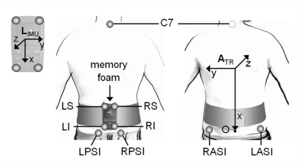
The athletes were equipped with an IMU (FreeSense, Sensorize Ltd, Italy) containing a 3D accelerometer and a 3D gyroscope (± 6 g and±500 deg·s-1 of full range, respectively; 100 samples·s-1) providing linear acceleration and angular velocity components, respectively along and about the axes of a unit-embedded frame (IMU local reference frame: LIMU). The
IMU was mounted so that its axes were parallel to the trunk anatomical axes (Figure 1). The unit data were transmitted via Bluetooth® to a laptop computer. Careful attention was paid to the fixation of the IMU on the lower back trunk (L2 level) to limit its oscillations relative to
the skeleton.19 To this aim, an ad-hoc elastic belt was used along with a memory foam material placed between the paravertebral muscles and the IMU.
To validate the IMU estimates, a nine-camera stereophotogrammetric system (Vicon MX3, Oxford, UK, 200 samples·s-1) was used. Four retro-reflective markers were attached to the IMU and five on the subjects’ trunk (Figure 1) to determine the orientation of the unit and of the trunk, which was considered to be rigid and described as a line joining shoulder and hip joint centers.9 A synchronization task (sudden trunk flexion-extension from standing) was performed at the beginning of each trial.
Data analysis
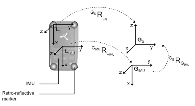
To remove random noise, marker and IMU data were low-pass filtered using a 40-points moving average filter (smooth function, loess method, Matlab®, MathWorks, MA). A global inertial reference frame (GIMU) was defined in a static phase aligning the x-axis of
LIMU with the gravity vector (Figure 2). The orientation matrix of LIMU with respect to GIMU
was computed through an adaptive Kalman filter,22 ad hoc designed to combine the information provided by the accelerometer and the gyroscope during the static and non-static phases of the analyzed motor task. The ratio between two noise sources associated to the two sensors (rgyro/acc) was adaptively modified during the sprint start to suit the characteristics of
the movement. In particular, a threshold (t) was defined for the difference between predicted and measured values of the system state,22 i.e. the IMU inclination in the sagittal plane. Below t, rgyro/acc was set to use the accelerometer and the IMU inclination was computed
through a quaternion based approach;23 above t, rgyro/acc increased in favour of the gyroscope,
and the unit inclination was estimated by integrating the angular velocity around the y-axis, with the initial conditions obtained by the accelerometer-based estimates. Ad hoc trials were
performed to select the following initial values of the Kalman filter parameters:24 t = 0.5 deg, and rgyro/acc equal to 1 and 100 deg·s·m-1, below and above the threshold t, respectively.
To obtain reference values, a stereophotogrammetric local reference frame (LS) was
defined (Figure 2) and its orientation in the stereophotogrammetric global reference frame (GS) was obtained in each instant of time. In order to compare the orientation of LS and LIMU
in the same global reference frame, the latter had to be expressed with respect to GS. To this
aim, the time-invariant rotation matrix relating the stereophotogrammetric and the IMU global reference frames was calculated, assuming that LS was coincident with LIMU at time
zero (Figure 2). To compare the IMU orientation with the whole trunk orientation, a trunk anatomical frame (ATR) was defined (Figure 1) and its orientation with respect to GS obtained
in each instant of time. Finally, Tait–Bryant angles (axis mobile rotation sequence: yxz) were calculated with respect to GS, from the orientation of LIMU, LS and of the whole trunk ATR.
The rotation about the y-axis, referred to as angular displacement β, was further considered. Four phases were identified using β: on your marks (OYM), transition (TNS), set (SET), and pick-up (PCK) (Figure 3). The average β values during OYM and SET (βOYM and
βSET), the variation from βOYM to βSET (Δβ), and the peak angular velocities during TNS and
PCK (ωTNS and ωPCK) were computed from each curve. The absolute difference between each
parameter set, as obtained from LIMU and LS (IMU accuracy), and from LIMU and ATR
(IMU-trunk consistency), was then evaluated (ΔIMU and ΔTR).
Statistical analysis
To assess both IMU accuracy and IMU-trunk consistency, the β curve as obtained from LIMU was compared to those obtained from LS and ATR, respectively. To this aim, the
difference between βOYM obtained from the LIMU and ATR was computed (βOFF = 18±4 deg)
trunk curves, and the Pearson’s correlation coefficient (r) and the Root Mean Square Difference (RMSD) were computed to assess the curve similarity. RMSD was also expressed in percentage of Δβ (RMSD%). The same coefficients were assessed for the PCK phase alone
(rPCK, RMSDPCK, RMSD%PCK), which is the most affected by inertial factors. After a normal
distribution test (Shapiro-Wilk test), the effect of the factor athlete was verified on all parameters and absolute differences by a repeated-measures one-way ANOVA, and descriptive statistics (mean±Standard Deviation (SD)) was performed (SPSS Inc. 17.0, Chicago, IL, USA; alpha = 0.05). Moreover, to assess the agreement between IMU and stereophotogrammetry, Bland–Altman analysis of ΔIMU and ΔTR was performed,25,26 and the
absence of heteroscedasticity27 was verified.
Results
For the IMU accuracy, β curves from LIMU and LS presented a RMSD% lower than
4±3 % and a high correlation (r) during the whole trial and the PCK phase alone (Table 1). After removing βOFF, which was found to be different among athletes (P = .001), similar
results were obtained for the IMU-trunk consistency (Table 1).
All parameters and absolute differences proved to be normally distributed. While all estimated parameters differed among athletes (P < .001), no statistical differences resulted for
ΔIMU and ΔTR (P > .15). ΔIMU and ΔTR for the angular displacement parameters (βOYM, βSET,
Δβ) were lower than 1 deg and 4 deg, respectively, while Estimated values of ΔIMU and ΔTR
for ωPCK were found to be lower than 9 and 6 deg·s-1, and decreased to 8 and 4 deg·s-1 for
ωTNS (Table 2). Bland–Altman plots indicated a good agreement for both IMU accuracy and
IMU-trunk consistency. Symmetry of the confidence interval of ΔIMU and ΔTR was observed
heteroscedasticity was found for all parameters included in ΔIMU and ΔTR (overall average
correlation = 0.09±0.41).
Discussion
The reliability of a lower-trunk mounted IMU in estimating the trunk inclination and angular velocity during the sprint start was assessed. Although in the present study, athletes were forced to complete the task in a relatively short distance, the average peak angular velocity of the trunk (ATR: ωPICK-UP = 198±31 deg·s-1) was comparable with previous
literature results7,13 (170±60 deg·s-1 and 186±48 deg·s-1).
The accuracy of the estimate of the IMU rotation about one of its local axes was supported by the agreement between the unit and the reference estimates. Such agreement proves that the main limitations concerning the use of IMUs19,20 do not have a detrimental effect on the unit inclination estimate, even during the most jerked phase of the sprint start. In particular, the efficacy of the implemented adaptive Kalman filter is attested by the strong curve similarity,28 which also suggests that the site and method of the unit attachment were effective in limiting the effects of the soft tissue movements.
The IMU and trunk inclinations were in agreement during the whole trial and during the PCK phase alone. However, they presented an initial offset, βOFF, which had a negligible
variability over repeated measurements, but was subject-dependent. This offset (18±4 deg) depends on the trunk model commonly used by coaches and adopted in the study, i.e. rigid trunk. After βOFF removal, ΔTR increased moving from the OYM (0±1 deg) to the SET phase
(4±4 deg), indicating a change in the athletes’ kyphosis and neck extension and further supporting the inadequateness of a rigid trunk model. On the one hand, these results suggest the importance of trunk movement in determining the sprint start performance given the key role of the spine muscles during explosive motor tasks,29 in terms of strength as well as of
control. On the other hand, a comparison between the IMU orientation and the lower trunk, where the unit was located, would reasonably reduce, or even reset βOFF, and would be
independent from changes in the upper spine. Indeed, strong correlations (r) obtained for all athletes and trials suggest that tracking only the lower trunk should not prevent describing the overall start strategy. Moreover, from a field point of view, the variation and rate of variation of the inclination are considered far more interesting than the relevant absolute values.
In conclusion, the study shows that a single IMU positioned on the lower back trunk provides reliable angular displacements and angular velocity in the sagittal plane during both the block start and the beginning of the pick-up phase of the sprint. The results support the possibility for coaches to be supplemented with a reliable and wearable instrument to collect information about the sprint start performance of their athletes directly in the field.
Acknowledgements
The authors wish to acknowledge Dr. Marco Donati for his technical support in developing the Kalman filter, and John McCamley for his contribution to the refinement of the manuscript.
References
1. Čoh M, Tomažin K, Stuhec S. The biomechanical model of the sprint start and block acceleration. Facta Univ Ser Phys Ed Sport. 2006;4(2):103-114.
2. Harland MJ, Steele JR. Biomechanics of the sprint start. Sports Med. 1997;23(1):11-20. 3. Schot PK, Knutzen KM. A biomechanical analysis of four sprint start positions. Res Q
Exerc Sport. 1992;63(2):137-147.
4. Fortier S, Basset FA, Mbourou GA, Faverial J, Teasdale N. Starting block performance in sprinters: a statistical method for identifying discriminative parameters of the performance and an analysis of the effect of providing feedback over a 6-week period. J
Sports Sci Med. 2005;4(2):134-143.
5. Mero A, Komi PV, Gregor RJ. Biomechanics of sprint running - a review. Sports Med. 1992;13(6):376-392.
6. Kugler F, Janshen L. Body position determines propulsive forces in accelerated running. J
Biomech. 2010;43(2):343-348.
7. Slawinski J, Bonnefoy A, Ontanon G, et al. Segment-interaction in sprint start: analysis of 3D angular velocity and kinetic energy in elite sprinters. J Biomech. 2010;43(8):1494-1502.
8. Mero A, Komi PV. Reaction-time and electromyographic activity during a sprint start. Eur
J Appl Physiol Occup Physiol. 1990;61(1-2):73-80.
9. Jones R, Bezodis I, Thompson A. Coaching sprinting: expert coaches' perception of race phases and technical constructs. Int J Sport Sci Coach. 2009;4(3):385-396.
10. Slawinski J, Bonnefoy A, Leveque JM, et al. Kinematic and kinetic comparisons of elite and well-trained sprinters during sprint start. J Strength Cond Res. 2010;43(8):1494-1502..
11. Čoh M, Jošt B, Škof B, Tomažin K, Dolenec A. Kinematic and kinetic parameters of the sprint start and start acceleration model of top sprinters. Acta Univ Carol Gym. 1998;28:33-42.
12. Mero A, Luhtanen P, Komi PV. A biomechanical study of the sprint start. Scand J Sport
Sci. 1983;5:20-28.
13. Natta F, Decker L, Boisnoir A. Caractérisation des comportements posturo-cinétiques en sprint. Paris:INSEP. Rapport du projet de recherche MJSVA n° 03-006. http://195.154.95.153:81/archimede/ExploreOneUpload.do;jsessionid=C4906B0B3D45 3D1195C326817AAC1576?vref=664. Published 2006. Accessed May 22, 2012.
14. Purcell B, Channells J, James D, Barret R. Use of accelerometers for detecting foot-ground contact time during running. Proc. SPIE. doi:10.1117/12.638389
15. Bergamini E, Picerno P, Pillet H, Natta F, Thoreux P, Camomilla V. Estimation of temporal parameters during sprint running using a trunk-mounted inertial measurement unit. J Biomech. 2011;45(6):1123-1126.
16. Channels J, Purcell B, Barrett R, James D. Determination of rotational kinematics of the lower leg during sprint running using accelerometers. Proc. SPIE.
doi:10.1117/12.638392
17. Faber GS, Kingma I, Bruijn SM, van Dieen JH. Optimal inertial sensor location for ambulatory measurement of trunk inclination. J Biomech. 2009;42(14):2406-2409. 18. Wong WY, Wong MS. Trunk posture monitoring with inertial sensors. Eur Spine J.
2008;17(5):743-753.
19. Forner-Cordero A, Mateu-Arce M, Forner-Cordero I, Alcantara E, Moreno JC, Pons JL. Study of the motion artefacts of skin-mounted inertial sensors under different attachment conditions. Physiol Meas. 2008;29(4):N21-N31.
20. Liu W, Nigg BM. A mechanical model to determine the influence of masses and mass distribution on the impact force during running. J Biomech. 2000;33(2):219-224.
21. Sabatini AM. Quaternion-based extended Kalman filter for determining orientation by inertial and magnetic sensing. IEEE Trans Biomed Eng. 2006;53(7):1346-1356.
22. Jurman D, Jankovec M, Kamnik R, Topic M. Calibration and data fusion solution for the miniature attitude and heading reference system. Sens Actuators A Phys. 2007;138(2):411-420.
23. Favre J, Jolles BM, Siegrist O, Aminian K. Quaternion-based fusion of gyroscopes and accelerometers to improve 3D angle measurement. Electron Lett. 2006;42(11):612-614. 24. Mazzà C, Donati M, McCamley J, Picerno P, Cappozzo A. An optimized Kalman filter
for the estimate of trunk orientation from inertial sensors data during treadmill walking.
Gait Posture. 2012;35(1):138-142.
25. Bland JM, Altman DG. Agreement between methods of measurement with multiple observations per individual. J Biopharm Stat. 2007;17(4):571-582.
26. Atkinson G, Nevill AM. Statistical methods for assessing measurement error (reliability) in variables relevant to sports medicine. Sports Med. 1998;26(4):217-238.
27. Nevill AM, Atkinson G. Assessing agreement between measurements recorded on a ratio scale in sports medicine and sports science. Br J Sports Med. 1997;31(4):314-318. 28. Cohen J. Statistical power for the behavioral sciences. 2nd ed. Hillsdale, NJ: Lawrence
Erlbaum Associates; 1988:75-105.
29. Bobbert MF, Van Soest AJ. Effects of muscle strengthening on vertical jump height: a simulation study. Med Sci Sports Exerc. 1994;26(8):1012-1020.
Figure 1 – IMU, belt, memory foam material, and marker location. The markers placed on
the IMU (LI, LS, RI, RS), trunk (C7) and pelvis, (RPSIS, LPSIS, RASIS, LASIS) are indicated, as well as the trunk anatomical reference frame, ATR: O: midpoint between LPSIS
and RPSIS; x-axis: joining C7 and O, positive downward; z-axis: directed as the vector product between the x-vector and the vector LASIS-RASIS; y-axis: orthogonal to the x-z plane.
Figure 2 – Local (L) and global (G) frames for the inertial unit (IMU) and the
stereophotogrammetric system (S). S S
G L
R : orientation of the marker frame built on the IMU (LS) with respect to the stereophotogrammetric global reference frame (GS); S IMU
G L
R :
orientation of the local IMU frame (LIMU) with respect to the IMU global reference frame
(GIMU);
S IMU
G G
R
: orientation of the IMU global reference frame (GIMU) with respect to theFigure 3 – Raw acceleration (A) and angular velocity (B) data, for a randomly chosen subject
and trial, measured along and about the IMU local axes (LIMU, depicted in the bottom left
corner of panel A: x axis (solid line), y axis (dashed line) and z axis (dotted line). Angular displacement β (C), obtained for the same subject and trial from the IMU local frame, LIMU,
(solid line), from the stereophotogrammetric local frame, LS, (dashed line) and from the trunk
anatomical reference frame, ATR, (dotted line). Static phases (OYM, SET, shaded intervals)
and non-static phases (TNS, PCK, white intervals) are shown. The angular displacement parameters (βOYM, βSET and Δβ) are also indicated. β was considered to be zero when the unit
Table 1 – Curve similarity analysis: mean±SD of the correlation coefficient (r), the Root
Mean Square Difference (RMSD) and the RMSD expressed in percentage of Δβ (RMSD%),
computed to assess IMU accuracy and IMU-trunk consistency, relative to the whole task and to the PCK phase alone.
IMU accuracy IMU-trunk consistency r 0.994 ± 0.013 0.998 ± 0.002 rPCK 0.995 ± 0.015 0.998 ± 0.001 RMSD [deg] 3 ± 2 3 ± 2 RMSD% [%] 4 ± 3 5 ± 3 RMSDPCK [deg] 3 ± 3 3 ± 2 RMSD%PCK [%] 5 ± 3 6 ± 4
Table 2 – Parameter analysis: descriptive statistics (mean ± SD) of each estimated parameter
and of the absolute differences between the IMU and the stereophotogrammetric sets of parameters, for both IMU accuracy (ΔIMU) and IMU-trunk consistency (ΔTR).
LS LIMU ATR IMU accuracy IMU-trunk consistency ΔIMU ΔTR βOYM [deg] -29 ± 6 -29 ± 6 -29 ± 6 1 ± 1 0 ± 1 βSET [deg] 12 ± 7 12 ± 6 9 ± 6 1 ± 1 4 ± 4 Δβ [deg] 43 ± 8 43 ± 7 39 ± 5 1 ± 1 4 ± 4 ωTNS [deg/s] 98 ± 18 106 ± 19 93 ± 18 8 ± 4 4 ± 6 ωPCK [deg/s] -192 ± 29 -201 ± 25 -198 ± 31 9 ± 6 6 ± 13