Université de Montréal
Determination of viral load and integration status of HPV 16
in normal and LSIL exfoliated cervical cells
par
Otelinda de Morais
Département de microbiologie et immunologie Faculté de Médecine
Mémoire présenté à la Faculté de Médecine en vue de l'obtention du grade de Mâitrise
en Microbiologie et Immunologie
Montreal, QC Novembre 2009
© Otelinda de Morais, 2009 Université de Montréal
University of Montreal Faculty of Superior Studies
This entitled memory:
Determination of viral load and integration status of HPV 16
in normal and LSIL exfoliated cervical cells
Presented by: Otelinda de Morais
Was evaluated by a jury composed from the following professors: Dr. Claude Lemieux-président rapporteur
Dr. Francois Coutlée--directeur de recherche Dre. Lise St-Jean-membre du jury
RÉSUMÉ
L’intégration du génome du virus papilloma humain (VPH) a été reconnu jusqu’`a récemment comme étant un événnement fréquent mais pourtant tardif dans la progression de la maladie du col de l’utérus. La perspective temporelle vient, pourtant, d’être mise au défi par la détection de formes intégrées de VPH dans les tissus normaux et dans les lésions prénéoplasiques.
Notre objectif était de déterminer la charge virale de VPH-16 et son état physique dans une série de 220 échantillons provenant de cols uterins normaux et avec des lésions de bas-grade. La technique quantitative de PCR en temps réel, méthode Taqman, nous a permis de quantifier le nombre de copies des gènes E6, E2, et de la B-globine, permettant ainsi l’évaluation de la charge virale et le ratio de E6/E2 pour chaque spécimen. Le ratio E6/E2 de 1.2 ou plus était suggestif d’intégration. Par la suite, le site d’intégration du VPH dans le génome humain a été déterminé par la téchnique de RS-PCR.
La charge virale moyenne était de 57.5±324.6 copies d'ADN par cellule et le ratio E6/E2 a évalué neuf échantillons avec des formes d’HPV intégrées. Ces intégrants ont été amplifiés par RS-PCR, suivi de séquençage, et l’homologie des amplicons a été déterminée par le programme BLAST de NCBI afin d’identifier les jonctions virales-humaines. On a réussi `a identifier les jonctions humaines-virales pour le contrôle positif, c'est-à-dire les cellules SiHa, pourtant nous n’avons pas detecté d’intégration par la technique de RS-PCR dans les échantillons de cellules
cervicales exfoliées provenant de tissus normaux et de lésions de bas-grade. Le VPH-16 est rarement intégré dans les spécimens de jeunes patientes.
Mots Clés : Virus Papilloma Humain, LSIL, Intégration HPV, Charge Virale, PCR en temps réel, RS-PCR, PCR-séquençage, HPV16.
ABSTRACT
Integration of human papillomavirus (HPV) has, until recently, been a frequent but late event in cervical carcinogenesis. The temporal view has, however, been challenged lately as integrated forms of HPV have been detected even in normal and preneoplastic lesions.
Our objective was to describe HPV 16 load and physical state in a series of 220 normal and low grade cervical samples. We used quantitative real-time PCR, Taqman method, targeting E6, E2 and B-globin to calculate the HPV 16 load and the E6/E2 ratio in each sample. An E6/E2 ratio of 1.2 was used as a surrogate marker of integration. The site of integration was determined by restriction site PCR.
Results show that the average viral load was 57.5±324.6 copies of DNA per cell, while E6/E2 ratio identified 9 samples with integrants. These integrants underwent amplification by restriction site PCR, followed by sequencing and nucleotide blast to identify the human-viral junctions. In conclusion, although it was possible to identify viral-host junctions with the integration positive control, that is, the SiHa cell line, the exfoliated cells of normal and low grade cervical lesions were negative for integration site by RS-PCR. HPV-16 is seldom integrated in specimens from young patients.
Key Words : Human Papillomavirus 16, LSIL, HPV integration, Viral load, real-time PCR, RS-PCR, PCR sequencing, HPV16.
SCIENTIFIC PUBLICATIONS
FROM THE AUTHOR
Impaired learning and LTP in mice expressing the carboxy terminus of the Alzheimer amyloid precursor protein.
Nalbantoglu J, Tirado-Santiago G, Lahsaïni A, Poirier J, Goncalves O, Verge G, Momoli F, Welner SA, Massicotte G, Julien JP, Shapiro ML.
Nature. 1997 May 29;387(6632):500-5.
Interleukin 1 enhances growth factor-dependent proliferation of the clonogenic cells in acute myeloblastic leukemia and of normal human primitive hemopoietic precursors. Hoang T, Haman A, Goncalves O, Letendre F, Mathieu M, Wong GG, Clark SC. J Exp Med. 1988 Aug 1;168(2):463-74.
Interleukin-6 enhances growth factor-dependent proliferation of the blast cells of acute myeloblastic leukemia.
Hoang T, Haman A, Goncalves O, Wong GG, Clark SC. Blood. 1988 Aug;72(2):823-6.
The genetic consequences of the Thy- mutation to CHO cells. Meuth M, Gonçalves O, Trudel M.
Basic Life Sci. 1985;31:297-312. Review.
Structural alterations of the aprt locus induced by deoxyribonucleoside triphosphate pool imbalances in Chinese hamster ovary cells.
Goncalves O, Drobetsky E, Meuth M. Mol Cell Biol. 1984 Sep;4(9):1792-9
Structure of mutant alleles at the aprt locus of Chinese hamster ovary cells. Nalbantoglu J, Goncalves O, Meuth M.
J Mol Biol. 1983 Jul 5;167(3):575-94.
A selection system specific for the Thy mutator phenotype. Meuth M, Gonçalves O, Thom P.
TABLE OF CONTENTS
RÉSUMÉ ... iii
ABSTRACT ... v
SCIENTIFIC PUBLICATIONS ... vi
TABLE OF CONTENTS ... vii
LIST OF FIGURES ... xi
Literature Review... xi
Article ... xiii
Figure 1: ... xiii
Figure 2: ... xiii
LIST OF TABLES ... xiv
Literature Review... xiv
Article ... xv
Table 1: ... xv
LIST OF ABBREVIATIONS ... xvi
DEDICATE... xvii
ACKNOWLEDGMENTS ... xviii
LITERATURE REVIEW ... xix
Introduction ... 1
1. Basics of Human Papillomavirus Virology ... 3
1.1 History... 3
1.2 Taxonomy of papillomaviruses... 5
1.2.1 Family Classification ... 5
1.2.2 Genotype Classification ... 6
1.2.3 Phylogenetic Classification ... 8
1.2.3.1 Genus or Site of Infection ... 9
1.2.3.2 Species: Oncogenic versus non-oncogenic HPV types ... 10
1.2.4 Clinical Association: Host-Site-Disease ... 11
1.3.1-Virion Structure ... 12
1.3.2-Genome Structure and Organization ... 14
1.4-Normal Infectious Cycle ... 15
1.4.1 Attachment, Entry, and Uncoating ... 16
1.4.2 Virus Replication and Life Cycle ... 17
1.4.2.1 Productive Infection ... 17
1.4.2.1.1 Early Stage of Productive Infection ... 18
1.4.2.1.2 Late Stage of Productive Infection ... 18
1.4.3 Regulation of Viral Gene Expression ... 20
1.4.3.1 Viral Transcription ... 20
1.4.3.2 The Long Control Region (LCR) ... 20
1.4.4 HPV Protein Functions ... 21
1.4.4.1 Regulatory Proteins E1 & E2 ... 22
1.4.4.1.1 In Viral Replication... 22
1.4.4.1.2 In Genome Segregation... 23
1.4.4.1.3 In Viral Transcription ... 24
1.4.4.2 Proliferatory Role of E6 and E7 in HPV Productive Life cycle ... 25
1.4.4.2.1 Basics of E6 & E7 ... 25
1.4.4.2.2 HPV-Infected Epithelial Differentiation ... 26
1.4.4.2.3 Role of E6 & E7 in Cell Cycle Progression ... 27
1.4.4.2.3.1.1 E7–pRb Model ... 27
1.4.4.2.3.2.1 Normal DNA damage Response ... 31
1.4.4.2.3.2.2 E6-p53 Model ... 31
1.4.4.2.3.2.3 E6 associates with Bak and Bax ... 31
1.4.4.3 The E4 and E5 Proteins ... 33
1.4.4.3.1 The E5 Protein ... 33
1.4.4.3.2 The E4 Protein ... 34
1.4.4.4 Structural Proteins L1-L2 ... 35
1.5 Virus Assembly and Release ... 35
1.6 Abnormal Proliferative Infection ... 35
2.1 HPV acquisition and transmission ... 36
2.2 Prevalence of HPV Infection ... 37
2.2.1 Definition ... 37
2.3.1 HPV Clearance ... 40
2.3.2 HPV Persistence ... 42
3. Development of cervical cancer ... 42
3.1 Pap Test ... 42
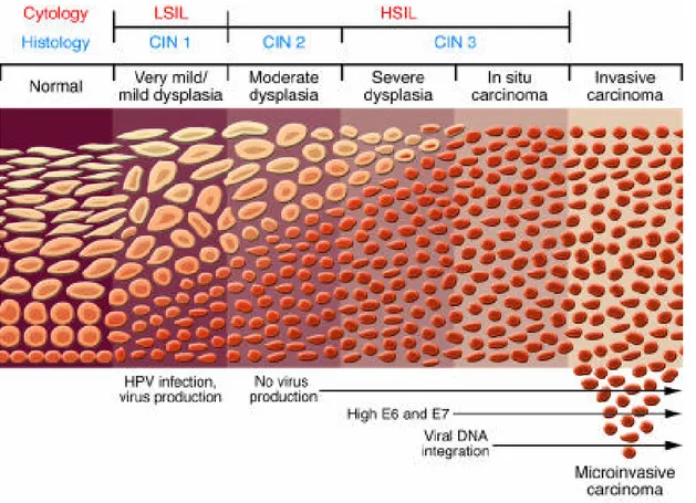
3.2 Pre-Cancerous Abnormalities ... 43
3.2.1 Classification Systems ... 43
3.2.2 Microscopic Lesion Progression ... 44
3.2.2.1 Low Grade CIN... 45
3.2.2.2 High Grade CIN ... 45
3.2.3 Molecular Progression of Lesions ... 46
4. Cancer of the Cervix ... 47
4.1 Cause of Cervical Cancer ... 47
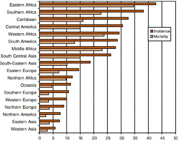
4.2 Burden of Cervical Cancer to Humanity ... 48
4.3 The Cervical Transformation Zone ... 49
4.4 Histopathology ... 50
4.5.1 Definition of high risk-low risk HPV ... 51
4.5.2 Low risk HPV ... 51
4.5.3 High risk HPV... 52
4.5.3.1 Human Papillomavirus Type 16 ... 53
4.6 Invasive cervical cancer ... 54
4.6.1 Persistence of HPV Infection ... 54
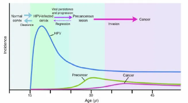
4.6.2 Relationship among incidences of cervical HPV infection, precancer, and cancer ... 55
5. Determinants of clinical progression of HPV infection ... 56
5.1 Environmental Factors ... 56 5.1.1 Infectious Agents ... 56 5.1.1.1 Herpes ... 56 5.1.1.2 Chlamydia trachomatis ... 57 5.1.1.3 HIV/AIDS ... 57 5.1.1.4 Cervical inflammation ... 58
5.1.2 Hormonal Contraceptive Use ... 58
5.1.3 Tobacco Smoking ... 60
5.2 Genetic or Host Factors ... 60
5.2.1 Evidence of Immune Response ... 61
5.2.2.1 MHC Complex ... 63
5.2.2.2 p53 Polymorphism ... 63
5.3 Viral Factors in Progression to Malignancy ... 64
5.3.1 Genotype- High risk-low risk differences ... 64
5.3.1.1In Vitro Properties of E6 and E7 Proteins ... 65
5.3.1.3 E6 Oncoprotein ... 66
5.3.1.3.1 E6 complexes E6AP and p53 ... 66
5.3.1.3.2 p53 dependent repression of transcription ... 67
5.3.1.3.4 E6 Activates Telomerase ... 67
5.3.2 HPV Viral Polymorphism ... 68
5.3.3 HPV Viral Load ... 69
5.3.4 Integration of Viral DNA ... 70
5.3.4.1 In Cervical cancer and in Lesions ... 70
6.1 Southern hybridisation ... 72
6.2 PCR-based methods ... 73
6.2.1 Real-Time QT-PCR-an indirect method ... 73
6.2.2.1 Restriction Site PCR ... 76 STUDY OBJECTIVE ... 79 ARTICLE ... 81 DISCUSSION ... 116 CONCLUSION ... 144 REFERENCES ... 146
LIST OF FIGURES
Literature Review
Figure 1:
Model of the human papillomavirus showing the arrangement of capsid proteins Reference: http://www.bris-ac.uk/biochemistry/gaston/HPV/hpv_information.htm
Figure 2:
Phylogenetic tree containing the sequences of 118 papillomavirus types
Reference: deVilliers et al.Classification of papillomaviruse.Virology 2004;324:17-27 Figure 3:
Linear representation of the HPV16 genome
Reference: http://www.ircm.qc.ca/microsites/hpv/en/390.html Figure 4:
Genome organization of human papillomavirus type 16
Reference: Schiffman M et al.Human papillomavirus and cervical cancer. The lancet 2007; 370:890-907
Figure 5:
HPV genome and its expression within the epithelium
Reference: Schiffman M et al. Human papillomavirus and cervical cancer. The lancet 2007; 370:890-907
Figure 6:
Interaction of cellular proteins with HPV E7
Reference: Wise-Draper TM et al. Papillomavirus E6 and E7 proteins and their cellular targets. Front Biosci 2008; 13: 1003-17
Figure 7:
Representation of cellular proteins affected by HR-E6
Reference: Tungteakkhun SS et al. Cellular binding partners of the human papillomavirus E6 protein. Arch Virol 2008; 153:397-408
Figure 8:
Major steps in the development of cervical cancer
Reference: Schiffman M et al.Human papillomavirus and cervical cancer. The lancet 2007; 370:890-907
Figure 9:
Progression from a benign cervical lesion to invasive cervical cancer Reference: http://www.jci.org/articles/view/28607/figure/2
Figure 10:
Cancer of the uterine cervix: age-standardised (world) incidence and mortality rates per 100 000 (all ages) in 18 world regions
Reference: Sankaranarayanan R, Ferlay ME J. Worldwide burden of gynaecological cancer: The size of the problem. Best Practice & Research Clinical Obstetrics & Gynaecology 2006;20:207-225
Figure 11:
High-risk types of HPV have been identified in a wide range of malignancies
Reference: Smith JS, Lindsay L, Hoots B, Keys J, Franceschi S, Winer R, et al. Human papillomavirus type distribution in invasive cervical cancer and high-grade cervical lesions: a meta-analysis update. Int J Cancer 2007;121:621-32
Figure 12:
Relationship among incidences of cervical HPV infection, precancer, and cancer Reference: http://www.jci.org/articles/view/28607/figure/3
Figure 13:
Regulation effect of HPV transforming proteins
Reference: Thomison J, et al. Human papillomavirus: molecular and cytologic/histologic aspects related to cervical intraepithelial neoplasia and carcinoma. Human Pathology 2008;39:154-166 Figure 14:
Schematic of the procedure used to detect integration from normal and LSIL cervical specimens with RS-PCR
Figure 15:
Schematic of RS-CR, involving two rounds of PCR, followed by sequencing of amplicons Figure 16:
Representative gel electrophoresis of RS-PCR amplification products
Figure 17: Blast Nucleotide Alignment of SiHa sample no. 75 with integration occurring at nucleotides 3134 of E2 sequence
Figure 18A:
Amplification of L1-E2 Deletion Artefact in F268 amplified with NP4 and RSO1 primers Figure 18 B:
PCR Amplification of L1- E2 Deletion Artefact in specimen F268 amplified with NP5 and RSO3 primers
Figure 19:
Sequence homologous to HPV 16 & Human DNA, depicting difficulty in interpretation of NCBI BLAST results
Article
Figure 1:
Log-transformed HPV loads (HPV DNA copies per cell) at recruitment and at follow-up visits among HPV-positive women for the 4 genotypes studied.
Figure 2:
Correlation matrix for viral load measurement of four combined HPV types (HPV-16,18,31,45) at accrual and follow-up
Figure 3:
Combined HPV(16,18,31,45) clearance stratified by tertiles of viral load assuming multiple events per individuals (unit of analyses is infection)
Figure 4:
Predicted ROC curve between the viral load (continuous) at visit t-1 and persistent infection at visit (t) within specified periods of follow-up
LIST OF TABLES
Literature Review
Table I:
Phylogenetic and Epidemiologic Classification of HPV types
Reference : Munoz N, Bosch FX, de Sanjose S, et al. Epidemiologic classification of human papillomavirus types associated with cervical cancer. N Engl J Med 2003; 348:518-527.
Table II:
Function of HPV proteins
Reference: Thomison J, et al. Human papillomavirus: molecular and cytologic/histologic aspects related to cervical intraepithelial neoplasia and carcinoma. Human Pathology 2008; 39:154-166 Table III:
Prevalence and incidence rates of infection for the most frequently detected HPV genotypes in the McGill Concordia Cohort under Study
Reference: Richardson H, et al.The natural history of type-specific human papillomavirus infections in female university students. Cancer Epidemiology, Biomarkers & Prevention 2003;12:485-490
Table IV:
Proportion of cervical cancer caused by the carcinogenic HPV types
Reference: Schiffman M et al.Human papillomavirus and cervical cancer. The lancet 2007; 370:890-907
Article
Table 1:
Between-visit correlation (r) of HPV load measurements by HPV type at entry and follow-up visits
Table 2:
Geometric means of HPV viral loads as a function of selected characteristics at the first occurrence of positivity for a given HPV type
Table 3:
Hazard ratios of HPV clearance from cox regression models, stratified by various levels of viral load
Table 4:
Odd ratios for associations between persistent HPV at visit (t) and viral load at visit (t-1) within specified periods of follow-up, McGill-Concordia cohort study(GEE model with exponential coefficients)
Table 5:
Interference of background human DNA in quantitation of HPV-16 DNA with HPV-16 E6 and HPV-16 E2 real-time PCR assays
LIST OF ABBREVIATIONS
AIDS: Acquired immune deficiency syndrome APOT: Amplification of papillomavirus oncogene transcript ATPase: Adenosine triphosphatase
ASCUS: Atypical squamous cells of undetermined significance CIN: Cervical intraepithelial neoplasia
CIS: Carcinoma in situ
COPV: Canine oral papillomavirus
DIPS: Detection of integrated papillomavirus sequences
DNA: Deoxyribonucleic acid
E6-AP: E6 Associated protein EGF: Epidermal growth factor
EV: Epidermodysplasia Verruciformis GAPDH: Glyceraldehyde 3-phosphate dehydrogenase HIV: Human immunodeficiency virus HLA: Human leukocyte antigen
HPV: Human papillomavirus
HR-HPV: High risk human papillomavirus HR-E6: High risk E6 protein
HSIL: High-grade squamous intraepithelial lesions hTERT: Human telomerase reverse transcriptase
ICC: Invasive cervical carcinoma
IFN: Interferon
LSIL: Low-grade squamous intraepithelial lesions LCR: Long control region
mRNA: messenger ribonucleic acid MHC: major histocompatibility complex ORF: Open reading frame
ORF(E): Early open reading frame ORF(L): Late open reading frame
Pap test: Papanicolaou test
PCR: Polymerase chain reaction pRB: Retinoblastoma protein
PV: Papillomavirus
QT-PCR: Quantitative PCR
ROPV : Rabbit oral papillomavirus
RS-PCR: Restriction site polymerase chain reaction RSOs: Restriction site oligonucleotides
SCC: Squamous cell carcinoma SIL: Squamous intraepithelial lesion S-phase: Synthesis phase
T-cell: Thymus cell
DEDICATE
This memoir is dedicated to my beloved husband, and to the love of our lives, Nancy, Katie and Kevin, in appreciation for the constant encouragement, help and support provided (without a complaint), during the course of my study.
ACKNOWLEDGMENTS
My deepest thanks to my research director, Dr Coutlée, for his availability, his useful comments and expert guidance throughout the program.
Muito Obrigada!
My gratitude goes also to the attending research staff, Helene Voyer, Pierre Forest, and Simon Gagnon for introducing me to the lab, answering my questions, and for the helpful discussions.
LITERATURE REVIEW
Introduction
Over 100 types of human papillomavirus (HPV) have been identified based on DNA sequence analysis [1]. Of these, 40 infect the anogenital region and have been classified according to their oncogenic potential into high-risk and low-risk types. High-risk papillomaviruses have been found to be the single most important risk factor of cervical cancer [2], with HPV 16, the most frequent oncogenic type, accounting for over 50% of cervical cancer[3, 4]. Most women are infected with the HPV virus shortly after sexual debut, with prevalence reaching a maximum around 25 years of age. Prevalence decreases rapidly thereafter, as most HPV infections become latent or are cleared by the host immune system [5-7].
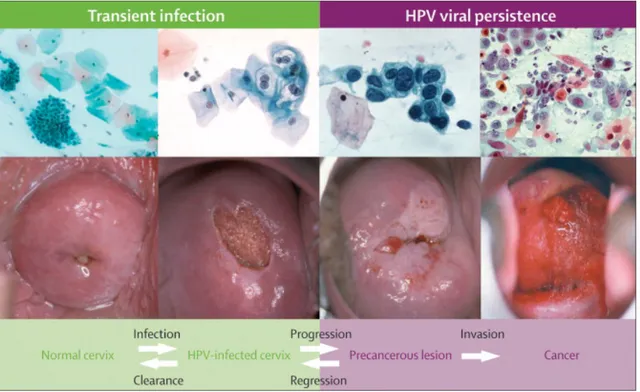
Thus while genital HPV infection is the most common sexually transmitted infection, cancer of the cervix is an uncommon outcome of a high-risk HPV infection. Recent research suggests viral load and viral integration as potential markers for cervical disease progression [8, 9].
Association between increasing viral load of HPV 16 and increasing severity of cervical lesions has been found [10-13]. Conversely, smaller amount of HPV16 DNA in women with HSIL compared to those with LSIL has also been reported [14, 15].
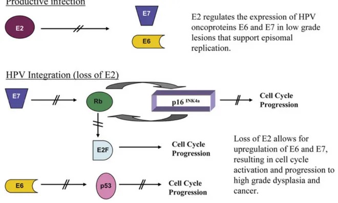
The physical status of high-risk HPV also promises to be a risk marker to evaluate progression of cervical lesions to cancer [16-18]. HPV integration into the cellular genome usually disrupts the E1 and E2 genes, E2 being the preferential site of
integration [19, 20]. The disruption of the E2 regulatory gene, due to integration, results in lack of expression of the E2 protein with subsequential upregulation of the oncogenic E6 and E7 proteins [18, 21]. The continuous overexpression of the E6 and E7 proteins contributes to malignant transformation.
The role that integration plays in malignant transformation is still being questioned. Initial studies on viral integration found viral DNA to be integrated into the host genome in nearly all cases of cervical carcinomas and cervical carcinoma cell lines [22-26], whereas the HPV genome was usually in episomal form in benign and low-grade cervical intraepithelial lesions[16, 25, 27].
This temporal view of integration has, however, been challenged recently as some investigators [15, 28-30] have detected HPV integration even in preneoplastic lesions. In these studies, viral load and viral integration were assessed with qualitative or quantitative real-time PCR targeting the E2 and E6 gene.
The aim of this study was to quantitatively assess, by real time PCR, amplification of E2/E6 sequences in exfoliated cells from normal and LSIL cervical specimens, from which to evaluate viral load and E6/E2 ratio. An E6/E2 ratio of 1.2 or greater was suggestive of integration in nine samples. Restriction site PCR (RS-PCR), a technique that allows retrieval of human–viral junctions, followed by DNA sequencing, however, did not confirm integration site in these potential integrants, although it did identify integration in the positive control of SiHa cultured cells, and some integration artefacts.
1. Basics of Human Papillomavirus Virology
1.1 HistoryThe papillomaviruses are a very diverse family of non-enveloped double-stranded DNA viruses. These small DNA tumour viruses are found in a wide variety of higher vertebrates including mammals, reptiles, and birds [31, 32]. Papillomaviruses infect both mucosal and epithelial cells and induce cellular proliferation giving rise to malignant or benign tumours (warts, papillomas).
The common wart has been described since ancient times, and is characteristic of cutaneous and mucosal epithelial infections. Ciuffo at the beginning of the 20th century demonstrated cell-free filtrates from warty lesions to transmit the disease leading him to conclude that warts are related to an infectious agent [33].
The first papillomavirus was identified in cottontail rabbits in 1933, but progress on the study of human papillomavirus (HPV) infection was delayed for many decades because the virus could not be propagated in cell culture[34].
In 1956, Koss described the morphological aspects of cells from warty lesions of the cervix, coining the term koilocytic atypia [35]. It took another 20 years, however, before researchers were able to demonstrate that this morphological appearance was due to HPV infection [36-38]. The morphological features of koilocytic atypia, which include perinuclear cytoplasmic clearing, peripheral condensation of cytoplasmic filaments, with nuclear enlargement and
hyperchromasia, have since been confirmed to be diagnostic for effect of the HPV virus and as the direct result of the viral genome replication.
The papillomavirus (PV) were studied less intensively in the 1950s and 1960s. Nevertheless, there were two important advances, namely, the physiochemical analysis of the virions and the demonstration that papillomavirus replication was closely associated with the differentiation process of the infected epithelium [39]. Papillomaviruses have indeed proven difficult to propagate in vitro because these viruses replicate in stratified squamous epithelium, which is not mimicked in monolayer cultures. Also, the species specific nature of HPV has thus far also prevented the adaptation of authentic HPV infection to experimental animals, although some useful animal papillomavirus models have since been described. With the development of molecular cloning technique in the 1970s, however, investigators were able to study the biological and biochemical properties of papillomavirus genomes. Sequencing of the cloned Papillomavirus genomes identified open reading frames and the function of the viral genes was determined by reverse genetics, and this resulted in a revived interest in papillomavirus research [40, 41]. Since, DNA sequence analysis has led researchers to recognize that papillomaviruses are a very diverse group with over 100 human members [31]. During the past decade, it has been determined that a subset of HPV types is closely linked with certain human cancers, most notably, cancer of the cervix. Interest has therefore been focused on this specific subgroup of HPVs which are associated with genital lesions. Of the 40 HPV which infect the anogenital tract, approximately 15 have been found in cervical cancers in a higher percentage than controls, while others are found rarely in cervical cancer, and this has given rise to the distinction between high-risk and low-risk HPV types.
Whilst studies have determined the interaction between HPV and the epithelial host cell, have identified the HPV protein functions and recognized the molecular targets of infected cells, ongoing research seeks to understand the natural history of the infection, to determine the biological properties of the different HPV types, and to identify the role of the nonviral and viral factors in the pathogenesis of cervical disease that may influence the outcome of an HPV infection.
1.2 Taxonomy of papillomaviruses
1.2.1 Family Classification
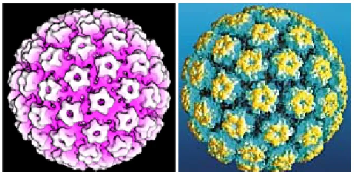
Early systems of classification lumped papillomavirus, polyomavirus and simian vacuolating virus, into a family grouping collectively known as the papovavirus family. This was based on identification of a common genetic structure: all 3 have a small, circular, double-stranded DNA genome (episome) that replicates in the host cell nucleus, and releases a non-enveloped virion with an icosahedral protein capsid as in Figure 1[31].
Figure 1: Atomic Model of the Human papillomavirus showing the arrangement of capsid proteins Later it was recognized, however, that the papillomaviruses were distinct from the other 2 members of this group. The papillomavirus genome
ranges from 6900 up to about 8000 base pairs in length, 60% larger than the polyomavirus genome. As such, the capsid is 55 nm in diameter, rather than 40 nm. The genomes are organized differently and except for the helicase motif of the PV E1 protein, do not share any major nucleotide or amino acid sequences [31]. These discoveries led to the reclassification of papillomaviruses as a distinct family by the International Committee on the Taxonomy of Viruses. HPVs are now
officially recognized as members of the Papillomaviridae family.
1.2.2 Genotype Classification
The common warts and lesions of Epidermodysplasia verruciformis (EV), which contain large quantities of viral particles, provided enough material to isolate viral DNA genomes. Initially, as more and more types of viruses were identified, researchers in the field agreed on a taxonomic system based on numbering, with each subsequent type receiving the next higher number[31]. For instance, HPV1 is an abbreviation for human papillomavirus type 1. An isolate was accepted as a new type based on liquid hybridisation analysis.
Since the early 1980s, however, when the first full genomes of several papillomaviruses were cloned, nearly all known papillomavirus genomes have been sequenced and compiled into a database such as GenBank and EMBL databases[42] This has allowed for a new classification, at the International Papillomavirus Workshop in 1995, based on nucleotide sequence of the L1 gene.
This new classification, based on DNA sequence, includes to date over 100 genotypes. Genotypes are defined as sharing between 71% and 89% identical nucleotide sequences with other HPV types in the L1 open reading frame, which is the most conserved ORF in the papillomavirus genome [43]. Subtypes have between 90% and 98% sequence identity to a prototype sequence, and variants of a genotype have <2% sequence difference in the coding regions [44].
Further conventional cloning of complete genomes has been difficult due to either limited amounts of sample available or because the viral DNA sequence is toxic to the vector systems used in cloning. This has led to an increased use of PCR amplification of overlapping fragments to obtain viral DNA genomes. These are distinguished by the mention cand, as for example, HPVcand (number.) PCR amplification with degenerate primers mainly of the L1 ORF has identified a few hundred potentially PV novel types [45].
1.2.3 Ph Phyloge papillom 2 below Figure 2: Ph modified ve constructed an HPV typ outermost s semicircular 7, 40, 43, an hylogenetic enetic anal mavirus typ w. hylogenetic tree ersion of the Ph using the Treev pe; c-numbers r semicircular sym r symbol refers t nd cand91 togeth c Classifica lysis of th pes based e containing the hylip version 3.5 view program of refer to candida mbols identify p to papillomaviru her form the HPV
ation he L1 sequ on how clo e sequences of 572 and based o f the University ate HPV types. apillomavirus g us species. To gi V species 8 in th uences has osely relate 118 papillomav on a weighted v of Glasgow. The All other abbr genera, e.g. the g
ive an example t he genus alpha-p
s identified ed they are
virus types. The version of the ne e numbers at the reviations refer genus alpha-pap taken from the u papillomavirus. d clear gro e genetical e L1 ORF sequ eighbor-joining e ends of each o to animal papi pillomavirus. Th upper part of the
oupings of lly [31], Fi
uences were use analysis. The tr of the branches i illomavirus type he number at the e figure, the HPV f the igure ed in a ree was dentify es. The e inner V types
Phylogenetic criteria have thus led to the taxonomic levels of family, genus, species, and the previously defined types, subtypes and variants. The sixteen different genera have less than 60% similarity of the L1 gene sequence. Species share between 60% and 70% of L1 ORF identical sequences.
These groupings are relatively consistent with observable papillomavirus phenotypes, including species of origin, tissue tropism, and association with benign versus malignant lesions.
1.2.3.1 Genus or Site of Infection
Based on DNA sequence and protein homologies, the relations between HPV genotypes can be expressed in the form of phylogenetic trees. Tissue tropism of the HPVs is reflected in the grouping of species within a genus. As such, genital mucosal human papillomaviruses are grouped into the genus Alpha-Papillomavirus, although the genus contains a few viruses that are tropic for cutaneous sites and cause common warts. The Alpha-Papillomavirus, however, share certain life cycle features common to this genus that differ from that of other cutaneotropic viruses.
The Beta papillomaviruses are evolutionary distinct from the Alpha genus and seemingly cause asymptomatic infections in the general population. The Beta Papillomavirus have been most frequently isolated from cutaneous epithelium, particularly among patients affected with a rare inherited disorder termed epidermodysplaqsia verruciformis (EV).
While the taxon genus encompasses PV types that have adapted to a particular tissue type and location, this is not absolute. Thus, within the genus Alpha-Papillomavirus, HPV16 is found not only in the genital mucosa, but can also be found in the mucosa of the oropharynx, and in genital cutaneous epithelium.
1.2.3.2 Species: Oncogenic versus non-oncogenic HPV types
The genus Alpha-Papillomavirus which groups genital HPV genotypes is further divided into evolutionarily related subgroups called species [44]. Thus, within a genus, distinct genomic sequences having identical or very similar biological and pathological properties belong to the same species. The sequence-based taxonomy therefore groups the HPV types with known cancer association at the species level. The HPV types that have most often been associated with cervical cancer and its precursor lesions have been evolutionarily clustered into species 5, 7, and 9. Fifteen HPV types are considered to be carcinogenic or high-risk types: 16, 18, 31, 33, 35, 39, 45, 51, 52, 56, 58, 59, 68, 73, and 82. Three are classified as probable high-risk types: 26, 53 and 66. In contrast, HPV types in species 10 have almost no association with invasive cancer. Consequently, these HPV types have been called low-risk types. HPV types 6, 11, 40, 42, 43, 44, 54, 61, 70, 72, 81, and HPV 89 are classified as low-risk types [4].
Further, there is good agreement between epidemiologic classification and the classification based on phylogenetic grouping as seen in Table I below.
TableI: Phylogenetic and Epidemiologic Classification of HPV types. The epidemiologic classification of these types as probable high-risk types is based on zero controls and one to three positive cases.
1.2.4 Clinical Association: Host-Site-Disease
Papillomaviruses have often been classified primarily according to the host species they infect and the sites or diseases with which they are associated. Of the more than 100 human papillomaviruses types that have been identified, they fall into two groups, cutaneous and mucosal HPVs. Mucosal types are associated with oropharyngeal and cervical lesions.
1.3 Ge 1.3.1-V All pap small, n squamo The vir molecul size bou coat), co A linea organiz been va control Figure netic Org Virion Stru pillomaviru nonenvelop ous epitheli rion partic le of doub und to cell omposed o ar represen ed in open ariably ref region (LC e 3: Linear anization ucture uses share ped, icosa ial cells, m cles are ble strande lular histon of 72 penta ntation of n reading ferred to a CR). r represent genes (L e a numbe ahedral DN mucosal and 52-55 nan ed circular nes and co americ caps f the HPV frames (O as the upst tation of th L1-L2), lon er of char NA viruse d cutaneou nometers i r DNA of ontained w somers. V genome ORF’S) an tream regu he HPV16 g ng control racteristics s that rep us. in diamete approxima within a cap e is depic nd a non-c ulatory reg genome; ea region (LC . Papillo licate in t er and con ately 8000 psid (or sp ted in fig coding reg gion (URR arly genes CR). omaviruses the nucleu ntain a si 0 base pair herical pro gure 3 be ion which R) or the (E1-E6), l s are us of ingle rs in otein elow, h has long late
The capsid proteins (L1 and L2) are virally encoded by the late open reading frames. The L1 protein is the major structural element, and has a molecular weight of approximately 55 kDa. The L1 protein represents approximately 80% of the total viral protein, whereas L2 is a minor virion protein component, and has a molecular size of approximately 70 kDa. Infectious virions contain 360 copies of the L1 protein organized into 72 capsomeres[46]. A single L2 molecule may be present in the centre of the pentavalent capsomeres at the virion vertices [46, 47]. Both proteins play an important role in mediating efficient virus infectivity.
1.3.2-Genome Structure and Organization
The genomes of the more than 100 human papillomaviruses types have been molecularly cloned and sequenced in their entirety. As the genomic structure of papillomaviruses shares many common features, the genetic map of HPV16 in figure 4 below illustrates the overall genetic organization of HPV genomes.
Figure 4: Genome organization of human papillomavirus type 16
The HPV16 genome (7904 bp) is shown as a black circle with the early (p97) and late (p670) promoters marked by arrows. The six early ORFs [E1, E2, E4 and E5 (in green) and E6 and E7 (in red)] are expressed from either p97 or p670 at different stages during epithelial cell differentiation. The late ORFs [L1 and L2 (in yellow)] are also expressed from p670, following a change in splicing patterns, and a shift in polyadenylation site usage [from early polyadenylation site (PAE) to late polyadenylation site (PAL)]. All the viral genes are encoded on one strand of the double-stranded circular DNA genome. The long control region (LCR from 7156–7184) is enlarged to allow visualization of the E2-binding sites and the TATA element of the p97 promoter. The location of the E1- and SP1-E2-binding sites is also shown.
All of the viral open reading frames (ORFs) are transcribed by one strand.The coding strand contains approximately 10 designated translational ORF that are classified as either early (E) or late (L) ORF, based on their location within the
genome[44]. The early region of the viral genome encodes for proteins E6, E7, E1, E2, E4, E5, which are implicated in DNA replication, transcription, and cellular transformation. The late ORF composed of L1 and L2 encode the viral capsid proteins. Downstream of the late L2 capsid gene ORF is the ~850bp LCR that contains no ORF but contains the sequence elements required for regulation of gene expression, replication of the genome and its assembly into virus particles. The viral E proteins are transcribed from the early promoter whereas the L proteins are transcribed principally from the late promoter. Viral early genes are expressed in undifferentiated and intermediately differentiated keratinocytes, whereas the products of the late genes, the capsid proteins L1 and L2, are expressed only in productively infected differentiated cells [48]. The function of the individual ORF, whose properties have been well characterized, is described in more detail below in section 1.4.4 entitled HPV Protein Functions.
1.4-Normal Infectious Cycle
The papillomaviruses are highly species-specific and also have a specific tropism for squamous epithelial cells. Therefore, all papillomaviruses obligatorily complete their life cycle in the epithelial tissue that they infect.
The human papillomaviruses establish productive infections only within stratified epithelium of the anogenital tract (and of skin and the oral cavity), eventually producing virions from the lysis of dying epithelial surface. As the infected cell moves towards the epithelial surface, the different stages of the virus life cycle are tightly linked to the differentiation program of the epithelial tissue and there is a coordinated timely expression of the different viral gene products.
1.4.1 Attachment, Entry, and Uncoating
The epithelial basal layer of the uninfected epidermis contains cells that are mitotically active. As the surface cells exfoliate, it is the continual division of the basal cells that allows for renewal of the epidermis.
It is believed that papillomavirus infection begins when PV particles gain access to the basal keratinocytes or cervical epithelial cells. This occurs most likely through microwounds or damage of the epithelial sheet [49, 50], although some papillomaviruses are thought to infect sites where access to the basal layer is already naturally facilitated, as at the base of the hair follicle, or sites where the columnar and stratified epithelial cells meet each other (such as the cervical or anal transformation zone).
The receptor by which papillomaviruses bind and enter the cells has not been clearly identified, however, alpha6- integrin has been proposed as a candidate receptor and heparin sulphate may also be involved [51, 52]. Following binding, papillomaviruses are taken into the cell relatively slowly, and for HPV 16, the virus seems to penetrate the cell by clathrin-dependent receptor-mediated endocytosis [53-55].
Inside the cell, there is papillomavirus uncoating and release of the virion occurring most likely by the disruption of intracapsomeric disulfide bonds (in lysosomes). The L2 minor capsid protein facilitates the transfer of the viral DNA to the nucleus where it undergoes transcription and replication.
1.4.2 Virus Replication and Life Cycle 1.4.2.1 Productive Infection
Papillomaviruses establish productive infections only within stratified epithelium and the viral life cycle is closely linked to the differentiation program of the
infected epithelial cell, as depicted in figure 5. Productive infection occurs in warts and in CIN 1 lesions of the cervix. The productive infection of cells by the
papillomaviruses can be divided into early and late stages.
Figure 5: HPV genome and its expression within the epithelium
The key events that occur following infection are shown diagrammatically on the left. The epidermis is shown in colour with the underlying dermis being shown in grey. The different cell layers present in the epithelium are indicated on the left. Cells in the epidermis expressing cell cycle markers are shown with red nuclei. The appearance of such cells above the basal layer is a consequence of virus infection, and in particular, the expression of the viral oncogenes, E6 and E7. The expression of viral proteins necessary for genome replication occurs in cells expressing E6 and E7 following activation of p670 in the upper epithelial layers (cells shown in green with red nuclei). The L1 and L2 genes (yellow) are expressed in a subset of the cells that contain amplified viral DNA in the upper epithelial layers. Cells containing infectious particles are eventually shed from the epithelial surface (cells shown in green with yellow nuclei). In cutaneous tissue, this follows nuclear degeneration and the formation of flattened squames. The timing and extent of expression of the various viral proteins are summarized using arrows at the right of the Figure. The consequence of expressing viral gene products in this ordered way is shown on the far right.
1.4.2.1.1 Early Stage of Productive Infection
Following access to the basal layer (cycling cells), the viral genome will replicate with the cellular DNA during S-phase. The genomes will be divided equally between daughter cells, and each infected basal cell thus accumulates a stable but low copy number of episomes, in the order of 50-100 copies per cell. This type of non-vegetative DNA replication is thought to require the expression of the viral replication proteins, E1 and E2, and possibly E5. Papillomavirus gene expression of the immediate early proteins E6 and E7 is maintained at minimal in the dividing basal cells.
1.4.2.1.2 Late Stage of Productive Infection
In the normal epithelium, suprabasal cells normally complete the cell cycle and begin the process of differentiation in order to produce the protective barrier of the skin or mucosa[56]. However, in HPV-infected epithelium, the cells in the suprabasal layers continue dividing and lose the normal differentiation phase [57]. The expression of E6 and E7 viral proteins is upregulated in the HPV-infected suprabasal cells, in contrast to the basal cells. The high level of expression of the E6 and E7 proteins, both of which exhibit pleiotropic effects (as discussed below), induces the host DNA replication machinery. This allows vegetative DNA replication to occur, followed by expression of the virus capsid proteins (L1 and L2) in the highly differentiated cells, producing genomes to be packaged into
capsids [58]. Also, although nuclei are degraded in normal differentiating epithelia, in HPV- infected epithelium, nuclei are present in all layers.
In this phase, E1 and E2 play a critical role. E2 protein is required for the initiation of viral DNA replication and genome segregation. In addition, E2 can also act as a transcription factor and can regulate the viral early promoter P97 in HPV16 and control expression of the viral oncogenes E6 and E7. The E7 of HPV16 has been shown to be necessary and sufficient to induce suprabasal DNA synthesis. The E5 oncoprotein also contributes quantitatively to this property.
The mechanism(s) which upregulate the switch from plasmid maintenance to vegetative viral DNA replication are not known. The switch may involve the presence or absence of controlling cellular factors in differentiating keratinocytes. In addition, or alternatively, the relative levels of viral factors, such as E1 or E2, or their modification, may change in terminally differentiating keratinocytes. Few studies have examined the mode of vegetative viral DNA replication in differentiated cells.
1.4.3 Regulation of Viral Gene Expression 1.4.3.1 Viral Transcription
Papillomavirus transcription is tightly regulated by the differentiation state of the infected squamous epithelial cell.
Papillomavirus transcription is complex. Whilst multiple promoters generate the various mRNA species, the mRNAs also undergo alternate and multiple splice patterns, resulting in diverse mRNA species in different cells. The major promoter active for HPV 16, in nonterminally differentiated cells, is P97 which directs the expression of E6 and E7 as well as several other early gene products.
1.4.3.2 The Long Control Region (LCR)
Each papillomavirus LCR (also referred to as the URR) contains constitutive enhancer elements that have some tissue or cell type specificity. These constitutive enhancer elements are responsive to cellular factors as well as to virally encoded transcription regulatory factors. Binding of these factors to the URR modulates viral replication and viral gene transcription. Binding sites have been identified for the virally encoded E2 regulatory proteins and the origin of DNA replication that binds the E1 replication factor, as well as for the cellular factors AP1, Oct1, and YY1, among others.
The cis-responsive elements play an important role in initial expression of the viral genes after virus infection and may otherwise be important in the maintenance of viral latency.
1.4.4 HPV Protein Functions
As mentioned previously, the viral genome is divided into early and late open reading frames (Table 2). The early open reading frames encode 6 proteins related to regulation of DNA replication and cell proliferation [59, 60]. The early open reading frames are E1, E2, E3, E4, E5 and E6. The late open reading frames are L1 and L2, and encode proteins related to the viral capsid [60].
The roles of the viral gene products have been most thoroughly worked out for the Alpha HPV types, in particular, the high-risk types associated with cervical cancer. The functions of each of the early and late virally encoded proteins are summarized in Table II and discussed in more detail in the appropriate sections below.
1.4.4.1 Regulatory Proteins E1 & E2
The regulatory proteins, E1 and E2, modulate transcription and replication.
1.4.4.1.1 In Viral Replication
Establishment of the viral genome as a stable episome in the proliferating basal cell layer requires the expression of the viral replication proteins E1 and E2. The molecular basis for the role of E1 and E2 in replication is well understood. The E1 gene product is a 73 kDa protein and is expressed at very low levels in the basal cells. The E1 protein binds weakly to the six E1 specific DNA binding sites located within the viral origin of replication. The E2 protein associates with E1 primarily through its N-terminus and also binds to DNA as a dimer through its C-terminus. The complexing of E2 with E1 increases the affinity of the E1 protein to the E1 binding sites in the LCR. The resultant E1-E2 complex induces localized distortion at the viral origin. As additional E1 molecules are recruited at the viral origin, the E2 protein is eventually displaced. This gives rise to a hexameric complex with helicase activity. Subsequently, the DNA unwinds providing the template for DNA synthesis.
The replicating proteins, E1 and E2, are also necessary for the replication of the viral episomes above the basal cell layer. As the infected cell migrates to the epithelial surface, activation of the late promoter (P670 in HPV 16), dependent on cellular differentiation, results in increased levels of E1 and E2. As the levels of E1 and E2 proteins increase, viral genome amplification occurs in the suprabasal cells, producing genomes to be packaged into infectious virions.
1.4.4.1.2 In Genome Segregation
The E2 proteins are well conserved among the papillomaviruses. The E2 protein consists of a transactivating domain at the N-terminal and of a sequence specific DNA binding and dimerization domain located in the carboxy terminal region of the protein. These two domains are separated by an internal hinge region.
The DNA binding domain of E2 recognizes a palindromic motif in the long control region (LCR) of the viral genome. In the case of HPV 16, there are four such E2 specific binding sites in the non coding region of the viral genome.
In addition to the full length E2’s critical role in viral DNA replication, the product of the E2 ORF is also important in genome segregation. As the basal cells of the epithelium undergo mitosis, it is thought that the viral genome replicates in synchrony with the cellular DNA during S-phase. It has been reported that E2 plays an important role in anchoring the viral episomes to mitotic chromosomes or to the mitotic spindle (for the high risk genotypes) thereby ensuring correct division of the episomes between the daughter cells [61, 62]. E2’s crucial role in segregation thus allows episomes to be maintained long term within replicating cells at a constant level.
1.4.4.1.3 In Viral Transcription
E2 transcriptional regulation has been well studied for HPV infecting the genital tract. E2 acts as a transcriptional factor, activating or repressing the viral early promoter (P97 in HPV16), thus controlling expression of the viral oncogenes E6 and E7. At low levels, E2 acts as a transcriptional activator. At E2 high levels, E2 represses oncogene expression by displacing SP1 transcriptional activator from a site adjacent to the early promoter.
The ability of E2 to either repress or activate early viral gene expression according to its abundance is thought to result from differences in the affinity of E2 for its various binding sites [63]. High levels of E2 acts to downregulate the expression of E6 and E7 genes in experimental systems. In HPV16 it is thought that binding site 4 is the primary site that is occupied when E2 is present at low levels and that binding to this site and to binding site 3 leads to promoter activation [64]. As E2 increases in abundance, occupancy of the remaining sites leads to the displacement of basal transcription factors, such as Sp1 and TBP (TATA-box-binding protein), that are necessary for promoter activation[65]. It appears that the increase in E2 expression that is important in stimulating viral genome amplification will lead eventually to the down regulation of the E6/E7 expression and to the eventual loss of the replicative environment necessary for viral DNA synthesis.
In addition to binding at its cognate sites, the E2 transcriptional activation function is required for E2 mediated promoter repression. Specific conservative point mutations within the E2 transactivation domain that eliminates E2 mediated transcriptional activation, also eliminates E6/E7 promoter repression [66, 67]. The
specific cellular transcription or chromatin remodelling that may mediate the repression has not yet been identified.
1.4.4.2 Proliferatory Role of E6 and E7 in HPV Productive Life cycle
1.4.4.2.1 Basics of E6 & E7
The first open reading frames in the HPV early region, E6 and E7, comprise the two main oncogenes of HPV.
The E6 proteins, from both the low and the high risk types, are approximately 150 amino acids in size and contain two zinc fingers with the characteristic motif Cys-X-X-Cys. Following HPV infection of the epithelial basal cell, the high risk E6 protein is one of the first early viral genes to be expressed, and can be found both in the nucleus and in the the cytoplasm.
The E7 protein of the high risk HPV is a small nuclear protein of 100 amino acids which has been shown to bind zinc through its single binding motif and is phosphorylated by casein kinase II.
1.4.4.2.2 HPV-Infected Epithelial Differentiation
The basal cells of the normal epithelium are mitotically active cells. As the basal or first parabasal cell divides, one cell maintains the basal population, while the other migrates upward to become the superbasal cell layer. The suprabasal cells exit the cell cycle and begin the process of differentiation to become the protective barrier that is normally provided by the skin or mucosa [56].
A number of model systems have been used to examine the papillomavirus productive life cycle during in vivo infection. Following experimental inoculation of mucosal epithelial tissue by ROPV (rabbit oral papillomavirus) or COPV (canine oral papillomavirus), there is an increase in cell proliferation in the basal and as well in the suprabasal cells [68, 69], leading to mature wart formation within 4 weeks post infection. In HPV-infected keratinocytes, there is stimulation of cell cycle progression, and as a result, expected normal terminal differentiation of the epithelium does not occur [57]. Following natural HPV infection, there is minimal activity of the E6 and E7 genes in the basal cell layer. The low activity of the E6 and E7 viral proteins drives the infected basal cell to divide, producing a small number of infected basal cells. The increase in proliferation of infected basal cells and the viral stimulation of suprabasal cells to re-enter the cell cycle, subsequently increases the number of virus producing cells.
The basic mechanism by which papillomaviruses stimulate cell cycle progression is well known. Basically, the E6 and E7 gene products target an abundance of cellular functions, with the most important interactions being what may be termed the E6-p53 and E7-pRb model.
1.4.4.2.3 Role of E6 & E7 in Cell Cycle Progression
Vegetative papillomavirus replication occurs in the more differentiated cells of the epithelium. These cells, however, are no longer dividing. Although the E1 and E2 proteins necessary for viral replication are coded by the virus, the virus is dependent on cell for all other enzymes necessary for its replication. These proteins are normally only expressed in S-phase during cellular DNA replication. Papillomaviruses have thus evolved, through E6 and E7 oncoproteins, a mechanism that activates the cellular genes necessary for their replication. E7 inactivates retinoblastoma tumor suppressor and related pocket-proteins which results in increased levels of p53, followed by G1 cell cycle arrest or apoptosis. E6 by promoting p53 degradation counters the acitivity of E7 and allows for activation of the cell DNA machinery necessary for viral replication.
1.4.4.2.3.1 Role of E7
1.4.4.2.3.1.1 E7–pRb Model
The main cellular target of E7 is the tumour suppressor protein pRb. Normally, the hypophosphorylated form of pRb binds to and inactivates the transcriptional regulator E2F. As a transcriptional regulator, the E2F molecule is important in the activation of genes necessary to enter S-phase. In normal cells, complex formation between pRb and E2F thus prevents the cell from entering the S-phase.
As a result of papillomavirus infection, however, the HPV E7 protein binds to the protein pRb [70], resulting in dissociation of the pRb protein from the E2F
transcriptional factors [71]. The released E2F transcriptional factors stimulate cells to pass from the G1 phase of the cell cycle to the stage of DNA replication. Thus E7 binding to pRb results in loss of pRb function which leads to E2F release, and subsequently basal and parabasal cell proliferation in the absence of external growth factors.
Apart from the dissociation of the pRb/E2F complexes, the binding of E7 to the protein pRb also causes a sharp decrease in the stability of the pRb protein and its rapid proteosomal degradation [72].
As a result of E7-pRb interaction, cell cycle progresses, and the tumour suppressor protein p53 also increases. The p53 tumour suppressor protein has numerous functions. Its principal role, however, is that of a transcriptional regulator required for the expression of a number of genes involved in cell cycle regulation and apoptosis.
1.4.4.2.3.1.2 E7 associates with other cellular proliferation proteins
In addition to pRb, E7 complexes with the pRb related pocket proteins, p107 and p130 [73], thereby exerting its transforming activities.
E7 also associates with other proteins involved in cellular proliferation, such as histone diacetylases [74], components of the AP1 transcription complex [75] and the cyclin-dependent kinase inhibitors p21 and p27 [76].
Although the property of the E7 viral oncoprotein to complex pRb would appear to account, at least in part, for induction of DNA synthesis and cellular proliferation, genetic studies indicate, however, that complex formation between E7 and the pocket proteins, including pRb, is not sufficien for its immortalization and transforming functions, suggesting the existence of additional E7 cellular targets relevant to cell transformation.
Figure 6 below provides a list of additional targets to which E7 has been shown to bind, although the relevance of such interactions is not yet clear.
Figure 6. Schematic representation of the HPV 16 E7 protein and interaction of E7 with cellular proteins. Conserved regions (CR) 1-3 are indicated and exhibit homologies with Adenovirus E1A and SV40 large T antigen. A consensus casein kinase phosphorylation site within CR2 is denoted by a black dot. Regions that harbor binding domains for cellular proteins are indicated. These include within the N-terminus a strong interaction domain for the retinoblastoma protein family, as well as domains for the binding of p300 and p600 . The C-terminus contains a weak interaction domain for the retinoblastoma protein family and an E2F binding domain, as well as domains for the binding of the p21CIP1 and p27KIP1 cyclin/cdk inhibitors, hTID-1 (168), BRG1 (169), TATA binding protein (170), Mi-2beta (46) , M2
pyruvate kinase and acid alpha-glucosidase. Asterisks indicate cellular proteins that interact with both high and low risk HPV E7.
1.4.4.2.3.2 Role of E6
1.4.4.2.3.2.1 Normal DNA Damage Response
Cells normally respond to DNA damage or to genotoxic agents by increasing the level of p53 protein within the cell. The higher level of p53 within the cell will signal growth arrest in the G1 phase of the cell cycle, or even apoptosis. Therefore, intracellular level of p53 is part of a cell defense mechanism which allows for either the DNA damage to be repaired before initiation of a new round of DNA replication or allows the removal of the cell by apoptosis [77].
1.4.4.2.3.2.2 E6-p53 Model
A primary role of the E6 protein is its association with the cellular tumour suppressor p53. In the case of high risk types, the E6 oncoprotein binds to p53 and stimulates its degradation by forming a complex with an ubiquitin ligase, the human protein E6AP [78]. The degradation of p53 is thought to prevent growth arrest or apoptosis in response to E7 mediated cell cycle entry in the upper epithelial layers.
1.4.4.2.3.2.3 E6 associates with Bak and Bax
The role of E6 protein in proliferation is further emphasized by the finding that it also associates with the proapoptotic proteins Bak [79] and Bax [80]. As an anti-apoptotic protein, E6 allows cellular progression and prevents death of the infected replicating cells.
1.4.4.2 A varie relevanc possible virus ho found to Relevan factors, Figure 7: the influen E6 has be 2.3.2.4 Oth ty of other ce to trans e that the ost cell fu o bind to o nt function subheadin Representati nce of HR-E6 een shown to b her E6 cellu r E6 cellul sformation binding of unctions un over 12 dif n of some ng genotyp ion of cellula 6 is at the lev bind. ular targets lar targets n or immo f E6 to so nrelated to fferent cel of these p pe. ar proteins aff el of transcrip s have been ortalization ome of the o cellular lular prote proteins is ffected by HR ption; protein n identified n has not y ese targets transform eins, as dep discussed R-E6. ‘‘Asteri ns without an d, however yet been c might con ation. The picted in f under the isk’’ designa asterisk are th r physiolog clarified. ntribute to e E6 has b figure 7 be e heading v ates proteins w hose to which gical It is o the been elow. viral where h
HR-1.4.4.3 The E4 and E5 Proteins
1.4.4.3.1 The E5 Protein
The HPV E5 proteins are required for optimal growth. In tissue culture, various HPV E5 genes have been shown to have some modest transforming activities. In transgenic mice, HPV16 E5 expressed in basal keratinocytes can alter the growth and differentiation of stratified epithelia and induce epithelial tumors at high frequency.
Although the biochemichal mechanism by which the E5 gene of HPV exerts its growth stimulatory effects have not yet been fully elaborated, it may involve interactions with the EGF receptor or the 16 kd subunit of the vacuolar ATPase, each of which has been shown to bind HPV E5 proteins. Interaction of HPV E5 protein with the 16kd subunit of the vacuolar ATPase can inhibit acidification of endosomes.
The E5 protein also binds to platelet-derived growth factor β receptor and colony stimulating factor 1 receptor [81], and is believed to be necessary for amplification of the viral genome [82] possibly related to the expression of polyadenylation sequences that regulate viral gene expression for all early ORFs [83].
There is also some evidence that E5 helps prevent cell apoptosis after DNA damage [84]. E5, however, is not expressed in most HPV-positive cancers, suggesting that if the E5 gene does stimulate cell proliferation in vivo, it probably functions in benign papillomas and not in cancer.
E5 protein might also participate in the initiation of the carcinogenic process or in some other aspects of the viral-host interaction relevant to the pathogenesis of the HPV infection. Indeed, some data implicate E5 in the downregulation of major histocompatibility complex (MHC) class II antigen expression which may aid the infected cells to evade the host immune system [85].
1.4.4.3.2 The E4 Protein
Although E4 is located in the early region of the viral genome, it is nevertheless a protein that exerts its action in the viral replication cycle. The expression of E4 is necessary for the production of the L2 protein, one of the 2 capsid structural proteins. The E4 protein is the most abundant protein in benign warts, and is expressed at relatively high levels in differentiated squamous cells [83]where viral packaging and assembly occur. In cultured epithelial cells, the E4 proteins are associated with the keratin cytoskeleton. The HPV16 E4 protein induce collapse of the cytokeratin network causing condensation of tonofilaments at the cell periphery and perinuclear cytoplasmic clearing which results in the morphological appearance of a koilocyte [83]. It is possible that this disruption facilitates the release of viral particles from superficial squamous epithelial cells [86, 87].
In addition, E4 may have a role in supporting amplification of the viral genome [88]. Reiterating, the available data thus point to the possibility that E4 may contribute to vegetative DNA replication or to altering the cellular environment in a manner that may favour virus synthesis or perhaps virus release.
1.4.4.4 Structural Proteins L1-L2
The structural proteins L1 and L2 compose the viral capsid [89].
1.5 Virus Assembly and Release
Little is yet known of papillomavirus assembly and release. The virus is not cytolytic. Virus particles are only observed in the granular layer of the epithelium and not at lower levels. Release of the virion particles occurs in the granular layers of the mucosal epithelium or the cornified layers of the keratinized epithelium. Viral release probably follows cell death thereby increasing the invisibility of HPVs to the immune system.
1.6 Abnormal Proliferative Infection
Ocasionally, the tight regulation between viral gene expression and epithelium differentiation is lost. In contrast to a differentiated and virally productive phenotype as that which occurs in warts and low grade lesions, in a proliferative infection there is apparent morphological evidence of increased abnormal proliferation of the basal cells. E6 and E7 are overexpressed in proliferating basaloid cells that overtake the epithelium and produce lesions. Ongoing research seeks to identify among viral, host, and environmental factors the mechanism that mediates loss of E2 control of E6/E7 expression.
2-Natural History and Epidemiology of Cervical HPV Infection
2.1 HPV acquisition and transmissionHPV is acquired by sexual transmission and this has been strongly confirmed by studies involving initially virginal women [90]. HPVs in the anogenital tract are transmitted mainly by skin-to-skin or mucosa-to-mucosa contact with infected epithelium of cervical, vaginal, vulvar, penile or anal origin. It is presumed that HPV infections are easily transmitted through microscopic lesions in the skin or the mucosa.
Some studies, however, report that on occasion, HPVs are transmitted through a non-sexual mode of transmission, namely, through vertical transmission from parent to unborn child, by fomites and by skin contact [91].
The probability of infection per sexual act is not known. However, a recent study
on the McGill Concordia Cohort of young female students, the same cohort being studied for the current report on integration, has estimated the probability of HPV transmission per coital act among newly forming couples by using stochastic computer simulation. The HPV transmission probability per act was found to range anywhere from 5-100%, leading the authors to conclude HPV to be more transmissible than either HIV or herpes virus [92]. There is also evidence to suggest that the amplitude of sexual transmissibility possibly varies among HPV types and also among populations [93].Due to their common transmission avenue, several HPV types can be transmitted from the same partner. This results in a high proportion of simultaneous infections with several different HPV types when individuals of either sex are sampled in the general population.
Epidemiological studies suggest, that in addition to the sexual behaviour of both men and women, genetic and environmental susceptibility factors such as age, use of barrier contraceptives, co-infections, and male circumcision are related to the acquisition and transmission of HPV[94].
2.2 Prevalence of HPV Infection 2.2.1 Definition
HPV prevalence can be defined as the percentage of individuals with detectable infection at a given point or period in time. Because the infection due to HPV is subclinical, prevalence estimates will vary based on the method of detection (cytology, colposcopy, biopsy, or HPV DNA detection). PCR-based methods yield the highest prevalence estimates of HPV DNA in the genital tract, and identify between 1.5%-44.3% of genital HPV infections in otherwise normal Pap smears [95, 96]
However when cervical specimens are taken from these women during follow-up surveys, the majority of infections is found to be transient. Thus total exposure to HPV infection is difficult to measure not only due to detection method but also because HPV DNA detection is usually transient. Serologic assays used to detect serum antibodies to certain viral proteins have also been insensitive. Moreover, titers of antibodies induced by natural infection are quite low. Thus, the true extent of HPV infection is thought to be underestimated.
The prevalence of HPV infection also varies between countries. In the United States, the annual incidence of HPV infection has been reported to approach 6.2
million per year, and has an estimated prevalence of 20 million [97], with genital HPV infection considered to be the most common sexually transmitted viral infection [94, 98].
2.2.2 Age-specific prevalence of HPV DNA
HPV incidence peaks soon after women initiate sexual activity, figure 8 below. Prevalence of infection ranges from approximately 25% to 40% [99, 100] in women 15 to 25 years of age. Subsequently, there is a lower incidence of HPV infection with age perhaps due to immune response, or otherwise due, to decreased HPV exposure and/or developing resistance to infection.
In some populations there is an increase in detection of HPV DNA in women over 60 years of age [101]. It is believed this peak of HPV prevalence around the age of menopause could perhaps represent persistent infections acquired at a younger age, could result from reexposure or otherwise be a cohort effect.
2.2.3 Type-specific prevalence of HPV DNA
In general, high risk types tend to be detected more frequently than low-risk types, and infection with one or more of the more than 40 genital HPV genotypes is a common occurrence among sexually active women [6]. HPV16 is the most common type detected among cytologically normal women [102].
More sp 621 fem and pre genotyp [103]. Table III genotype In agree type (7% cohort o pecifically male univer evalence/ pes with th : Prevalence es in the McG ement with %) at enrol of young fe y, for the p rsity stude incidence he highest e and inciden Gill Concord h results in llment and emale univ population ents were f rates we t incidence nce rates of i dia Cohort u the genera d also prese versity stud under stu followed fo ere determ e rates in infection for under Study al populati ents the hig dents.
udy, the M or 24 mon mined. Re this cohor
r the most fre
on, HPV-1 ghest incid McGill Con nths at 6-m esults for rt are in ta equently det 16 is the m dent rate (5 cordia Coh month interv the 10 H able III be tected HPV most prevale .2) in this hort, vals, HPV elow ent