Institut de Génie Biomédical Faculté de Médecine Université de Montréal
Laboratoire de Mathématiques Appliquées aux Systèmes Ecole Centrale Paris
Anatomo-functional magnetic resonance imaging of
the spinal cord and its application to the
characterization of spinal lesions in cats
Julien COHEN-ADAD
A thesis submitted in fulfillment of the requirements for the degrees of: Docteur of Philosophy from the Université de Montréal
Docteur of Philosophy from the École Centrale Paris
Université de Montréal – Ecole Centrale Paris
Imagerie par résonance magnétique anatomo-fonctionnelle de la moelle épinière et application à la caractérisation des lésions spinales chez le chat
Julien COHEN-ADAD
Jury
President Richard D. HOGE Université de Montreal, Canada Advisors Serge ROSSIGNOL Université de Montréal, Canada
Habib BENALI INSERM / Université Paris 6, France Reviewers Karla MILLER University of Oxford, UK
Cyril POUPON CEA Neurospin, France
Members Rachid DERICHE INRIA Sophia Antipolis, France Stéphane LEHÉRICY Hopital Pitié-Salpêtrière, France A. Robert LEBLANC Université de Montreal, Canada
Contents
Contents ... i
List of Tables ... vii
List of Figures... ix
Glossary ... xiii
Remerciements ... xv
Abstract... xvii
Résumé ... xix
Publications arising from this work ... xxi
1. Introduction ...1
2. Literature review ...3
Spinal cord anatomy and function ... 3
Anatomy of the cord ... 3
Simple reflexes ... 6
Vascular arborisation of the spinal cord ... 6
Spinal cord injury... 7
Imaging the spinal cord white matter with MRI... 10
Principles of DW-MRI... 10
DW-MRI of the spinal cord ... 14
Strategy towards reducing susceptibility artifacts in DW-MRI... 17
Applications to spinal cord injury... 25
Functional MRI of the spinal cord... 27
Principle of fMRI... 27
Applications in the spinal cord ... 29
3. Rationale...35
Synthesis of past studies ... 35
Objectives ... 35
Methodology... 37
Animal preparation ... 37
Ex vivo spinal cord ... 38
DW-MRI acquisition ... 39
DW data processing ... 42
FMRI acquisition ... 43
4. Article #1: In vivo DTI of the healthy and injured cat spinal cord at high
spatial and angular resolution ...47
Preface ...47
Abstract ...48
Introduction ...48
Material and methods ...49
General protocol ...49
Animal preparation...50
MRI Acquisition...50
Data processing ...51
Results ...54
Visualisation of anatomical tracts...54
Spinal cord injury ...57
Discussion ...59
Originality of the work ...60
Limitation of the study ...61
Towards an accurate diffusion direction representation...64
Conclusion...65
Acknowledgement...65
5. Article #2: Detection of multiple pathways in the spinal cord using q-ball
imaging...67
Preface ...67
Abstract ...68
Introduction ...68
Material and methods ...69
Ex vivo acquisition...69
In vivo acquisition...70
Q-ball estimation ...70
Data processing ...72
Results ...72
Detection of multiple pathways...72
Comparison between DTI and QBI ...75
In vivo spinal cord ...76
Impact of b-value...78
Impact of diffusion direction sampling ...80
Discussion ...80
Benefits of QBI for the spinal cord ...80
Validation ...81
Impact of voxel shape...81
Local HARDI reconstruction... 82
Perspectives ... 82
Acknowledgement ... 83
6. Article #3: Investigations on spinal cord fMRI of cats under ketamine...85
Preface ... 85
Abstract... 86
Introduction... 87
Material and methods... 88
Animal preparation and stimulation protocol ... 88
MRI acquisition ... 88
Data analysis ... 90
Measurements of end-tidal CO2... 91
Results... 92
Detection of task-related signal in the spinal cord... 92
Variability within cat ... 95
Noise characteristics ... 97
BOLD dependence on CO2 level ... 100
Discussion... 101
Acquisition parameters and image quality... 101
Lateralization of the activation ... 102
Spatial specificity... 103
Intensity of stimulation ... 103
Modelling the haemodynamic response... 104
CO2 basal state and BOLD signal... 104
Conclusion ... 105 Acknowledgement ... 105
7. Discussion ...107
Summary of results ... 107 Other contributions ... 108 Limitations... 108 Ongoing work ... 109 Future directions ... 1118. General conclusion ...115
A. Guideline for imaging the spinal cord of cats and humans ...117
DW-MRI of the cat thoraco-lumbar spinal cord... 117
B. Article #4: Impact of realignment on spinal functional MRI time series...129
Abstract ...129 Introduction ...129 Methods...130 Subjects ...130 Acquisition ...131 Realignment...131 Analysis ...131 Results ...133Estimation of transformation matrix...133
Quantification of cardiac variance...133
Discussion ...134
Realignment algorithms...134
Impact of GLM analysis on spinal fMRI data...135
Limitation of the study ...136
Conclusion...136
Acknowledgment...136
C. Article #5: Characterization of cardiac-related noise in fMRI of the cervical
spinal cord...137
Abstract ...137 Introduction ...138 Methods...139 Spinal imaging...139 Physiological monitoring...141 Data analysis...141 Results ...144Quantification of signal change at the cardiac frequency...144
Localization of signal change at cardiac frequencies ...146
Intra- and inter-session reliability...146
Spatial ICA decomposition and identification of cardiac components...148
Pattern of signal change relative to the cardiac systole ...149
Discussion ...150
Quantification of cardiac-induced signal change ...150
Source and localization of cardiac effects ...150
Temporal pattern of cardiac effects ...151
Intra- and inter-session reliability...152
Limitations of the study...152
Conclusion...153
D. Article #6: Activation detection in diffuse optical imaging by means of the
general linear model ...155
Abstract... 155
Introduction... 156
Methods ... 158
Hemodynamic and optical modelling ... 158
The general linear model ... 160
Adjusting the method with simulated data... 164
Modelling of drift components subspace ... 165
Filtering the subspace ... 166
Results... 170
Task-related signal detected... 170
High resolution time shift ... 173
Towards hemodynamic response estimation ... 174
Discussion... 176
Physiological structure and filtering ... 176
Model estimation ... 177
Shift method... 177
Limitation of the filtering method... 178
Limitation of the shift method ... 178
HRF estimation ... 179
Conclusion ... 179
Acknowledgments ... 180
List of Tables
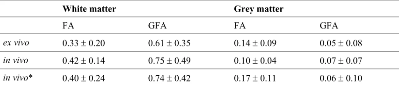
Table 5.1. FA and GFA quantifications... 76
Table 6.1. Weighting table of activation between adjacent vertebras ... 94
Table 6.2. Quantification of static and temporal SNR in the spinal cord ... 99
Table 6.3. F-score results of linear regression between betas and noise parameters ...99
Table B.1. Ratio of cardiac variance before and after realignment ... 134
List of Figures
Figure 2.1. Central and peripheral nervous system in human... 4
Figure 2.2. Human spinal cord... 4
Figure 2.3. White matter tracts in the spinal cord... 5
Figure 2.4. Monosynaptic and polysynaptic reflexes ... 6
Figure 2.5. Vascular supply in the spinal cord... 7
Figure 2.6. Injured spinal cord... 8
Figure 2.7. Principle of axonal sprouting... 9
Figure 2.8. Principle of diffusion-weighted MRI ... 11
Figure 2.9. Multi-shot EPI acquisition... 19
Figure 2.10. Parallel imaging acquisition ... 20
Figure 2.11. Reduced FOV acquisition ... 20
Figure 2.12. Non-CPMG FSE acquisition ... 21
Figure 2.13. LSDI acquisition... 22
Figure 2.14. Radial acquisition ... 23
Figure 2.15. DW-MRI in spinal cord compression... 26
Figure 3.1. Complete and partial lesion on a cat... 38
Figure 3.2. Ex vivo spinal cord in gelatine ... 39
Figure 3.3. Susceptibility artifacts in the thoracic region ... 40
Figure 3.4. Benefits of respiratory gating ... 41
Figure 3.5. Distortion correction using the reversed gradient method... 42
Figure 4.1. Cat in spinal coil... 50
Figure 4.2. Distortion correction – variance maps... 52
Figure 4.3. Selective tractography – regions of interest ... 54
Figure 4.4. Tensor map of the healthy spinal cord of cat... 55
Figure 4.5. Fibre tractography of the healthy spinal cord of cat... 56
Figure 4.6. Selective tractography – results... 56
Figure 4.7. Histological slices of spinal cord injury ... 57
Figure 4.8. FA quantification along the injured spinal cord ... 57
Figure 4.9. Tractography of the injured spinal cord ... 59
Figure 4.10. Selective tractography of the injured spinal cord ... 59
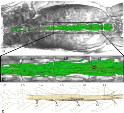
Figure 5.1. Axial PD image showing segmented white and grey matter... 72
Figure 5.2. Mapping of q-ball ODF in the spinal cord ... 73
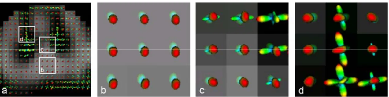
Figure 5.3. Evolution of diffusion maxima across slices... 74
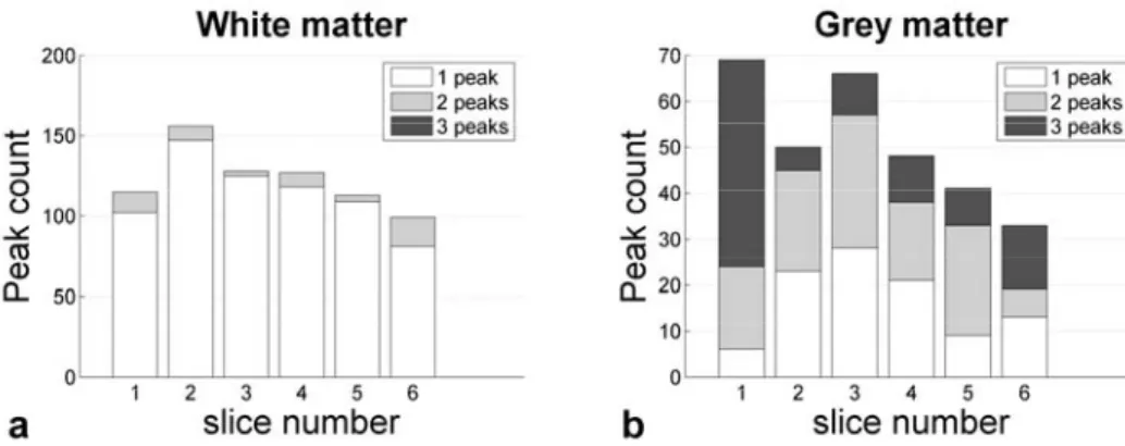
Figure 5.4. Number of diffusion directions in spinal cord white and grey matter... 74
Figure 5.5. Comparison of DTI and QBI... 75
Figure 5.6. QBI results from in vivo data ... 77
Figure 5.7. Impact of b-value on ODF estimation ... 78
Figure 5.9. Impact of q-space sampling ...79
Figure 6.1. Mask used for the GLM ...89
Figure 6.2. Fixed-effect results in seven cats ...93
Figure 6.3. Quantification of T-scores along the spinal cord of the nine cats...94
Figure 6.4. Quantification of responses across runs for four cats ...95
Figure 6.5. T-map across runs selected from one cat ...96
Figure 6.6. Percent signal change in function of stimulation intensity in two cats ...96
Figure 6.7. Typical raw EPI obtained in one cat ...98
Figure 6.8. Temporal and static SNR in the brain and spinal cord...98
Figure 6.9. Quantification of percent signal change and CO2 across time ...100
Figure 6.10. Percent signal change in function of CO2 level...101
Figure 6.11. Rigid-body registration parameters... 101
Figure A.1. Cat positioning for DW scans ...118
Figure A.2. Slice positioning for DW in cat...119
Figure A.3 Typical T2-weighted SE-EPI in cat ...119
Figure A.4.Principle of super-resolution MRI...120
Figure A.5. Typical sagittal FSE image in cat...121
Figure A.6. Typical axial FSE in a cat with left hemilesion ...122
Figure A.7. Typical T1-weighted image in cat ...123
Figure A.8. Coil sensitivity mapping on a water phantom...124
Figure A.9. Slice positioning for fMRI acquisition in human...125
Figure A.10. Typical T2-weighted GE-EPI in human ...126
Figure A.11. Typical T1-weighted image in human ...127
Figure A.12. Typical SWI in human ...128
Figure B.1. Diagram of the analysis method for run i and volume j. ...132
Figure B.2. Comparison of transformation matrix and cardiac signal ...133
Figure B.3. Effect of realignment on cardiac signal distribution map...134
Figure C.1. Field of view for sagittal acquisition in human ...140
Figure C.2. Flowchart of data processing...142
Figure C.3. Cardiac mapping in the cervical spinal cord ...145
Figure C.4. Intra-subject variability of cardiac contribution to fMRI signal...147
Figure C.5. Inter-subject variability of cardiac contribution ...147
Figure C.6. Variance map of an ICA component related to post-systolic phase...148
Figure C.7. Independent component related to cardiac cycle...149
Figure D.1. Principle of the shift method ...164
Figure D.2. Paradigm and modelled response to a series of stimuli...165
Figure D.3. Correlations between drifts and protocol ...166
Figure D.4. Computed T-score in function of correlation threshold ...167
Figure D.5. Error on β estimation as a function of basis size...168
Figure D.6. Detectability in function of effect size ...169
Figure D.7. Shift method on simulated data...169
Figure D.9. Shift method applied to real data... 171
Figure D.10. HbR and HbO2 peaking time ... 172
Figure D.11. Spatial mapping of estimated β for each pair ... 173
Figure D.12. Spatial mapping of temporal delay in haemodynamic response... 174
Glossary
ADC Apparent diffusion coefficient A-P Antero-posterior
ATP Adenosine triphosphate
BOLD Blood oxygenation level dependent C Cervical
CBF Cerebral blood flow CNS Central nervous system CPG Central pattern generator
CPMG Carr–Purcell–Meiboom–Gill
CST Corticospinal tract
DOI Diffuse optical imaging DTI Diffusion tensor imaging DW Diffusion-weighted EPI Echo planar imaging
FA Fractional anisotropy
fMRI Functional magnetic resonance imaging
FOV Field of view
FRT Funk-Radon transform
FSE Fast spin echo
FWHM Full width at half maximum
GLM General linear model
GRAPPA Generalized autocalibrating partially parallel acquisitions HARDI High angular resolution diffusion imaging
HbO2 Oxyhaemoglobin
HbR Deoxyhaemoglobin HRF Haemodynamic response function
ICA Independent component analysis
LSDI Line scan diffusion imaging L Lumbar MRI Magnetic resonance imaging MRO2 Metabolic rate of O2
ODF Orientation distribution function
QBI Q-ball imaging
PD Proton density
RARE Rapid acquisition with relaxation enhancement R-C Rostro-caudal
ReST Reticulospinal tract
R-L Right-left ROI Region of interest
RST Rubrospinal tract
S Sacral SAR Specific absorption rate SCI Spinal cord injury
SCT Spinocerebellar tract
SE Spin echo
SEEP Signal enhancement by extravascular proton density
SH Spherical harmonics
SNR Signal-to-noise ratio
STT Spinothalamic tract
T Tesla (in the context of MRI)
T Thoracic (in the context of spinal cord)
TE Echo time
TR Repetition time
Remerciements
Bizarrement la section « remerciements » d’un manuscrit de thèse est celle qui est la plus lue, mais aussi celle qui est généralement écrite après acceptation du manuscrit. Cette incongruité confère aux remerciements une propriété singulière : celle de pouvoir diffuser des sentiments vrais et non censurés.
For de cette liberté d’expression, et par ordre chronologique d’apparition, j’aimerais d’abord remercier Habib de m’avoir accordé sa confiance, de m’avoir reçu dans son laboratoire parisien et présenté avec enthousiasme un projet de doctorat passionnant, de m’avoir accueilli au sein de son équipe fort sympathique (pas seulement au sens de souffrir ensemble), mais aussi pour sa générosité, sa bonne humeur, son bon humour (ou tout au moins compatible avec le mien), sa présence physique et virtuelle (e.g., courriel, visioconférence) et son insatiable curiosité scientifique donnant lieu à de nombreuses investigations mathématico-physiologiques.
J’aimerais également remercier Habib de m’avoir présenté à Serge, mon deuxième papa dans cette aventure académique, à qui je suis extrêmement reconnaissant de m’avoir ouvert la porte de son laboratoire montréalais, enseigné une rigueur scientifique à la fois plaisante et efficace, et transmis sa passion pour l’étude de la moelle épinière, dans un milieu comme les neurosciences où le cerveau règne en monarque. Serge m’a également enseigné une philosophie de vie que j’ai petit à petit adoptée. Par exemple, sa faculté de renvoyer les corrections d’un article en moins de 3 heures ou de répondre aux courriels en moins de 3 minutes. Grâce à lui j’ai réalisé que limiter la procrastination peut avoir des effets bénéfiques sur la santé d’un projet, qu’il soit d’ordre scientifique ou d’ordre privé. Au-delà des sciences, la musique fut également la glie de notre amitié, et je garde un excellent souvenir de notre Spinal Chord Ensemble.
Il n’y a pas de mots pour décrire l’influence que peuvent avoir des directeurs de thèse sur le cours d’une vie, mais elle est immense. Je suis conscient de la chance phénoménale qui m’a été donnée d’avoir été guidé par Serge et Habib, sur le plan professionnel mais aussi et surtout sur le plan humain.
Je tiens à remercier Cyril Poupon et Karla Miller d’avoir accepté la révision du présent manuscrit, et à Rick Hoge, Stéphane Lehéricy, Rachid Deriche d’avoir fait partie du jury. Merci à Alain Herment et à Trevor Drew pour leur accueil respectif au sein du Laboratoire d’Imagerie Fonctionnelle et du Groupe de Recherche sur le Système Nerveux Central, ainsi que pour leur support, encouragements et humour très apprécié, respectivement « franchouillard » et « british ». Merci également à Julien Doyon pour son accueil et son support continu au sein de l’Unité de Neuroimagerie Fonctionnelle. Merci à Christian Saguez pour m’avoir autorisé à suivre son programme de l’école doctorale de Centrale Paris, tout comme Robert Leblanc pour son accueil au sein de l’Institut de Génie Biomédical de l’Université de Montréal. Merci également à
Louise Bélanger, Géraldine Carbonel et Catherine Lhopital de nous avoir aidé à gravir les nombreux échelons bureaucratiques afférents à une cotutelle de thèse.
Merci à Rick Hoge, Cyril Poupon et Alexandre Vignaud pour leurs précieux conseils m’ayant permis d’améliorer les acquisition IRM, Rachid Deriche, Maxime Descoteaux et Aurobrata Ghosh pour leurs contributions en imagerie à haute résolution angulaire, Pierre Rainville, Mathieu Piché et Guoming Xie pour leur collaboration en imagerie spinale chez l’humain, Rick Hoge et Claudine Gauthier pour leur collaboration sur l’hypercapnie, Henrik Lundell pour sa collaboration sur l’art de corriger les artefacts de susceptibilité magnétique, Jonathan Brooks pour sa collaboration en imagerie fonctionnelle, Frédéric Lesage, Sarah Chapuisat, Benoît Briau, Eric Beaumont et Simon Dubeau pour leur collaboration en imagerie optique.
Durant les trois dernières années, collègues et amis m’ont beaucoup apporté tant sur le plan scientifique que sur celui du pur divertissement. Au risque d’en oublier, je remercie pèle mêle du côté français : Mélanie, pour ses facultés de tétrapilectomie1 qu’elle ne cesse
d’investiguer, Guillaume pour son aptitude à filtrer ses mauvais calembours, Vincent pour sa capacité à accepter les miens, Saad pour m’avoir accueilli si chaleureusement de nombreuses fois à Oxford, Arnaud, Caroline, Sarah, David, Rémy, Stéphane et Alexandre. Outre atlantique, j’ai particulièrement apprécié les réunions biblio autour d’un « spécial bonjour » et les tournées des cottages québécois avec Pierre et Christophe. Au CRIUGM, j’étais sensible aux fredonnements plaisants de Vo An du bureau d’à côté, à la salsa trépidante d’Oury du bureau de l’autre côté, ainsi qu’aux nombreuses rencontres avec Cécile, Olivia, Ronan, Gaëlle, Johane, Mathieu, Mathieu, Mathieu, Mathieu, Mathieu. La vie au GRSNC fut parsemée de retraites au grand air, sorties anniversaires au Maiko sushi, symposiums, et autres prétextes pour encourager une synergie de convivialité et d’échange scientifique. Je remercie tout particulièrement Hugues, Janyne, Greg, Alain, Hugo, Claude, Maurice, Philippe, René avec qu’il a été fort agréable de passer ces moments agréables.
Enfin, et sur une note plus affective, un grand merci à Claudine, ma mère, mon père, ma petite sœur, Manu, Fred, et autres proches pour leur soutien sans faille, leur amitié et/ou amour. À la manière du Yin et du Yang, en plus d’un environnement scientifique efficace, le bon déroulement d’une thèse nécessite un épanouissement de soi, une sérénité et stabilité autant psychique qu’intellectuelle, pour se concentrer sur la substantifique moelle dont il est question. Un peu comme le Graal du goût parfait dans l’art culinaire, j’ai su trouver le parfait dosage avec ceux qui m’entourent, pour les années passées, celles à venir, et je leur dis merci.
1 L’art de couper les cheveux en quatre
Abstract
Spinal cord injury has a significant impact on quality of life since it can lead to motor (paralysis) and sensory deficits. These deficits evolve in time as reorganisation of the central nervous system occurs, involving physiological and neurochemical mechanisms that are still not fully understood. Given that both the severity of the deficit and the successful rehabilitation process depend on the anatomical pathways that have been altered in the spinal cord, it may be of great interest to assess white matter integrity after a spinal lesion and to evaluate quantitatively the functional state of spinal neurons.
The great potential of magnetic resonance imaging (MRI) lies in its ability to investigate both anatomical and functional properties of the central nervous system non-invasively. To address the problem of spinal cord injury, this project aimed to evaluate the benefits of diffusion-weighted MRI to assess the integrity of white matter axons that remain after spinal cord injury. The second objective was to evaluate to what extent functional MRI can measure the activity of neurons in the spinal cord.
Although widely applied to the brain, diffusion-weighted MRI and functional MRI of the spinal cord are not straightforward. Various issues arise from the small cross-section width of the cord, the presence of cardiac and respiratory motions, and from magnetic field inhomogeneities in the spinal region. The main purpose of the present thesis was therefore to develop methodologies to circumvent these issues. This development notably focused on the optimization of acquisition parameters to image anatomical, diffusion-weighted and functional data in cats and humans at 3T using standard coils and pulse sequences. Moreover, various strategies to correct for susceptibility-induced distortions were investigated and the sensitivity and specificity in spinal cord functional MRI was studied. As a result, acquisition of high spatial and angular diffusion-weighted images and evaluation of the integrity of specific spinal pathways following spinal cord injury was achieved. Moreover, functional activations in the spinal cord of anaesthetized cats were detected.
Although encouraging, these results highlight the need for further technical and methodological development in the near-future. Being able to develop a reliable neuroimaging tool for confirming clinical parameters would improve diagnostic and prognosis. It would also enable to monitor the effect of various therapeutic strategies. This would certainly bring hope to a large number of people suffering from trauma and neurodegenerative diseases such as spinal cord injury, tumours, multiple sclerosis and amyotrophic lateral sclerosis.
Keywords : Magnetic resonance imaging, diffusion tensor imaging, functional MRI, spinal cord, injury, animal.
Résumé
Les lésions de la moelle épinière ont un impact significatif sur la qualité de la vie car elles peuvent induire des déficits moteurs (paralysie) et sensoriels. Ces déficits évoluent dans le temps à mesure que le système nerveux central se réorganise, en impliquant des mécanismes physiologiques et neurochimiques encore mal connus. L’ampleur de ces déficits ainsi que le processus de réhabilitation dépendent fortement des voies anatomiques qui ont été altérées dans la moelle épinière. Il est donc crucial de pouvoir attester l’intégrité de la matière blanche après une lésion spinale et évaluer quantitativement l’état fonctionnel des neurones spinaux.
Un grand intérêt de l'imagerie par résonance magnétique (IRM) est qu’elle permet d’imager de façon non invasive les propriétés fonctionnelles et anatomiques du système nerveux central. Le premier objectif de ce projet de thèse a été de développer l’IRM de diffusion afin d’évaluer l'intégrité des axones de la matière blanche après une lésion médullaire. Le deuxième objectif a été d'évaluer dans quelle mesure l’IRM fonctionnelle permet de mesurer l'activité des neurones de la moelle épinière.
Bien que largement appliquées au cerveau, l’IRM de diffusion et l’IRM fonctionnelle de la moelle épinière sont plus problématiques. Les difficultés associées à l’IRM de la moelle épinière relèvent de sa fine géométrie (environ 1 cm de diamètre chez l’humain), de la présence de mouvements d’origine physiologique (cardiaques et respiratoires) et de la présence d’artefacts de susceptibilité magnétique induits par les inhomogénéités de champ, notamment au niveau des disques intervertébraux et des poumons. L’objectif principal de cette thèse a donc été de développer des méthodes permettant de contourner ces difficultés. Ce développement a notamment reposé sur l'optimisation des paramètres d'acquisition d'images anatomiques, d’images pondérées en diffusion et de données fonctionnelles chez le chat et chez l’humain sur un IRM à 3 Tesla. En outre, diverses stratégies ont été étudiées afin de corriger les distorsions d’images induites par les artefacts de susceptibilité magnétique, et une étude a été menée sur la sensibilité et la spécificité de l’IRM fonctionnelle de la moelle épinière. Les résultats de ces études démontrent la faisabilité d’acquérir des images pondérées en diffusion de haute qualité, et d’évaluer l’intégrité de voies spinales spécifiques après lésion complète et partielle. De plus, l’activité des neurones spinaux a pu être détectée par IRM fonctionnelle chez des chats anesthésiés.
Bien qu’encourageants, ces résultats mettent en lumière la nécessité de développer davantage ces nouvelles techniques. L’existence d’un outil de neuroimagerie fiable et robuste, capable de confirmer les paramètres cliniques, permettrait d’améliorer le diagnostic et le pronostic chez les patients atteints de lésions médullaires. Un des enjeux majeurs serait de suivre et de valider l'effet de diverses stratégies thérapeutiques. De telles outils représentent un espoir immense pour nombre de personnes souffrant de traumatismes et de maladies
neurodégénératives telles que les lésions de la moelle épinière, les tumeurs spinales, la sclérose en plaques et la sclérose latérale amyotrophique.
Mots-clés : Imagerie par résonance magnétique, imagerie du tenseur de diffusion, IRM fonctionnelle, moelle épinière, lésion, animal.
Publications arising from this work
Journal articles
1. Cohen-Adad, J., Hoge, R.D., Leblond, H., Xie, G., Beaudoin, G., Song, A., Krueger, G., Doyon, J., Benali, H., Rossignol, S., 2009. Investigations in functional MRI of the spinal cord of cats. Neuroimage 44, 328-39.
2. Piché, M., Cohen-Adad, J., Khosh Nejad, M., Perlbarg, V., Xie, G., Beaudoin, G., Benali, H., Rainville, P., Characterization of cardiac-related noise in fMRI of the cervical spinal cord. Magn Reson Imaging (in press).
3. Cohen-Adad, J., Descoteaux, M., Rossignol, S., Hoge, R.D., Deriche, R., Benali, H., 2008. Detection of multiple pathways in the spinal cord using q-ball imaging. Neuroimage, 42, 739-749.
4. Cohen-Adad, J., Benali, H., Barrière, G., Leblond, H., Hoge, R.D., Rossignol, S., 2008. Développement clinique de l’IRM du tenseur de diffusion de la moelle épinière dans un contexte de lésion médullaire. IRBM, 29, 255-260.
5. Cohen-Adad, J., Benali, H., Hoge, R.D., Rossignol, S., 2008. In vivo DTI of the healthy and injured cat spinal cord at high spatial and angular resolution. Neuroimage 40, 685-697.
6. Cohen-Adad, J., Chapuisat, S., Doyon, J., Rossignol, S., Lina, J.M., Benali, H., Lesage, F., 2007. Activation detection in diffuse optical imaging by means of the general linear model. Med Image Anal 11, 616-629.
Conference articles
1. Cohen-Adad, J., Rossignol, S., Benali, H., Hoge, R.D., 2008. In vivo assessment of axonal disruption using diffusion magnetic resonance imaging. In: 31st Canadian Medical and Biological Engineering Conference, Montreal, Canada. Oral presentation (in press). 2. Cohen-Adad, J., Descoteaux, M., Rossignol, S., Hoge, R.D., Deriche, R., Benali, H.,
2008. Detection of multiple pathways in the spinal cord white matter using q-ball imaging. In: IEEE International Symposium on Biomedical Imaging, Paris, France, 1380. Oral presentation.
3. Cohen-Adad, J., Benali, H., Rossignol, S., 2007. Methodology for MR diffusion tensor imaging of the cat spinal cord. Conf Proc IEEE Eng Med Biol Soc 1, 323-326. Oral presentation.
4. Cohen-Adad, J., Piche, M., Rainville, P., Benali, H., Rossignol, S., 2007. Impact of realignment on spinal functional MRI time series. Conf Proc IEEE Eng Med Biol Soc 1, 2126-2129. Poster.
5. Chapuisat, S., Cohen-Adad, J., Grova, C., Lina, J.M., Rossignol, S., Doyon, J., Benali, H., Lesage, F., 2007. Application of the general linear model to hemodynamic response estimation in diffuse optical imaging. In: SPIE Photonics North, Ottawa, Canada. Poster.
Conference abstracts
1. Cohen-Adad, J., Gauthier, C., Brooks, J.C.W., Leblond, H., Hoge, R.D., Fisher, J.A., Beaumont, E., Dubeau, S., Lesage, F., Doyon, J., Benali, H., Rossignol, S., 2009. Venous effect in spinal cord fMRI: insights from intrinsic optical imaging and laser speckle. In: 14th Annual Meeting of the Organization for Human Brain Mapping (HBM), San Fransisco, USA.
2. Brooks, J.C.W., Cohen-Adad, J., Gauthier, C., Hoge, R.D., Rossignol, S., 2009. Studying the sensorimotor pathways: brain - brainstem - spinal cord imaging. In: 14th Annual Meeting of the Organization for Human Brain Mapping (HBM), San Fransisco, USA.
3. Cohen-Adad, J., Ghosh, A., Leblond, H., Descoteaux, M., Deriche, R., Benali, H., Rossignol, S., 2009. Comparison of DTI and Q-Ball imaging metrics in a cat model of spinal cord injury. In: 14th Annual Meeting of the Organization for Human Brain Mapping (HBM), San Fransisco, USA.
4. Gauthier, C., Cohen-Adad, J., Brooks, J., Fisher, J.A., Rossignol, S., Hoge, R.D., 2009. Comparison of hypercapnia-induced BOLD changes in the brain and spinal cord. In: 14th Annual Meeting of the Organization for Human Brain Mapping (HBM), San Fransisco, USA.
5. Cohen-Adad, J., Leblond, H., Ghosh, A., Descoteaux, M., Deriche, R., Benali, H., Rossignol, S., 2009. Evaluation of q-ball metrics for assessing the integrity of the injured spinal cord. In: 17th Annual Meeting of the International Society for Magnetic Resonance in Medicine (ISMRM), Honolulu, Hawaii
6. Cohen-Adad, J., Rossignol, S., Hoge, R.D., 2009. Slice-by-slice motion correction in spinal cord fMRI: SliceCorr. In: 17th Annual Meeting of the International Society for Magnetic Resonance in Medicine (ISMRM), Honolulu, Hawaii
7. Gauthier, C., Cohen-Adad, J., Brooks, J., Rossignol, S., Hoge, R.D., 2009. Investigation of venous effects in spinal cord fMRI using hypercapnia. In: 17th Annual Meeting of the International Society for Magnetic Resonance in Medicine (ISMRM), Honolulu, Hawaii
8. Cohen-Adad, J., Lundell, H., Rossignol, S., 2009. Distortion correction in spinal cord DTI: What’s the best approach? In: 17th Annual Meeting of the International Society for Magnetic Resonance in Medicine (ISMRM), Honolulu, Hawaii
9. Lundell, H., Cohen-Adad, J., 2009. Point spread function mapping for distortion correction of spinal cord diffusion weighted MRI. In: 17th Annual Meeting of the International Society for Magnetic Resonance in Medicine (ISMRM), Honolulu, Hawaii 10. Cohen-Adad, J., Descoteaux, M., Barrière, G., Hoge, R.D., Deriche, R., Benali, H.,
Rossignol, S., 2008. Characterization of the healthy and injured spinal cord using q-ball imaging. In: 8th international congress of the IRME, Paris, France. Poster presentation. 11. Xie, G., Piché, M., Cohen-Adad, J., Kosh Nejad, M., Perlbarg, V., Hoge, R.D., Benali,
H., Rossignol, S., Rainville, P., 2008. Characterizing and controlling for physiological noise in human cervical spinal fMRI in response to noxious stimuli. In: Society for Neuroscience, Washington, DC, USA, (in press).
12. Cohen-Adad, J., Descoteaux, M., Barrière, G., Hoge, R.D., Deriche, R., Benali, H., Rossignol, S., 2008. Characterization of the healthy and injured spinal cord using q-ball imaging. In: Pathogenesis and Mechanisms of White Matter Injury Workshop, Krakow, Poland. Poster presentation.
13. Cohen-Adad, J., 2008. Diffusion Tensor Imaging and Tractography of the spinal cord in animals and humans. In: Symposium on spinal Cord: Function, Repair and Rehabilitation after Injury, Montreal, Canada. Oral presentation.
14. Barrière, G., Cohen-Adad, J., Benali, H., Rossignol, S., 2008. Locomotor recovery after spinal lesions in the cat using behavioral and imaging techniques. In: Spinal Cord : Function, Repair and Rehabilitation after Injury, Montreal, Canada.
15. Barrière, G., Cohen-Adad, J., Benali, H., Rossigol, S. 2008. A dual lesion paradigm to study spinal cord injury (SCI) in cats using electrophysiological and imaging methods. CDRF’s 3rd Spinal Cord Symposium, Atlanta, May 9-11, USA.
16. Cohen-Adad, J., Descoteaux, M., Deriche, R., Rossignol, S., Hoge, R.D., Benali, H., 2008. Q-ball imaging of the spinal cord. In: 16th Annual Meeting of the International Society for Magnetic Resonance in Medicine (ISMRM), Toronto, Canada, 569. Oral presentation.
17. Cohen-Adad, J., Gauthier, C., Benali, H., Rossignol, S., Hoge, R.D., 2008. BOLD signal responses to controlled hypercapnia in human spinal cord. In: 16th Annual Meeting of the International Society for Magnetic Resonance in Medicine (ISMRM), Toronto, Canada, 3403.
18. Cohen-Adad, J., Benali, H., Hoge, R.D., Rossignol, S., 2007. In vivo assessment of spinal cord integrity by diffusion tensor imaging. In: Society for Neuroscience, San Diego, USA, 37, 77.10.
19. Cohen-Adad, J., Hoge, R.D., Benali, H., Rossignol, S., 2007. In vivo diffusion MRI of the cat spinal cord following injury. In: Symposium on Motor Systems, San Diego, USA. 20. Cohen-Adad, J., Benali, H., Hoge, R.D., Leblond, H., Barrière, G., Doyon, J., Rossignol,
S., 2007. Spinal cord in vivo diffusion tensor imaging of healthy human and spinalized cats. In: Organization for Human Brain Mapping, Chicago, USA, S887.
21. Cohen-Adad, J., Benali, H., Hoge, R.D., Rossignol, S., 2007. In vivo diffusion tensor imaging of the spinal cord. In: Canadian Physiological Society, Mont Ste-Anne, Quebec, Canada.
22. Cohen-Adad, J., Perlbarg, V., Doyon, J., Hoge, R.D., Beaudoin, G., Song, A., Leblond, H., Provencher, J., Rossignol, S., Benali, H., 2006. Methodology to study functional MRI of the cat lumbar spinal cord. In: Organization for Human Brain Mapping, Florence, Italy, S2851.
23. Cohen-Adad, J., Benali, H., Rossignol, S., 2006. A functional MRI methodology to map the neuronal activity in the spinal cord of cats. In: 28th International Symposium of the Groupe de recherche sur le système nerveux central et le Centre de recherche en sciences neurologiques: "Computational Neuroscience", Montreal, Canada.
Research reports
1. Cohen-Adad, J., 2006. Y a-t-il une variation de la densité de protons associée à l’Imagerie par Résonance Magnétique fonctionnelle ? Université de Montréal, Montréal, Canada (20 pages).
2. Cohen-Adad, J., 2006. Etude des interactions somato-viscérales. Université de Montréal, Montréal, Canada (17 pages).
3. Cohen-Adad, J., 2006. L’énigme des ondes de Mayer : faits et modèles. Université de Montréal, Montréal, Canada (26 pages).
Invited speaker
1. Imaging spinal cord injury and white matter damage. 7th International Symposium on
Experimental Spinal Cord Repair and Regeneration, Brescia, Italy, 2009.
3. Diffusion Tensor Imaging and tractography of the spinal cord in animals and humans. 2nd
Symposium on Spinal cord: Function, Repair and Rehabilitation after injury, Montreal, Canada, 2008.
4. DW-MRI and fMRI of the spinal cord. DRCMR center, Copenhagen, Denmark, 2008. 5. MRI of the spinal cord: from white matter organization to neuronal activity. Mass.
General Hospital / Harvard University, Boston, USA, 2008.
6. Diffusion Tensor Imaging: Principles and Applications. Functional Neuroimaging Unit, Université de Montréal, Montreal, Canada, 2008.
7. Diffusion and functional MRI of the spinal cord. BIC/MNI, McGill University, Montreal, Canada, 2008.
8. New advances in DTI of the spinal cord. Emory University / Georgia Tech, Atlanta, USA, 2007.
9. Diffusion and functional MRI of the spinal cord. Université de Montréal, Montreal, Canada, 2007.
10. Diffusion MRI of the spinal cord. INSERM / CHU Pitié-Salpêtrière, Paris, France, 2007. 11. Spinal cord imaging: some investigations. FMRIB, Oxford University, Oxford, UK,
À mes parents, à ma soeur.
1. Introduction
Spinal cord injury (SCI) induces motor (paralysis) and sensory deficits of varying severity through the disruption of specific ascending and descending spinal pathways. These deficits evolve in time as reorganisation of the central nervous system occurs, both at local and supraspinal sites. This functional compensation may involve various anatomical, physiological and neurochemical mechanisms still under investigation (Rossignol, 2006b). Both the severity of the deficit and the successful rehabilitation process depend on the type and number of axonal tracts that have been altered, as well as changes in the intrinsic properties of the spinal cord. Hence, it may be of great interest to assess white matter integrity at the site of a lesion, to identify anatomical tracts that have been damaged or spared and to quantify the activity of spinal neurons to examine the functional state of the cord.
In clinical routine, evoked potentials and electromyographic measurement already allow to evaluate the integrity of spinal pathways and intrinsic circuits. However, these approaches give only an indirect indication of the actual anatomic integrity. The great potential of magnetic resonance imaging (MRI) lies in its ability to investigate both anatomical and functional properties of biological samples non-invasively. To address the problem of spinal cord injury, MRI appears to be a candidate of choice to evaluate the morphological integrity of the cord, as well as its functional characteristics. The present project aimed to evaluate the benefits of diffusion-weighted (DW) MRI to assess the integrity of remaining white matter axons after spinal cord injury. The second objective was to evaluate to what extent functional MRI (fMRI) can measure the activity of spinal neurons, to be able to observe changes in their activity following injury. These changes could either be related to a spontaneous intrinsic reorganization of spinal circuitry or result from exogenous sensory inputs that aim to improve the recovery of function such as treadmill locomotor training.
However, DW-MRI and fMRI of the spinal cord are not straightforward. Various issues arise from the small cross-section width of the cord, the presence of cardiac and respiratory motions, and from magnetic field inhomogeneities in the spinal region. The purpose of the present thesis was to develop methodologies to circumvent these issues. This work has been conducted is three different laboratories, whose expertises strongly influenced my research priorities. I have been co-supervised by Dr. Serge Rossignol, member of the Groupe de Recherche sur le Système Nerveux Central, Université de Montréal (Montréal, Canada) and a Canada Research Chairholder on Spinal Cord, whose team studies spinal cord neurophysiology in the context of locomotor rehabilitation. I have also been co-supervised by Dr. Habib Benali, based at the Laboratoire d’Imagerie Fonctionnelle – UMR_S 678 INSERM / Université Pierre et Marie Curie (Paris VI), Pitié-Salpêtrière Hospital (Paris, France) whose team’s main expertise is to develop methods for the analysis of MRI data. Those two laboratories also collaborate closely with Dr. Richard Hoge, based at the Unité de Neuroimagerie Fonctionnelle,
Centre de Recherche de l’Institut Universitaire de Gériatrie de Montréal (Montréal, Canada), whose main expertise is MR physics. The work presented in this thesis therefore emerges from the input of those three distinctive fields. This conjunction has been essential for conducting such a multi-disciplinary project.
The manuscript is organized as follows. In chapter 2, a literature review will present past studies related to the spinal cord morphology and basic functions and studies related to DW- and fMRI of the spinal cord. Chapter 3 will synthesize past studies related to the topic of interest. Objectives, hypotheses and methods will emerge and will guide the reader through the following chapters. Three published articles presented as the core of this thesis will be provided in chapters 4, 5 and 6. Chapter 7 will briefly summarize the achieved work, discuss its originality and limitations. It will also present additional contributions to this thesis as well as ongoing work and future directions.
2. Literature review
Equation Section 2
Spinal cord anatomy and function
The present work aims to use MRI to identify spinal pathways, to characterize spinal lesions and to detect neuronal activity evoked by peripheral nerve stimulation in cats. Therefore, the following literature review will focus on these considerations only. For the description of the cord anatomy and function however, the human model will mainly be presented given cat and human share many similarities in terms of spinal structure and function. Also, most spinal MRI studies have been conducted in human, justifying its foreground position. This review has been inspired by the textbooks by W. Kahle et al. (Kahle and Frotscher, 2005) and E.D. Schwartz et al. (Schwartz and Flanders, 2007) and by other publications.
Anatomy of the cord
The spinal cord is part of the central nervous system (CNS) and participates to the active and passive control of sensorimotor functions (e.g., locomotion, motility, pain perception). An illustration of the CNS is provided in Figure 2.1. The length of the spinal cord is much shorter than the length of the vertebral column around it and its relative size evolves with the age. The human spinal cord extends from the medulla oblongata and ends near the first or second lumbar vertebra, terminating in a fibrous extension known as the filum terminale. It is about 44 cm long in humans. The diameter in cross-section is about 1 cm diameter and ovoid-shaped, with an enlargement in the cervical and lumbar regions. In cross-section, the peripheral region of the cord contains white matter axons forming ascending and descending tracts to and from supraspinal regions. In contrast to the brain, the spinal cord white matter surrounds the butterfly shaped grey matter containing neurons and glial cells. The grey matter surrounds the central canal, which is an anatomic extension of the ventricular system of the brain and also contains cerebrospinal fluid (CSF). Illustration of human spinal cord is provided in Figure 2.2.
The human spinal cord is divided into 31 different segments. Motor nerve roots exit in the ventral aspects and sensory nerve roots enter in the dorsal aspects. The ventral and dorsal roots later join to form paired spinal nerves, one on each side of the spinal cord. There are 31 spinal cord nerve segments in a human spinal cord: 8 cervical (C) segments (the 1st cervical nerve exits the vertebral column above the C1 vertebra and the others below the C1-C7 vertebral bodies), 12 thoracic (T) segments (thoracic nerves exit the spinal column below T1-T12 vertebral bodies), 5 lumbar (L) segments (lumbar nerves exit spinal column below L1-L5), 5 sacral (S) segments (sacral nerves exit spinal column below S1-S5) and 1 coccygeal segment (coccygeal nerves exit spinal column at the coccyx).
Figure 2.1. Central and peripheral nervous system in human
The CNS includes the brain (1) and the spinal cord (2). Peripheral nerves (3) connect the CNS to muscles and skin and form the brachial plexus (4) and the lumbo-sacral plexus (5). Spinal ganglions (6) are located at the entrance of afferent fibres and contain cell bodies. Adapted from (Kahle and Frotscher, 2005).
Figure 2.2. Human spinal cord
This figure illustrates the human spinal cord and its relationship with surrounding structures in the spinal canal. Adapted from (Schwartz and Flanders, 2007).
In the cat there are 8 cervical spinal segments, 13 thoracic spinal segments, 7 lumbar spinal segments, 3 sacral spinal segments and 5 coccygeal spinal segments. The spinal cord ends around the S1 vertebral body level. A description of the cat spinal anatomy is provided in (Fletcher and Malkmus, 1999; Hudson and Hamilton, 1993). When imaging the cat spinal cord, the following anatomical and physiological characteristics were useful for the rostro-caudal localization and for the identification of physiological fluctuations in fMRI time series. The cranial and caudal edges of the cat right kidney reach level L1 and L4 vertebral bodies respectively, and cranial and caudal edge of the cat left kidney reach level L2 and L5 vertebral bodies respectively.
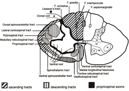
White matter tracts are illustrated in Figure 2.3. They mainly consist of ascending and descending tracts. Ascending tracts are involved in the perception of sensory inputs and include the dorsal column, the spinocerebellar tracts (SCT) and the spinothalamic tracts (STT). Descending tracts are responsible for the motor control and include the corticospinal tracts (CST), the rubrospinal tract (RST), the reticulospinal tract (ReST) and the vestibulospinal tract (VST). Partial lesions involving specific tracts therefore lead to specific functional deficits as shown in (Anderson et al., 2007; Barriere et al., 2008; Frigon and Rossignol, 2006; Jiang and Drew, 1996).
Figure 2.3. White matter tracts in the spinal cord
The left side of the drawing shows major white matter tracts in the human spinal cord. The right side shows the lamination of the grey matter. Adapted from (Schwartz and Flanders, 2007).
Simple reflexes
To provide a better understanding of the stimulations, this section presents the concept of mono- and polysynaptic reflexes. Afferent fibres of the dorsal root – whose cell bodies are located in the spinal ganglion – transmit sensory signals from the periphery to neurons located in the dorsal horn of the cord. Those signals are processed first at the spinal level but are also sent to supraspinal regions through collaterals of the afferents or through projecting interneurons whose axons ascend the spinal cord (see Figure 2.4a). At the spinal level, the information reaches motoneurons in the ventral horn either directly (monosynaptic pathways as in Figure 2.4b) or through interneurons (di-, tri-, or polysynaptic pathways as in Figure 2.4c). When motoneurons are activated, muscles will contract unless the cholinergic transmission is blocked by agents such as pancuronium which acts at the neuromuscular junction.
Figure 2.4. Monosynaptic and polysynaptic reflexes
This figure illustrates the transmission of afferent signal to supraspinal regions (a) and reflexes via monosynaptic (b) and polysynaptic pathways (c). Adapted from (Kahle and Frotscher, 2005).
Vascular arborisation of the spinal cord
Given that functional magnetic resonance imaging (fMRI) relies on the recording of haemodynamic changes related to neuronal activity, it is worth describing the vascular architecture providing the blood supply in the spinal cord. An illustration of the spinal cord vascular supply is shown in Figure 2.5. Arterial supply in the cord is provided by vertebral arteries and segmental arteries. Vertebral arteries give rise to two posterior spinal arteries which course along the dorsal side of the cord. At about C2 vertebral body, vertebral arteries merge to form the anterior spinal artery. This artery courses along the medio-ventral side of the cord. Segmental arteries give rise to radicular arteries which supply blood to the cord’s roots. Draining veins have a distribution similar to that of the arteries. The venous network gives rise to one anterior spinal vein and two posterior spinal veins.
Figure 2.5. Vascular supply in the spinal cord
Left panel shows the general arterial supply and venous drainage. Right panel shows the cross-sectional distribution of arteries. Source: http://www.frca.co.uk/.
Spinal cord injury
Spinal cord injury (SCI) has a significant impact on the quality of life since it can lead to motor deficits (paralysis) and sensory deficits. Throughout the world, about 2.5 million people live with SCI2. To date, there is no consensus for fully rehabilitative cure in SCI,
although numerous therapeutic approaches have shown benefits (Rossignol et al., 2007; Thuret et al., 2006). It is thus of great importance to develop tools that will improve characterization of spinal lesions as well as the integrity of remaining spinal tracts to eventually establish better prognosis after spinal injury.
Spinal lesions notably induce inflammation, oedema and necrosis at the site of injury (see Figure 2.6). Apart from the lesion, secondary pathological processes including ischemia, inflammation and excitotoxic events may also occur. These so-called ‘secondary injuries’ follow a first spinal trauma and can be located several vertebral levels from the epicentre of the lesion (Park et al., 2004; Tator and Fehlings, 1991). In the following paragraphs we will briefly describe some spinal anatomo-functional changes triggered by SCI and that are of interest for the present thesis.
2 http://www.campaignforcure.org
Figure 2.6. Injured spinal cord
The epicentre is characterized by dead cells and surrounded by scar tissue. The lesion could expend with time due to apoptosis and lead to secondary injuries. The lesion directly damages local axons but some closely axons could be spared. Demyelination occurs and further damages axons in physical contact to the injury. This process which last for a few days is called Wallerian degeneration. Adapted from (Schwartz and Flanders, 2007).
Wallerian degeneration
Following spinal lesions, nerve fibres may be disconnected from their cell bodies. As a result, these axons degenerate within a few days. This process is known as Wallerian degeneration. Axon segments which are no longer linked to cell bodies always show degeneration. However, axon collaterals proximal to the lesion can survive. This phenomenon was first observed in 1850 by Waller (Waller, 1850). A recent interpretation of the phenomenon is proposed in (Beirowski et al., 2005). Downstream effects of Wallerian degeneration is the degeneration of ascending and descending pathways in the spinal cord white matter that could potentially be detrimental to intrinsic spinal programs, notably those involved in locomotion. Axonal regrowth and remyelination
At the site of injury, fibrous scar tissue and myelin are associated with a large number of molecules that inhibit axonal regrowth. It has been shown that limiting the inhibitory effects of these molecules facilitates axonal regeneration, therefore improving recovery of function (Klapka et al., 2005; Schwab, 2004). Besides direct axon damage, some axons may also loose their myelin sheath thereby hampering the conduction of action potentials. Other approaches tend to induce remyelination by several means (McDonald and Belegu, 2006), including cell grafts (stem cells) or interference with immunological mechanisms (Schwartz and Yoles, 2006). Spinal reorganization
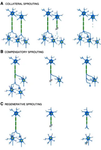
Not only does the spinal cord possess repair mechanisms that could be enhanced, but plastic reorganizations may occur to compensate the loss of functions. For instance, there may be regeneration of damaged axons or collateral sprouting of axons in spared
descending/ascending pathways or primary afferents (Kerschensteiner, 2007). These new growths can project through different spinal paths as they are forming new circuits and adopt unpredicted trajectories as they by-pass the scar tissue (Bareyre et al., 2004; Fouad and Pearson, 2004). This process may play various roles that are illustrated in Figure 2.7. Therefore, apart from directly acting on the lesion site, a large number of studies took advantage of the spinal cord’s capacity to change its intrinsic morphological and functional properties. As an illustration of those powerful mechanisms, a cat that has undergone complete spinal cord injury can recover a spinal locomotion after several weeks (Rossignol, 2006b).
Given that both the severity of the deficit and the successful rehabilitation process depend on the type and number of axonal tracts that have been altered, it may be of great interest to assess the white matter integrity at the site of a lesion, to identify anatomical tracts that have been damaged and to quantify the activity of spinal neurons to evaluate the functional state of the cord. Moreover, monitoring the efficiency of the above-mentioned therapeutic strategies is needed to get them accepted in clinical routine. The purpose of the next section is to present a non-invasive technique that appears to be suitable to reach these crucial goals.
Figure 2.7. Principle of axonal sprouting
New axonal pathways and synaptic terminals can appear in the spinal cord following injury. These new growths can play various roles, notably by innervation of collateral neurons (A), by an accrue number of connections for compensation (B) or by regeneration within an injured axon (C). Adapted from (Schwartz and Flanders, 2007).
Imaging the spinal cord white matter with MRI
Principles of DW-MRI
Magnetic resonance imaging (MRI) is based on absorption and reemission of a radiofrequency signal from resonating hydrogen protons (Purcell et al., 1946). This allows to image water molecules which constitute a great proportion of biological tissues. Proton density (PD), longitudinal (T1) and transverse (T2) decays give rise to various contrasts which are used
to emphasise specific anatomical structures (e.g., white matter and grey matter). Classical MRI sequences are useful to detect various types of pathologies such as tumours, oedema, ischemia, compression, brain atrophy and multiple sclerosis. Although very useful for many widely-used applications, these types of sequence do not enable specific imaging of white matter axons which would be of interest for assessing the number of disrupted axons and the progression of Wallerian degeneration. The following section will cover an MRI-based technique to image the white matter more specifically.
Diffusion-weighted signal
Diffusion-weighted (DW) MRI allows the measurement of water diffusion anisotropy in biological tissues (Beaulieu, 2002; Hagmann et al., 2006; Le Bihan et al., 2001; Mori and Zhang, 2006). Since the fibrous morphology of white matter restricts water diffusion along white matter axons, DW-MRI allows the indirect localisation of white matter tissue (Douek et al., 1991). The basics of DW-MRI are as follows: water molecules are in constant motion in biological tissues. This state is called Brownian motion. When a particular pulse sequence is applied, it is possible to quantify the extent of water displacement in a given direction (Stejskal and Tanner, 1965). This sequence consists of magnetic gradients applied before and after a 180° refocusing pulse. For motionless molecules, the dephasing induced by the first gradient is compensated by the application of the second gradient. No signal is attenuated. However, if water molecules move during the application of this pair of gradients, spins are dephased more rapidly and signal decreases as a function of the magnitude of displacement. The magnitude by which the diffusion signal is weighted is dictated by the so-called b-value. This value depends on the square of the gradient strength and on the diffusion time interval ∆t as defined by
2 2 2
3
b=γ δ g ⎛⎜∆ −t δ ⎞⎟
⎝ ⎠ (2.1)
where g is the diffusion-encoding gradient, δ is the duration of the encoding gradient, ∆t is the delay between application of and γ is the gyromagnetic ratio. DW images hence show hypersignal where water molecules are more static and an apparent diffusion coefficient (ADC) maps can be established. Based on the latter physical process, DW-MRI can be used to quantify axon disruption, myelin integrity and axon swelling (Thurnher and Bammer, 2006a). Principle of DW-MRI is illustrated in Figure 2.8.
Figure 2.8. Principle of diffusion-weighted MRI
Simplified timeline of RF pulse and gradient events in a standard DW sequence. For motionless molecules, the dephasing induced by the first gradient is compensated by the application of the second gradient: MR signal is mainly T2-weighted. However, if water molecules move during the application of this pair of gradients, spins are dephased more rapidly and signal decreases as a function of the magnitude of displacement.
Diffusion tensor imaging (DTI) and tractography
By weighting the diffusion signal in several directions, it is possible to compute a parametric model representing the main diffusion direction at each voxel (Basser et al., 1994). This type of model is described by a tensor and is usually represented graphically as a 3D ellipsoid. This technique is known as Diffusion Tensor Imaging (DTI). The purpose of DTI is to estimate the so-called diffusion tensor D from the following equation
b D
A e= − ⋅ (2.2)
where A stands for the signal attenuation and b for the diffusion weighting, as defined in Equation (2.1). After multiple regressions are performed to retrieve D, the latter matrix is
diagonalized using singular value decomposition to get the three eigenvalues. From each tensor’s eigenvalues, several indices characterising the diffusion can be computed. These include fractional anisotropy (FA) and mean diffusivity. Also, the first and second eigenvalues can be of interest as they have been shown to be more specifically informative to characterize some pathologies, as compared to anisotropic measurement (Gulani et al., 2001; Kinoshita et al., 1999; Ono et al., 1995; Song et al., 2002b).
Having modelled the diffusion tensor, it is possible to reconstruct global axonal pathways by linking every voxel with a similar eigenvector to its neighbours. This procedure, known as fibre tractography, allows 3-D mapping of white matter architecture in the mammalian brain (Basser et al., 2000; Ciccarelli et al., 2003; Conturo et al., 1999; D'Arceuil et al., 2007; Descoteaux et al., 2007b; Dyrby et al., 2007; Oppenheim et al., 2007). It should be mentioned however that reconstructed fibre bundles do not represent real axonal tracts but trace the path where water diffuses preferentially. Hence, tractography results should be handled with care since they may not represent the real pathway of axons (Basser et al., 2000; Johansen-Berg and Behrens, 2006). For instance, false negatives could be induced by the presence of crossing fibres which would artificially decrease the FA and stop the tracking procedure. Inversely, false positives could be induced by fibrous structures such as scar tissue in which water diffusion also has anisotropic properties (Schwartz et al., 2005d). Probabilistic methods allow better flexibility in the quantification of white matter directionality and have therefore the potential to limit the number of false negatives (Behrens et al., 2003). Another limitation of tractography is the impossibility of distinguishing anterograde from retrograde conduction (i.e., descending or ascending pathways in the cord).
Application of tractography in the spinal cord demonstrated the feasibility of retrieving major longitudinal pathways, i.e., axon bundles oriented in the rostro-caudal (R-C) direction (Bilgen et al., 2005; Ciccarelli et al., 2007; Ellingson et al., 2007b; Fenyes and Narayana, 1999; Gullapalli et al., 2006; Maier, 2007; Wheeler-Kingshott et al., 2002). Tracking of spinal nerves was also possible using DTI (Benner et al., 2008). An important motivation for tracking specific pathways in the spinal cord is the ability to obtain quantitative measurements along those pathways, to correlate the integrity of specific tracts with the severity of functional deficits (Gullapalli et al., 2006; Schwartz et al., 2005a). To successfully perform these measurements, white and the grey matter in the spinal cord must be segmented accurately, despite the limited spatial resolution in the axial plane. For this purpose, methods based on fuzzy-logic technique have been suggested to automatically and efficiently segment white and grey matter on DTI maps (Ellingson et al., 2007a). Other studies also demonstrated the ability to detect collateral fibres in the ex vivo human spinal cord using the second eigenvector of the diffusion tensor (Mamata et al., 2006) or using high angular resolution diffusion imaging techniques (Berens, 2006). Interestingly, the latter study demonstrated particular diffusion properties in the dorsal horn of spinal cord injured rats, such as increased diffusivity in the plane orthogonal to dorso-ventral tracts.
HARDI and q-space techniques
Diffusion tensors quantify mean diffusion within the space of a voxel, i.e., on the order of the millimetre. Knowing that axon diameter is on the order of the micrometer, the computed tensor only gives a macroscopic quantification of the diffusion process. The result is a macroscopic integration including every axon localised in the given voxel. If axons were homogeneously aligned within a voxel, the first eigenvector of the tensor would accurately approximate their direction (Lazar and Alexander, 2003). However, the tensor model is not capable of resolving multiple fibre orientations within one voxel (Campbell et al., 2005; Tuch et al., 2002). Although the use of the second eigenvector has been proposed as a means of resolving crossing fibres in the brain (Wiegell et al., 2000) and in spinal cord (Maier and Mamata, 2005; Mamata et al., 2006), there is some restriction imposed by the tensor itself. Indeed, the three eigenvectors are, by definition orthogonal. Thus, when the primary direction is defined by the first eigenvector – longitudinal fibres in the case of the spinal cord, the second eigenvector is limited in terms of degrees of freedom since its direction is necessarily on the plane orthogonal to longitudinal fibres. In the presence of non orthogonal fibres, the usual way of decomposing the tensor (i.e., in an orthogonal fashion) becomes less efficient (Hagmann et al., 2006).
To overcome this issue, model-free approaches have been proposed to measure the microscopic diffusion without constraining its representation. These methods are known as diffusion spectrum imaging (Wedeen et al., 2005) and have already demonstrated benefits for imaging the brain (Schmahmann et al., 2007). However, long acquisition times are required to adequately sample of q-space and retrieve the three-dimensional diffusion profile. To reduce acquisition times, sampling of q-space in one direction has been proposed (Callaghan et al., 1988), allowing the distinction of diffusion properties of various types of axons (Assaf et al., 2000; Ong et al., 2008). Another approach is to sample the q-space restricted in a single sphere. This method is known as high angular resolution diffusion imaging (HARDI). Some popular HARDI reconstructions include q-ball imaging (QBI) methods (Campbell et al., 2005; Tuch, 2004; Zhan and Yang, 2006) and deconvolution methods (Alexander, 2005; Jian and Vemuri, 2007; Tournier et al., 2004). Originally proposed in (Tuch, 2004), QBI reconstructs the diffusion orientation distribution function (ODF) directly from the raw HARDI measurements on a single sphere using the Funk-Radon transform (FRT). This FRT can be solved analytically, efficiently and robustly with the spherical harmonic (SH) basis (Anderson, 2005; Descoteaux et al., 2007a; Hess et al., 2006). QBI has already shown benefits for imaging micro-diffusion properties in the brain (Mukherjee et al., 2008; Tuch et al., 2005).
Anisotropy and white matter
A few remarks should be made regarding the interpretation of diffusion anisotropy in healthy and injured white matter, given that its origin is still poorly understood. Anisotropy could originate from the myelin sheath, axonal membrane and neurofibrils, or a combination of them (Biton et al., 2007). Although water anisotropy is often attributed to the presence of
myelin around white matter axons (Chenevert et al., 1990), it was shown that similar diffusion anisotropy is observed both in nonmyelinated and myelinated nerves, suggesting that myelin is not a necessary determinant for diffusion anisotropy (Beaulieu and Allen, 1994). Being able to accurately model the diffusion process would increase the accuracy of the estimated ADC (Beaulieu, 2002). Better modelling would allow to distinguish several confounding parameters such as axonal density (Jespersen et al., 2007) and size (Assaf et al., 2008; Ong et al., 2008), oedema (Sen and Basser, 2005), tissue motion (Summers et al., 2006), myelination state (Beaulieu and Allen, 1994) and partial volume effect (Smith et al., 2008). Recently, methodological frameworks have been proposed to quantitatively validate extracted diffusion directions with the actual directions observed through light microscopy in histological samples (Choe et al., 2008; Golabchi et al., 2004). These frameworks represent essential tools for the community developing new methods for white matter imaging.
DW-MRI of the spinal cord
ChallengesSpinal cord DW-MRI has been the subject of intense research for the last ten years (Bammer and Fazekas, 2003; Clark and Werring, 2002; Ducreux et al., 2007; Lammertse et al., 2007; Maier, 2007; Thurnher and Bammer, 2006a; Vargas et al., 2008). Although widely applied to the brain, this method is challenging at the spinal level because of: (i) the small size of the cord relative to the brain (~1 cm diameter in the human) requiring higher spatial resolution and thus decreasing the signal-to-noise ratio (SNR), (ii) physiological motions (respiration, cardiac), which may bias ADC estimation (Kharbanda et al., 2006) and create ghosting artifacts (Clark et al., 2000), (iii) partial volume effects, which are more problematic in the cord due to the surrounding cerebrospinal fluid (CSF) (Nunes et al., 2005), (iv) chemical-shift artifacts arising from the epidural fat and other nearby structures and (v) geometric distortions arising from magnetic field inhomogeneities in nearby inter-vertebral disks and lungs. The latter point is particularly challenging in DW-MRI since standard sequences based on echo-planar imaging (EPI) are very sensitive to this type of artifact (Ardekani and Sinha, 2005; Heidemann et al., 2003; Jeong et al., 2006; Voss et al., 2006).
Coil
Receiving coils deserve careful consideration for the improvement of SNR, which is a particularly important requirement in spinal cord MRI given the need for high spatial resolution. This brief review on coils used for spinal cord imaging also applies to other types of sequences than DW-MRI, such as MRI relaxometry and functional MRI. To image the human spinal cord, people have used standard multi-channel spine coils (Brooks et al., 2008; Dietrich et al., 2001; Ducreux et al., 2007; Holder et al., 2000; Kharbanda et al., 2006; Mamata et al., 2006; Thurnher and Bammer, 2006a; Voss et al., 2006; Wilm et al., 2007), 8-channel head coils – for the upper cervical cord only (Jeong et al., 2005; Ohgiya et al., 2007a; Tsuchiya et al., 2005),