Université de Montréal
Maternal Nutrition and The Risk of Preeclampsia
Présenté par:
Hairong Xu
Département de médecine sociale et préventive
Faculté de Médecine
Thèse présentée à la faculté des études supérieures
en vue de l’obtention du grade de
Ph.D. en santé publique
Feb, 2011
Université de Montréal Faculté des études supérieures
Maternal Nutrition and The Risk of Preeclampsia
Présentée par : Hairong Xu
A été évaluée par un jury composé des personnes suivantes:
Président-rapporteur: Dre Lise Goulet
Directeur de recherche: Dr William D Fraser Codirectrice de recherche : Dre Bryna Shatenstein, Membre du jury: Dre Jennifer O’Loughlin
La prééclampsie est responsable du quart des mortalités maternelles et est la deuxième cause de décès maternels associés à la grossesse au Canada et dans le monde. L’identification d’une stratégie efficace pour la prévention de la
prééclampsie est une priorité et un défi primordial dans les milieux de recherche en obstétrique. Le rôle des éléments nutritifs dans le développement de la
prééclampsie a récemment reçu davantage d’attention. Plusieurs études cliniques et épidémiologiques ont été menées pour déterminer les facteurs de risque alimentaires potentiels et examiner les effets d’une supplémentation nutritive dans le développement de troubles hypertensifs de la grossesse.
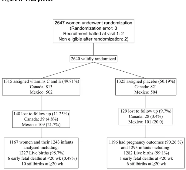
Pour déterminer les effets de suppléments antioxydants pris pendant la grossesse sur le risque d’hypertension gestationnelle (HG) et de prééclampsie, un essai multicentrique contrôlé à double insu a été mené au Canada et au Mexique (An International Trial of Antioxidants in the Prevention of Preeclampsia – INTAPP). Les femmes, stratifiées par risque, étaient assignées au traitement expérimental quotidien (1 gramme de vitamine C et 400 UI de vitamine E) ou au placebo. En raison des effets secondaires potentiels, le recrutement pour l’essai a été arrêté avant que l’échantillon complet ait été constitué. Au total, 2640
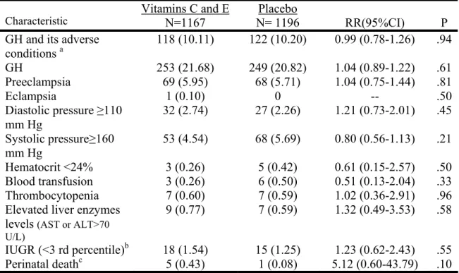
femmes éligibles ont accepté d’être recrutées, dont 2363 (89.5%) furent incluses dans les analyses finales. Nous n’avons retrouvé aucune évidence qu’une supplémentation prénatale de vitamines C et E réduisait le risque d’HG et de ses effets secondaires (RR 0,99; IC 95% 0,78-1,26), HG (RR 1,04; IC 95% 0,89-1,22) et prééclampsie (RR 1,04; IC 95% 0,75-1,44). Toutefois, une analyse
secondaire a révélé que les vitamines C et E augmentaient le risque de « perte fœtale ou de décès périnatal » (une mesure non spécifiée au préalable) ainsi qu’une rupture prématurée des membranes avant terme.
Nous avons mené une étude de cohorte prospective chez les femmes enceintes recrutées dans l’INTAPP afin d’évaluer les relations entre le régime alimentaire maternel en début et fin de grossesse et le risque de prééclampsie et d’HG. Un questionnaire de fréquence alimentaire validé était administré deux fois pendant la grossesse (12-18 semaines, 32-34 semaines). Les analyses furent faites séparément pour les 1537 Canadiennes et les 799 Mexicaines en raison de l’hétérogénéité des régimes alimentaires des deux pays. Parmi les canadiennes, après ajustement pour l’indice de masse corporelle (IMC) précédant la grossesse, le groupe de traitement, le niveau de risque (élevé versus faible) et les autres facteurs de base, nous avons constaté une association significative entre un faible apport alimentaire (quartile inférieur) de potassium (OR 1,79; IC 95% 1,03-3,11) et de zinc (OR 1,90; IC 95% 1,07-3,39) et un risque augmenté de prééclampsie. Toujours chez les Canadiennes, le quartile inférieur de consommation d’acides gras polyinsaturés était associé à un risque augmenté d’HG (OR 1,49; IC 95% 1,09-2,02). Aucun des nutriments analysés n’affectait les risques d’HG ou de prééclampsie chez les Mexicaines.
Nous avons entrepris une étude cas-témoins à l’intérieur de la cohorte de l’INTAPP pour établir le lien entre la concentration sérique de vitamines antioxydantes et le risque de prééclampsie. Un total de 115 cas de prééclampsie et 229 témoins ont été inclus. Les concentrations de vitamine E ont été mesurées
de façon longitudinale à 12-18 semaines (avant la prise de suppléments), à 24-26 semaines et à 32-34 semaines de grossesse en utilisant la chromatographie liquide de haute performance. Lorsqu’examinée en tant que variable continue et après ajustement multivarié, une concentration de base élevée de γ-tocophérol était associée à un risque augmenté de prééclampsie (quartile supérieur vs quartile inférieur à 24-26 semaines : OR 2,99, IC 95% 1,13-7,89; à 32-34 semaines : OR 4,37, IC 95% 1,35-14,15). Nous n’avons pas trouvé de lien entre les concentrations de α-tocophérol et le risque de prééclampsie.
En résumé, nous n’avons pas trouvé d’effets de la supplémentation en vitamines C et E sur le risque de prééclampsie dans l’INTAPP. Nous avons toutefois trouvé, dans la cohorte canadienne, qu’une faible prise de potassium et de zinc, tel qu’estimée par les questionnaires de fréquence alimentaire, était associée à un risque augmenté de prééclampsie. Aussi, une plus grande concentration sérique de γ-tocophérol pendant la grossesse était associée à un risque augmenté de prééclampsie.
Mots-clés: Prééclampsie, Hypertension gestationnelle, Vitamines C et E, Alimentation maternelle, Tocophérol, Étude clinique, Étude de cohorte, Étude cas-témoins
ABSTRACT
Preeclampsia (PE) accounts for about one-quarter of cases of maternal mortality, and ranks second among the causes of pregnancy-associated maternal deaths in Canada and worldwide. The identification of an effective strategy to prevent PE is a priority and fundamental challenge in obstetrics research. The role of nutritional factors in the etiology of PE has recently received increased attention. Many clinical and epidemiological studies have been conducted to investigate potential dietary risk factors for PE and to examine the effects of nutritional supplementation on the development of hypertensive disorders of pregnancy.
To investigate the effects of prenatal antioxidant supplementation on the risk of gestational hypertension (GH) and PE, a double blind, multicenter trial (The International Trial of Antioxidants for the Prevention of Preeclampsia – the INTAPP trial) was conducted in Canada and in Mexico. Women were stratified by their risk status and assigned to daily experimental treatment (1 gram vitamin C and 400 IU vitamin E) or to placebo. Due to concerns about potential adverse effects, recruitment for the trial was stopped before the full sample had been achieved. A total of 2640 consenting eligible women had been recruited at that point with 2363 women (89.5%) included in the final analysis. We found no evidence that prenatal supplementation of vitamins C and E reduced the risk of GH and its adverse conditions (RR: 0.99, 95% CI 0.78-1.26), GH (RR 1.04, 95% CI 0.89-1.22), and PE (RR 1.04, 95% CI 0.75-1.44). However, in a secondary
analysis, we found that vitamins C and E increased the risk of ‘fetal loss or perinatal death’ (a non-pre-specified outcome) as well as preterm premature rupture of membranes (PPROM).
We conducted a prospective cohort study on pregnant women enrolled in the INTAPP trial to investigate the associations between maternal diet in early and late pregnancy and the risk of PE and GH. A validated food frequency
questionnaire (FFQ) was administered twice during pregnancy (12-18 weeks, 32-34 weeks). Analyses were conducted separately for 1537 Canadian and 799 Mexican women as there were significant heterogeneities in various nutrient intakes between the two countries. Among Canadian women, after adjusting for pre-pregnancy body mass index (BMI), treatment group, risk stratum (high versus low) and other baseline risk factors, we found that the lowest quartiles of potassium (OR 1.79, 95% CI 1.03-3.11) and zinc (OR 1.90, 95% CI 1.07-3.39) intake were significantly associated with an increased risk of PE. Also in Canadian women, the lowest quartile of polyunsaturated fatty acids was associated with an increased risk of GH (OR 1.49, 95% CI 1.09-2.02). None of the nutrients analyzed were found to be associated with PE and GH risk among Mexican women.
We further conducted a case control study ancillary to the INTAPP trial to assess the relationship between plasma concentration of antioxidant vitamins and the risk of PE. A total of 115 PE cases and 229 matched controls were included. Vitamin E concentrations were measured longitudinally at 12-18 weeks (prior to supplementation), 24-26 weeks, and 32-34 weeks of gestation using
high-performance liquid chromatography (HPLC). When examined as a continuous variable, and after multivariate adjustment, elevated baseline γ-tocopherol concentrations were associated with an increased risk of PE (OR 1.35, 95% CI 1.02-1.78). Analyses of repeated measurements indicated that elevated γ-tocopherol levels were associated with an increased risk of PE (highest vs. lowest quartile at 24-26 weeks: OR 2.99, 95% CI 1.13-7.89; at 32-34 weeks: OR 4.37, 95% CI 1.35-14.15). We found no associations between α-tocopherol concentrations and the risk of PE.
In summary, we found no effects of vitamins C and E supplementation on the risk of PE in the INTAPP trial. However, in the Canadian cohort we found that lower intakes of potassium and zinc as estimated by the FFQ were associated with an increased risk of PE. Moreover, higher plasma concentration of γ-tocopherol during pregnancy was associated with an increased risk of PE.
Key words: Preeclampsia, Gestational Hypertension, Vitamins C and E,
TABLE DES MATIÈRES
RÉSUMÉ... iii
ABSTRACT ... vi
TABLE DES MATIÈRES ... ix
LISTE DES TABLEAUX ... xii
LISTE DES ABRÉVIATIONS ... xv
REMERCIEMENTS... xvii
INTRODUCTION... 1
STUDENT’S CONTRIBUTION... 2
CHAPTER 1 LITERATURE REVIEW ... 4
1.1 Causal mechanisms of PE ... 4
1.2 Involvement of nutritional factors in the pathogenesis of PE ... 8
1.3 Macronutrients and risk of PE... 9
1.3.1 Energy and diet composition... 9
1.3.2 Fiber ... 11
1.3.3 Protein intake ... 12
1.3.4 Lipid intake ... 13
1.4 Micronutrient and risk of PE... 16
1.4.1 Calcium ... 16
1.4.2 Sodium ... 17
1.4.3 Vitamins C and E ... 18
1.4.4 Vitamin A... 20
1.4.5 Folate (Folic acid) ... 21
1.4.6 Vitamin D... 23
1.4.7 Magnesium... 24
1.4.8 Other micronutrients ... 25
1.5 Obesity, weight gain and risk of PE... 25
1.6 Other risk factors of PE... 28
1.6.1 Genetic and epigenetic factors and risk of PE ... 28
1.6.2 Life style factors and risk of PE... 29
1.6.2.1 Smoking, alcohol use and PE... 29
1.6.2.2 Physical activity and PE... 31
1.6.3 Pregnancy related factors and risk of PE ... 32
1.6.4 Psychosocial factors and risk of PE ... 33
1.6.5 Pre-existing medical conditions ... 34
1.6.6 Environmental chemicals and risk of PE ... 35
1.7 Dietary measurements... 35
1.7.1 Field methods for assessing dietary measurements ... 35
1.7.2 Selection of methods for dietary measurement... 38
1.7.3 FFQs in epidemiological studies... 39
1.8 Summary ... 42
CHAPTER 2 OBJECTIVES AND HYPOTHESES ... 45
2.1 Objectives... 45 2.1.1 General objective: ... 45 2.2.2 Specific objectives: ... 45 2.2 Hypotheses ... 45 2.2.1 Hypothesis I: ... 45 2.2.2 Hypothesis II: ... 46 2.2.3 Hypothesis III:... 46 CHAPTER 3 METHODOLOGY ... 47 3.1 Study design ... 47 3.1.1 Objective I... 47 3.1.2 Objective II... 49 3.1.3 Objective III ... 49 3.2 Outcomes... 50 3.3 Independent variables... 50 3.3.1 Treatment allocation... 50 3.3.2 Nutritional variables... 51
3.3.3 Food Frequency Questionnaire (FFQ) ... 51
3.3.4 FFQ validation in Mexico ... 53
3.3.5. Plasma concentration of Vitamin E... 53
3.3.6 Covariates... 54
3.4 Data management and quality assessment ... 54
3.4.1 Nutrient intake data ... 54
3.5 Statistical analysis ... 55 3.5.1 Objective I... 56 3.5.2 Objective II... 57 3.5.3 Objective III ... 59 3.6. ETHICAL CONSIDERATIONS ... 60 CHAPTER 4 ARTICLE I ... 61
An international trial of antioxidants in the prevention of Preeclampsia (INTAPP trial) ... 62 Source of funding... 63 Condensation... 64 ABSTRACT... 65 Introduction ... 66 Methods... 67 Results ... 73 Discussion ... 75 Acknowledgements ... 80 References ... 81
CHAPTER 5 ARTICLE II... 93
Maternal nutrient intake and the risk of hypertensive disorders in pregnancy ... 94
Abstract ... 95
Introduction ... 96
Methods... 97
Assessment of nutrient intake ... 98
Study outcomes ... 100
Statistical analysis ... 101
Results ... 102
Discussion ... 106
References ... 113
CHAPTER 6 ARTICLE III ... 125
Case control study of Plasma concentration of Tocopherols in relation to the risk of preeclampsia ... 126 Abstract ... 127 Introduction ... 128 Methods... 129 Results ... 133 Discussion ... 136 References ... 141 CHAPTER 7 DISCUSSION... 153
7.1 Nutrition and PE... 153
7.2 Application of FFQ in epidemiological studies ... 161
7.3 Methods for analyzing repeated dietary measurements ... 162
7.4 Strengths and limitations... 164
7.5 Recommendations and future directions... 168
REFERENCES... 172
APPENDIX ... xx
Food Frequency Questionnaires... lvi Role of nutrition in the risk of preeclampsia... cii An international trial of antioxidants in the prevention of preeclampsia (INTAPP) ... cxxii
LISTE DES TABLEAUX
Figure 1. Hypothetical framework on Pathogenesis of Preeclampsia... xx Figure 2. Multivariate analysis approach for nutrient intakes during pregnancy and the risk of GH and PE. ... xxi Figure 3. Analytical framework for the case control study of plasma tocopherol concentrations in relation to the risk of PE ... xxii
ARTICLE I
Figure 1. Trial profile... 86 Table 1 Women’s baseline demographic and obstetric characteristics by
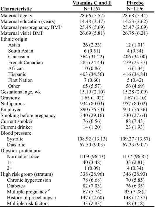
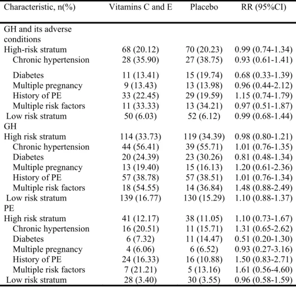
treatment group ... 87 Table 2. Primary outcomes ... 89 Table 3. Primary outcome, gestational hypertension, and preeclampsia stratified by risk at enrolment... 90 Table 4. Secondary maternal outcomes ... 91 Table 5. Secondary Neonatal outcomes ... 92
ARTICLE II
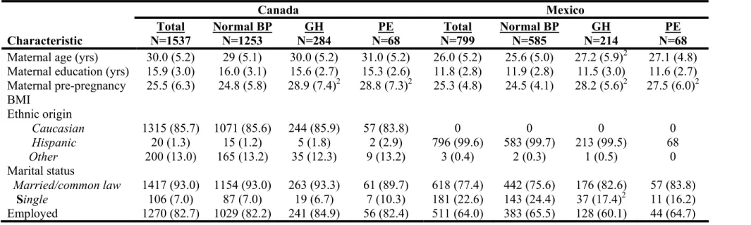
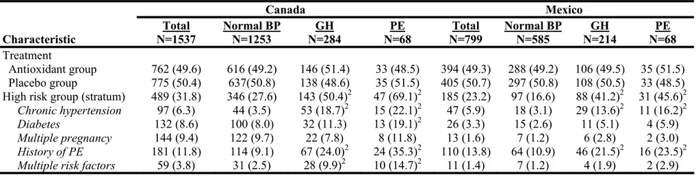
Table 1. Maternal Dietary intake from Food Frequency Questionnaire (FFQ) administered at trial entry (12-18 weeks of gestational age) and in the third trimester (32-34 weeks of gestational age) in Canada and Mexico ... 117 Table 2. Socio-demographic and clinical characteristics of total cohort, women with hypertensive disorders, and women with normal blood pressure in Canada and Mexico... 119 Table 3. Treatment allocation, risk status at trial entry, and vitamins or mineral supplementation of total cohort, women with hypertensive disorders, and women with normal blood pressure ... 121 Table 4. Unadjusted Odds ratios of dietary nutrients intake (lowest quartile vs other quartiles, 12-18 weeks of gestational age) in association with preeclampsia
(PE) and gestational hypertension (GH) in Canadian and Mexican pregnancy cohorts ... 123
ARTICLE III
Table 1 Socio-demographic and clinical characteristics of PE cases and
normotensive controls at trial entry (12-18 weeks of gestationl age) ... 146 Table 2. Plasma concentrations of antioxidant vitamins among preeclamptic women and normotensive controls... 147 Table 3. Plasma concentrations of antioxidant vitamins among preeclamptic women and normtensive controls stratified by treatment group... 148 Table 4. Baseline plasma concentrations of tocopherols in relation to the risk of preeclampsia... 150 Table 5. Repeated measurements of concentrations of tocopherols in the relation to the risk of preeclampsia ... 151
APPENDIX
Table A: A summary of RCTs of certain micronutrient supplementations during pregnancy and the risk of Preeclampsia... xxiii Table B. Nutrients estimated by Food Frequency Questionnaire (FFQ) and average of three non-consecutive Food Records (3D-FR) (FFQ validation study in Canada) ... xxvi Table C. Association between nutrients estimated by Food Frequency
Questionnaire (FFQ) and three non-consecutive Food Records (3D-FRs)- (FFQ validation study in Canada)... xxviii Table D. Proportions (%) of participants ranked into the same quartile of the distribution according to nutrient estimates obtained from the Food Frequency Questionnaire (FFQ) and three non-consecutive Food Records (3D-FR) (FFQ validation study in Canada)... xxx Table E: Nutrients estimated by Food Frequency Questionnaire (FFQ) and three non-consecutive Food Recalls... xxxii Table F. Pearson’s correlation coefficients between FFQ and the 24-hour recalls for energy and selected nutrients (FFQ validation study in Mexico)... xxxiv
Table G. Proportions (%) of participants ranked into the same tertile of the distribution according to nutrient estimates obtained from the Food Frequency Questionnaire (FFQ) and three non-consecutive Food Recalls (FFQ validation study in Mexico) ... xxxvi Table H : Baseline characteristics of women included in the analysis of INTAPP trial and women lost to follow up ... xxxvii Table I: The risk of GH or PE according to quartile distributions of nutrient intakes estimated from FFQ administered at 12-18 weeks of gestational age ... xxxix Table J: Unadjusted Odds ratios of dietary nutrients intake in association with preeclampsia (PE) and gestational hypertension (GH) in Canadian and Mexican pregnancy cohorts (FFQ administered at 12-18 weeks of gestational age) ... xliv Table K: The risk of GH or PE according to quartile distributions of nutrient intakes estimated from FFQ administered at 32-34 weeks of gestational age ... xlix Table L. Unadjusted Odds ratios of changes in nutrient intakes (standardized as Z score) in association with preeclampsia (PE) and gestational hypertension (GH) in Canadian and Mexican cohorts ... liv
LISTE DES ABRÉVIATIONS
APOB: Apolipoprotein BBMI: Body Mass Index
CIHR: Canadian Institute of Health Research CPEP: Calcium for Preeclampsia Prevention CRF: Case Report Form
FFQ: Food Frequency Questionnaire GH: Gestational Hypertension GSH-Px: Glutathione Peroxidase HLA: Human Leukocyte Antigen
HPLC: High-Performance Liquid Chromatography IFN-γ: Interferon-gamma
IMMS: Instituto Mexicano del Seguro Social
INTAPP: International Trial of Antioxidants in the Prevention of Preeclampsia IQ: Intelligence quotient
IUGR: Intrauterine Growth Restriction
LCPUFA: Long Chain Polyunsaturated Fatty Acids LDL-C: Low-Density Lipoprotein Cholesterol
NAD(P)H: Nicotinamide Adenine Dinucleotide Phosphate-Oxidase NO: Nitric Oxide
NOS: Nitric Oxide Synthase OR : Odds Ratio
PAI: Plasminogen Activator Inhibitor PE: Preeclampsia
PPROM:Preterm Premature Rupture Of Membranes PROM: Premature Rupture Of Membranes
PUFA: Polyunsaturated Fatty Acids RR : Relative Risk
sEng: soluble Endoglin
sFlt1: Soluble fms-Like Tyrosine Kinase 1 TCC: Trial Coordinating Center
VCAM-1: Vascular Cell Adhesion Molecule 1 VEGF: Vascular Endothelial Growth Factor VLDL: Very-Low-Density Lipoprotein 95% CI : 95% Confidence Interval
REMERCIEMENTS
Je voudrais remercier tous ceux qui m’ont assistée et inspirée au cours de mes études doctorales.
À commencer par mon superviseur, Dr William Fraser, que je remercie profondément pour son mentorat, ses conseils et sa direction qui m’ont offerts une expérience extraordinaire tout au long du chemin à partir des débuts de ma recherche. Son énergie constante ainsi que son enthousiasme étaient pour moi une source de motivation qui a rendu ma vie de chercheur facile et gratifiante. Surtout, et ce qu’il me fallait de plus, Dr Fraser m’a conféré son encouragement et son appuie, sans broncher, et de maintes façons. Son intuition exceptionnelle de chercheur scientifique, un oasis d’idées et de passions, a inspiré et enrichi ma vie d’étudiante, de chercheur, et de future femme de science.
J’offre mes remerciements à Dr Bryna Shatenstein pour ses conseils, son encadrement, et sa contribution essentielle à ce projet de recherche. L’originalité de ses idées m’a nourrie intellectuellement et m’a mené à une maturité d’esprit dont je bénéficierai pour de nombreuses années à venir.
En particulier, je suis endettée à Dr. Zhong-Cheng Luo pour ses excellents conseils, ses discussions scientifiques, la supervision de mes travaux. Dr Luo a généreusement offert de son temps si précieux pour lire ma thèse et faire part de ses commentaires critiques et judicieux.
Je remercie amplement Dr Pierre Julien pour sa contribution significative à ce projet de recherche et pour ses commentaires constructifs sur cette thèse.
Les Drs Suzanne Tough, Lise Goulet and Jennifer O'Loughlin méritent un remerciement spécial en tant que membres et directeurs du comité d’études.
Mes collègues au Centre de recherche en périnatalité du CHU Ste-Justine (Drs François Audibert, Nils Chaillet, Shuqin Wei, Isabelle Krauss) m’ont accueillie chaleureusement dans leur milieu de travail. J’adresse un
remerciement particulier à Yuquan Wu, Fabienne Simonet, Spogmai Wassimi, et An Na pour leur amitié et leur aide durant les cinq années passées.
De tout mon cœur, merci à ma famille pour leur amour et leur soutien, quoi qu’il m’arrive dans la vie; cette dissertation aurait simplement été impossible sans eux. À mes parents, Xingxin Xu and Guidi Xu, je dois une gratitude immesurable pour leur dévotion. Typiques d’une famille chinoise, mes parents travaillèrent fort pour subvenir aux besoins de la famille et furent tout dans la mesure du possible pour que je puisse atteindre ce niveau d’études. Ils ne se sont jamais plaints, malgré des temps difficiles. Je ne pourrais en demander plus, et il n’y a pas de mots pour décrire l’amour infaillible qu’ils me portent. Je suis fière également des talents de ma sœur Haixia, modèle que je suivis inconsciemment dans mon adolescence et qui m’a toujours portée d’excellent conseil.
J’ai la chance inouïe d’avoir deux anges parfaits, Justin et Jake. Vous êtes tous deux le plus beau cadeau de ma vie.
Un remerciement spécial a mon mari, pour son amour et son soutien pendant mes études doctorales.
Je voudrais mentionner l’Initiative stratégique de formation en recherche en santé de la reproduction (ISFRSR) des Instituts de recherche en santé du Canada (IRSC) qui ont participé au soutient de ce projet de recherche.
Et finalement, je porte ma reconnaissance à tous ceux qui figuraient dans la réalisation de ma thèse, et mes excuses à tous ceux dont je n’ai pas mentionné le nom individuellement.
INTRODUCTION
Preeclampsia (PE), defined as pregnancy-induced hypertension and
proteinuria, is a syndrome that is unique to human pregnancy, affecting between 2% and 8% of pregnancies.(1-3) It accounts for 10%-15% of direct maternal deaths in low- and middle income countries as well as in high income
countries.(4, 5) Gestational hypertension (GH), especially PE, is a frequent cause of low birth weight (<2500g) in infants and thereby perinatal deaths through both preterm delivery and intrauterine growth restriction (IUGR).(6-10) Since
delivery is the only known cure, PE is a leading cause of indicated premature delivery(11) and accounts for about 15% of infants with growth restriction.(12) As many as 60% of extremely low birth weight (< 800g) infants suffer learning disabilities and low IQ(13), increasing the hidden costs of the disease. The study of the etiology, prevention and outcomes of PE and other hypertensive disorders of pregnancy remains a research priority. Effective prevention of PE would have major health benefits and result in considerable savings to health care budgets.
It has long been suggested that diet may play a role in PE. Much of the clinical and basic research into the nutritional causes of the hypertensive disorders of pregnancy has paralleled research on the etiology of hypertension, focusing on individual nutrients such as calcium, sodium, magnesium, and fatty acids. Until now, the effects of diet and specific nutrients on the hypertensive disorders of pregnancy have rarely been studied in a prospective cohort. Dietary assessment methods have not often been validated for use among pregnant women. The present research represents one of the very few studies to date that
have comparatively assessed the role of diet in the etiology of PE among women living in different geographic settings– in this case, in Canada and in Mexico - both in early pregnancy and in late pregnancy. In addition, our goal was to assess the role of diet on the development of PE at different stages of gestation. We believe that this study makes a novel contribution to understanding of the role of maternal nutrient intakes and supplementation in early and late pregnancy on the risk of PE.
STUDENT’S CONTRIBUTION
The studies described in this thesis were conducted in the context of a research program that involved a number of researchers. As a PhD student in this program, I played a key role in all of the studies described. With respect to the INTAPP trial, I played a leading role in preparation and modification of Case Report Forms and Standard Operation Procedures, the data management and adjudication of the primary outcomes (Gestational hypertension and
Preeclampsia), and the planning and execution of data analysis. I played a key role in the Data Safety and Monitoring Committee, making significant
contributions to the work of that committee including: 1) preparation of the report of potential adverse events associated with vitamins C and E in the literature; and 2) conducting the interim analysis regarding the adverse events observed in the INTAPP trial. With respect to the preparation of the tools for the nutritional surveys, I worked closely with nutritional experts in the INTAPP team and made significant contributions to FFQ validations study in Canada
including preparing of the statistical analysis plan and conducting data analysis. Regarding the ancillary study of nutrient intakes during pregnancy and risk of hypertensive disorders, I played a leading role in the conceptualization of the study, data management and quality assessment of the FFQ data, preparation of study analysis plan and conduct of the study analysis.
I worked closely with our colleagues at Québec Lipid Research Center and played a leading role in the conceptualization and implementation of the case control study of plasma tocopherol concentrations in relation to PE risk. The main specific responsibilities for this study were: 1) study design and preparation of study protocol; 2) implementation of the study including identifying cases and controls for laboratory measurements; and 3) preparing the statistical analysis plan and conducting data analysis using appropriate statistical models.
I drafted all manuscripts listed in the present dissertation and was the primary author for each of the manuscripts.
CHAPTER 1 LITERATURE REVIEW
1.1 Causal mechanisms of PEPE is a multisystem disorder that is specific to human pregnancy and only can be resolved by delivery. Generally, the etiology of PE can be conceptualized in two broad categories: PE of placental origin and PE of maternal origin.(14) PE of placental origin arises from a hypoxic placenta and progresses in two stages described as pre-clinical (poor placentation) and clinical features.(14) PE of maternal origin arises from the interaction between a normal placenta and maternal constitutional factors such as microvascular disease, chronic
hypertension, obesity, inflammation or diabete that predispose the woman to the condition. Thus pregnancy may represent a metabolic and vascular ‘stress test’ that unmasks latent cardiovascular risk.(15) However, involvement of both placental and maternal constitutional factors is very common in the development of PE and these broad categories are likely not mutually exclusive. Most patients are somewhere on a continuum between these two etiologic pathways.
Several etiologic theories of PE have been proposed and extensively investigated. (14), (16-21) During normal pregnancy, cytotrophoblasts invade the maternal decidua and spiral arteries and completely remodel the maternal spiral arteries into large capacitance vessels with low resistance. In
preeclampatic pregnancies, shallow endovascular cytotrophoblast invasion of the spiral arteries results in a hypoxic and dysfunctional placenta, and the release of factors such as cytokines, growth factors and certain chemicals into the maternal
circulation.(14-26) These maternal circulating factors mediate endothelial dysfunction, leading to the clinical signs of PE. Increased levels of factor VIII-related antigen, total and cellular fibronectin, thrombomodulin, endothelin, and disturbances of the prostacyclin to thromboxane A2 ratio all support the
hypothesis that systemic endothelial dysfunction plays a central role in the pathogenesis of PE.(27-36) Several lines of evidence support the hypothesis that the abnormal placentation may play a role in inducing an alteration in the balance of circulating levels of angiogenic/antiangiogenic factors such as vascular endothelial growth factor (VEGF), free placental growth factor (PlGF), soluble fms-like tyrosine kinase (sFlt1) and soluble endoglin (sEng), which contributes to endothelial cell dysfunction in the maternal vasculature.(19, 37-40) Recent studies suggest that women with clinically established PE have
significantly lower levels of PlGF and VEGF compared with gestational age-matched normotensive controls.(16, 41-46) Circulating sFlt1, a receptor binding VEGF and PlGF, is significantly increased before the onset of PE.(46-48) However, it remains unclear whether impaired placental perfusion initiates symptoms such as hypertension, endothelial dysfunction, and increased sFlt1 expression, or whether inadequate placental development occurs initially and is followed by a pathological rise in sFlt1 expression and secretion.(49)
The role of oxidative stress in the pathogenesis of PE is also increasingly recognized.(21, 50, 51) Oxidative stress is an imbalance between pro-oxidant and antioxidant forces, resulting in an accumulation of free radicals or reactive oxygen or reactive nitrogen species. Deleterious effects of free radicals include
lipid peroxidation, oxidative damage to bimolecules, and cellular dysfunction. It has been hypothesized that hypoxia stimulates the activity of xanthine or
nicotinamide adenine dinucleotide phosphate-oxidase (NAD (P)H) in placenta, which leads to superoxide generation. Oxidative stress likely contributes to maternal endothelial cell activation, enhanced apoptosis of trophoblast, and is believed to underlie the intense vasoconstriction and procoagulant state of PE.(14) (16-21) Markers of oxidative stress, such as isoprostanes and
malondialdehyde, are increased in plasma,(52, 53) small arteries(54) and decidua basalis(55) of women with PE.
Experimental and epidemiological data support the role of maternal-fetal immune maladaptation in the etiology of PE.(56-59) There are reports of altered immune status in PE.(60-62) A significantly lower proportion of T-helper cells was demonstrated in women who later developed PE.(60) Deposition of immunoglobulin (IgM), complement (C3), and fibrin has been observed in the walls of spiral arteries in women who develop PE.(61, 63) Studies have shown that mothers lacking most or all activated killer cell immunoglobulin-like receptors (KIRs, AA genotype) when the fetus had HLA-C (human leukocyte antigens) were at a substantial risk of PE.(63) These findings are supported by epidemiological studies investigating the relationship between parity, paternity and the risk of PE.(64, 65) It has been demonstrated that multiparity is associated with a reduced risk of PE, which suggests an immune tolerance phenomenon. Interestingly, some studies suggest that the protective role of primiparity is lost
with the change of partner, suggesting that primpaternity, rather than primparity, is related to the risk of PE. (64, 65)
PE is associated with an increase in systematic inflammatory responses. The causes of these responses remain unknown. One attractive concept is that placental ischemia and reperfusion with oxidative stress may induce the higher proliferation of cytotrophoblasts and increase the deportation of
syncytiotrophoblasts.(14, 20) Thus, the altered balance between proliferation and apoptosis of trophoblasts may cause aponecrotic or even necrotic release of trophoblasts, accentuating maternal inflammatory burdens. It has been reported that there are increased amounts of trophoblast debris, comprised of
syncytiotrophoblast membrane microparticles, cytokeratin fragments, and soluble fetal proteins in maternal circulation in women with PE. (14), (16-21) Enhanced activation of cytokine mediators of apoptosis (especially interferon, tumour necrosis factor) have been found in PE. (14), (16-21) It is well known that severe PE and eclampsia have a familial tendency. Nilsson and colleagues reported a heritability of 31% for PE and 20% for GH.(66) Chesley et al. reported a 26% incidence of PE in daughters of women with PE compared to only an 8% incidence in the daughters-in-law.(67) It seems that a number of maternal susceptibility genes or perhaps fetal genes may contribute to the
pathogenesis of PE by interacting with the maternal cardiovascular or hemostatic systems, or by regulating endothelial activation and inflammatory responses.(68-71)
In summary, there are numerous theories of the pathogenesis of PE. (Appendix: Figure1) These different underlying mechanisms are not mutually exclusive, but rather likely interactive. A vast array of initiating agents and multiple pathogenic mechanisms have been implicated in the development of PE, including increased systematic vascular resistance, enhanced platelet aggregation, activation of coagulation systems and endothelial dysfunction.
1.2 Involvement of nutritional factors in the pathogenesis of PE The role of maternal diet in the etiology of PE has recently received increased attention. Information largely derived from studies external to pregnancy indicates that certain nutrients may be involved in several important steps in the current proposed concepts of the pathogenesis of PE.(71-74) Several nutrients, in particular, omega-3 (n-3) fatty acids, antioxidants, folic acid, and L-arginine have important roles in modulating endothelial function.(71, 72) Higher intake or supplementation of these nutrients is associated with the decreased expression of endothelium adhesion molecules (VCAM-1), but increased levels of endothelium dependent vasodilation and nitric-oxide production.(75-80) The influence of these nutrients on endothelial function is multiple and complex, including inhibition of monocyte adhesion and platelet activation, and improvement of vasodilation and blockage of lipid oxidation.(71, 72, 74)
Nutrients can affect oxidative stress by increasing or decreasing free radicals or antioxidants, by providing substrates for the formation of free radicals, or by modulating functions of antioxidant enzymes. For instance, lipids are extensively involved in the generation of free radicals.(81) Antioxidants (vitamin C, E, alpha
or beta-carotene, copper, selenium, zinc, etc.) can directly or indirectly scavenge free radicals or function as essential substrates or cofactors for the adequate functioning of antioxidant enzymes. Therefore, adequate dietary antioxidant intake is crucial for maintaining pro-oxidant and antioxidant balance as some nutrients are not synthesized in humans.
Compelling evidence suggests that nutrients may modify certain
inflammatory responses.(82-85) For example, nutrients can affect the production of monocyte tumor necrosis factor-α (e.g. antioxidants and fatty acids), modulate pro-inflammatory cytokine production and actions (e.g. iron, fatty acids), or activate genes involved in the inflammatory responses (e.g. polyunsaturated fatty acids). (82-85)These mediators are essentially implicated in the pathogenesis of PE such as trophoblast apoptosis, inflammatory response and endothelial activation.
It has also been suggested that nutrients such as trace elements, fatty acids and folic acid can contribute to insulin resistance, a risk factor for PE.(86-89) Both experimental and epidemiological studies have indicated that n-3 fatty acids can improve glucose tolerance and prevent insulin resistance.(90, 91)
1.3 Macronutrients and risk of PE 1.3.1 Energy and diet composition
It is proposed that high-energy diets can affect endothelial function and inflammatory responses by activating oxidative stress-responsive transcription factors, inflammatory cytokine production and the expression of adhesion molecules.(92, 93) Abnormal lipid metabolism can be present in women with
mild or severe PE: these anomalies are characterized by increased levels of triglycerides, low-density lipoprotein cholesterol (LDL-C), LDL-III (small dense lipoprotein) and apolipoprotein A-I.(94, 95)
A large case-control study was conducted in Jerusalem, involving 180 women with PE and 360 healthy controls who were matched for country of origin, parity, month of delivery, age, year of immigration and years of schooling. A dietary history was obtained at the time of delivery. Results indicated that preeclamptic women had significantly lower intakes of protein, fat and energy. However, further investigations suggested that these differences might be
secondary to the disease rather than causal.(96) Atkinson et al. carried out a case-control study in Zimbabwe using a crude (simple/qualitative/non-quantitative) food frequency questionnaire (FFQ) and found no significant differences between 180 women with PE and 194 normtensive controls.(97) Only a few prospective population-based studies have examined the relationship between energy intake and the risk of PE, and they have yielded inconsistent findings.(98, 99) A US study evaluated diet using a 24-hour dietary recall at 13-21 weeks gestation in 4157 women who had been enrolled in a randomized controlled trial of calcium supplementation in the prevention of PE.(99) There was no evidence of an increased risk of PE in women with a higher intake of energy. Moreover, there was no difference between cases and controls in the intake of any of the 28 nutrients that were studied.(99) A Norwegian team administered a
semi-quantitative FFQ to 3771 women at 17-19 weeks of gestation.(98) The risk of PE was increased among women with a high energy intake (adjusted OR: 5.4, 95%
CI: 2.3 –12.4, for the 4th quartile) and a high intake of polyunsaturated fatty acids (adjusted OR: 2.3, 95% CI: 1.1-4.6). Differences persisted even after adjusting for age, smoking and body mass index (BMI). Moreover, the authors observed a stronger association for early onset PE. The discrepancy between these studies may be partially explained by the methods used to estimate dietary intake, the time in pregnancy at which diet is assessed, different definitions of PE and GH, or population differences (i.e. lifestyle, heterogeneity in nutrient intake, socio-demographic factors). It is worth pointing out that in both the Norwegian(98) and American Studies, (99) women who later developed PE had a higher pre-pregnancy body weight, suggesting the potential role of energy balance before pregnancy in the development of PE.
1.3.2 Fiber
Evidence derived from randomized controlled trials indicates that dietary fiber may have beneficial effects on plasma lipid and lipoprotein profiles, postprandial glucose metabolism, insulin sensitivity and blood pressure.(100, 101)The clinical data on the role of fiber in pregnancy are however quite limited.
In 1991, Skajaa et al. found no differences in mean daily fiber intake during the third trimester between PE cases and controls.(102) Frederick et al.
conducted a case-control study of 172 preeclamptic women and 339
normotensive controls to explore the relation between PE risk and maternal intake of dietary fiber, potassium, magnesium and calcium. They reported that fiber intake was inversely associated with the risk of PE. (103) They found that women with fiber intake in the highest quartile (>24.3g/day) had a reduction in
the risk of PE (OR 0.46, 95% CI 0.23-0.92) compared to the lowest quartile (<13.1g/day). In this study, the FFQs were administered at the end of pregnancy, therefore the possibility of recall bias can be not excluded.(103) More recently, Qiu et al. carried out a prospective cohort study of 1,538 pregnant women in Washington State, in which a 121-item FFQ was administered at a mean
gestational age of 13.1 weeks. The adjusted relative risk of PE for women in the highest (>21.2 g/day) vs. the lowest quartile (<11.9 g/day) was 0.28 (95% CI 0.11-0.75). (104) The authors observed similar magnitudes of associations for the highest vs. the lowest quartiles of water-soluble fiber (RR 0.30; 95% CI 0.11-0.86) and insoluble fiber (RR 0.35; 95% CI 0.14-0.87).(104) Furthermore, mean triglyceride concentrations were significantly lower and high-density lipoprotein cholesterol concentrations were nonsignificantly higher for women in the highest quartile compared to those in the lowest quartile.(104) Additional well designed cohort studies and clinical trials are needed to further explore the role of fiber as well as of obesity, insulin resistance, and dyslipidemia in the development of PE.
1.3.3 Protein intake
It has been suggested that certain amino acids such as arginine, citrulline, glycine, taurine, and histidine, as well as small peptides that directly scavenge oxygen free radicals are essential for normal endothelial vasomotion.(71, 72) However, epidemiological studies have not yielded compelling evidence to support an association between protein deficiency and the increased risk of PE.(96-99) Furthermore, trials of protein supplementation have failed to demonstrate a reduction in the risk of PE.(105, 106) The effects of high protein
supplementation (protein/energy supplementation in which the protein content of the supplement provided >25% of its total energy content) on pregnancy
outcome were assessed in a Cochrane systematic review. No significant benefits of protein supplementation were observed.(107) Another systematic review to assess the effects of the balanced protein-energy supplementation on pregnancy outcomes (protein content less than 25% of total energy content) showed no effects on pregnancy outcomes including the risk of PE.(108) It should be noted that the trials included in these systematic reviews had methodological flaws. Alternate treatment allocation rather than a solid randomization method was used, and a large proportion of women were lost to follow up for the primary outcome.
On the other hand, it has been hypothesized that high protein diets may increase the risk of PE by contributing to oxidative stress via increased homocysteine production and increased whole-body nitric oxide (NO) production from nitric oxide synthase (NOS) induction.(109) However, a published meta analysis showed that, in three trials involving 384 women, energy/protein restriction had no effect on pregnancy-induced hypertension or PE, despite the fact that women who were overweight or who exhibited high weight gain significantly reduced weekly maternal weight gain and mean birth weight.(110)
1.3.4 Lipid intake
Several studies have documented dyslipidemia in women with PE. Reduced HDL (111, 112) and increased triacylglycerols (113), LDL cholesterol (114, 115) and small dense LDL (116) were demonstrated in women with PE. Increases in
serum triglycerides and free fatty acids among women who later developed PE were evident before 20 weeks of gestation.(117)
Increased levels of polyunsaturated and total free fatty acids, and other lipids and reduced (n-3) fatty acids have been observed in women with PE.(118, 119) One study prospectively assessed dietary fatty acid intake and fatty acid
composition in maternal, fetal and umbilical blood.(120) Maternal blood was sampled in a large cohort of women at less than 16 and at 22-32 weeks of
gestation, and within 24 hours of delivery. A subset of women underwent dietary assessment in each trimester. The results showed that there were no differences between groups (GH with or without proteinuria vs normotensive women) in maternal fatty acid and nutrient intake at 16 and 32 weeks of gestation. After delivery, levels of essential fatty acids, including 18:2 (n-6) Linoleic acid and 18:3 (n-3) α-Linoleic acid were significantly lower, whereas the sum of (n-6) long-chain polyenes (polyunsaturated fatty acids with 20 or more carbon atoms and three or more double bonds) were significantly higher in hypertensive women compared to controls.(120) In another prospective study, an increased intake of polyunsaturated fatty acids was demonstrated in women who later developed PE.(98)
Omega-3 (n-3) fatty acids have been suggested to have a preventive effect on early delivery and hypertensive disorders of pregnancy.(121, 122) Omega-3 (n-3) fatty acids are known to reduce fasting and postprandial triglycerides and to decrease platelet and leukocyte reactivity. It has been suggested that high-dose n–3 fatty acid intake could reduce maternal thromboxane A2 synthesis and
enhance maternal refractoriness to angiotensin II, which may reduce the risk of PE.(123) Low erythrocyte levels of omega-3 fatty acids and high levels of omega-6 fatty acids, particularly arachidonic acid, appear to be associated with an increased risk of PE.(124) Wang et al. observed a significantly lower level of total n–3 and n–6 polyunsaturated fatty acids in women with PE.(125) However, recent clinical trials failed to detect any significant effect of fish oil
supplementation on PE risk in women at high risk of GH.(126-129) Interestingly, a recent study reported that dietary intake in polyunsaturated fatty acids (PUFAs: n-3 and n-6) was positively correlated with glutathione peroxidise (GSH-Px) activity in healthy pregnant women. (130) The author suggested that increased GPx activity may be a response to the increased oxidative stress generated by the relatively higher concentrations of PUFAs.(130) Moreover, a recent prospective study indicated that the odds ratio for hypertensive disorders presented a U-shaped curve across different intake levels of n-3 long-chain polyunsaturated fatty acids (n-3 LCPUFA). (131) The authors concluded that excessive
consumption in early pregnancy of n-3 LCPUFA or other nutrients (e.g. vitamin A, D, E) found in liquid cod-liver oil may increase the risk of developing hypertensive disorders in pregnancy.(131) However, Horvath et al. carried out a meta analysis of randomized controlled trials to evaluate the LCPUFAs on pregnancy outcomes.(132) There was no evidence of an effect of LCPUFAs on the rate of pregnancy-induced hypertension or PE.
1.4 Micronutrient and risk of PE 1.4.1 Calcium
Calcium is the micronutrient that has been most extensively studied in relation to PE. Numerous studies have demonstrated reduced levels of serum or urinary calcium in PE.(133-136) Several epidemiological studies indicate an association between low dietary intake of calcium and increased risk of PE.(103, 137)
Encouraged by the results of observational studies, a number of controlled trials have been conducted to confirm the beneficial effects of calcium
supplementation, but with conflicting results.(138-140) A recent large trial investigated whether calcium supplementation of pregnant women with low calcium intake reduced PE and preterm delivery.(141) Calcium supplementation was associated with a small reduction in the incidence of PE and/or eclampsia (4.1% versus 4.5%; RR 0.91, 95% CI 0.69-1.19), early onset PE and/or
eclampsia (RR 0.77; 95% CI 0.54-1.11) and GH (RR 0.96, 95% CI 0.86-1.06), however, null effects were not excluded. A life table analysis indicated that effects on PE and/or eclampsia were evident by 35 weeks of gestation (1.2% in calcium group versus 2.8% in placebo group, P = .04). Furthermore, calcium supplementation was associated with a reduced risk of eclampsia (RR 0.68, 95% CI, 0.48-0.97) and severe GH (RR 0.71, 95% CI 0.61-0.82).(141) Overall, there was a statistically significant reduction in the severe preeclamptic complications index including any of the following: severe PE, early onset PE, eclampsia, placental abruption, HELLP syndrome (hemolysis, elevated liver enzymes, and
low platelet count), or severe GH (RR 0.76, 95% CI 0.66-0.89, life-table analysis, log rank test P =.04).(141) Hofmeyr et al. recently conducted a meta analysis of 12 randomized controlled trials, including 15,528 women, in which 66% women had a low dietary calcium intake and 96% women were at low risk for GH or PE.(142) The dose of calcium administered varied from 1.5 to 2.0 grams per day. Calcium supplementation significantly reduced the risk of high blood pressure (11 trials, 14,946 women: relative risk 0.70, 95% CI 0.57-0.86), PE (12 trials, 15,206 women: RR 0.48, 95% CI 0.33-0.69), and maternal death or serious morbidity was reduced (four trials, 9732 women: RR 0.80, 95% CI 0.65-0.97). The effect was greatest for women at high risk for hypertensive disorders of pregnancy (five trials, 587 women: RR 0.22, 95% CI 0.12-0.42) and for those with low baseline calcium intake (seven trials, 10,154 women: RR 0.36, 95% CI 0.18-0.70).(142) However, HELLP syndrome was increased in the calcium supplementation group compared to the placebo group (two trials, 12,901 women: RR 2.67; 95% CI 1.05-6.82). There were no differences in neonatal outcomes such as preterm birth or stillbirth or death before discharge from hospital.(142)
1.4.2 Sodium
A Cochrane review indicated that manipulating sodium intake does not affect the frequency of PE.(143) In addition, a study in Japan indicated that a low-salt diet is not only ineffective for the prevention of PE, but also accelerates volume depletion in PE.(144) A reduction in sodium intake may cause a significant reduction in the intake of energy, protein, carbohydrates, fat, calcium and other
nutrients.(145) Therefore, based on recent evidence, salt restriction is not recommended in pregnancy.
1.4.3 Vitamins C and E
Vitamins C and E are two essential nutrients that can scavenge free radicals and constitute a strong line of defence in delaying or preventing ROS-induced cellular damage. Vitamin C (ascorbic acid) is an essential water-soluble vitamin, and serves as a non-enzymatic antioxidant by delivering a hydrogen atom with a single electron to a reactive oxygen molecule. Adequate dietary intake is
required to prevent oxidative stress. Vitamin E is the major peroxyl radical scavenger in biological lipid phases, such as membranes or LDL. Its antioxidant action has been ascribed to its ability to chemically act as a lipid-based free radical chain-breaking molecule, thereby inhibiting lipid peroxidation and oxLDL formation.(146) Vitamins C and E also play a key role in the modulation of enzymes involved in the vascular endothelial damage known to contribute to the pathophysiological mechanisms of the clinical expression of PE.(147) In vitro and in vivo studies demonstrate a synergistic effect between the two vitamins.(148)
Numerous studies have reported low levels of vitamin C in women with PE.(149-151) A case- control study (99 women with PE compared with 99 controls) found that women with both elevated oxidized LDL and low vitamin C concentration had a 9.8 fold risk of PE (95% CI 3.0-32.2).(151) Sagols et al. reported that plasma levels of ascorbic acid and serum antioxidant activities were significantly decreased in mild and severe PE compared to normtensive
controls.(152) Serum alpha-tocopherol levels were significantly decreased only in severe PE.(152) Furthermore, a case control study using a semi-quantitative FFQ found that women who consumed <85mg of vitamin C daily, as compared with others, experienced a two-fold risk of PE. Women with plasma ascorbic acid less than 34.6 micromol/liter had a 3.8-fold increased risk of PE, compared with those in the highest quartile. Analyses were adjusted for maternal age, parity, pre-pregnancy BMI, and energy intake.(153)
A reduced level of vitamin E in association with PE has been reported in some,(154-156) but not in all studies.(149, 150, 157-160) Reduced levels of vitamin E have been most consistently demonstrated in severe cases of PE.(152, 154, 161, 162) The variation across studies may be explained by the fact that concentrations of lipid soluble vitamin E had not been adjusted for lipid
concentrations although the elevation of total cholesterol and triglycerides is one of characteristics of PE.(163) Moreover, the measurement of plasma vitamin E is a far from satisfactory estimate of the focal site of vitamin E activity, namely the cell membrane. To date, no study has measured vitamin E concentrations in red cell membrane or those in any other tissue in women with PE. It is striking that the diversion from normal values correlated with severity of disease in the reports describing either lowered or elevated plasma vitamin E concentration in women with PE.(152, 154, 156, 164)
The first clinical trial to investigate the effects of vitamin C and E on the risk of PE was conducted by a UK research group.(165) Patients were included in the study if they were at increased risk of PE, as defined by abnormal uterine artery
Doppler waveform or by past history of the disease. Among women who were at risk, the investigators reported a reduction in PE in the group with
supplementation of vitamin C (1000 mg/day) and vitamin E (400 IU alpha-tocopherol/day) for (RR 0.39; 95% CI 0.17-0.90). The ratio of PAI-1
(plasminogen activator inhibitor-1, a marker of endothelial cell activation) to PAI-2 (a marker of placental function) was significantly decreased in the
vitamin-treated group. High-risk women who developed PE in the placebo group had lower plasma vitamin C concentrations (p<0.002) compared with normal pregnant controls and these returned to normal levels on supplementation.(166) Plasma concentrations of the isoprostane, 8-epi-prostaglandin F2alpha, a marker of lipid peroxidation were raised in the high-risk placebo group but fell to concentrations comparable to low risk subjects after vitamin C and E
supplementation.(166) Another small trial of women who were considered as being at high risk on the basis of their clinical history found no evidence of benefits with the same antioxidants.(167)
1.4.4 Vitamin A
The role of vitamin A and β-carotene (pro-vitamin A) in pregnancy induced hypertension and PE is also a subject of controversy. Many clinical studies have found significantly lower levels of vitamin A and β-carotene in preeclamptic women than in healthy women.(168-172) However, the decreased levels of retinol and β-carotene might be secondary to disease as a part of an acute phase reaction rather than the results of a causal relationship. Further studies are needed to determine the temporal relationship between carotenoids and the risk
of adverse pregnancy outcomes. High dose of vitamin A could be toxic and there is concern about its teratogenicity. (173-177) Given the fact that it is unlikely that a safety threshold of vitamin A consumption in early pregnancy will be established over the next few years, it will therefore be ethically difficult to conduct human trials to assess the effects of vitamin A supplementation in early pregnancy on the risk of PE.
1.4.5 Folate (Folic acid)
Folate is the generic term for this water-soluble B-complex vitamin. It functions as a coenzyme in single-carbon transfers in the metabolism of amino acids and nucleic acids, and is therefore required by all cells for growth. Folic acid (pteroylmonoglutamic acid, or PGA), which is the common form used in vitamin supplements and fortified food products, is the most oxidized and stable form of folate. Most naturally occurring folates, called food folate, are
pteroylmonoglutates, which contain one to six additional glutamate molecules joined in a peptide linkage to the γ-carboxyl of glutamate.
The importance of adequate folate supply during pregnancy and lactation is increasingly recognized. It has been suggested that folic acid from food intake and routine supplementation may be sufficient during the periconception period, but larger doses may be required in early gestation, in particular for women with higher risk of adverse pregnancy outcomes (e.g. PE). Folate may reduce the risk of developing PE by improving endothelial function at both the placental and systemic levels,(178) or by lowering homocysteine, a risk factor for PE.(179)
Epidemiologic studies have found that supplementation of multivitamins containing folic acid was associated with reduced risk of PE.(180),194) Bodnar et al. examined the association between regular use of multivitamins containing folic acid at <16 weeks' gestation and the risk of PE in 1,835 women in
Pittsburgh, Pennsylvania between 1997 and 2001.(180) They found that regular use of multivitamins containing folic acid was associated with a 45% reduction in PE risk compared with nonusers (OR 0.55; 95% CI 0.32-0.95). Hernandez-Diaz et al. also observed a significant reduction of risk for GH after
supplementation of multivitamins containing folic acid (adjusted OR 0.55; 95% CI 0.39- 0.79).(181) Wen et al. carried out a prospective cohort study of 2951 women in Ottawa and Kingston, Canada. They found that supplementation with multivitamins containing folic acid in the early second trimester was associated with increased serum folate, lowered plasma homocysteine, and reduced risk of PE (adjusted odds ratio 0.37; 95% CI 0.18-0.75).(182) Catov et al. examined the associations between supplementation of multivitamin containing folate or folate only during a 12-week periconceptional period, using data from the Danish National Birth Cohort.(183) They found that regular use of periconceptional multivitamin containing folate use was associated with a 20% reduction of the risk of PE among normal-weight women. However, such a reduction was not observed for folate only supplements.(183) Furthermore, Ray and Mamdani found that there is a small reduction in the rates of PE in Canada after folic acid food fortification in 1998 (prevalence ratio 0.96; 95% CI 0.94- 0.98).(184)
Evidence from trials assessing the effects of folate supplements on the risk of PE, is very limited. Taylor et al. conducted a randomized trial to assess the effects of supplementation of elemental iron (65 mg/day) and folic acid (350 µg/day) on adverse pregnancy outcomes in 48 healthy pregnant women. They found no effect of iron-folic acid supplementation on the risk of PE.(185) Charles et al. re-analysed data from a large randomised controlled trial performed between 1966 and 1967 and found that the risk of PE was lower in groups receiving the supplementation of folic acid 200µg/day and 5mg/day compared to the placebo group.(186)
1.4.6 Vitamin D
The immunomodulatory properties of the hormonal vitamin D system could potentially have beneficial effects for successful maintenance of pregnancy.(187) Impaired vitamin D metabolism is demonstrated in preeclamptic pregnancy.(187, 188) Therefore, ensuring adequate vitamin D status/intake could potentially contribute to the prevention of PE.(189)
Studies exploring the role of maternal vitamin D status in adverse pregnancy outcomes are scarce. Bodnar et al. conducted a nested case-control study of pregnant women followed from less than 16 wk gestation to delivery (1997-2001) to assess the association of maternal serum 25(OH) D levels with the risk of PE.(190) Their results indicated that serum 25(OH) D concentrations in early pregnancy were lower in women who subsequently developed PE compared with controls. There was a monotonic dose-response relationship between serum 25(OH) D concentration at <22 week of gestation and the risk of developing PE.
A 50-nmol/liter decline in 25(OH)D concentration doubled the risk of
developing PE (adjusted OR 2.4, 95% CI 1.1-5.4). Newborns of preeclamptic women were twice as likely as control newborns to have 25(OH)D less than 37.5 nmol/liter (adjusted OR 2.2, 95% CI 1.2-4.1).(190) A recently published study by Haugen et al. examined the association between vitamin D intake during pregnancy and the risk of PE in 23,423 nulliparous pregnant women taking part in the Norwegian Mother and Child Cohort Study.(191) They found that the odds ratio of PE for women with a total vitamin D intake of 15-20 [mu]g/d was 0.76 (95% CI 0.60-0.95) compared those with less than 5 [mu]g/d. Moreover, they reported a 27% reduction in the risk of PE (OR 0.73; 95% CI 0.58-0.92) for women taking 10-15 [mu]g/d vitamin D supplements as compared with no supplements. However, no association was found between vitamin intake from the diet alone and the risk of PE. (191) There may be a correlation between vitamin D supplementation and intake of other nutrients (e.g. calcium, Omega-3 fatty acid).(191) Further studies with data on other nutrients intake and vitamin D status will be necessary to further disentangle the effect of each on PE risk.
1.4.7 Magnesium
Magnesium is an essential mineral needed by humans in relatively large amounts. It is crucial for regulating temperature and protein synthesis and maintaining electrical potential in nerves and muscle membranes. A prospective observational study used a FFQ to assess diet at 30 weeks of gestational age and found no difference in magnesium intake in Danish women who developed PE compared to controls.(102) Observational studies suggest that supplementation
with magnesium is associated with a reduced risk of PE.(192) However, a Cochrane systematic review of randomized trials found no evidence of a benefit of magnesium supplementation on the risk of PE.(193) The methodological quality of trials included in the review was poor.
1.4.8 Other micronutrients
Certain trace elements are essential co-factors for adequate activation of antioxidant enzymes. These trace elements (e.g., copper, iron and selenium) are directly implicated in oxidative/anti-oxidative balance - a key pathogenic process in PE, and are highly dependent on dietary habits and supplements.(194-196) Serum concentrations of magnesium, copper and zinc have been reported to be significantly lower in PE compared with controls.(136, 197)
Epidemiological studies have suggested that deficiencies of zinc, iron, and selenium are associated with an increased risk of PE. However, randomized trials have failed to demonstrate a beneficial effect of trace elements
supplementation in the prevention or management of PE.(198-200) Little information is available with respect to the specific roles of these trace elements in early pregnancy for PE susceptibility.
1.5 Obesity, weight gain and risk of PE
Obesity is an independent risk factor for PE.(17) The link between obesity and PE is complex. The metabolic changes in obese women, such as increased lipid availability, higher cholesterol and triglyceride levels, or insulin resistance may lead to a derangement of the VLDL/toxicity-preventing activity balance and
enhance cytokine-mediated oxidative stress, subsequently leading to endothelial cell dysfunction.(201-203) Moreover, elevated cardiac output with compensatory vasodilation in women with obesity may also lead to endothelial cell dysfunction. Most observational studies consistently demonstrate that maternal obesity or a higher prepregnancy BMI is associated with an increased risk of PE or GH.(204-207)
Bodnar et al. reported that pre-pregnancy adiposity is a strong independent risk factor for PE.(204) The authors further explored the dose-dependent relationship between pre-pregnancy BMI and the risk of PE. The results
indicated that PE risk rises through most of the BMI distribution. Compared with women with a BMI of 21, the risk of PE doubled for women at BMI of 26, and nearly tripled risk for those at a BMI of 30.(204) A systematic review,
identifying three cohort studies in 1.4 million women (from US, Sweden, the Netherlands, Latin America, the Caribbean, Taiwan, and the UK), demonstrated that the risk of PE typically doubled with each 5-7 kg/m2 increase in
pre-pregnancy body mass index.(205)
The prevalence of obesity is rapidly increasing worldwide and the epidemic is especially pronounced in women of child bearing age.(208, 209) It has been reported that the prevalence of pre-pregnancy obesity increased by 69% over a 10-year period, from 13% in 1993–1994 to 22% in 2002–2003.(209) As obesity confers a significant risk for PE, the epidemic of obesity therefore will
undoubtedly increase the incidence of PE. Wallis et al. analyzed public-use data from the National Hospital Discharge Survey and reported that rates of PE and
GH had increased by 25% and 184% respectively from 1987 to 2004.(210) Thus, public health programs to promote the reduction of overweight or obesity as well as further research to evaluate their effectiveness are needed to suppress the epidemic of obesity and thereby the significant rise in PE.
Over the past 25 years, several authors have demonstrated a significant association between excessive weight gain and hypertensive disorders of pregnancy.(206, 207, 211-213) Brennand et al. showed that obese women with excessive weight gain had a higher prevalence of PE (14.9%) than obese women with low (3.7%) or acceptable (6.3%) weight gain.(214, 215) Saftlas et al. did not find an association between excessive weight gain and the risk of PE although the risk of transient hypertension was increased more than twofold among women in the highest quartile of the weight gain index (OR 2.55; 95% CI 1.66-3.92).(215) A prospective population-based cohort study by Cedergren of 245,526 singleton term pregnancies showed that obese women with low
gestational weight gain had a decreased risk for PE (OR 0.52; 95% CI 0.42-0.62) compared to those with excessive weight gain. There was a 2-fold increased risk for PE among average and overweight women with excessive weight gain.(216) Kiel et al. carried out a population-based cohort study of 120,251 pregnant obese women to examine the associations between gestational weight change and adverse outcomes.(217) The authors reported that, among overweight or obese pregnant women, gestational weight gain of less than the currently recommended 15 lb was associated with a significantly lower risk of PE. The authors concluded that limited or no weight gain in obese pregnant women has favourable
pregnancy outcomes. Langford et al. conducted a population-based cohort study to examine the association between gestational weight gain and adverse
outcomes among overweight women (BMI 26.0-29.0 kg/m2).(218) Compared to women who gained 15-25 lbs, women who gained <15 lbs were 0.8 (95% CI 0.6-1.0) times as likely to have PE, but women who gained >25 lbs were 1.7 (95% CI 1.5-1.9) times as likely to have PE.(218) The Institute of Medicine (IOM) has recently revised guidelines for healthy ranges of weight gain in pregnancy for overweight or obese women: 15-25 lb of weight gain for
overweight (BMI 25-29.9 kg/m2), and 11-20 lb of weight gain for obese women (>30kg/m2).(219) Continued research and changes in health policy should promote the implementation of the new guidelines and determine its impact on health care.
1.6 Other risk factors of PE
Other risk factors including life style factors, environmental factors, genetic factors, psychosocial factors, and pregnancy related factors were reviewed in the following sections.
1.6.1 Genetic and epigenetic factors and risk of PE
Studies have suggested that a family history of PE nearly tripled the risk of PE. (220) Some ethnic groups, like African-American and Hispanic women in the US, have a higher incidence of hypertensive disorders of pregnancy compared to white women.(221) Various candidate genes implicated in thrombophilia, haemodynamics, cytokines, oxidative stress, lipid metabolism,
angiogenesis, and invasion were identified. (222) Epigenetic features are also implicated in the pathogenesis of PE. It has been described that medically assisted procreation increased the risk of PE. (58) For non-imprinted genes, epigenetic alterations are also possible in PE. For instance, methylation alterations of the SER-PINB5 and SERPINA3 promoters have been demonstrated recently in PE.(222)
1.6.2 Life style factors and risk of PE 1.6.2.1 Smoking, alcohol use and PE
Smoking is associated with a variety of adverse pregnancy outcomes, but paradoxically it has a protective role against hypertensive disorders of pregnancy. Previous studies suggest that women who smoke during pregnancy have a
reduced risk of PE compared to non-smokers, even when confounders are carefully controlled.(99),(223-225) Ioka et al. conducted a retrospective cohort study and did not find evidence of a protective effect of cigarette smoking on the risk of PE.(226) Lain et al. found that smoking during pregnancy is associated with reduced cellular fibronectin and increased intracellular adhesion molecule-1.(227) The authors further suggested that the negative association of smoking and PE may be mediated, in part, by the interaction of changes in endothelial activation that are the results of pregnancy and changes that are the result of smoking. Smoking may result in a decrease in basal endothelial activation and a stronger perturbation may be required among smokers to achieve the endothelial activation that is present in preeclamptic women. Results from previous studies on whether or not smoking before pregnancy may also reduce the risk of PE are