ACADEMIE UNIVERSITAIRE WALLONIE-EUROPE UNIVERSITE DE LIEGE
FACULTE DE MEDECINE VETERINAIRE
DEPARTEMENT CLINIQUE DES ANIMAUX DE COMPAGNIE ET DES EQUIDES SERVICE D’IMAGERIE MEDICALE
EVALUATION DE NOUVELLES PROCEDURES GUIDEES PAR
IMAGERIE SUR LA COLONNE LOMBO-SACREE
DU CHIEN
ASSESSMENT OF NEW IMAGING-GUIDED PROCEDURES
OF THE LUMBOSACRAL SPINE
IN DOGS
Annalisa Pia Liotta
THESE PRESENTEE EN VUE DE L’OBTENTION DU GRADE DE DOCTEUR EN SCIENCES VETERINAIRES
TABLE OF CONTENT
•! LIST OF ABBREVATIONS 5
•! I. INTRODUCTION
I.1. Interventional radiology 7I.1 a: Introduction to interventional radiology 8
I.1 b: Interventional spinal pain management in human medicine 11
I.2. Degenerative lumbosacral stenosis in dogs 14
I. 2. a: Anatomy of the lumbosacral space 15
I. 2. b: Motion pattern of the lumbosacral junction and pathophysiology of degenerative lumbosacral stenosis 19
I. 2. c: Signalement, history and clinical signs 20
I. 2. d: Diagnostic imaging of degenerative lumbosacral stenosis 21
I. 2. e: Treatments 29
References 32
•! II. AIMS OF THIS PROJECT
II.1: General and specific aims 41II.1.a: General aim of the project 42 II.1.b: Specific aims of the project 42
•! III. ARTICLES
III. 1: Feasibility of ultrasound-guided epidural access at the lumbosacral space in dogs 44Summary 45
Material and methods 47
Results 50
Discussion 57
References 60
III. 2: Technique, difficulty and accuracy of computed tomography-
guided epidural and intra-articular facet joint injections in dogs 62
Summary 63
Introduction 64
Material and methods 65
Results 69
Discussion 74
References 78
III. 3: Computed tomography-guided epidural and facet joint
corticosteroid injections in dogs: importance of contrast medium injection and
clinical safety 81
Summary 82
Introduction 83
Material and methods 84
Results 87
Discussion 95
References 100
•! IV. DISCUSSION, CONCLUSIONS AND GENERAL PROSPECTS
IV. 1: General discussion and specific conclusions 103
References 119
•! V. SUMMARY/RESUME
V. 1: Summary 124 V. 2: Résumé 125 References 132•! VI. ACKNOWLEDGEMENTS
133
LIST OF ABBREVATIONS
Anulus fibrosus (AF)
Cerebrospinal fluid (CSF) Computed tomography (CT)
Computed tomographic myelography (CTM) Degenerative lumbosacral stenosis (DLSS) Endplates (EPs)
Epidural steroid infiltration (ESI) Fine needle aspiration (FNA) Intervertebral disc (IVD)
Intervertebral disc degeneration (IVDD) Lumbosacral (L-S)
Lumbar vertebra (L)
Magnetic Resonance Imaging (MRI) Nucleus pulposus (NP)
Sacral vertebra (S) Ultrasound (US)
I. 1. a: Introduction to interventional radiology:
Interventional radiology is a branch of modern medicine, which provides image guidance to gain access to organs for diagnostic and therapeutic procedures. The first developing branch of interventional radiology concerned the cardiovascular field with angiography being one of the most important applications of interventional radiology in adult and pediatric patients (Kaufman, 2014; Kandasami et al., 2016). Nowadays, in both human and veterinary medicine, imaging-guided procedures are performed in many other fields, essentially with a diagnostic or therapeutic aim (Vignoli et Saunders, 2011; Mahnken et al., 2013; Sainani et al., 2013).Theoretically, all the imaging modalities that provide a good anatomic spatial resolution of the target and surrounding anatomical structures can be used to perform interventional imaging.A good visualization of the surrounding anatomical structures is the key of imaging-guidance procedures, with the intrinsic advantage of avoiding critical anatomical structures, compared to the “ blind” technique (Mahnken et al., 2013). Among the different techniques, fluoroscopy, ultrasound (US), or computed tomography (CT)-guidance are well-established methods, while magnetic resonance (MRI)-guidance has been performed only more recently (Mahnken et al., 2013; Kaufman, 2014; Mikhail et al., 2015).
1) Fluoroscopy provides real-time images with high spatial resolution. It is used during musculoskeletal procedures such as percutaneous osteosynthesis or intraarticular drug injections. It can also be used in conjunction with the administration of contrast medium, making this technique particularly suitable for vascular interventions. However, it is a 2D technique, has a low soft tissue contrast resolution and exposes patient and operator to radiation (Mahnken et al., 2013).
2) The great advantage of US is the real-time visualization of the needle insertion, low cost and absence of radiation exposure. However, its field of view is limited and bone or air can limit the field of application. It is therefore considered the technique of choice for superficial targets, especially for musculoskeletal procedures (Mahnken et al., 2013; Amber et al., 2014; Wilson et al., 205; Orlandi et al., 2016). US-guidance can either be freehand or with needle guidance (Vignoli et Saunders, 2011; Mattoon et Nyland, 2015). The first technique requires more experience, but it is the most versatile. The needle guidance system, on the other hand, uses a biopsy guide attached to the transducer, which makes visualization and insertion of the needle easier. However, the fixed angle of the needle can cause physical limitations making this technique less versatile (Vignoli et Saunders, 2011; Mattoon et
Nyland, 2015). US guidance can also be indirect. This means that the features of the target of interest (size, depth, proximity with other organs) and the needle insertion pathway (depth and angle of insertion) are determined with US, but the insertion of the needle is performed blindly (Vignoli et Saunders, 2011; Mattoon et Nyland, 2015).
3) In contrast, 3-D reconstructions, typical of cross-sectional imaging modalities such as CT or MRI, allow a better visualization of the target and therefore a better planning of the path to the target (Schwarz and Puchalski, 2011; Mahnken et al., 2013).
However, CT is not a real-time visualization technique. The introduction of CT-fluoroscopy combines the advantages of both fluoroscopy and CT and allows the reduction of the procedure’s duration, real-time visualization of critical anatomical structures and faster images reconstruction. In contrast to conventional CT-guidance, radiation exposure of the operator cannot be avoided and it is more expensive (Mahnken et al., 2013; Paik, 2014).
4) Besides imaging without ionizing radiation, MR imaging offers advantages such as high soft-tissue contrast, multiplanar imaging without reconstructions, and the ability to measure multiple physical or functional parameters (including flow, perfusion, diffusion, and temperature). However, rapid acquisition sequences and a more open magnet design are required to permit easy access to the patient during the procedure (Westbrook et al., 2011; Campbell-Washburn et al., 2015). Flexible transmit and receive coils have been specially designed to allow access to patients for interventions (Westbrook et al., 2011). Low field permanent magnets are considered the best from the access point of view, but on the other hand their use is limited because of image quality and acquisition time (Westbrook et al., 2011). The restricted availability of MR scanners and special coils, and the need of dedicated non-ferromagnetic biopsy instruments are therefore limiting the use of MRI-guided procedures in both human and veterinary patients (Vignoli et Sanders, 2011; Mahnken et al., 2013). Moreover, ferromagnetic materials such as orthopedic protheses or identification chips can be present within the patient and are associated with large imaging artefacts preventing imaging of the area of interest (Gavin, 2009a).
In addition to these classic imaging-guided procedures, the injection of contrast medium is usually suggested to verify the correct position of the needle during different kind of procedures (Johnson et al., 1999; Watanabe et al., 2002; Bartynski et al., 2005).
Imaging-guided procedures can be used as diagnostic or therapeutic means (or as both) or to guide anesthetic procedures, increasing the likelihood of reaching the anesthetic target. The most common imaging-guided diagnostic procedures are fine needle aspiration
(FNA) or biopsy. Ultrasound or CT-guided biopsy or FNA are well-established methods, while MRI-guidance has only been performed more recently. In human medicine, the imaging-guided therapeutic procedures are essentially used in interventional oncology, in the musculoskeletal field, and for pain management (Mahnken et al., 2013; Lee et al., 2016).
a) Thermal ablation is one of the main fields of interventional oncology (Beland et Mayo-Smith, 2014). Its basic aim is to decrease tumor size or to destroy tumor cells by means of heat. Applied imaging-guided techniques of thermal ablation are radiofrequency ablation, laser interstitial thermotherapy, microwave ablation, and high-intensity focused US (Beland et Mayo-Smith, 2014). Another commonly performed therapeutic imaging-guided technique of local tumor ablation is percutaneous ethanol injection. Injection of ethanol leads to tumoral tissue necrosis and local secondary fibrosis (Beland et Mayo-Smith, 2014).
b) In the musculoskeletal field, imaging-guided procedures can vary from local ablation of an osteoid osteoma to percutaneous vertebroplasty or osteoplasty (Irani et al., 2014). Moreover, imaging techniques, more commonly CT or fluoroscopy, are commonly used to guide minimally invasive fracture reductions.
c) In the anesthetic field, imaging-guided procedures, essentially US-guided, are widely used to perform epidural anesthesia (Grau et al., 2001; Karmakar et al., 2009; Bauer et al., 2012) or local plexus blocks (Marhofer et al., 2005; Mejia-Terrazas et al., 2015; Sehmbi et al., 2015; Seidel et al., 2015; Amini, 2016; Neal, 2016). The increased popularity of US-guided epidural anesthesia has been attributed to a more accurate estimation of the depth of the epidural space, and a more optimal determination of the needle path especially in cases of vertebral canal malformations, in cases of obesity, or in obstetric patients where hormonal changes can influence spinal and epidural anatomy (Grau et al., 2001; Karmakar et al., 2009; Bauer et al., 2012). Moreover, US guidance has made nerve blocks a technically feasible, safe, and efficacious option, allowing surgery to be performed without general anesthesia (Mejia-Terrazas et al., 2015; Sehmbi et al., 2015; Seidel et al., 2015).
More specifically, in veterinary medicine interventional radiology has predominantly been used in the diagnostic field, and US or CT-guided FNA or biopsies are commonly performed (Vignoli et al., 2004; Schwarz et Saunders, 2011; Vignoli et Saunders, 2011; Mattoon et Nyland, 2015). Moreover, in recent years, the combined use of several imaging techniques has provided superior diagnostic information. For instance, the US-guided injection of contrast medium is commonly performed to improve the safety and the quality of
other diagnostic studies such as pyelography, portography, lymphography, peritoneography (Mattoon et Nyland, 2015) or myelography (Etienne et al., 2010).
More recently, imaging-guided therapeutical procedures have been described as in human medicine. The target of these procedures is quite various, involving all the fields of veterinary medicine. For instance, US-guided ethanol injections for the treatment of hyperparathyroidism, hepatic or renal cysts/abscesses, or cervical tumors, as well as US-guided radiofrequency heat ablation have been described (Ahmed et al., 2003; Zatelli et al., 2005; Agut et al., 2008; Mattoon et Nyland, 2015). In horses, many studies have been performed focused on the description of US-guided techniques to perform neurological (Audigié, et al., 2004; Pease et al., 2012; Depecker et al., 2014; Mackay, 2014), anesthetic (Morath et al., 2013; O’Neil et al., 2014) or orthopedic procedures (Perrin et al., 2015; Levis et al., 2016; Withcomb et al., 2016). Recently, there also has been an increased interest in therapeutical interventional procedures of the spine in dogs, leading to the description of many imaging-guided techniques (Levy et al., 2014; Mackenzie et al., 2014; Kneissl et al., 2015). These preliminary studies describe and test the feasibility of imaging-guided techniques for the injection of therapeutic molecules perineurally, within the intra-articular facet joints, or within the intervertebral disc (IVD).
As in human medicine, imaging-guidance is used in dogs and cats to perform anesthetic procedures and improve their success rates. The US anatomy of different plexus and nerves (brachial plexus, femoral nerve, sciatic nerve), as well as US-guided anesthetic procedures have been described (Anson et al., 2013; Viscasillas et al. 2014; Guilherme et Benigni, 2008; Campoy et al., 2010; Echeverry et al., 2010; Haro et al., 2011; Gregori et al., 2014; Anson et al., 2015).
I. 2. b: Interventional spinal pain management in human medicine:
Low back pain and radiculopathy are common debilitating diseases in human medicine and they are commonly treated by percutaneous injections of corticosteroids within the epidural space (Wilkinson et Cohen, 2013). The mechanism of action of corticosteroids is not completely understood. Suppression of prostaglandin synthesis, decreased formation of inflammatory leukotrienes, decreased edema formation, osmotic dilution and washout of inflammatory cytokines, as well as local enhancement of the blood flow to ischemic nerve roots are among the proposed mechanisms of action (Wilkinson et Cohen, 2013). The target
of these injections can be the facet joints or the epidural space (Peh, 2011; Wilkinson et Cohen, 2013). “Blind” techniques can be used, but with a variable success rate depending on the target. For instance, it has been showed that inappropriate needle position can occur in 20-40% of epidural injections (Watanabe et al., 2002; Bartynski et al., 2005). Therefore, many imaging-guided percutaneous techniques have been developed to decrease the failure rate, involving fluoroscopy, CT or US (Silbergleit et al., 2001; Watanabe et al., 2002; Hoeltje et al., 2013).
Among them, CT-guidance is the preferred modality to perform both facet joint and epidural injections, because the precise and safe needle insertion into the target allows a high accuracy (Hoeltje et al., 2013).
The choice of the technique depends on the target position and different approaches have been used. For facet joint injections, the orientation angle of the needle depends on the conformation of the articular processes (Silbergleit et al., 2001; Watanabe et al., 2002; Hoeltje et al., 2013).For epidural injections, a translaminar (synonymous: interlaminar) or a transforaminal access can be used (figure 1). When performing the translaminar access the needle is inserted dorsoventrally in a sagittal plane , passing through the interarcuate ligament to reach the interarcuate space between two adjacents vertebrae. For the transforaminal access, the needle is inserted in an oblique direction (dorsolaterally-ventromedially) to reach the intervertebral foramen. It allows injected molecules to spread more cranially and is the preferred modality in cases of bilateral or multiple spinal compressions, but it is associated with a high likelihood of subarachnoid contamination. On the other hand, the transforaminal access is the most target-specific and is the preferred modality in cases of lateralized or foraminal neural compressions, but it is associated with a high likelihood of inadvertent vascular puncture (Silbergleit et al., 2001; Watanabe et al., 2002; Hoeltje et al., 2013).
Figure 1: Figure illustrating the translaminar (synonymous interlaminar) or transforaminal access. Adapted fromhttp: //updates.pain-topics.org/2012/01/har ms-of-epidural-steroid-injections. html !
The interpretation of data relative to epidural steroid injections (ESI) is difficult and literature remains controversial about which access should be considered the most effective (Watanabe et al., 2002; De Palma et al., 2005; Carrage et al., 2008; Roberts et al., 2009; Staal et al., 2009; Wilkinson et Cohen, 2013). Commonly, substances injected in the epidural space and facet joints are betamethasone, triamcinolone and methylprednisolone (Watanabe et al., 2002; Peh, 2011; Wilkinson et Cohen, 2013). Local anesthetics such as lidocaine or bupivacaine can also be added, with possible long-term benefits (Watanabe et al., 2002). Different amounts can be injected at the discretion of the physician. For instance, dose of triamcinolone and methylprednisolone can vary respectively between 5 and 80 mg and between 40 and 80 mg. However, no significant differences have been found between patients receiving different doses, concerning pain relief (Owlia et al., 2007; Whynes et al., 2012). Furthermore, no absolute recommendations are established according to the frequency of injections. A spaced procedure interval of 2 weeks is usually recommended and up to 2-3 injections are usually performed (Watanabe et al., 2002; Peh 2011; ). Repetition of the series can be performed with different suggested intervals varying from 2 to 6 months (Watanabe et al., 2002; Peh 2011).
I. 2. a: Anatomy of the lumbosacral space:
(Adapted from: Fletcher, 1993; Pelagalli et Botte, 1999; De Lahunta et Glass, 2009) The lumbosacral (L-S) junction is defined as the bone and connective tissue that surrounds the cauda equina. Therefore, the anatomy of this region is complex and many different structures can be identified.
-Bone structures:
The bone structures surrounding the L-S junction are the last lumbar verterbra (L) and the three sacral vertebrae (S), which are fused in the adult and form a unique bone called the sacrum. The nervous and vascular structures pass through the intervertebral foramina that are usually formed by the cranial and caudal vertebral notch of two adjacent vertebrae. Given the fusion of the sacral vertebrae, the sacral intervertebral foramina are not present, but are replaced by the dorsal and ventral sacral foramina. Some anatomic variants exist and occasionally a supernumerary or a transitional vertebra can be present. At the L-S junction a transitional vertebra has mixed morphological features of both a lumbar and a sacral vertebra and it is more commonly present in the German Shepherd dogs and Greater Suisse Mountain dogs (Damur-Djuric et al., 2006).
-Neurological structures:
The conus medullaris is the last segment of the spinal cord, tapering into an elongate cone approximately at the level of the 6th
-7th
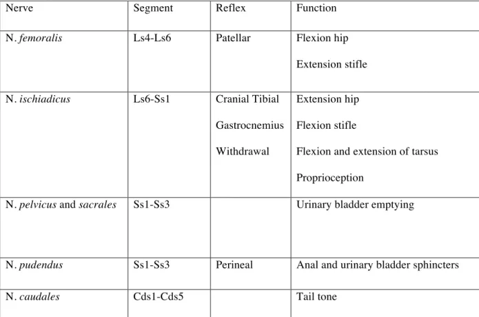
lumbar vertebra (L). Caudally to the conus medullaris the spinal cord is reduced to a terminal filament (filum terminale) (Fletcher, 1993; Pelagalli et Botte, 1999). L6, L7, S1-S3 and the first to the fifth caudal spinal nerve roots stream caudally to the conus medullaris to reach their respective intervertebral foramen. Collectively, these roots are referred to as the cauda equina (figure 2). The peripheral nerves originating from the cauda equina are the n. femoralis, n. ischadicus, n. pelvicus, n.sacralis, n. pudendus, and n.caudales. Their clinical significance is illustrated in Table 1.
Figure 2: Enlarged dorsal view of the terminal spinal cord in dogs.
Adapted From: http://vanat.cvm.umn.edu/neurLab2/SpCdGross. (17/11/2015).
(1Cd= Fisrt sacral nervous root; 1S= First sacral nervous root; 5L= Fifth lumbar nerve; 5Cd= Fifth caudal spinal segment; 7L= Seventh lumbar nerve; Cd= Spinal caudal segment; L4= Fourth lumbar vertebra; L5= Fifth Lumbar vertebra. L6= Sixth lumbar vertebra; L7= Seventh lumbar vertebra ).
Table 1. Cauda Equina: origin and clinical function of the spinal nerves.
Nerve Segment Reflex Function
N. femoralis Ls4-Ls6 Patellar Flexion hip Extension stifle
N. ischiadicus Ls6-Ss1 Cranial Tibial Gastrocnemius Withdrawal
Extension hip Flexion stifle
Flexion and extension of tarsus Proprioception
N. pelvicus and sacrales Ss1-Ss3 Urinary bladder emptying
N. pudendus Ss1-Ss3 Perineal Anal and urinary bladder sphincters
N. caudales Cds1-Cds5 Tail tone
(N.= nerve, Ls= Lumbar spinal segment, Ss= Sacral spinal segment, Cds= Caudal spinal segment)
-Meningeal structures:
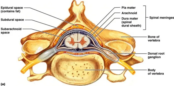
Within the vertebral canal, the nervous system is surrounded by the meninges, which differentiate into 3 layers: the dura mater, the arachnoidea, and the pia mater. The dura mater is the most superficial layer and caudally envelops the filum terminale of the spinal cord. Before the dura mater constricts around the filum terminale, it forms a sac (dural sac) extending 1-2 cm beyond the end of the spinal cord, the dura mater spinalis. The epidural space separates the dura mater spinalis from the periosteum lining the vertebral canal. This space contains fat and the internal vertebral venous plexus. The latter lies on the floor of the vertebral canal. The epidural space is crossed by spinal roots traversing the vertebral canal to reach the intervertebral foramina. The arachnoidea is the intermediate layer and is separated from the pia mater, which is in direct contact with the spinal cord, by the subarachnoid space in which the cerebrospinal fluid (CSF) flows (figure 3).
The presence of the spinal cord with its surrounding meninges is at the L-S junction is variable. The conus medullaris or only the dural sac, which is accompanied by the spinal roots forming the cauda equina, can be present. The simultaneously presence of these three
structures at the L-S junction is variable according to the breed, with small dogs having a relatively longer spinal cord and a dural end-sac extending well into the sacrum (Lang, 1988).
Figure 3: Transverse image of one vertebra, showing the relationship between the bone, the spinal cord, the spinal meninges and the spinal nerve. Adapted from http: //www.apsubiology.org/ anatomy/ 2010/2010_Exam_Reviews/Exam_4_Review / CH _ 12 _ Gross _ Anatomy _ of _ the _ Spinal_Cord. htm (17/11/2015)
-Connective structures:
The vertebral bodies are interconnected by the IVD. The outer layer of the IVD is the anulus fibrosus (AF), a poorly vascularised and barely innervated structure, which consists in a dense network of multiple, organized concentric fibrous lamellae that run obliquely into the adjacent intervertebral bodies. The internal aspect of the IVD is the nucleus pulposus (NP), an avascular and not innervated bean-shaped structure, essentially composed of water. Between the AF and the NP a transition zone is identified, which presents intermediate histological features between the adjacent layers passing from a fibrous to a more mucoid/cartilaginous structure (Bergknut et al., 2013). The IVD is bordered cranially and caudally by the cartilaginous endplates (EPs) that are strongly connected with the inner aspect of the AF, whereas its outer aspect forms connections directly with the bony vertebral body epiphyses through the Sharpey’s fibres (Bergknut et al., 2013). Adjacent to the cartilaginous EPs there is a densely woven vascular network, which plays an essential role in supplying the IVD with nutrients (Bergknut et al., 2013). The vertebrae are connected at the level of adjacent cranial and caudal articular processes by means of a facet joint consisting of a synovial joint capsule surrounding the articular processes. The L-S vertebral canal is further
stabilized by the ligament longitudinale dorsale and by the ligament longitudinale ventrale, running on the dorsal and ventral aspect of the vertebral bodies respectively, and by the ligamentum flavum (synonymous: interarcuate ligament or yellow ligament) between the arches of adjacent vertebrae (figure 4).
Figure 4: Parasagittal image of the lumbar vertebral column showing the vertebral ligaments Adapted from http://slideplayer.com/slide/3468966/ (22/04/2015)
I. 2. b: Motion pattern of the lumbosacral junction and pathophysiology of
degenerative lumbosacral stenosis:
Terminology regarding cauda equina dysfunction is confusing: the term L-S disease refers to all the diseases affecting the L-S region, whilst the term cauda equina syndrome refers to neurological dysfunction, originating from compression, destruction, or displacement of the nerve roots of the cauda equina (Ramirez et Thrall, 1998; Thomas et Dewey, 2008). Therefore, many diseases can cause the cauda equina syndrome: vertebral malformation, idiopathic stenosis, discospondylitis, neoplasia, IVD disease, sacral osteochondrosis, vasculopathy, trauma, fractures, and degenerative lumbosacral stenosis (DLSS) (Meij et Bergknut, 2010).
multifactorial, not completely understood, ethiopathogenesis, involving particularly the complex motion pattern of this area (Meij et Bergknut, 2010). Biomechanics of the L-S area have been investigated (Benninger et al., 2004). The main types of motion, centered at the L-S IVD, are flexion and extension, whilst lateral and rotational movements seem less important (Benninger et al., 2004). Some studies showed that abnormal motion is influenced by the angle of the articular joint processes, suggesting the articular process tropism as a possible cause of abnormal axial rotation and of increased torsional stress on the IVD (Seiler et al., 2002; Rossi et al., 2004). Moreover, several studies have evaluated how flexion and extension can affect the degree of neural compression, suggesting an intermittent stenosis secondary to gait movement in dogs affected by DLSS (Lang, 1988; Jones et al., 1999; Benninger et al., 2004; Benninger et al., 2006; Gradner et al., 2007; Reynolds et al., 2014).
Besides the genetic tropism or angle of articular processes, other causes such as L-S transitional vertebra (Fluckiger et al., 2006) or osteochondrosis of the sacrum (Lang et al. 1992) have been shown to predispose to cauda equina syndrome. Furthermore, a recent study identified a skeletal and morphological variability of the L-S junction in German Shepherd dogs suggesting a primary L-S stenosis in this breed (Ondreka et al., 2013). The abnormal motion patterns, independently of the underlying causes, and the secondary loss or lack of loadbearing proprieties have been proposed as the initial mechanism leading to IVD degeneration (IVDD). The secondary decrease in IVD width would lead to an ulterior shift of the load bearing from the central to the peripheral part of the spine, such as the ventral aspect of the vertebral bodies and the articular processes. This instability process would finally result in IVD herniation (Hansen Type I), secondary to the chronic IVDD, and in a compensatory proliferation of the surrounding structures (hypertrophy of the interarcuate ligament, epidural fibrosis, thickening of the capsules of the articular processes, osteophytes, and ventral spondylosis) with secondary stenosis of the vertebral canal and intervertebral foramina (De Risio et al., 2000; Meij et Bergknut, 2010).
I. 2. c: Signalement, history and clinical signs of degenerative lumbosacral
stenosis:
Typically middle-aged large-breed dogs and especially German Shepherd dogs and working dogs have a high predisposition to develop DLSS (De Risio et al., 2000; Meij et Bergknut, 2010), increasing the suspicion of a genetic predisposition. Indeed, cauda equina
syndrome was among the main causes of euthanasia of military German Shepherd and Belgian Shepherd dogs (Moore et al., 2001). At presentation, owners complain about the dog’s difficulty to rise, sit or lie down, jump or climb and about urinary or fecal incontinence (De Risio et al., 2000; Sharp et Wheeler, 2005; Thomas et Dewey, 2008; Meij et Bergknut, 2010).
The main neurological finding is pain at the L-S region, evoked by hyperextending the tail or hyperextending the caudal lumbar spine (De Risio et al., 2000; Sharp et Wheeler, 2005; Thomas et Dewey, 2008; Meij et Bergknut, 2010). The origin of the pain is multifactorial. It can be classified as radicular pain (originating from nerve root entrapment), meningeal pain (irritation of the meninges), osteoarthritic pain (degeneration of the periosteum, ligament longitudinale dorsale or joint capsules) or, to a lesser extent given the weak innervation of the IVD, discogenic pain (degeneration or tearing of the AF) (De Risio et al., 2000). Pain may be manifested or exacerbated during exercise and may result in intermittent unilateral or bilateral lameness. The intermittent status, referred to as neurogenic intermittent claudication, is caused by the dilation of the radicular blood vessel secondary to exercise, causing/worsening the underlying interforaminal stenosis. (De Risio et al., 2000) The neurological examination can also highlight proprioceptive and voluntary motor deficits (De Risio et al., 2000; Sharp et Wheeler, 2005; Thomas et Dewey, 2008; Meij et Bergknut, 2010). The evaluation of spinal reflexes (including patellar, cranial tibial, gastrocnemius, withdrawal and perineal reflexes) is compatible with a lower motor neuron disease: decreased to absent withdrawal and gastrocnemius reflexes, hyperreflexive or normal patellar reflexes, decreased to absent perineal reflex, and proprioceptive deficits (De Risio et al., 2000; Sharp et Wheeler, 2005; Thomas et Dewey, 2008; Meij et Bergknut, 2010).
I. 2. d: Diagnostic imaging of degenerative lumbosacral stenosis:
Diagnosis of DLSS is based on history, neurological assessment, and correlation of clinical findings and ancillary diagnostic imaging findings. Because of the anatomical features and the complexity of the disease’s pathogenesis, several diagnostic-imaging techniques have been described to evaluate the L-S vertebral canal (Ramirez et Thrall, 1998; De Risio et al., 2000; Meij et Bergknut, 2010). Some of these techniques, such as epidurography, discography, and vertebral sinus venography were used in the past, but they are not routinely performed nowadays because of the possible complications and their
inability to assess the L-S structures entirely. These techniques have been replaced by CT and MRI, which are now considered the gold standard for diagnosis of DLSS (Meij et Bergknut, 2010). Radiography and myelography, however, can still be considered an important tool for the diagnosis of DLSS, if advanced modalities are not available or declined by the owner for financial reasons.
-Conventional and position-dependent (“dynamic”) radiography:
In the past, many studies focused on the possibility of diagnosing DLSS with survey radiography, using different projections with neutral, flexion or extension of the L-S spine. The latter projections are also called stress, positional or “dynamic” projections. The use of the world “dynamic” is debatable. In fact “dynamic” is referred to as something characterized by constant change or movement. When “dynamic” projections are performed, the spine is not moving but it is in a static neutral, flexed or extended position. Therefore the word positional or position-dependent would be more appropriate. However, in literature the world “dynamic” is still widely used. Measurements performed on radiographs in flexed and extended positions aimed to highlight the possible abnormal motion pattern and the eventual instability of the L-S junction and to establish normal values for radiographic pattern (Mattoon et Koblik, 1993; Schmid et Lang 1993). However, results were controversial. For instance one study showed no difference in the degree of sub-luxation of the sacrum between normal and affected German Shepherd dogs (Schimd et Lang, 1993), whilst another study showed that a logistic model based on radiographic parameters (neutral L-S angle>170°, extension angle> 165°, a flexion angle >185°, a total range of motion <20°, a L-S point of intercept cranial to mid-body of L7 or caudal to S1) was able to discriminate normal from affected dogs with an overall accuracy rate of 86% (Mattoon et Koblik, 1993). Recently, it has been shown that the accuracy in detecting the cranial margin of the sacrum on x-rays is only fair (Blume et al., 2015) and this should be taken into account when evaluating the position of the sacrum. However radiography has to be considered the first step in normal work-up for dogs with clinical signs compatible with cauda equina compression (Ramirez et Thrall, 1998; De Risio et al., 2000; Meij et Bergknut, 2010). Common radiographic findings in dogs affected by DLSS are L-S decreased IVD space, decreased intervertebral L-S foraminal space, spondylosis, degenerative joint disease of the articular processes, and ventral displacement “telescoping” of the sacrum (figure 5). These results are however not specific and a normal radiographic examination does not rule out the presence of L-S disease.
Furthermore, it has been demonstrated that there is no correlation between neurological and radiographic findings (Scharf et al., 2004). Indeed, the main limitation of radiography is the poor ability to assess soft tissue, but on the other hand its great advantage is the possibility to rule out other causes of cauda equina syndrome, such as discospondilitis, fracture, luxation or neoplasia of the bone structures (Ramirez et Thrall, 1998; De Risio et al., 2000; Meij et Bergknut, 2010).
Figure 5: Radiographic image of the lumbosacral region (latero-lateral projection). This dog presented lumbosacral spondylosis (arrows), sclerosis of the lumbosacral vertebral endplates (arrowsheads) and decreased lumbosacral intervertebral foramen size. These findings are common in dogs affected by degenerative lumbosacral stenosis (cranial of the dog to the reader’s left; dorsal of the dog to the top of the image).
-Myelography:
Myelography consists in the injection of contrast medium to visualize the subarachnoid space and to delineate the spinal cord contours (Roberts et Selcer, 1993). In the past, it was the modality of choice to investigate spinal cord compression (Ramirez et Thrall, 1998), which is usually visualized as an interruption and/or displacement of the contrast medium column. However, the accuracy of myelography for detecting L-S diseases is variable. First of all, it only allows detection of dural sac compressions. It has been demonstrated that the dural sac can extend beyond the sacrum in 80% of dogs, but its exact length cannot be assessed before contrast medium injection (Lang, 1988). Intervertebral foramina stenosis and compression of the spinal roots cannot be assessed with myelography and therefore a normal myelographic study does not rule out cauda equina compression (Ramirez et Thrall, 1998). A study focused on flexion-extension myelography (“dynamic” views) of the L-S region showed that size, shape and diameter of the dural sac was constant among the different views in normal dogs but highlighted that a variable degree of
compression was visible in flexion/extension compared to the neutral position in affected dogs (Lang, 1988). These “dynamic” views can therefore increase the sensitivity of myelography (Lang, 1988). It has been reported that myelography can increase accuracy of CT for the detection of spinal compression (Shimizu et al., 2009). Thus, in cases of non-diagnostic conventional CT examination, these two techniques can be used in conjunction to perform a CT myelography (CTM), i.e. a CT examination after the injection of contrast medium in the subarachnoid space.
-Computed Tomography:
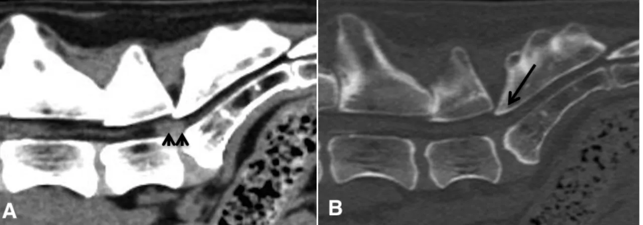
Computed tomography is considered with MRI one of the gold standard procedures for the diagnosis of DLSS (Ramirez et Thrall, 1998; Meij et Bergknut, 2010). When performing a CT examination, different image reconstruction algorithms can be chosen. A medium spatial frequency algorithm associated with the use of a soft tissue window is considered the best to assess the soft tissue structures. Instead, high spatial frequency algorithms and a bone window are used to assess body parts with inherently wide contrast object such as bone (Schwarz et O’Brien, 2011). A specific CT protocol for the examination of the spine with dogs in dorsal recumbency has been recommended (100-120 kilovolts, 200 milliampere/ second, slice width of 1-2 millimeters) (Seiler et al., 2011). In dogs affected by DLSS common findings using are disc herniation, hypertrophy of the interarcuate ligament and of the joint capsules, vertebral spondylosis, EPs sclerosis, sacral osteochondrosis, or ventral displacement of the sacrum (figure 6) (Ramirez et Thrall, 1998; Meij et Bergknut, 2010).
Figure 6: Computed Tomography images (sagittal multiplanar reconstruction) of the lumbosacral region of dog affected by degenerative lumbosacral stenosis. In the soft tissue window image (A), a severe disc protrusion with loss of epidural fat is visible (arrowsheads). In the bone window image (B) the ventral displacement of the roof of the sacrum (arrows), called as “telescoping” is evident (cranial of the dog to the reader’s left; dorsal of the dog to the top of the image).
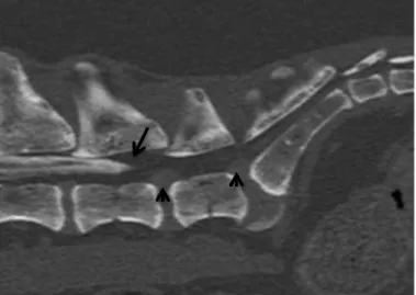
Moreover, it has been shown that intravenous injection of iodinated non-ionic contrast medium can help in the visualization of soft tissue, increasing the assessment of neural compression by soft tissue structures (Jones et al., 1999). CT is faster and less expensive than MRI but its main disadvantages, beside the use of ionizing radiation, is its poor ability to assess soft tissue structures, such for example spinal cord and other nervous structures (Ramirez et Thrall, 1998; Meij et Bergknut, 2010). To increase the accuracy in detecting spinal cord compression, CTM can be performed combining the advantages of myelography and CT (figure 7). However, as for myelography the results of CTM depend on the length of the dural sac (figure 8).
Figure 7: Computed tomographic-myelography image of the lumbosacral region (sagittal multiplanar reconstruction, bone window, neutral position) in a dog affected by degenerative lumbosacral stenosis. The contrast medium fills the subarachnoid space of the lumbar spine and dural sac (myelography), but there is a focal loss of contrast medium visualization at the lumbosacral junction (arrows). This finding is indicative of spinal cord compression. Also notice the severe vertebral spondylosis affecting the cranial lumbar spine and the degenerative joint disease of the articular process (cranial of the dog to the reader’s left; dorsal of the dog to the top of the image).
Figure 8: Computed tomographic- myelography image of the L-S region (sagittal multiplanar reconstruction, bone window, neutral position). Discal protrusions (arrowsheads) are visible at L6-L7 and L7-S1, associated with L7-S1 spondylosis. In this dog, the dural sac filled by contrast medium ends at the level of the mid-body of L6, limiting the usefulness of the injection of contrast medium in the subarachnoid space to assess spinal cord compression.
Similarly to CT and myelography, CTM examination of the L-S region can be performed with the spine in flexion and extension. The comparison between these views and the neutral position can point out a different degree of compression, usually more important during the extension of the LS spine, highlighting the intermittent nature of the compression (figure 9).
Figure 9: Computed tomographic-myelography images of the lumbosacral region (sagittal multiplanar reconstruction, bone window) in a dog affected by degenerative lumbosacral stenosis. Images’acquisition has been performed with the lumbosacral spine in neutral position (A), in flexed position (B) and in extended position (C). The intermittent nature of the spinal cord compression, visible as a focal loss of contrast medium column, is noticed: the most severe compression is visible in the extended position, whilst no compression is visible in the flexed position (cranial of the dog to the reader’s left; dorsal of the dog to the top of the image).
-Magnetic Resonance Imaging:
In human medicine, MRI is considered the modality of choice for the detection of cauda equina compression. Flexion-extension studies have been used to highlight intermittent foraminal stenosis, in cases where no stenosis was visible in the neutral position despite the presence of significant clinical signs (Weishaupt et al., 2000).
To perform an MRI examination of the spine different sequences can be chosen to highlight different anatomic and pathologic aspects (Gavin, 2009b). In T2-weighted images both fluid and fat are hyperintense. Bright fluid in images is desirable as most pathologic abnormalities have an increased fluid signal (Gavin, 2009b). In STIR sequences there is a uniform loss of fat signal, whilst fluid-filled structures are displayed as bright on a generalized dark background, making the identification of pathologic lesions easier (Gavin, 2009b). In T1-weighted sequences fat is hyperintense and fluid is hypointense. Abnormal tissue has often an increased vascular supply, therefore the administration of intravenous contrast medium leads to an increased signal intensity (Gavin, 2009b). More specifically on T2-weighted images, normal IVDs have a high NP signal surrounded by a medium AF signal. Epidural fat has very high signal intensity and appears bright white. The signal intensity is related to the concentrations of matrix hyaluronic acid and glycosaminoglycans, which in turn attract and hold water. Substances with highest glycosaminoglycans concentration, such as the IVD, will have a prominent T2 signal. On T1-weighted sequences,
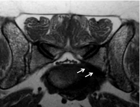
the normal IVD is of uniform medium signal intensity, slightly greater than that of the spinal cord, nerve roots and bone marrow. Common findings in dogs affected by DLSS are IVD degeneration and protrusion, loss of epidural fat or subarachnoid space visualization, and dural sac or nerve root compression (figures 10 and 11) (Ramirez et Thrall, 1998; De Risio et al., 2000; Meij et Bergknut, 2010). However, studies focused on the comparison between MRI and clinical or surgical observations reported that the degree of compression determined by MRI at the time of presentation was not proportional to disease severity (Jones et al., 2000; Mayhew et al., 2002; Suwankong et al., 2006).
Figure 10: Transverse T2-w magnetic resonance image of the lumbosacral region. Notice the complete obliteration of the left intervertebral foramen by severe lateral spondylosis (arrows) and the secondary absence of visualization of the foraminal epidural fat. Ventral spondylosis is also visible (right of the dog to the reader’s left; dorsal of the dog to the top of the image). Courtesy of Dr. V. De Busscher and Dr. Finck, ChesterGates.
Figure 11: Sagittal T2-w magnetic resonance image of the lumbosacral canal. There is a generalized loss of T2-w hyperintensity of the last lumbar intervertebral discs associated with protrusion dorsally within the vertebral canal. At the lumbosacral junction the intervertebral disc protrusion(arrowsheads) is more important and there is a secondary loss of epidural fat visualization and dorsal displacement of the cauda equina roots (cranial of the dog to the reader’s left; dorsal of the dog to the top of the image). Courtesy of Dr.V. De Busscher and Dr. Finck, ChesterGates
-Computed Tomography versus Magnetic Resonance Imaging:
In general CT is the gold standard to evaluate bone structures, given the superior spatial resolution and the possibility to acquire thinner slices. Also, it is faster and cheaper than MRI (Da Costa et Samii, 2010). To increase visualization of spinal cord compression, myelography in conjunction with CT (i.e CTM) can be performed. However, this technique increases the duration of the procedure and the risk of complications associated with myelography (Da Costa et Samii, 2010). On the other hand, MRI has a better soft tissue resolution with a better assessment of all neural structures, as well as ligaments, joint capsule, and IVD and myelography is not unnecessary (Da Costa et Samii, 2010). Studies comparing agreement between different imaging modalities have been performed, focusing on different regions of the spinal cord and different diseases. For instance, a study comparing CT versus MRI in the diagnosis of DLSS showed a high agreement between these two modalities (Suwankong et al., 2006). Another study evaluated the association between diagnostic findings with MRI and CT and outcome after surgical treatment. Both MRI and CT were effective to identify neural compression, but no significant association between results of imaging studies and postoperative outcome was identified (Jones et al., 2000). In summary, considering its better soft tissue resolution, MRI can be considered the modality of choice for the detection of cauda equina compression. However, from a practical point of view, in veterinary medicine MRI systems are less available than CT scanners and more expensive. Therefore, CT examination of the L-S spine, especially if associated with myelography, can be considered a good alternative for the diagnosis of DLSS.
I. 2. e: Treatments:
Both conservative and surgical treatments have been proposed in cases of DLSS ( De Riso et al., 2000; Sharp et Wheeler, 2005; Dewey, 2008; Meij et Bergknut, 2010).
-Conservative treatment:
The aim of conservative treatment is to reduce pain. It is based on body weight reduction, change in exercise pattern, predominantly by introducing a regular walking activity of short duration to maintain muscle tone, and the use of nonsteroidal anti-inflammatory drugs per os (De Riso et al., 2000; Meij et Bergknut, 2010). However, toxicity of non steroidal anti-inflammatory drugs is reported, especially affecting the gastro-intestinal and renal system. Indeed, gastric ulcers and nephropathy are the most common reported
complications (Khan et Mclean, 2012). A controversial opinion exists concerning the use of systemic corticosteroids because of their collateral effects (Meij et Bergknut, 2010). In a recent study, medical treatment in dogs affected by DLSS resulted in an overall success rate of 55% (De Decker et al., 2014). Lumbosacral ESI have also been proposed with an improvement in the 79% of the patients. However, in this retrospective study dogs’ improvement was assessed by the owners following a prescheduled table and a systematic neurological evaluation was not performed (Janssens et al., 2009).
-Surgical treatment:
The aim of surgical treatment is to decompress the cauda equina and to free entrapped nerve roots. Candidates for surgical treatment are dogs with moderate to severe clinical signs (pain and neurologic deficits), unresponsive to conservative treatment (Meij et Bergknut, 2010). Among the suggested surgical techniques, the preferred modality is usually the dorsal laminectomy, which is sometimes followed by additional procedures when further decompression is required, such as partial discectomy consisting of dorsal fenestration (or dorsal annulectomy), nuclear pulpectomy (or nucleotomy), foraminotomy, and rarely, facetectomy (Sharp et Wheeler, 2005; Dewey, 2008; Meij et Bergknut, 2010; Saulnier-troff et al., 2014). When ventral subluxation of S1 is present, stabilization by fixation and fusion can be suggested (Sharp et Wheeler, 2005; Dewey, 2008; Meij et Bergknut, 2010).Outcome after decompressive surgery ranges from good to excellent (Meij et Bergknut, 2010), but urinary and fecal incontinence are poor prognostic predictors with usually no improvement despite surgery (De Risio et al., 2001; Meij et Bergknut, 2010). Moreover, working dogs have less favorable results, probably related to their activity (Meij et Bergknut, 2010). Different intra, early or late postoperative complications have been described with surgical treatment. For instance, formations of adhesions between the nerves and the surrounding soft tissue or intra-articular fractures have been described with laminectomy and fixation-fusion technique (Sharp et Wheeler, 2005; Jeffery et al., 2014).
-Conservative vs surgical treatment:
(Adapted from De Risio, 2000; Dewey, 2008; Meij et Bergknut, 2010)
Current treatments in dogs affected by DLSS are based primarly on severity of clinical signs. In a dog with pain, but in absence of neurological deficits, medical treatment is considered the first option. Besides the administration of NSAIDs, gabapentin can also be used as pain neuromodulator. Conservative treatment does not resolve the underlying problem and it tends to be eitheer transiently effective or ineffective.
In presence of moderate to severe lumbosacral pain, unsuccessful medical treatment or in presence of neurological deficits the surgical treatment could be the best option. The choice of the surgical method (dorsal laminectomy vs fuxion-fixation or foraminotomy) should be done after the imaging assessment of the spinal cord compression. The outcome of surgical treatment is good to ecellent (73% to 93%), even if complications such adhesions, implant failure, fracture of the articular process have been described. Urinary incontinence is considered a poor prognostic factor.
References
AGUT A., SOLER M., LAREDO F.G., PALLARES F.J., SEVA J.I. Imaging diagnosis-Ultrasound-guided ethanol sclerotherapy for a simple renal cyst. Vet Radiol Ultrasound, 2008, 49:65-7
AHMED M., WEINSTEIN J., LIU Z., AFZAL K.S., HORKAN C., KRUSKAL J.B., GOLDBERG S.N. Image-guided percutaneous chemical and radiofrequency tumor ablation in an animal model. J Vasc Interv Radiol, 2003, 14:1045-52
AMBER KT., LANDY DC., AMBER I., KNOPF D., GUERRA J., Comparing the accuracy pf ultrasound versus fluoroscopy in glenohumeral injections: a systematic review and meta-analysis. J Clin Ultrasound, 2014, 42: 411-416
AMINI R., KARTCHNER JZ., NAGDEV A., ADHIKARI S. Ultrasound-guided Nerve Blocks in Emergency Medicine Practice, J Ultrasound Med, 2016, In press
ANSON A., GIL F., LAREDO F.G., SOLER M., BELDA E., AYALA M.D., AGUT A. Correlative ultrasound anatomy of the feline brachial plexus and major nerves of the thoracic limb. Vet Radiol Ultrasound, 2013, 54:185-93
!
ANSON A., LAREDO F. G., GIL F., SOLER M., BELDA E., AYALA M.D., AGUT A. Comparison of two techniques for ultrasound-guided axillary brachial plexus blockade in cats. J Feline Med Surg. 2015, 17:476-85
AUDIGIE F., TAPPREST J., DIDIERLAURENT D., DENOIX J.M. Ultrasound-guided atlanto-occipital puncture for myemography in the horse. Vet Radiol Ultrasound. 2004, 45:340-4.
BARTYNSKI W.S., GRAHOVAC S.Z., ROTHFUS W.E. Incorrect needle position during lumbar epidural steroid administration: inaccuracy of loss of air pressure resistance and requirement of fluoroscopy and epidurography during needle insertion. Am J Neuroradiol, 2005, 26, 502-505.
BAUER M., GEORGE J.E., 3RD, SEIF J., FARAG E. Recent advances in epidural analgesia. Anesthesiol Res Pract, 2012, doi:10.1155/2012/309219.
BELAND M.D, MAYO-SMITH W.W. Image-guided tumour ablation: basic principles. In: Kaufman J.A, Lee M. (Eds), Vascular and Intervention Radiology. Elsevier: Philadelphia, 2014, 547-553.
BENNINGER M.I., SEILER G.S., ROBINSON L.E., FERGUSON S.J., BONEL H.M., BUSATO A.R., LANG J. Three-dimensional motion pattern of the caudal lumbar and lumbosacral portions of the vertebral column of dogs. Am J Vet Res, 2004, 65, 544-551. BENNINGER M.I., SEILER G.S., ROBINSON L.E., FERGUSON S.J., BONEL H.M., BUSATO A.R., LANG J. Effects of anatomic conformation on three-dimensional motion of the caudal lumbar and lumbosacral portions of the vertebral column of dogs. Am J Vet Res, 2006, 67, 43-50.
BLUME L.M., WORTH A.J., COHEN E.B. BRIDGES J.P. HARTMAN A.C. Accuracy of radiographic detection of the cranial margin of the dorsal lamina of the canine sacrum. Vet Radiol Ultrasound 2015; 56:579-88.
CAMPBEL-WASHBUM AE., FARANESH AZ., LEDERMAN RJ., HANSEN MS. Magnetic Resonance Sequences and Rapid Acquisition for MR-guided Interventions. Magn Reson Imaging Clin N Am, 2015, 23: 669-79.
CAMPOY L., BEZUIDENHOUT A.J., GLEED R.D., MARTIN-FLORES M., RAW R.M., SANTARE C.L., JAY A.R., WANG A.L. Ultrasound-guided approach for axillary brachial plexus, femoral nerve, and sciatic nerve blocks in dogs. Vet Anaesth Analg, 2010, 37, 144-153.
CARRAGEE EJ., HURWITZ EL., CHENG I., CARROLL L., NORDIN M., GUZMAN J., PELOSO P., HOLM L.W, COTHE P., JONHONSON SH., VAN DER VELDE G., CASSIDY D., HALDEMAN S.. Treatment of neck pain: injections and surgical interventions: results of the bone and joint decade 2000-2010 task force on neck pain and its associated disorders. Spine. 2008, 33:s153-169
DA COSTA R.C., SAMII V.F. Advanced imaging of the spine in small animals. Vet Clin North Am Small Anim Pract, 2010, 40, 765-790.
DAMUR-DJURIC N., STEFFEN F., HASSIG M., MORGAN J.P., FLUCKIGER M.A. Lumbosacral transitional vertebrae in dogs: classification, prevalence, and association with sacroiliac morphology. Vet Radiol Ultrasound, 2006, 47, 32-38.
DE DECKER S., WAWRZENSKI L.A., VOLK H.A. Clinical signs and outcome of dogs treated medically for degenerative lumbosacral stenosis: 98 cases (2004-2012). J Am Vet Med Assoc, 2014, 245, 408-413.
DE LAHUNTA A, GLASS E. Veterinary neuroanatomy and clinical neurology. Saunders Elsevier: Missouri, 2009. 1-505
DE RISIO L., THOMAS W.B., SHARP N.J. Degenerative lumbosacral stenosis. Vet Clin North Am Small Anim Pract, 2000, 30, 111-132, vi.
DE RISIO L., SHARP N.J., OLBY N.J., MUNANA K.R., THOMAS W.B. Predictors of outcome after dorsal decompressive laminectomy for degenerative lumbosacral stenosis in dogs: 69 cases (1987-1997). J Am Vet Med Assoc, 2001, 219, 624-628.
DEPALMA MJ., BHARGAVA A., SLIPMAN CW. A critical appraisal of the evidence for selective nerve root injection in the treatment of lumbosacral radiculopathy. Arch Phys Med Rehabil., 2005, 86:1477-1483.
DEPECKER M., BIZON-MERCIER C., COUROUCÉ-MALBLANC A. Ultrasound-guided atlanto-occipital puncture for cerebrospinal fluid analysis on the standing horse. Vet Rec, 2014, 174 (2):45.
DEWEY CW. Disorders of the cauda equina. In: Dewey CW. A pratical guide to canine and feline neurology. 2nd Edition. Iowa: Wiley-Blackwell, 2008; 389-404.
ECHEVERRY D.F., GIL F., LAREDO F., AYALA M.D., BELDA E., SOLER M., AGUT A. Ultrasound-guided block of the sciatic and femoral nerves in dogs: a descriptive study. Vet J, 2010, 186, 210-215.
ETIENNE A.L., PEETERS D., BUSONI V. Ultrasonographic percutaneous anatomy of the caudal lumbar region and ultrasound-guided lumbar puncture in the dog. Vet Radiol Ultrasound, 2010, 51, 527-532.
FLETCHER TF. Spinal cord and Meninges. In: Evans HE. Miller's anatomy of the dog. Philadelphia: Saunders; 1993; p. 800-827
FLUCKIGER M.A., DAMUR-DJURIC N., HASSIG M., MORGAN J.P., STEFFEN F. A lumbosacral transitional vertebra in the dog predisposes to cauda equina syndrome. Vet Radiol Ultrasound, 2006, 47, 39-44.
GAVIN P.R (a). Equipment consideration and selection. In: Gavin P.R and Bagly R.S., Practical Small Animal MRI. Wiley-Blackwell: Iowa, 2009, 21-22.
GAVIN P.R (b). Sequence Selection. In: Gavin P.R and Bagly R.S., Practical Small Animal MRI. Wiley-Blackwell: Iowa, 2009, 8-9.
GRADNER G., BOCKSTAHLER B., PEHAM C., HENNINGER W., PODBREGAR I. Kinematic study of back movement in clinically sound malinois dogs with consideration of the effect of radiographic changes in the lumbosacral junction. Vet Surg, 2007, 36, 472-481. GRAU T., LEIPOLD R., CONRADI R., MARTIN E., MOTSCH J. [Ultrasonography and peridural anesthesia. Technical possibilities and limitations of ultrasonic examination of the epidural space]. Anaesthesist, 2001, 50, 94-101.
GREGORI T., VISCASILLAS J., BENIGNI L. Ultrasonographic anatomy of the sacrococcygeal region and ultrasound-guided epidural injection at the sacrococcygeal space in dogs. Vet Rec, 2014, 175, 68.
GUILHERME S., BENIGNI L. Ultrasonographic anatomy of the brachial plexus and major nerves of the canine thoracic limb. Vet Radiol Ultrasound, 2008, 49, 577-583.
HAN HJ., KIM J.Y., JANG H.Y., LEE B., YOON JH., JANG S.K., CHOI SH., JEONG SW. Fluoroscopic-guided intradiscal oxygen-ozone injection therapy for thoracolumbar intervertebral disc herniation in dogs. In vivo 21:609-614.
HARO P., GIL F., LAREDO F., AYALA M.D., BELDA E., SOLER M., AGUT A. Ultrasonographic study of the feline sciatic nerve. J Feline Med Surg, 2011, 13, 259-265. HOELTJE J., BRUENING R., KASTLER B., et al. Interventional Pain Management. In: Mahnken AH, Wilhem KE, Ricke J (eds). CT- and MR-Guided Interventions in Radiology. Springer Berlin Heidelberg; 2013: 363-419.
IRANI F.G., BUY X., GANGI A. Musculoskeletal interventions. In: Kaufman J.A, Lee M. (Eds), Vascular and Intervention Radiology. Elsevier: Philadelphia, 2014, 527-546.
JANSSENS L., BEOSIER Y., DAEMS R. Lumbosacral degenerative stenosis in the dog. The results of epidural infiltration with methylprednisolone acetate: a retrospective study. Vet Comp Orthop Traumatol, 2009, 22, 486-491.
JEFFERY N.D., BARKER A., HARCOURT-BROWN T. What progress has been made in the understanding and treatment of degenerative lumbosacral stenosis in dogs during the past 30 years? Vet J, 2014, 201, 9-14.
JOHNSON B.A., SCHELLHAS K.P., POLLEI S.R. Epidurography and therapeutic epidural injections: technical considerations and experience with 5334 cases. AJNR Am J Neuroradiol, 1999, 20, 697-705.
JONES J.C., SHIRES P.K., INZANA K.D., SPONENBERG D.P., MASSICOTTE C., RENBERG W., GIROUX A. Evaluation of canine lumbosacral stenosis using intravenous contrast-enhanced computed tomography. Vet Radiol Ultrasound, 1999, 40, 108-114.
JONES J.C., BANFIELD C.M., WARD D.L. Association between postoperative outcome and results of magnetic resonance imaging and computed tomography in working dogs with degenerative lumbosacral stenosis. J Am Vet Med Assoc, 2000, 216, 1769-1774.
KANDASAMI D., GAMANAGATTI S., GUPTA AK. Pediatric Interventional Radiology: Vascular Interventions. Indian J Pediatr, 2016. In press.
KAUFMAN J.A. Vascular Interventions. In: Kaufman J.A, Lee M. (Eds), Vascular and Intervention Radiology. Elsevier: Philadelphia, 2014, 56-67.
KARMAKAR M.K., LI X., HO A.M., KWOK W.H., CHUI P.T. Real-time ultrasound-guided paramedian epidural access: evaluation of a novel in-plane technique. Br J Anaesth, 2009, 102, 845-854.
KHAN S.A., MCLEAN M.K. Toxicology of frequently encountered nonsteroidal anti-inflammatory drugs in dogs and cats. Vet Clin North Am Small Anim Pract, 2012, 42, 289-306.
KNEISSL S., BREIT S., WILLMITZER F., THALHAMMER J., DENGG S. Dispersal pattern of injectate following CT-guided perineural infiltration in the canine thoracolumbar spine: a cadaver study. Vet Radiol Ultrasound, 2015, 56, 212-219.
LANG J., HÄNI H., SCHAWALDER P. A sacral lesion resembling osteochondrosis in the german shepherd dog. Vet Radiol Ultrasound, 1992, 33, 69-76.
LANG J. Flexion-Extension myelography of the canine cauda equina. Veterinary Radiology 1988, 29, 242-257.
LEE SR., KILCOYNE A., KAMBADAKONE A., ARELLANO R. Interventional oncology: pictorial review of post-ablation imaging of liver and renal tumors. Abdom Radiol (NY), 2016. In press.
LEVY M., GASCHEN L., RADEMACHER N., BRAGULLA H. Technique for ultrasound-guided intraarticular cervical articular process injection in the dog. Vet Radiol Ultrasound, 2014, 55, 435-440.
LEWIS D., SCOTT M., FISCHER C.D., BOND S.L., LEGUILLETTE R. Feasibility for ultrasound-guided injection of the collateral ligaments of the distal interphalangeal joint in horses. Vet Radiol Ultrasound. 2016, doi: 10.1111/vru.1234
MACKENZIE S.D., CASWELL J.L., BRISSON B.A., GAITERO L., CHALMERS H.J. Comparison between computed tomography, fluoroscopy, and ultrasonography for guiding percutaneous injection of the canine intervertebral disc. Vet Radiol Ultrasound, 2014, 55, 571-581.
MACKENZIE S.D., BRISSON B.A., GAITERO L., CASWEL J.L. PENTING L., SINCLAIR M., CHALMERS H.J. Distribution and short- and long-term effects of injected gelified ethanol into the lumbosacral intervertebral disc in healthy dogs. Vet Radiol Ultrasound, 2016. 57: 180-190.
MAHNKEN A.H., WILHEM K.E., RICKE J. (eds). CT- and MR-Guided Interventions in Radiology. Springer Berlin Heidelberg; 2013. 1-571.
MARHOFER P., GREHER M., KAPRAL S. Ultrasound guidance in regional anaesthesia. Br J Anaesth, 2005, 94, 7-17.
MATTOON J.S., KOBLIK P.D. Quantitative survey radiographic evaluation of the lumbosacral spine of normal dogs and dogs with degenerative lumbosacral stenosis. Vet Radiol Ultrasound, 1993, 34, 194-206.
MATTOON J.S., POLLARD R., WILLS T., NYLAND T.G. Ultrasound-guided aspiration and biopsy procedure. In: Nyland T.G., Mattoon J.S. (Eds), Small animal diagnostic ultrasound. Saunders: Philadelphia, 2015, 30-48.
MAYHEW P.D., KAPATKIN A.S., WORTMAN J.A., VITE C.H. Association of cauda equina compression on magnetic resonance images and clinical signs in dogs with degenerative lumbosacral stenosis. J Am Anim Hosp Assoc, 2002, 38, 555-562.
MACKAY RJ. Developments in ultrasound-guided techal puncture in horses. Vet Rec. 2014,174:43-4.
!
MEIJ B.P., BERGKNUT N. Degenerative lumbosacral stenosis in dogs. Vet Clin North Am Small Anim Pract, 2010, 40, 983-1009.
MEJIA-TERRAZAS G.E., GARDUNO-JUAREZ MDE A., LIMON-MUNOZ M., TORRES-MALDONADO A.S., CARRILLO-ESPER R. [Bilateral brachial plexus block. Case report and systematic review]. Cir Cir, 2015, 83, 312-318.
MIKHAIL AS., PARTANEN A., YARMOLENKO P., VENKATESAN AM., WOOD BJ. Magnetic Resonance-Guided Drug Delivery. Magn Reson Imaging Clin N Am, 2015, 23: 643-655.
MOORE G.E., BURKMAN K.D., CARTER M.N., PETERSON M.R. Causes of death or reasons for euthanasia in military working dogs: 927 cases (1993-1996). J Am Vet Med Assoc, 2001, 219, 209-214.
MORATH U, LUYET C, SPADAVECCHIA C, STOFFEL MH, HATCH GM. Ultrasound-guided retrobulbar block in horses: a cadaveric study. Vet Anaesth Analg. 2013 Mar; 40 (2):205-11.
!
NEAL JM. Ultrasound-guided Regional Anesthesia and Patient Safety: Update of an Evidence-based Analysis. Reg Anesth Pain Med, 2016, 41: 195-204
!
ONDREKA N., AMORT K.H., STOCK K.F., TELLHELM B., KLUMPP S.W., KRAMER M., SCHMIDT M.J. Skeletal morphology and morphometry of the lumbosacral junction in German shepherd dogs and an evaluation of the possible genetic basis for radiographic findings. Vet J, 2013, 196, 64-70.
O’NEILL HD., GARCIA-PEREIRA FL, MOHANKUMAR PS. Ultrasound-guided injection of the maxillary nerve. Equine Vet J, 2014, 46:180-4
ORLANDI D., CORAZZA A., ARCIDIACONO A., MESSINA C., SERAFINI G., SCONFIENZA LM., SILVESTRI E., Ultrasound-guided procedures to treat sport-related muscle injuries. Br J radiol, 2016, 89: 2015048
OWLIA M.B., SALIMZADEH A., ALISHIRI G., HAGHIGHI A. Comparison of two doses of corticosteroid in epidural steroid injection for lumbar radicular pain. Singapore Med J, 2007, 48:241-245.
PAIK NC. Radiation dose reduction in CT fluoroscopy-guided lumbar interlaminar epidural steroid injection by minimizing preliminary planning imaging. Euro Rad, 2014, 24: 2109-17 PEASE A., BEHAN A., BOHART G. Ultrasound-guided cervical centesis to obtain cerebrospinal fluid in the standing horse. Vet Radiol Ultrasound, 2012, 53:92-5.
PELAGALLI G. V., BOTTE V. Anatomia Veterinaria sistematica e comparata, Edi Ermes: Milano, 1999, 514p.
PERRIN R., DIGUET A.C., CANTET P., BAILLY C., BROGNIEZ L., DUGDALE A., NISOLLE J.F., VANDEWEERD J.M. Ex vivo assessment of an ultrasound-guided injection technique of the navicular bursa in the horse. Anat Histol Embryol, 2015, doi: 10.1111/ahe.12220.
RAMIREZ O., 3RD, THRALL D.E. A review of imaging techniques for canine cauda equina syndrome. Vet Radiol Ultrasound, 1998, 39, 283-296.
REYNOLDS D., TUCKER R.L., FITZPATRICK N. Lumbosacral foraminal ratios and areas using MRI in medium-sized dogs. Vet Comp Orthop Traumatol, 2014, 27, 333-338.
ROBERTS R.E., SELCER B.A. Myelography and epidurography. Vet Clin North Am Small Anim Pract, 1993, 23, 307-329.