IMAGES IN INTERVENTION
Transcatheter Mitral Valve Replacement
Guided by Echocardiographic
–CT
Scan Fusion
Early Human Clinical Experience
Augustin Coisne, MD, P
HD,
a,b,c,dFrançois Pontana, MD, P
HD,
b,c,d,eThomas Modine, MD, P
HD,
aArnaud Sudre, MD,
aPatrizio Lancellotti, MD, P
HD,
f,gRebecca T. Hahn, MD,
hOmar K. Khalique, MD,
hJuan F. Granada, MD,
iDavid Montaigne, MD, P
HD,
a,b,c,dErwan Donal, MD, P
HD
jB
oth pre-operative planning and
periproce-dural guidance of transcatheter mitral valve
replacement (TMVR) are based on the
inde-pendent analysis of transesophageal
echocardiogra-phy (echo) and computed tomograechocardiogra-phy scanner (CT)
images (
1
). Echo-CT fusion (Vivid E95, GE
Health-care, Horten, Norway) (
Figure 1
,
Video 1
) is an
inno-vative tool that allows the visualization of both
images in the same visual perspective on the echo
screen in the operating room through the fusion of
the pre-operative CT and the periprocedural live
3-dimensional
transesophageal
echocardiography.
Echo-CT fusion thus provides more comprehensive
periprocedural
guidance
and
navigation
by
improving visualization and communication within
the entire heart team.
In our experience, this is particularly beneficial in
3
steps
during
TMVR
procedures:
1)
catheter
crossing of the annular plane avoiding the
sub-valvular apparatus; 2) position check of the delivery
system before and during
final deployment; and 3)
left ventricular out
flow tract impact after
deploy-ment of recapturable and retrievable prosthesis
(
Figures 2 and 3
). Echo-CT fusion seems, therefore, a
promising live tool in perioperative imaging of
TMVR.
ADDRESS FOR CORRESPONDENCE:
Dr. Augustin
Coisne, CHU Lille, Department of Clinical
Physi-ology and Echocardiography, Heart Valve Center,
Lille, France. E-mail:
[email protected]
.
Twitter:
@AugustinCoisne
.
ISSN 1936-8798/$36.00
https://doi.org/10.1016/j.jcin.2020.02.011
From theaCHU Lille, Department of Clinical Physiology and Echocardiography– Heart Valve Center, Lille, France;bUniversité deLille, European Genomic Institute for Diabetes (E.G.I.D), FR 3508, Lille, France;cInserm UMR 1011, Lille, France;dInstitut Pasteur
de Lille, Lille, France;eCHU Lille, Department of Cardiovascular Radiology, France;fUniversity of Liège Hospital, GIGA
Cardio-vascular Sciences, Departments of Cardiology, Heart Valve Clinic, CHU Sart Tilman, Liège, Belgium;gGruppo Villa Maria Care and
Research, Anthea Hospital, Bari, Italy;hCardiovascular Research Foundation, Columbia University Medical Center/NY
Presbyte-rian Hospital, New York, New York;iCardiovascular Research Foundation, Columbia University Medical Center, New York; and jCardiologie, CHU Rennes, INSERM 1099, Université Rennes-1, Rennes, France. Dr. Modine has been a consultant for Abbott and
Medtronic. Dr. Hahn is Chief Scientific Officer for the Echocardiography Core Laboratory at the Cardiovascular Research Foun-dation for multiple industry-sponsored trials, for which she receives no direct industry compensation; has received speaker fees from Boston Scientific Corporation, Baylis Medical, Edwards Lifesciences, and Medtronic; and has been a consultant for Abbott Structural, Edwards Lifesciences, Gore & Associates, Medtronic, Navigate, and Philips Healthcare. Dr. Khalique has served on the Speakers Bureau for Edwards Lifesciences; and has been a consultant for Boston Scientific and Abbott Structural. All other authors have reported that they have no relationships relevant to the contents of this paper to disclose.
Manuscript received December 9, 2019; revised manuscript received February 10, 2020, accepted February 11, 2020.
J A C C : C A R D I O V A S C U L A R I N T E R V E N T I O N S V O L .-, N O .-, 2 0 2 0 ª 2 0 2 0 B Y T H E A M E R I C A N C O L L E G E O F C A R D I O L O G Y F O U N D A T I O N
P U B L I S H E D B Y E L S E V I E R
pri n t& web 4 C =FPO
FIGURE 1 Step-by-Step Echo-CT Fusion Overview
(A) Alignment with computed tomography (CT) acquisition is thefirst mandatory step to ensure standardized and expected views where vertical axis crosses through the center of the mitral valve while the horizontal axis is parallel to the mitral valve. (B) 5 landmarks are then placed (2 mitral annulus points, 2 anterior/posterior points plus the aorta point. (C) An alignment step is then performed on a 3-dimensional volume of the mitral annulus set. (D) Finally, the same landmarks are placed on the 3-dimensional transesophageal echocardiography volume allowing (E) echo-CT fusion imaging.
Coisneet al. J A C C : C A R D I O V A S C U L A R I N T E R V E N T I O N S V O L .-, N O .-, 2 0 2 0
TMVR Guided by Echo-CT Fusion -2 0 2 0 :-–
-2
R E F E R E N C E
1. Bax JJ, Debonnaire P, Lancellotti P, et al. Transcatheter Interventions for Mitral regurgita-tion: multimodality imaging for patient selection and procedural guidance. J Am Coll Cardiol Intv 2019;12:2029–48.
KEY WORDS fusion imaging, mitral regurgitation, TMVR
APPENDIXFor a supplemental video, please see the online version of this paper.
pri n t& web 4 C =FPO
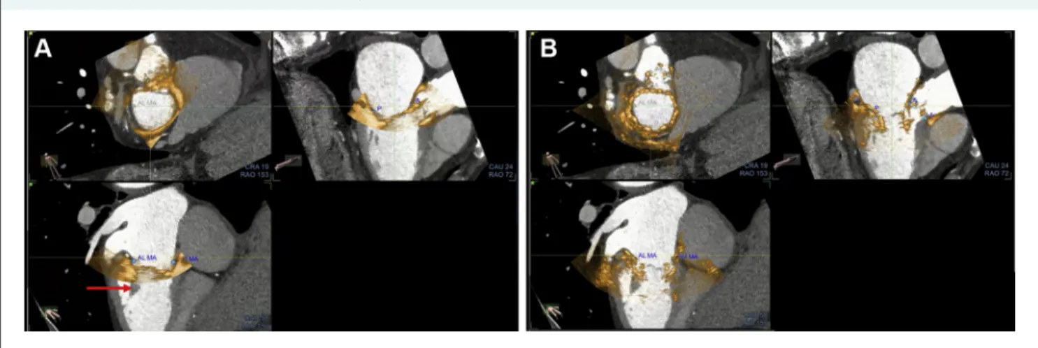
FIGURE 2 Example of Echo-CT Fusion With the Intrepid Prosthesis
Echo-CT fusion pre- (A) and post- (B) deployment of the Intrepid (Medtronic, Minneapolis Minnesota) prosthesis and its utility in crossing the mitral annulus plan with the prosthetic system while avoiding the subvalvular apparatus (red arrow). CT¼ computed tomography.
pri n t& web 4 C =FPO
FIGURE 3 Example of Echo-CT Fusion to Assess Neo-LVOT After TMVR With the Tendyne Prosthesis
(A) After the deployment, the amount of material and the shadowing of the device made the assessment of the neo-left ventricular outflow tract (neo-LVOT) difficult. (B) With echo-CT fusion, the assessment of the neo-LVOT (red arrow) may be more accurate to assess LVOT obstruction. This could be very useful for the Tendyne (Abbott Vascular, Santa Clara, California) prosthesis, which is fully recapturable and retrievable. CT¼ computed tomography; LVOT ¼ left ventricular outflow tract; TMVR¼ transmitral valve replacement.
J A C C : C A R D I O V A S C U L A R I N T E R V E N T I O N S V O L .-, N O .-, 2 0 2 0 Coisneet al.
-2 0 2 0 :-–- TMVR Guided by Echo-CT Fusion
3