S
Thiamine
Pyrithiamine
Oxythiamine
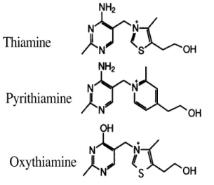
Fig. 1. Thiamine and two of its synthetic structural antimetabolites.
It has been known for many years that, in the nervous system, the reactions of the Krebs cycle are essential not only for cellular respiration, but also for the synthesis of a number of important neurotransmitters. Soon after Peters' formulation of the concept of "biochemical lesion", Mann and Quastel (16) reported that acetylcholine synthesis was diminished in the brain of thiamine-deficient pigeons. It was thus considered that the mpairment of pyruvate oxidation observed by Gavrilescu and Peters caused a decreased synthesis of acetyl coenzyme A and hence acetylcholine. However, during the following decades, it became increasingly evident that this "cholinergic lesion" was not the main cause for the irreversible lesions observed during thiamine deficiency (17-19). Likewise, the thiamine deficiency syndrome could not clearly be related to decreased synthesis of other neurotransmitters such as γ-amino-butyric acid (GABA) or glutamate (20).
A different approach was to suppose that thiamine, or one of its derivatives, could play a role in nerve activity, not related to the coenzyme function of ThDP. This idea was supported by several intriguing observations. In 1938, Minz (21) reported that, when the vagus nerve of an ox was stimulated, thiamine was released into the bathing medium. The phenomenon was studied in more detail by von Muralt and coworkers in the nineteen forties (see section V). The release of thiamine was observed especially in peripheral cholinergic nerve terminals, so that at the time some authors considered thiamine as "Vagusstoff II", acetylcholine being "Vagusstoff I" (22).
However, the implication of thiamine (if any) in cholinergic transmission remains poorly understood, except, of course, for the indirect role of ThDP in acetylcholine synthesis. On the other hand, Cooper and coworkers (23) provided evidence for a possible role of thiamine in nerve excitability in general and suggested that the triphosphorylated derivative of thiamine (thiamine triphosphate, ThTP) was the specific neuroactive substance. However, reliable estimations of the ThTP content in nervous tissue became available only after 1980. An additional complication is related to the fact that thiamine deficiency pre-dominantly affects the midbrain, thalamus, periventricular and periaqueducal regions, the cerebral cortex being relatively spared. In vulnerable regions, the metabolic flux, as measured by CO2 production, is decreased to a larger extent than the activity of ThDP-dependent enzymes in the thiamine-deficient brain. This suggests that other factors than a decrease in ThDP-dependent enzymes must account for decreased metabolic flux (14). In order to explain this selective vulnerability, excitotoxic processes and oxidative stress have recently been put forth (24-27).
A comprehensive survey on thiamine in the brain has been published by Haas in 1988 (28) and, in the present review, we wish to highlight some new developments in the biochemistry of thiamine phosphate esters and thiamine deficiency disorders in humans and animals. A final section will be devoted to some neurodegenerative diseases, such as Alzheimer's disease, where impairment of thiamine metabolism or of ThDP-dependent enzymes has been reported.
II. PHOSPHORYLATED DERIVATIVES OF THIAMINE:METHODS OF ANALYSIS AND DISTRIBUTION IN EXCITABLE TISSUES
In addition to free thiamine, three phosphate esters of thiamine have been described in nervous tissue: thiamine mono-phosphate (ThMP), ThDP and ThTP (Table 1) Their analysis having been extensively discussed (29,30), we shall only point out the main findings with special emphasis on the difficulties associated with the identification and quantification of ThTP. The vast majority of the analytical methods developed for the
quantitative analysis of thiamine and its phosphate esters in biological samples require their oxidation to fluorescent thiochromes, either before or after separation. At the present time, separation of thiamine or thiochrome derivatives by high-performance liquid chromatography (HPLC) has superseded other methods by combining reliability, rapidity and sensitivity, though, as we shall see, there are still problems with the identification of ThTP. While
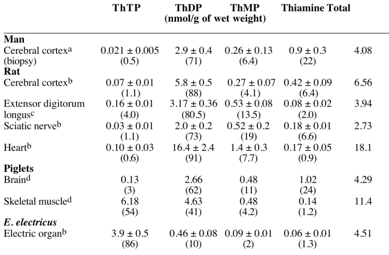
the cofactor ThDP is the prevailing thiamine compound in most tissues, ThTP accounts for only about 1% of total thiamine with the exception of skeletal muscle and electric organs. The proportion of ThMP and free thiamine is variable (1-20 % of total thiamine). Thiamine and ThMP seem to occur mainly in the cytosolic fraction in rat brain, while ThDP has a prevalently mitochondrial localization (35). Table 1. Thiamine derivatives in various excitable tissues.
____________________________________________________________________________ ThTP ThDP ThMP Thiamine Total
(nmol/g of wet weight)
____________________________________________________________________________ Man Cerebral cortexa 0.021 ± 0.005 2.9 ± 0.4 0.26 ± 0.13 0.9 ± 0.3 4.08 (biopsy) (0.5) (71) (6.4) (22) Rat Cerebral cortexb 0.07 ± 0.01 5.8 ± 0.5 0.27 ± 0.07 0.42 ± 0.09 6.56 (1.1) (88) (4.1) (6.4) Extensor digitorum 0.16 ± 0.01 3.17 ± 0.36 0.53 ± 0.08 0.08 ± 0.02 3.94 longusc (4.0) (80.5) (13.5) (2.0) Sciatic nerveb 0.03 ± 0.01 2.0 ± 0.2 0.52 ± 0.2 0.18 ± 0.01 2.73 (1.1) (73) (19) (6.6) Heartb 0.10 ± 0.03 16.4 ± 2.4 1.4 ± 0.3 0.17 ± 0.05 18.1 (0.6) (91) (7.7) (0.9) Piglets Braind 0.13 2.66 0.48 1.02 4.29 (3) (62) (11) (24) Skeletal muscled 6.18 4.63 0.48 0.14 11.4 (54) (41) (4.2) (1.2) E. electricus Electric organb 3.9 ± 0.5 0.46 ± 0.08 0.09 ± 0.01 0.06 ± 0.01 4.51 (86) (10) (2) (1.3) ____________________________________________________________________________ The percentage of each thiamine derivative compared to total thiamine is indicated
between brackets. a, calculated from (31), assuming that proteins account for 13 % of wet weight; b, according to (32); c, according to (33); d, according to (34).
ThDP seems more abundant in neuronal than in glial cell-enriched fractions (36).
The existence of ThTP was first suggested n rat liver (37) and then in other rat tissues ncluding brain (38), in baker's yeast (39) and in bacteria (40). The highest ThTP content is ound in pig skeletal muscle (in particular in young animals,41), and electric organs (42,32). Cultured cells of neuronal origin (cerebellar granule, neuroblastoma and PC-12 cells) have a
5-10 times higher ThTP content than cells of glial origin (43,44).
The intracellular location of ThTP is variable (41,42,32): in brain, heart and kidney it mainly occurs in the particulate fractions, while in liver, skeletal muscle and electric organs it is found in the soluble fraction. Matsuda and Cooper (45) suggested that ThTP is bound to synaptosomal membranes in rat brain, but subcellular fractioning of rat brain reveals only a slight enrichment of this fraction compared with
he myelin and mitochondrial fractions (35,46). ThTP, probably in a protein-bound form, is eleased from the rat brain particulate fraction after hypotonic lysis (47). While the role of ThDP as cofactor is textbook knowledge, the ole of ThTP is still hypothetical and many contro-versial results have been published. It is here-fore essential to critically examine these data with special emphasis on the reliability of he analytical method used. This is striking when we consider the high variability of the ThTP content found in rat brain by various methods Table 2). It varies from 0.02 to 0.6 nmol/g wet weight (30-fold) and would account for espectively 0.3 and 7.8 % of total thiamine. Similar extraction procedures were used in the wo extreme cases: homogenization of the rat brain in trichloroacetic acid, centrifugation and extraction of the acid with diethyl ether. In contrast to ATP, ThTP is relatively stable and he delay between sacrifice of the animal and nactivation of brain enzymes by trichloro-acetic acid is not critical provided it is reasonably short a few minutes, 48). It is also unlikely that nutritional factors could cause such differences n ThTP content, as animal chows are generally tandardized, and daily administration of massive doses of thiamine to rats for two weeks does not lead to a significant increase in brain ThTP content (49). Regional variations of hiamine derivatives in rat brain are small (50-52) and, in most instances, cerebral cortex was used. According to Pincus et al. (53), ThTP accounts for 11% of total thiamine in human post-mortem brain, but we were unable to detect any ThTP in human postmortem brain, in contrast to fresh biopsies (31). Similar discrepancies were also observed for the
determination of ThTP in blood. As already pointed out by Kawasaki (29), some authors found a high ThTP content in rat (50 nmol/L) and in human (54) blood, while others found only trace amounts (<8 nmol/L) and sometimes inconsistently (55-57). One study (55) reports higher ThTP concentrations in erythrocytes from alcoholic patients compared to the general population, but this was not confirmed by other groups (58-60). In erythrocytes ThTP is synthesized by adenylate kinase (61, section IV.3.), but the relationship between increased adenylate activity and alcoholism remains obscure. Most of these discrepancies are probably related to the analytical procedures used. Though, differen-ces between rat strains cannot be excluded, we believe that the main source of error in ThTP estimation is the presence of interfering compounds, fluorescent in the same range of wavelengths as thiochrome triphosphate.
In rat heart, after HPLC separation, we observed a contaminating compound with a retention time very close to that of ThTP (72). Its interference could easily be eliminated by replacing a filter fluorometer by a more selec-tive fluorescence spectrometer with narrower slit widths. In rat brain, a compound with the same retention as ThTP and labeled after administration of [35S]-thiamine to the animals was reported to be resistant to hydrolysis by thiamine triphosphatase (ThTPase) (63). Incubation of rat brain homogenates with pyrithiamine (Fig.1) also leads to the synthesis of a fluorescent compound after oxidation, with a retention time close to thiochrome tri-phosphate (72). After loading neuroblastoma Table 2. ThTP content in rat brain as determined by different methods.
____________________________________________________________________________ References Analytical method [ThTP] [ThTP]
employed (authors' units) (nmol/g ww)
____________________________________________________________________________ (64) RP-HPLCb 0.6 nmol/g ww (7.8) 0.6 (65) LC 0.4 nmol/g (2.8) 0.4 (66) LC 0.19 µg/g ww (5) 0.38 (67) Electrophoresis 0.12 µg/g ww (4.3) 0.24 (68) SP-HPLCa 0.1 nmol/g ww (1.6) 0.1 (69) PPC 0.044 µg/g ww 0.087 (70) Electrophoresis 0.04 µg/g ww (2.1) 0.079 (32) RP-HPLCb 0.07 nmol/g ww (1) 0.07
(35) IP-HPLCa 0.35 pmol/mg (0.4) 0.046 (34) SP-HPLCa 0.04 nmol/g ww (0.4) 0.04 (48) RP-HPLCb 0.039 nmol/g ww 0.039 (43) IP-HPLCa 0.21 pmol/mg (0.24) 0.027 (71) RP-HPLCb 0.02 nmol/g ww (0.3) 0.02 ____________________________________________________________________________ Data expressed by the authors in µg /g of wet weight (g ww) and in nmol/mg of protein were converted to nmol/g of wet weight assuming that proteins account for 13 % of rat brain wet weight. The numbers between brackets give, if available, the % of ThTP compared to total thiamine content of the tissue. IP-HPLC, ion-pairing HPLC; LC, low pressure liquid chromatography; PPC, paper partition chromatography; RP-HPLC, reversed phase HPLC; SP-HPLC, straight-phase HPLC. The upperscripts in the second column indicate whether TTP is eluted last (a) or first (b) of the thiamine compounds with the HPLC methods used.
cells with 14C-thiamine, the release of adioactivity into the extracellular medium ollowed by HPLC yielded two radioactive peaks: one coeluted with thiochrome, the other different from known thiamine compounds Bettendorff, unpublished results). These results how that thiamine-related compounds other han its phosphate esters may be produced in neuroblastoma cells but also in rat brain (71). In addition, the existence of hydroxyethylthiamine, an intermediate in the oxidation of pyruvate to acetyl-CoA, was shown in brain for instance 73). Therefore, in order to firmly establish the authenticity of ThTP in unknown samples, dentification on the basis of retention times and comparison with blanks obtained in the absence of oxidant are insufficient. ThTP should be purified on a Dowex or AG 50W-X8 cation exchange resin (43,47) and the fluorescence emission spectrum be determined. Hydrolysis of ThTP by ThTPases should also be tested
47,63,71).
In conclusion, it appears that in many tudies (especially earlier reports) the ThTP content of brain and nerve tissue was largely overestimated. In mammalian brain, ThTP probably does not amount to more than 0.3-0.4% of total thiamine. It remains true, how-ever, that the proportion of ThTP may be much higher in skeletal muscles and electric organs
see sections IV.3. and IV.4). III. THIAMINE TRANSPORT
The thiamine core molecule is synthesi-zed by most microorganisms and higher plants (74). n some animals, especially ruminants, bacterial ctivity in the gut may be responsible for most
of the thiamine supply (75). Thiamine is then taken up from the intestinal lumen through a high-affinity saturable process (Km = 4.4 µM), probably involving a thiamine/H+ antiport mechanism (76). A low-affinity transporter is also present (77,78). Due to the relatively low Vmax values for both transporters, lipid-soluble thiamine derivatives such as thiamine tetrahydrofurfuryl disulfide (79), benfotiamine (80,83) or thiamine isobutyryl disulfide (49) have a much higher bioavailability than thiamine.
Thiamine is present in blood plasma in the form of free thiamine and ThMP (57,84), while ThDP is the major form in ery-throcytes (56). Circulating thiamine concen-trations are only about 10 nM in humans, an order of magnitude less than in rats.
Thiamine and, to a lesser extent, ThMP (85,86) are transported across the blood-brain barrier by a carrier-mediated mechanism (87,88) which seems to be independent of energy metabolism (89). The rate of thiamine influx across the blood-brain barrier is close to its turnover in various parts of the rat brain and amounts to about 10 pmol/min/g of tissue (51,88). This is a rather low value and any decrease in thiamine transport capacity might lead to a net loss of vitamin from the brain. Cortex slices accumulated thiamine in a satur-able and energy-requiring process, suggesting that nerve cells actively pump thiamine (90). Neuroblastoma and glial cells possess a Na+ -independent high-affinity thiamine transporter with a Km of 35 nM for thiamine that is non-competitively inhibited by the Na+-channel activators veratridine, batrachotoxin and
aconitine (91). This effect is not antagonized by tetrodotoxin and seems independent of the presence of Na+-channels.
The rates of thiamine transport reported in brain cells are generally much lower (1-3 orders of magnitude) than in hepatocytes or kidney cells (78, 91-93). It is of interest to note that the high affinity carrier is also responsible for part of the release of intracellular thiamine in exchange for extra-cellular vitamin. It may thus contribute to the homeostasis of intracellular thiamine at high extracellular concentrations (93). The thiami-ne transport gene has been characterized in Saccharomyces cerevesiae (94,95) but not yet in mammalian cells. Thiamine taken up by the cells is rapidly phosphorylated to ThDP (91), which in turn binds to transketolase and, after transport into mitochondria (96), to PDH and OGDH. Phosphorylation of free thiamine pro-bably constitutes the driving force for secon-dary active transport of thiamine into neuro-blastoma cells and other cell types (78,91).
IV. METABOLISM OF PHOSPHORY-LATED DERIVATIVES OF THIAMINE
IV.1. Enzymatic synthesis of ThDP
ThDP is synthesized according to the reaction: thiamine + ATP ⇔ ThDP + AMP by a soluble, cytosolic and apparently ubiquitous enzyme called thiamine diphosphokinase (or thiamine pyrophosphokinase, ThDK, EC 2.7.6.2.). In rat brain, ThDK is present in all regions investigated (97,98), the highest activity being found in the cerebellum and pons, and the owest in cerebral cortex and midbrain. The ThDK gene has been isolated and characterized n Saccharomyces cerevisiae (99) and Schizosaccharomyces pombe (100).
ThDK, a homodimer of 46-56 kDa, was characterized or purified from various sources (31,101-108). In all instances (bacterial, yeast, mammalian and human brain enzyme), the apparent Km for thiamine is low (10-7-10-6 M) but for ATP it is of the order of 10-2 M. The pH optimum is broad and slightly alkaline.
The equilibrium of the reaction is very far owards the side of the reactants (103), But, in vivo, the equilibrium will be shifted to the right because ThDP binds with high affinity to cytoplasmic transketolase o r enters
mitochondria to bind to PDH or OGDH. Also, in cell types such as muscle fibers, the concentration of AMP is kept very low because of the presence of high amounts of adenylate kinase and creatine phosphate. In any event, the concentration of free ThDP in the cytosol is < 10-5 M (see the next section).
IV.2.A pivotal fast cytosolic ThDP pool
ThDP synthesized in the cytosol has seve-ral possible metabolic fates (Fig.2): binding to transketolase, transport into mitochondria, phosphorylation to ThTP or hydrolysis.
ThDP binds with high affinity to transketolase, a cytosolic enzyme. Bound cytosolic ThDP may therefore be relatively abundant in cell types that are rich in transketolase, such as hepatocytes and erythrocytes. However, in neurons and other aerobic cells, most of the ThDP is transported into the mitochondrial matrix, where it binds to PDH and OGDH complexes. ThDP uptake by isolated mitochondria is a saturable process with an apparent Km around 20 µM (96). Thiamine and ThMP can also be transported (109, 110), possibly in exchange for ThDP. The latter may be hydrolyzed to ThMP in the mitochondrial matrix (110), but there is no enzymatic hydrolysis of ThMP in mitochondria. Likewise, no synthesis of ThDP occurs in mitochondria, ThDK being exclusively cytosolic (111).
Free cytosolic thiamine Free cytosolic ThDP Free ThTP ThMP ATP AMP ATP ADP ThMP MITOCHONRION 1 2 5 5 6 7 3 ThDP bound to transketolase Bound ThTP ThDP bound to oxo acid dehydrogenases ThDP pool of mitochondrial matrix AMP ADP CYTOSOL 4 1
Fig. 2. Metabolism of thiamine and its phosphate esters in a neuronal cell (1, ThPK; 2, adenylate kinase; 3, ThDP kinase; 4, ThTPase; 5, ThDPase; 6,ThMPase; 7, mitochondrial
ransporter).
In the cytosol, ThDP can be hydrolyzed to ThMP by thiamine diphosphatases (ThDPases, ection IV.4). It is also likely that in some cell ypes, including hepatocytes and neurons, cytosolic ThDP can be phosphorylated to ThTP
see section IV.3).
Gaitonde and Evans (112) injected [14 C]-abeled thiamine to rats and found that, in the brain, the incorporation of radioactivity was slower into ThDP than into ThMP and ThTP. As ThDP is the most likely precursor of the two other derivatives (thiamine kinase activity has never been found in the brain), this result can only be understood if there is a smaller, faster pool of precursor ThDP, which in rat brain is essentially cytosolic (35). I n cultured neuroblastoma cells, the slow and fast ThDP pools coexist in the same cell type and are not the result of a different thiamine metabolism in glia and neurons (113).
The bulk of the ThDP bound to apoenzymes (transketolase, PDH and OGDH) is considered as the "slow turnover ThDP pool", hough the stabilities of the different coenzyme-apoenzyme complexes vary: ThDP dissociates aster from PDH than from OGDH and ransketolase (19,114). It can be estimated that he slow ThDP pool represents 90-95 % of the otal cellular ThDP and has a turnover of 6-20 h 35,113,115). For the smaller fast pool, the urnover is of the order of 1-3 h (113). It is not clear whether the fast pool consists only of cytosolic ThDP. An appreciable part may actually be located in the mitochondrial matrix 110). ThDP transport across the mitochondrial membrane is a relatively fast process (first order ate constant ≈ 1.7 min-1, 96). Thus, the putative mitochondrial fast pool may have a urnover rather close to the turnover of the cytosolic pool and the 2 pools should not necessarily be considered as separated.
In brain cells, cytosolic ThDP probably epresents not more than 15-20% of total cellular ThDP. About 10% of total ThDP appears in the S3 (soluble) fraction after differential centrifugation of a rat brain
homogenate (35). As other fractions also con-tain a cercon-tain amount of cytoplasm (especially in cell debris and synaptosomes), the pro-portion of cytosolic ThDP is more likely to be 15 %. It was also estimated (35) that 30-40% of this cytosolic ThDP is bound to transketolase. Therefore, free cytosolic ThDP should amount to 9-10% of cellular ThDP. Since the molarity of total ThDP in brain cells is around 14 µM, the concentration of free cytosolic ThDP should not excced 2 µM. This is quite a low concentration, especially if it is compared to Km values for ThDP-hydroly-zing enzymes (section IV-4).
IV.3. Enzymatic synthesis of ThTP
While there is little doubt that ThTP is present in a wide variety of organisms (section II), the mechanism of its enzymatic synthesis remains poorly understood. The very existence of a specific mechanism has been questioned since Kawasaki and coworkers found that ThTP synthesis could be catalyzed by adenylate kinase (EC 2.7.4.3) according to the reaction ADP + ThDP ⇔ AMP + ThTP (108, 117-120). However, ThDP is a poor substrate for adenylate kinase and this mechanism appears to be of importance only in cell types where adenylate kinase is very abundant, e.g. skeletal muscle and possibly electric organs.
In nerve cells and hepatocytes, the main mechanism for ThTP synthesis, as originally proposed by Eckert and Möbus in 1964 (121), would be ThDP + ATP ⇔ ThTP + ADP. The enzyme catalyzing this reaction was called ThDP-kinase (ThDP:ATP phosphoryltrans-ferase, EC 2.7.4.15) but until now it remains poorly characterized in nervous tissue.
After intracerebro-ventricular (71) or intraperitoneal (35) administration of labeled thiamine to rats, radioactivity rapidly appeared in ThTP, demonstrating that this compound is indeed synthesized in vivo in the brain. The in vivo synthesis of ThTP was also demonstrated in rat liver (122). It should be noted that the proportion of ThTP in the liver found by these authors was about 2.3% of total thiamine, substantially more than in brain. This is why several authors used liver (or yeast) rather than brain extracts to study the enzymatic mechanism of ThTP synthesis. ThDP kinase was partially purified and characterized from rat
iver, where it is essentially found in the upernatant (cytosolic) fraction (123). The authors measured the formation of labeled ThTP rom [14C]ThDP in the presence of ATP and Mg2 +. After a 100-fold purification, the specific activity was still low and two pH optima were ound (5.3 and 7.8). Interestingly, a sigmoid curve was obtained when the activity was plotted as a function of ThDP concentration and he activity was very low when the ThDP concentration was less than 10 µM. A few years atter, the same group reported a complete purification of ThDP kinase from brewer's yeast
124).
In brain extracts, no ThDP kinase activity could be demonstrated using ATP and free ThDP as substrates. In 1977, Ruenwongsa and Cooper (125), using a supernatant fraction from at liver, reported that labeled ThTP was formed rom [γ-32P]ATP. No addition of ThDP to the preparation was required and the authors concluded that ThDP bound to a protein was he substrate for ThTP synthesis. However, as hown later, the estimation of labeled ThTP was not correct because ThTP was not well eparated from ATP (126). Nonetheless, a purified ThDP-binding protein from liver was ater used as a substrate for ThTP synthesis catalyzed by brain extracts (127). The studied eaction was: ThDP-protein + ATP ⇔ ThTP-protein + ADP. The enzyme was purified from bovine brain starting with an acetone powder of a crude mitochondrial fraction and its molecular weight was estimated to be 103 kDa. The pecific activity of the purified enzyme was low and the yield was only 0.6 %. The enzyme was unstable and inactivated by sulfhydryl reagents. There was a controversy about the nature of the ThDP-protein complex used as substrate. According to Nishino et al. (127), this soluble protein (purified from a liver supernatant raction) was distinct from transketolase, which s a well-known cytosolic protein binding ThDP with high affinity. Voskoboyev and Chernikevich (128) reinvestigated the purification procedure and concluded that the only ThDP-binding protein in the rat liver upernatant fraction was transketolase and at east in rat liver, the ThDP-transketolase complex was not a substrate for ThDP kinase. So far, the mechanism described by Nishino et
al. in bovine brain has not been confirmed in other laboratories.
More recently, we reported a net synthesis of ThTP in a particulate fraction (synaptoneurosomes) isolated from rat brain (47) with either thiamine or ThDP as substra-te. The phosphate donor seemed to be ATP but we could not totally exclude the possibility that adenylate kinase was implicated in the synthesis. No net synthesis of ThTP was observed in rat brain cytoplasm, this was probably due to hydrolysis of newly formed ThTP by soluble ThTPase (see below). ThTP formed inside the microvesicles was protein-bound. An interesting property of this system is that the ThTP synthesized already reached its maximum concentration after 10 min, though only about 0.02 % of ThDP had been converted. The fact that it was not possible to raise ThTP synthesis above this level, suggests that the maximum amount of ThTP present in brain cells is limited by the availability of a ThTP-binding protein, any free ThTP in excess being rapidly hydrolyzed. This hypo-thesis is compatible with results obtained in cultured neuroblastoma cells: when these cells are incubated in the presence of 14C-thiamine a rapid incorporation of radioactivity into ThTP is observed, though no net synthesis occurs (35). Furthermore, chronic administra-tion of massive doses of thiamine to rats leads to a 10 % increase in brain ThDP content but ThTP levels are not affected (49). However, when 14C-thiamine was administered, brain ThTP had a high specific radioactivity (35). These results suggest that, at least in brain cells, ThTP levels are highly regulated.
In contrast, the enzymatic synthesis of ThTP seems to be faster than its hydrolysis only in the case of skeletal muscle and electric organs, where ThTP accumulates (Table 1). In some particular cases such as pig skeletal muscle or electric organ, ThTP is the most abundant of all thiamine compounds. There are, however, large differences between species and between different types of muscles (33). At least in skeletal muscle, it is likely that the main mechanism responsible for ThTP synthesis is the reaction catalyzed by adenylate kinase (118). The role of adenylate kinase, which is a cytosolic enzyme, is in agreement with the observation that ThTP is essentially found in the cytosol of skeletal muscle (34,41) and electric organ
32,42). The accumulation of large amounts of cytosolic ThTP may be interpreted as follows: he equilibrium of the reaction ADP+ThDP ⇔ AMP+ThTP will be shifted to the right because AMP reacts with ATP (which is in excess) to orm 2 ADP. Both reactions are catalyzed by adenylate kinase and the global reaction will be ThDP+ATP ⇔ ThTP+ADP. This equilibrium
will also be shifted to the right because the muscle contains high amounts of phosphocreatine and creatine-kinase: ADP + phosphocreatine ⇔ ATP + creatine. The global process will thus be ThDP + phosphocreatine → ThTP + creatine.
V.4. Enzymatic hydrolysis of phospho-rylated derivatives of thiamine
a) Thiamine triphosphatases (EC 3.6.1.28)
Until now, two types of ThTP-hydrolyzing enzymes (ThTPases) have been found in mammalian tissues: a soluble and a membrane-associated enzyme. Only the soluble enzyme has been purified and proven specific for ThTP 129). In addition, acid and alkaline phosphatases 130) as well as myosin (131) have a ThTPase activity, though ThTP does not seem to play a direct role in muscle contraction (132).
The specific soluble ThTPase seems to be present in most mammalian tissues (133,134) with the exception of the intestine (130). Its activity is very low in electric organs (32) and keletal muscle (135). The enzyme is activated by Mg2 + while Ca2 + is inhibitory. The opti-mum pH is around 9 but the enzyme is clearly distinct from alkaline phosphatase (130).
After purification to homogeneity, ThTPase eems to be a monomer with a molecular mass of 33.9 kDa as determined by gel exclusion chromatography and electro-phoresis (129). It is pecific for ThTP, al-though the interaction with he substrate is complex: reaction velocity plotted versus ThTP concentration yields a biphasic curve with a first saturation around 50 µM. In man (31) and rat brain (47) however, imple Michaelis-Menten kinetics with apparent Km values of 56 and 70 µM were obtained, about 10-fold lower than for membrane-associated ThTPases. It must however be ecalled that the concentration of free ThTP in he cytoplasm of neurons is less than 1 µM (47).
The membrane-bound ThTPase in brain has been studied by Barchi and coworkers
(134,136,137). It is found in all particulate fractions isolated from rat brain homogenates but it is more abundant in nuclear and synaptosomal fractions than in mitochondria. There was an absolute requirement for divalent metal cations but, in contrast to the soluble alkaline ThTPase, Ca2 + could replace Mg2 + (134). The optimal pH is 6.5 and the apparent Km for ThTP is high (1-2 mM). ATP and ADP are effective inhibitors: IC50 values are 20 µM for ATP, 75 µM for ADP and more than 5 mM for ThDP. A nonhydro-lyzable methylene phosphonate analog of ThTP was synthesized; it is a weak competitive inhibitor (KI = 3 mM). These results suggest that the active site has a much higher affinity for adenine nucleotides than for thiamine compounds. However, the enzyme could not be identified with any known ATPase. It remains possible that the enzyme is a nucleoside triphosphatase with broad substrate specificity. Histochemical evidence shows a colocalization of soluble and membrane-associated ThTPase in axolemma, presynaptic membranes, synaptic vesicles and Golgi apparatus (138).
As already pointed out above, the soluble ThTPase activity is low in skeletal muscles and electric organs but those tissues contain a membrane-associated ThTPase, first described in the electric organ of Electrophorus electricus (32,139,140,141) and a few years later in skeletal muscle (135). As for the brain membrane-associated ThTPase, this activity is found in particulate fractions, predominantly the "nuclear" fraction. The distinctive feature of this ThTPase is its strong activation by chaotropic anions (I- > SCN- > NO3- > Br- > Cl-), that is not observed for the brain enzyme (135). The ThTPase from E. electricus is irreversibly inhibited by the anion transport inhibitor 4,4'-diisothiocyanostilbene-2,2'-disulfonic acid (DIDS). The fact that anions protect against inhibition by DIDS (in particular, SO42- ions were effective at 5 mM) suggests that an anion-binding site is important for catalysis or regulation of the activity. ThTP also protects from inhibition by DIDS and the dissociation constant estimated from the protective effect was 0.25 mM. The optimum pH for the E. electricus ThTPase was around 6.5 in the absence of anions but in the presence of the
chaotropic activating anion NO3-, this value was hifted to more alkaline values. At low substrate concentrations, we observed deviation from ypical Michealis-Menten behavior, suggesting wo active sites with apparant Km values of 0.5 µM and 1.8 mM. However, after purification of he membranes on a sucrose gradient, we observed michaelian kinetics (Km of 0.2 mM, 140).
b. Thiamine diphosphatase(s) (EC 3.6.1. -)
Hydrolysis appears to be the main metabolic fate of ThDP in brain. The alternative oute (phosphorylation to ThTP) is quantitatively important only in skeletal muscles and electric organs. It is thus inte-resting to note hat a phosphohydrolase that is relatively pecific for ThDP has been purified and characterized from brain (142). GDP, IDP and UDP are also good substrates, while ATP and ADP are not. ThDP is the preferred substrate only in the presence of Mn2 + ions. In any case, a divalent metal cation (Mn2 +, Ca2 + or Mg2 +) s required for activity and ThDP is hydrolyzed at physiological pH. The enzyme was called B(brain)-type ThDPase.
In other organs such as the liver (143), ThDP can be hydrolyzed by a nucleoside diphosphatase (NDPase, EC 3.6.1.6). The latter catalyzes the hydrolysis of GDP, IDP and UDP, but not ADP; for ThDP, the activity was low at neutral pH but higher at pH 9. The NDPase purified from beef liver was referred to as the L(liver)-type enzyme but it is also present in brain together with the B-type ThDPase 144,31). The presence of the L-type enzyme may explain that the maximum ThDPase activity was found at pH 9, in human brain homogenates (31,145). The B-type ThDPase is membrane-bound and presumably associated with the endoplasmic reticulum and Golgi apparatus of neurons (146). Its molecular mass was estimated to be 75 kDa and the Km for ThDP was around 0.66 mM, well above the concentration of free ThDP in the neuronal cytosol (section IV.2).
In addition to the particulate B-type ThDPase, another ThDP hydrolase was partially purified from a supernatant fraction of sheep brain (147). In contrast to the L- and B-type enzymes, it also hydrolyzed ADP.
After separation of neuronal and glial cell-enriched fractions of rat brain, the activity at pH 9 was found to be 15-fold higher in the neuronal fraction (36). Determination of ThDPase in cultured cells suggested that the alkaline L-type enzyme is predominantly located in astrocytes while the B-type enzyme is found in neuroblastoma cells (31). This confirms earlier histochemical studies (138, 149-150) suggesting a preferentially neuronal and glial localization for respectively the B- and L-type enzymes. Other cytochemical studies showed the presence of ThDPase activity in myelin sheaths of central and peripheral fibers (151), probably as a result of axonal transport towards motor end plates. ThDPase was also found in synaptic vesiscles (152). ThDPase has been considered a marker for the Golgi apparatus (153-155), where it might play a role in protein glycosylation (156). ThDPase in the Golgi apparatus is probably of the B-type (146).
c. Thiamine monophosphatase
It has been known for a long time that ThMP can be hydrolyzed to thiamine by phosphohydrolases (ThMPases) present in brain and other organs. However, no phosphatase specific for ThMP has been characterized so far. Kiessling (157) prepared acetone powders from homogenates of liver, kidney and brain and studied the ThMPase activity of those crude preparations as a function of pH. In the liver, there was only a rather sharp optimum at pH 6.0 but, in kidney and brain, a second peak appeared at pH 9.0. A similar pattern was reported by Laforenza et al. (145), who used homogenates from autopsied humain brain supplemented with 1% Triton X-100. The acticity was highest at pH 9; at physiological pH, the enzymatic hydrolysis of ThMP is 10 times slower than ThDP hydrolysis. ThMPase is found in glial rather than neuronal fractions, in sharp contrast to ThDPase (36). Cytochemical data, however, show the presence of ThMPase associated with the plasma membrane of the synaptic glomeruli in the substantia gelatinosa of rat spinal cord (138). Note that the sections were incubated at acid pH (5.4-5.5). The very specific localization of this "acid ThMPase" was actually identical with that of the so-called "fluoride-resistant acid phosphatase" (158), a marker enzyme for transganglionic regulation of primary sensory neurons. The physiological
ubstrate for this enzyme is unknown but it is not likely to be ThMP. In those cytochemical tudies of the spinal cord, it is advantageous to use ThMP as substrate since, in contrast to β-glycerophosphate or p-nitrophenyl phosphate, it s not a good substrate for lysosomal acid phosphatase. To our knowledge, there are no cytochemical data concerning ThMPase activity at pH 9.0. It is possible that the ThMPase activity measured in brain homogenates is lost after the treatment (fixation etc.) needed for cytochemical studies.
V. EVIDENCE FOR A NON-COFACTOR ROLE OF THIAMINE COMPOUNDS
A possible non-cofactor role of thiamine derivatives in nervous tissue has been extensively discussed previously (23,159) and we shall highlight only the main points in the present survey.
Since the initial observation of Minz (21) in 1938 (see Introduction), several workers have confirmed that the electrical stimulation of some nerve preparations (such as the isolated frog pinal cord) induced a release of thiamine 22,160,161). Gurtner (162) reported evidence hat the stimulation of nerves results in a hydrolysis of thiamine phosphate esters, producing an excess of free thiamine, that is inally released. More recently we have ob-erved that intermittent light stimulation of the photosensitive baboon Papio papio, decreased ThTP levels in the occipital (visual) cortex, while ThMP levels were increased (48).
The meaning of these data remains uncertain, but they may be related to the well-known fact that repetitive electrical stimulation of neurons strongly accelerates ATP hydro-lysis as the Na+-K+-ATPase is stimulated in order to maintain the ionic gradients. Thus, the ATP]/[AMP] ratio may substantially decrease n the cytosol of neurons, which should impair he synthesis of ThDP: indeed, the equilibrium position of the reaction Thiamine + ATP ⇔ ThDP + AMP is shifted to the right only if the ATP]/[AMP] ratio is high (section IV.1). ThDP hydrolysis may thus become much faster than ts synthesis, causing an accumulation of ThMP and free thiamine in the cytosol and, in some cases, an important release of free thiamine into
he extracellular fluid.
Another hypothesis would consist in the activation of the rapid turnover pool of thiamine phosphate esters as a result of electrical stimulation, due to the consumption of ThDP or ThTP in cofactor-independent processes.
ThTP content in membrane vesicles prepared from rat brain positively correlates with chloride permeability (47,163,164). ThTP at physiological concentrations (<10 µM) activates a high-conductance chloride channel (300-400 pS) in cultured neuroblastoma cells when applied to the bath of excised inside-out patches (44). The role of these so-called maxi-Cl -channels has remained elusive so far. The fact that a delay of several minutes was required between addition of ThTP to the preparation and appearance of channel activity suggests the involvement of a chemical reaction such as a phosphorylation. This would be in agreement with the observation that once the channel is activated, removal of ThTP does not lead to inactivation.
Thiamine release is preferentially observed in the case of cholinergic transmission. Indeed, thiamine release is often associated with acetylcholine release (22) and several studies reported the facilitation of synaptic trans-mission by thiamine (165-169). At least some of these effects are presynaptic and result from a potentiation of acetylcholine release by thiamine. As these effects are rapid (minutes or less) they can hardly be related to increased acetyl-CoA synthesis through activation of PDH by newly formed ThDP. Indeed, thiamine transport into the cells and subsequent phosphorylation to ThDP are slow processes (91).
Interactions of thiamine compounds (in particular pyrithiamine) with acetylcholine re-ceptors (170,171), Na+-K+-ATPase (172), Na+ and K+ channels (173-175) have been reported, but in most cases unphysiologically or unpharmacologically high concentrations were required. A recent report shows that intracellular ThDP (≤ 1 mM) decreases the open probability of delayed rectifier K+ channels in cultured rat cortical neurons (174). These channels are also inhibited by extra-cellular thiamine tetrahydrofurfuryl disulfide (175). A similar effect of thiamine-related compounds (at high concentrations ≥ 1 mM), including thiamine antimetabolites such as pyrithiamine, might explain the slower rate of repolarization
of the membrane after an action potential at the ingle node of Ranvier in frog sciatic (176) and obster nerves (177).
VI. THIAMINE IN NEUROLOGICAL DISORDERS
Beriberi and Wernicke-Korsakoff syn-drome are considered to be the typical mani-estations of respectively alimentary and toxic alcohol-related) thiamine deficiency (178). In addition, impairment of thiamine-dependent enzymes or thiamine metabolism has been observed in various neurological diseases such as Alzheimer's disease (179-187), frontal lobe degeneration of the non-Alzheimer's type (188) and heriditary ataxias (189-192).
Leigh's disease (subacute necrotizing encephalo-myelopathy), a progressive and fatal neurodegenerative disease of childhood with histological lesions resembling those found in Wernicke-Korsakoff syndrome, was previous-ly attributed to a deficit in ThTP (53,193,194). Current views suggest that it is a hetero-geneous yndrome resulting from mito-chondrial DNA point mutations as well as res-piratory enzyme and PDH defects (195-198).
Thiamine-responsive megaloblastic anae-mia, an autosomal recessive disorder, is associated with a defect in thiamine transport or phosphorylation (199-201) and an increased ensitivity of transketolase to thiamine deficiency 202). It seems to be a homogeneous disease probably resulting from a mutation on chromosome 1 predisposing carriers to megaloblastic anaemia (203). Some forms of maple-syrup urine disease, an autosomal, ecessively transmitted deficiency in BCODH, are thiamine-responsive (204).
ThMP, the only thiamine phosphate ester ound in cerebrospinal fluid (205) is reduced in patients with Guamanian amyotrophic lateral clerosis and Parkinsonism-dementia (206). The motor cortex of these patients also has decreased TDPase activity (154).
Subclinical thiamine deficiency in humans is probably more widespread in developed countries than initially thought, especially in elderly people (207-211), as well as in some risk groups such as infants, pregnant and lactating women (212) and drug abusers (213).
VI.1. Beriberi and Wernicke-Korsakoff yndrome
a. Human studies
In man, beriberi is the most typical manifestation of nutritional thiamine deficien-cy (214,28). It is characterized by peripheral neuropathy, loss of eye movements, nystagmus and hyperaesthesia often complicated by cardiovascular manifestations (wet beriberi and the fulgurant Shoshin cardiac beriberi). In developed countries, beriberi is virtually non-existent, but thiamine deficiency related to chronic alcohol abuse leads to a severe impairment of brain function, known as Wernicke-Korsakoff syndrome (215,216).
Wernicke's encephalopathy is most often associated with alcoholism, but it can also occur as a result of general malnutrition (28) as observed in war prisoners (214), after gastrectomy (217), in drug abusers and in AIDS patients (218,219). Its clinical features include ophthalmoplegia, global confusional state, ataxia of gait and polyneuropathy. Central nervous symptoms (confusion, delusions, ataxia) are more prominent than in beriberi, hence the appellation cerebral beri-beri (214). The Korsakoff psychosis appears as a consequence of chronic thiamine deficiency in the alcoholic patient, generally after several episodes of acute Wernicke's encephalopathy. Clinical features are anterograde amnesia, disorientation, confabulation and learning defects, probably arising from irreversible diencephalic lesions. Administration of thiamine at an early stage of the encephalopathy leads to a rapid response to the vitamin, with the reversal of the acute manifestations of thiamine deficiency. At later stages, recovery of the patient is at best partial, leaving clinical sequelae (residual ataxia, horizontal nystagmus, Korsakoff psychosis), linked to cerebral lesions. The relationship between thiamine deficiency and Wernicke-Korsakoff syndrome is well documented (178,220-224), though synergistic effects of ethanol toxicity and liver disease may be combined with thiamine deficiency to produce neuronal lesions (225-227). Decreased thiamine and thiamine phosphates (especially ThMP) levels are observed in blood and cerebrospinal fluid in clinical cases of Wernicke's encephalopathy (228,229). This is most probably related to the bad nutritional status of chronic alcoholics as well as to lesions in the gastrointestinal tract interfering with the absorption of thiamine (230) and liver disease
58). Ethanol inhibits thiamine phospho-rylation n rat brain (231) and increases ThDP dephosphorylation in nervous tissue and kidney cells (232-234). Not all alcoholics develop Wernicke-Korsakoff syndrome and, on the basis of the existence of transketolase variants with a high Km for ThDP, a genetic sensitivity to hiamine deficiency has been suggested (235-237). Other studies (60,238) did not confirm the existence of different transketolase variants and cloning of the cDNA for human transketolase evealed the existence of only one allelic variant 6). However, post-translational modifications of ransketolase cannot be excluded. The prevalence of the Wernicke-Korsakoff syndrome s higher in Australia (2.8% of all autopsies) than n France or Germany (≤ 1%), though the per capita alcohol consumption is higher in the atter two countries (239). This suggests that environmental, dietary and maybe genetic
actors should be given more attention.
Transketolase, PDH and OGDH activities are reduced in the autopsied brain tissue from alcoholics with Wernicke's encephalopathy 240). Only the first two enzymes are decreased n alcoholics in the absence of Wernicke's encephalopathy, suggesting a cor-relation between OGDH activity and the neuro-psychiatric symptoms of this disease (241).
Though cerebellar and cortical lesions 28,242) are reported in Wernicke-Korsakoff yndrome, the most prominent and charac-eristic lesions (necrosis of nerve cells, reactive gliosis and blood vessel lesions) are found in the halamus and mammillary bodies (178,215,223). This selective vulnerability remains unexplained, especially as all neurons are supposed to have a high oxidative energy metabolism and thiamine derivatives are distributed relatively uniformly hroughout the human brain (31). In human postmortem brains, Pincus et al. (53) reported 5 imes higher total thiamine levels in mammillary bodies than in other regions, but this was not confirmed in a later study (31). In order to explain this selective vulnerability, animal models have been developed (221,243) and more recently, an increasing number of articles describing cellular models of thiamine deficiency have been published (202,244-256).
b. Experimental models of thiamine deficiency
After the initial studies of Peters on thiamine-deficient pigeons, mainly rats and, to a lesser extent, mice were used in the study of thiamine deficiency. When the rats were fed a thiamine-deficient diet it took about 4 weeks before the first symptoms appeared (28,257). The anatomical lesions produced were not very reproducible and less widespread than those observed in human Wernicke-Korsakoff syndrome. Better results were obtained when a thiamine-deficient diet was combined with administration of pyrithiamine (Fig.1), a synthetic thiamine antimetabolite. There are several possible mechanisms to explain the poisonous effects of pyrithiamine. It is a competitive inhibitor of thiamine transport (91,178,258) and, inside the cells, it is a potent inhibitor of ThPK (73,103,259). In contrast to other thiamine antagonists, such as oxy-thiamine which does not enter the brain (260), pyrithiamine easily crosses the blood-brain barrier. Pyrithiamine enters the brain more slowly than other organs but its elimination from the brain is also slower than from liver, kidney or muscles (73,259,261). Another possibility is that the diphosphorylated deriva-tive of pyrithiamine could block the activity of ThDP-dependent enzymes. This mechanism is however less likely as pyrithiamine, in contrast to oxythiamine, is a potent inhibitor, but a poor substrate for the diphosphokinase (102); it also appears that pyrithiamine diphosphate has a lower affinity for ThDP-dependent enzymes than oxythiamine diphosphate (259). Finally, a direct toxic effect of pyrithiamine cannot be excluded (72,262, section V).
In the pyrithiamine-treated rat, the first symptoms of thiamine deficiency appear after one week of pyrithiamine treatment: anorexia and weight loss are followed by ataxia, paralysis and loss of righting reflex, as well as learning and memory impairments (266-268). The animals develop seizures after 2 weeks, followed by death within several days. The lesions in thalamus and mammillary bodies resemble those encountered in Wernicke's encephalopathy in man; this justifies the use of pyrithiamine for the study of the histopatholo-gical and neurochemical aspects of thiamine deficiency (19,263). Recent studies on pyrithiamine-treated rats also show cortical damage in white matter tracts (264) and hippocampal modifications (265). In addition to central nervous system
esions, thiamine deficiency leads to retrograde axonal degeneration in peripheral nerves (269) and motor neuropathy (270).
Various studies have shown that in the rat, clinical symptoms of thiamine deficiency only appear when brain total thiamine stores are depleted by 50%. When the reduction reaches 80%, severe disturbances of posture and equilibrium occur (52,257,271,272). They are accompanied by irreversible pathologic lesions. Thiamine depletion is slower in brain than in iver, heart or kidneys (73,257,273), though the brain is more vulnerable. The energy metabolism is similarly decreased in all organs 274,275), but only the brain presents rreversible lesions (263,276,277).
In thiamine-deficient rat (52,273) or mouse brain (272) but also in cultured neuroblastoma cells (247), all thiamine derivatives, including ThTP, are reduced to a similar extent and at quite a similar rate. Two studies report no change in ThTP content in the thiamine-deficient amb brain (278) and even an increase in rat brain (70). In view of the above-mentioned echnical problems encountered with the dentification of ThTP in animal tissues and the elatively high turnover of ThTP compared to ThDP (35,113), these results should be taken with caution.
On a microscopic level, thiamine deficien-cy in the rat model is accompanied by early oedema involving glial cells (particularly astrocytes) and dendritic as well as neuronal processes, followed by haemorrhagic lesions probably resulting from an early breakdown of he blood-brain barrier (279-285). This is a very mportant point, as increased permeabi-lity of he blood-brain barrier is already observed in vulnerable regions prior to the onset of histological and neurological lesions. Thus, the ensitivity of the brain to thiamine de-ficiency is not restricted to neurons and glia, but endothelial cells are also affected, resulting in the upture of the blood-brain barrier. This view is currently supported by the observation that addition of pyrithiamine to cultured brain cortical capillary endothelial cells induces an ncrease in glucose consumption, lactate production and permeability of the cells to
ucrose (251)
Oedema has been interpreted as the result of the impairment of Na+-K+ -ATPase-dependent ion transport due to decreased ATP
production. However, Na+-K+-ATPase activity was increased at the early stages of thiamine deficiency in rat brain (286) and in neuroblastoma cells it decreased only at the very end (247). Furthermore, energy-rich compounds (phosphocreatine and ATP) are relatively unaffected in the brain of thiamine-deficient rats (19,257,275,287), and only one study reported a 10 - 30% reduction in regions with necrotic lesions but only at the very late stages of thiamine deficiency (280). Interes-tingly, a recent report showed that in thiamine-deficient rats apoptosis is essentially restricted to the thalamus, while necrosis seems to be prevalent in the other brain regions (288).
At the onset of neurological symptoms, disintegrating mitochondria and chromatin clumping are observed in the diencephalic structures (282). Similar changes were observed in neuroblastoma cells cultured in thiamine-deficient medium (247,248). Mito-chondria became electron-translucent, spheri-cally-shaped and abnormally large. The oxy-gen consumption decreased, the mitochondria were uncoupled, ATP levels decreased and lactate production increased. These changes were essentially reversible: one hour after addition of thiamine to neuroblastoma cells, the oxygen consumption was nearly normal, the respiratory control was restored and the mitochondria had a normal shape. A normal respiration (with respiratory control) could also be restored by succinate, that can be oxidized independently of ThDP-dependent enzymes (248,289), suggesting that mitochon-dria retain their functional integrity in thiamine deficient cells. Mitochondria isolated from thiamine-deficient rat brain also show a decreased respiratory control, when pyruvate or 2-oxoglutarate are used as substrates, while the ADP/O ratio remains unaffected with succinate (290,291).
Rapid functional recovery was also observed in thiamine-deprived cultured rat heart cells after addition of thiamine to the medium (250). These results suggest that impairment of mitochondrial respiration is the main cause of the biochemical lesion induced by thiamine deficiency. Such a decrease in mitochondrial respiration might be explained by decreased activities of the ThDP-dependent enzymes PDH and OGDH. This would lead to a slow-down of the Krebs cycle and to decreased production of
NADH, the substrate for the respiratory chain complex I.
Histochemical evidence suggests a hetero-geneous distribution of OGDH, PDH and ransketolase in rat brain, with no clear correlation to irreversible lesions (15,292-293).
A decrease in transketolase activity seems o be associated with decreased apoenzyme concentration in all brain regions, suggesting no elationship with the selective vulnerability of diencephalic structures (52,294). As ransketolase mRNA was not affected, thiamine deficiency either leads to decreased stability of he apoenzyme or inhibition of mRNA ranslation (294). Indeed, removal of ThDP from ransketolase reduces the stability of the apoform (295). Transketolase activity remains below control levels even after thiamine eplenishment and disappearance of the eversible symptoms of thiamine deficien-cy 19,296). Furthermore, the metabolic flux hrough the pentose phosphate shunt represents only 2.3% of total glucose utilization in rat brain 297) and is not decreased by thiamine deficiency (298) suggesting that the
ransketolase reaction is not rate-limiting.
Most authors agree on the fact that PDH activity is not much affected in the thiamine-deficient rat brain, and if there is a decrease at all, it occurs at later stages of thiamine deficiency and does not parallel pathologic vulnerability (14,19,221,299,300). Thus, the biochemical lesion appears to be more pecifically linked to the impairment of 2-oxoglutarate oxidative decarboxylation rather
han pyruvate oxidation.
Concerning OGDH, an early decrease is observed throughout the brain, while a ubsequent decrease is observed in vulnerable egions (221,301). In contrast to transketolase, OGDH apoprotein levels are not affected by hiamine deficiency, suggesting an impaired catalytic efficiency but no decrease in synthesis 252,301). Moreover, in contrast to non-vulnerable regions, OGDH activity in non-vulnerable egions does not completely return to normal after thiamine replenishment (221), suggesting rreversible damage of the enzyme complex
301).
Various studies reported changes in neurotransmitter metabolism in the thiamine-deficient rat (20,243,302,303). Though these changes may result in behavioral changes,
memory impairment, altered sleep pattern or thermal regulation, it is doubtful that they are also responsible for selective neuronal cell death in vulnerable regions.
At the current stage of our knowledge, the formation of irreversible brain lesions in pyrithiamine-treated rats or mice appears to involve the following cascade of biochemical events:
1) A general impairment of energy metabolism increases the generation of free ra-dicals, especially nitric oxide (NO), in blood-brain endothelial cells (304,305). Enhanced expression of endothelial NO-synthase accompanies the increase in blood-brain barrier permeability (305). However, this early enhancement of the expression of endothelial NO-synthase may be questioned as another study (306) showed that total NO-synthase is decreased in vulnerable regions in presymptomatic thiamine-deficient rats.
2) Alterations of the blood-brain barrier permit entry of immunoglobulins and iron into the brain parenchyma (excess iron probably originates from hemin of disrupted erythro-cytes). Note that the intrinsic iron in control animals is relatively abundant in vulnerable regions such as the thalamic nuclei (307).
3) Extraneuronal proteins, NO production and iron, together with the metabolic impair-ment itself, activate microglia to initiate an inflammatory response enhancing production of NO (305). This secondary and more impor-tant production of NO may be the key event leading to neuronal loss: NO reacts with super-oxide to produce peroxynitrite. The latter will cause neuronal energy depletion by impairing the mitochondrial respiration and/or calcium metabolism (308). At this stage, an important induction of ferritin occurs in microglia (305); it probably represents an important neuro-protective effect and an antioxidant defense as it stores iron in a form that will not generate reactive radicals. Nonetheless, reactive oxygen species probably have deleterious effects on neurons at this stage (309). Recent reports suggest that OGDH is highly sensitive to free radicals such as NO (310); this may lead to further impairment of neuronal metabolism and initiation of neuronal death. In addition, microglial and endothelial OGDH is particu-larly sensitive to inflammatory processes in combination with thiamine deficiency (253).
4) At later stages of thiamine deficiency, acidosis (311) and ATP shortage (280) will lead o progressive depolarization of neurons and activation of voltage-gated calcium channels 312) impairing conduction of action potentials and promoting calcium-linked cell death. The partial dissipation of ionic gradients will also cause a release of excitotoxic glutamate 24,25,282,313). This probably explains the appearance of seizures after prolonged pyrithiamine-induced deficiency in rats. In neurologically symptomatic deficient rats, in vivo microdialysis revealed a five-fold increase in extracellular glutamate but such an increase was only observed after the onset of convulsions 314). Treatment with MK 801, a selective NMDA receptor antagonist, resulted in ignificant attenuation of neuronal loss in some halamic nuclei (24). However, a comparable degree of neuroprotection was afforded by pretreatment with an anticonvulsant dose of diazepam, which has no effect on NMDA eceptors (314). Thus, the neuroprotective effect of MK 801 is at least partially due to its anticonvulsant properties.
Other biochemical events associated with ymptomatic stages in the pyrithiamine rat model of thiamine deficiency include ß-amyloid-precusor accumulation (315,316), and increased expression of immediate-early genes and ncreased levels of Fos- and Jun-like proteins 317). Collapse of cell energy metabolism, in addition to NMDA receptor-mediated excitotoxicity and convulsive activity may lead
o thalamic damage.
It remains to be seen whether this sequence of events also applies to Wernicke-Korsakoff yndrome in man. Indeed, seizures might be argely involved in late neuronal cell death in pyrithiamine-treated animals (314). In humans, eizures may occur during alcohol withdrawal but they are by no means a characteristic eature of the Wernicke-Korsakoff syndrome 318). Animal models combining alcohol consumption and mild thia-mine deficiency are an interesting alternative to pyrithiamine-only models (220). Finally, results obtained from one pecies cannot necessarily be extrapolated to other species. Thus, as recently shown, thiamine deficiency in guinea pigs, even at later stages, does not lead to significant neuronal loss (319). Blood-brain barrier abnormalities were observed but there was no sign of microglial activation.
These results strongly suggest a causal relationship between inflammatory processes and neuronal death in the rat. The relative ease with which humans can suffer from subclinical and acute thiamine deficiency (alcoholism, undernutrition, gastrointestinal disorders, aging...) stays in sharp contrast to the situation experienced in the rat model, where only the combination of thiamine-deficient diet and administration of pyrithiamine leads to repro-ducible brain lesions. Total thiamine content in brain and blood is much higher in rats than in humans (31), suggesting large differences in thiamine homeostasis between both species or higher thiamine-synthesizing capacity by intestinal bacteria in rats compared to man. VI.2. Neurodegenerative diseases and aging
As pointed out by Gibson et al. (320), Alzheimer's disease shares several common features with Wernicke-Korsakoff syndrome: severe cell loss in the nucleus basalis of Meynert, cholinergic deficits and memory loss. Recent studies have shown an accumulation of amyloid precursor protein immunoreactivity in vulnerable brain regions in pyrithiamine-induced thiamine deficiency in rats and mice (315). However, β-amyloid, a fragment of β-amyloid precursor protein which accumulates in Alzheimer's disease and Down's syndrome was not detected in thiamine-deficient brains (316,319). Presenilin, recently linked to the pathogenesis of Alzheimer's disease, remains unchanged (321). Tau, a major component of neurofibrillary tangles in Alzheimer brain, is increased in basal cholinergic forebrain nuclei of patients with Wernicke's encephalopathy and proven thiamine deficiency (322).
ThDP-dependent enzyme activities are decreased in the brain (184,185,320,323-325) and erythrocytes (326) of patients with Alzheimer's disease. Thiamine and its phos-phate esters are relatively uniformly distri-buted in human brain, but there is a tendency towards reduced total thiamine levels in the brain after 77 years (31). TDP levels are decreased by about 20 % in the post-mortem cortex of Alzheimer patients compared with matched controls (184,186). Interestingly, a recent report also suggests depleted plasma thiamine concentrations in patients with probable Alzheimer's disease but not with Parkinson's disease (327,328). According to the authors,
hese differences are not explained on the basis of nutritional deficits in Alzheimer patients and he reason for decreased thiamine levels in these patients remains unknown. In another study, circulating thiamine levels were not affected in Alzheimer patients (329). Normal brain thiamine content was observed in patients with Friedreich's ataxia and olivopontocerebellar atrophy (192) though thiamine concentration was significantly decreased in cerebrospinal fluid 330). Several studies also report a transient mprovement in cognition in patients with Alzheimer's disease after administration of large doses of thiamine (331,332) but these results emain controversial (333). A recent study howed that administration of thiamine etrahydrofurfuryl disulfide led to an mprovement in cognitive function in mildly affected Alzheimer patients only (79). Lipophilic hiamine derivatives have a greater bioavailability (see section III) than thiamine and hould therefore be preferred (334). Note that hiamine disulfide derivatives preferential-ly raise ThTP levels in brain (49), heart (335) and liver 122) presumably via incorporation into the fast ThDP pool (Fig. 2, 35,113).
A 40-50 % reduction of ThDP levels was observed in the cortex of patients with frontal obe degeneration of the non-Alzheimer's type fronto-temporal dementia, FTD), another primary degenerative dementia characterized by rontal lobe atrophy (188). In contrast to Alzheimer's disease, senile plaques are not a alient feature of FTD, which supports the absence of correlation between ThDP levels and abundance of senile plaques (186).
Free thiamine plasma levels are only about 10 nmol/L in humans, well below the Km for hiamine transport into mammalian cells (78). In addition, total thiamine levels are particularly ow in human blood and brain when compared with other species (31), which might explain why man is particularly prone to thiamine deficiency (31). Furthermore, age-related changes may favor the crossing of the boundary between normality and subclinical deficiency. The affinity of the thiamine transporter in rat mall intestine decreases with age (336). Total hiamine content decreases in human brain after he age of 77 (31) and brain OGDH was eported to be more sensitive to thiamine deficiency in aged mice (337). These factors
might explain the beneficial effects (increased activity, improved sleep pattern and general well-being) of thiamine supplementation in the elderly (208). Two recent studies show the protective effects of excess thiamine (> 10 µM) on cultured neuronal cells: thiamine protects retinal neurons against glutamate toxicity (338) and promotes the survival of hippocampal neurons in high cell density culture (339). It is not likely that thiamine exerts these trophic effects through its cofactor role. Indeed, media used for cell culture already contain a high concentration of thiamine (1-10 µM) and the intracellular ThDP concentration is high (113). Thiamine can be readily oxidized and might play a role as antioxidant at high concentrations. Thiamine (340) and particular-ly the negatively charged ThDP (341) inhibits the nonenzymatic formation of antigenic advanced glycation end-products (AGE end-products) by the reaction of the open chain aldehydic form of glucose with proteins, nucleotides, and other molecules. AGE products may combine with receptors and mediate perturbations in endothelial cells and vascular homeostasis. Furthermore AGE products receptor may mediate the effects of β-amyloid peptide in neurons and glial cells (342) and they are thus of special relevance to the pathology of Alzheimer's disease.
There was no significant correlation between brain ThDP levels and OGDH activity in Alzheimer patients (186). Though brain OGDH protein levels are decreased in AD, this decrease was less than the decrease in enzyme activity and no correlation between the two parameters was found (187). Inflammatory events leading to microglial activation (as observed in experimental thiamine deficiency, 305) and oxidative stress may play a crucial role in Alzheimer's disease (343,344) and lead to OGDH impairment as a consequence of free radical-mediated events (310). Similar mecha-nisms might be responsible for decreased PDH activities in the brain of patients with Alzheimer's disease. PDH is also thought to be sensitive to oxidative modifications of sulf-hydryl groups (345). OGDH activity is also decreased in Parkinson's disease (346) and hereditary ataxias (190,191).
VII. CONCLUSION
Most of the deleterious effects of thiamine deficiency appear to be linked to decreased