Bovine Respiratory Disease: from clinic to etiologic diagnosis, a short step
Prof. Hugues GuyotDVM, PhD, Dipl ECBHM
University of Liège – Faculty of Veterinary Medicine Clinical Department of Production Animals
Bovine Ambulatory Clinic Liège – BELGIUM
INTRODUCTION
Bovine Respiratory Disease, or BRD, are common in young cattle in the first year of life but occurs mainly before 6 months. A peak of incidence is noticed between 2-10 weeks, due to a decline of immunity. BRD is associated with the well-known shipping-fever that may come from cumulative stress. This condition can reach 5-20% case fatality rate and a morbidity rate up to 100%. Most of the time, the treatment administered is effective. In other cases, a relapse within 2 weeks, associated or not with mortality, can be observed.
BRD induces economic losses with treatments (antibiotics), prevention (vaccination) and mortalities. There are also zootechnical consequences with growth retardation, circulation of infectious agents, and increased sensitivity to other pathogens.
Cattle are more susceptible to BRD for many reasons. They have narrow upper airways that increase air speed and allow a deeper colonization of particles in the lower respiratory tract. There is also a link between the digestive and respiratory system in bovines. Inhalation of eructation gaz occurs regularly. The respiratory tract in cattle is more sensitive to endotoxins. In case of rumen acidosis, a paralysis of the mucociliary escalator is observed. Finally, cases such liver abscess or Vena Cava Caudale thrombosis, a metastatic pneumonia can follow.
BRD often begins with virus attack, which may be complicated with surroundings bacterial infections. Among the different viral aetiologies, Bovine Respiratory Syncytial Virus (B-RSV),
Parainfluenza virus (PI-3), Bovine Herpes Virus (BHV-1 or IBR), Bovine Viral Diarrhoea Virus
(BVD, for its immunosuppression effect) are the most frequent encountered. Adenovirus and
Coronavirus, BHV-4, Rhinovirus, Reovirus, and Enterovirus can also be present, in a lesser
extent. Among bacteriological causes, Mannheimia haemolytica, Pasteurella multocida,
Histophilus somni, Trueperella pyogenes, and Mycoplasma bovis (dispar) are commonly met. Chlamydophila and Salmonella dublin can sometimes also provoke pneumonias.
An onset of BRD can begin with only an increased rectal temperature. That is why it is recommended to the owner to check for temperature in the different batches of animals. After, other symptoms will appear, such as cough, nasal discharge, dyspnea, etc. BRD can be categorized in 4 clinical grades, from 1 to 4, according to the severity of the disease. Grade I does not need a treatment, as well as grade IV that is so severe that the animal may probably die within a few days. Other scores exist and they also take into account symptoms
such cough, rectal temperature, nasal discharge, etc. The grade or score of the disease is important to establish a treatment, a prognosis or an ancillary exam. Measurement of blood L-Lactate (with portable spectrophotometer, e.g. Accutrend-Plus, Cobas, ~150€ and ~3€/test) allows on the field the determination of the grade of BRD as well as prognosis. L-Lactate ≥ 4 mmol/L in calves with BRD (up to 13 months) is associated with grade IV BRD and poor prognosis (death within 3 days) with 95% sensitivity and 80% specificity (Coghe et al., 2000). Another study shows that L-Lactate > 3.7 mmol/L is associated with 44 times more risk to die in calves with BRD, compared to calves with L-Lactate <1.3 mmol/L (Buczinski et al., 2014).
BRD is clearly a multifactorial disease. Its management must include the resistance of animal (genetic, nutrition, vaccination), the environment (ventilation, hygiene) and the identification of the infectious agents. This conference is focused on the last topic.
IDENTIFY THE AGENT
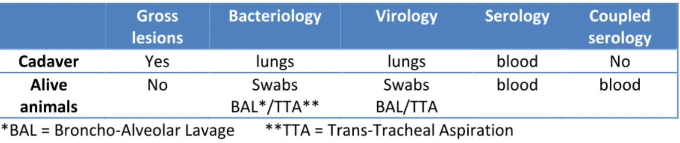
Identify the agent have 4 advantages: establish a diagnosis and prognosis, adapt treatment and provide vaccination to prevent troubles. First of all, the complete history of the farm must be undertaken, including season, age and origin of infected cattle, previous treatment or prophylaxis, previous results in the farm (serologies, necropsies, bacteriology and other assays). It is essential to realize new analyses on the farm because the epidemiology of BRD can vary according to year, season, and farm. Different ways are available to manage the identification of BRD pathogens (see Table 1).
Table 1: different tools and samples for identification of etiologic agents in BRD animals. Gross
lesions
Bacteriology Virology Serology Coupled
serology
Cadaver Yes lungs lungs blood No
Alive animals No Swabs BAL*/TTA** Swabs BAL/TTA blood blood *BAL = Broncho-Alveolar Lavage **TTA = Trans-Tracheal Aspiration
1. Cadavers
Cadaver can be sent to laboratory for necropsy, but the practitioner can conduct it on farm to evaluate gross lesions (emphysema, abscess). Some parts of the cadaver (e.g. lungs) can be sent to the laboratory for bacteriology or virology. Attention must be paid to the quality of cadaver, duration of the disease (acute mortality often associated with B-RSV, while chronic disease is more frequently Mycoplasma or BVD) and previous treatments administered by the veterinarian or the owner. Antibiotics treatments are frequently encountered in cadaver sent to laboratory. Animals examined on farm with grade IV BRD, associated with poor prognosis, can be
sacrificed prior to any treatment (sentinel animal) in order to go further in the etiologic diagnosis.
2. Alive animals a. Serology
Serology is made on serum (blood sampling on plain tube) using an ELISA method. In Belgium, B-RSV, PI-3, IBR (gE, gB), Adenovirus, BVD and
Mycoplasma bovis are available. A single serology is not useful, as this only
represents a contact with the pathogen and this is no evidence that the animal was infected by this pathogen.
Coupled serologies are more indicative of an active infection. Two blood samples (plain tubes) have to be made at 3-4 weeks interval. The first sampling must be realised during the acute phase of the disease. A multiplication of antibodies (Ab) titles per 2-3 times indicate a seroconversion. Samples can be done on treated animals (antibiotics) but not on vaccinated animals!
Limitations of serologies are, among others, the poor seroconversion rate, compared to lungs culture (from Broncho-Alveolar Lavage). Prefer this analysis at a herd level, in acute phase of BRD. Moreover, for B-RSV, serology is sometimes negative after infection (Van der Poel et al., 1997). In presence of maternal Ab (from colostrum), there is no or lesser seronconversion with usual commercial test (based on IgG fixation) because there is a more mucosal response (IgA) rather than humoral (IgG) response (Kimman et al., 1987). When using coupled serologies, the half-life of Ab must be added to the interpretation of the results, as the return to seronegativity can be long: 3-11 months according to the vaccination status of the animals (Fulton et al., 2004; Prado et al., 2006).
b. SWABS
A swab is a small piece of absorbent material attached to the end of a stick and used for collecting fluids or secretions in animals. They vary in sizes and composition (with/without culture medium, guarded or unguarded). They can be used for nasal sampling or naso-pharyngeal sampling (size differs). After disinfection of the muzzle, the swab is inserted in the nose for reaching the medial part of the eye commissure. Once in the nose, perform a rotation to coat the swab with nasal secretions. Put the swab on a culture medium (with guarded swabs, there is no culture medium), such as AMIES medium, and send immediately to the laboratory. Culture (bacteriology) and antibiogram can be performed on swabs, as well as Mycoplasma detection (including PCR) and virology (Antigen detection, viral isolation, PCR).
Nasal swabs are not predictive of lower respiratory airway pathogens, such
Mycoplasma (or Mannheimia) (Thomas et al., 2002). Comparing swab and
BAL, the negative predictive value, meaning the percentage of chance to have a negative BAL if swab culture is negative, is 67% and 33%, respectively for
Mannheimia and Mycoplasma (Godinho et al., 2007). Furthermore,
non-specific subclinical infections can be present in nasal secretions and
Pasteurella can be existent in nasal cavities of healthy animals (Allan et al.,
1985). Swab is quick, cheap, easy to perform, stressless but less accurate compared to BAL/TTA. Swab should be use as a herd diagnosis (take minimum 10 animals in an infected herd), and not in a single animal.
c. Broncho-Alveolar Lavage (BAL)
BAL needs a bit more material. A flexible tube (PTFE), with 2x4 mm diameter, and of 140 cm length is often used. A needle (14G) is inserted in one extremity (for connecting to a syringe). The set must be sterile. Sterile syringes and NaCl 0.9% complete the material. For this procedure, head of animals must be kept attached up. A disinfection of the muzzle is made and the catheter is introduced into the nose, in one movement. The larynx is pushed back and the catheter slides deeper. The animal coughs when the catheter arrives in the trachea. Thereafter, slide the catheter until blocking at the wedge position (3rd of 4th segment of bronchia). Finally, inject NaCl 0.9% (preferably at 37°C). The volume depends on the age: 3x60 ml maximum for adult cattle and 3x30 ml maximum for calves. After injection of NaCl, directly suck back the liquid that will appear foamy and cloudy (40-60% recovered). Place this liquid in a plain tube, store at 4°C but preferably send immediately to the laboratory. Bacteriology (isolation and/or PCR/ELISA), viral isolation (or PCR/ELISA) and cytology (sample on EDTA tube, examined as soon as possible) can be made on BAL samples.
BAL is more contaminated (by nasal cavities) compared to TTA. In some cases, the BAL is positive (culture) while TTA is negative. In other cases, less bacteria are found in TTA compared to BAL. BAL may not reflect exactly the bacteriology pattern of lower respiratory tract (Schreiber et al., 2000). To reduce the contamination of upper respiratory tract, a sterile AGAR can be used on the extremity of the catheter before introducing into the nasal cavity. BAL is indicated for calves (rather that for adult cattle due to the length of catheter).
d. Trans-Tracheal Aspiration (TTA)
TTA is performed using a long sterile intra-catheter (14G, length 600 mm) with needle (13G, length 70 mm) (e.g. Centracath, Vygon). The rest of material is similar to BAL (NaCl 0.9%, syringes, plain tube). For this procedure,
contention is very important. The head of the animal must be held up. A surgical preparation of the lower third of ventral neck, front of the trachea, is performed (shaving and disinfecting). A local anaesthesia is performed (2 ml Procaïne-HCl 4%) and a 5 mm incision is made on the skin, above the trachea which should be palpated. The needle is inserted, bevel down, between two rings of trachea, perpendicularly first and pushed down thereafter. The catheter is then introduced down in the trachea. NaCl is injected (same recommendation as for BAL) and recovered (~20% are recovered). Liquid obtained is placed into a sterile plain tube and sent immediately to the laboratory. Bacteriology (isolation and/or PCR/ELISA), viral isolation (or PCR/ELISA) and cytology (sample on EDTA tube, examined as soon as possible) can be made on TTA samples.
TTA should be avoided in calves due to the size of catheter and needle. This procedure is more stressful and invasive (calves have small trachea). This procedure is reserved for adult cattle, even though it is the most accurate.
e. General remarks for SWABS, BAL and TTA
Some criteria must be included when selecting an animal for these procedures. Table 2 summarizes these conditions.
Table 2: criteria for a successful etiologic diagnosis
Animal in acute phase
Avoid too severe cases (grade IV BRD) Animals NOT previously treated (antibiotics) Correct (4°C) and rapid (<24h) delivery to the lab
Regarding the storage and delivery of samples, some precautions should be observed. For swabs, make sure that the sample is dipped in a culture medium (AMIES). Indeed, guarded swabs do not include a culture medium (use the culture medium from another unguarded swab). If the sample is not directly sent to the lab, store it at 4°C (maximum 7°C). The sample must be cultured within 24 hours. To do this, it is advisable to send it to the lab the same day.
ENHANCE IMMUNITY
Immunity depends on multiple factors. Among these, nutrition, maternal immunity, vaccination, metaphylaxis, and factors promoting immunosuppression are those on which we can act.
1. Nutrition
Energy and protein supply, either for the dams, or for the calves must not be neglected. These intakes play a role in maintaining immunity. Trace elements (Selenium, Iodine, Copper and Zinc) and vitamins (A, D, E) must also be given in adequate amounts.
2. Transfer of colostral immunity (TCI)
More BRD cases occur when immunity of calves is decreasing (immunity gap). This corresponds to the fall of maternal Ab concentration that is not compensated by the
de novo synthesis of Ab by the calf. The correct transfer of colostral immunity is thus
of particular importance to keep a good level of Ab in calf’s blood.
Failure of TCI will get negative outcome on calves’ health. In dairy cattle, a decrease of average daily gain and subsequent milk production (1st lactation), as well as an increase of mortality and culling (1st lactation), is observed while failure of TCI (Robinson et al., 1988; DeNise et al., 1989). In beef cattle, failure of TCI will lead to an increased morbidity (especially respiratory troubles), calf mortality (pre-weaning), and decreased average daily gain, compared to cattle with correct TCI (Wittum and Perino, 1995). Other authors related that blood IgG concentration ≤ 800 mg/dl in young calves increase two times the risk of pneumonia. In the same way, administration of colostrum from a dam with mastitis increases three times the risk of pneumonia in calves (Virtala et al., 1999). It should be noted that increased intakes of IgG in calves from a herd with a correct status of TCI will not result in reduction of mortality and morbidity.
There are different ways to assess the success or failure of TCI. Both ways are focused on blood sampling in healthy calves at an age between 2 and 6 days of life. On the field, there is a semi-quantitative test based on the coagulation of gamma-globulins with glutaraldehyde (according to the method described by Sandholm, 1974). The test, developed by the Ambulatory Clinic of the University of Liège (Calf-IgG-Test) has a Se of about 92% and Sp of 71% (compared to the gold-standard Radial-Immuno-Diffusion, unpublished data). A coagulation time within one minute assumes an adequate transfer of immunity in the young healthy calf (blood IgG concentration >10.1 g/L). In the same way of the assessment of TCI, colostrum quality can be measured. A Californian Mastitis test can be first conducted to ensure that no subclinical or clinical mastitis is present. On the field, there is also a semi-quantitative test based on the coagulation of gamma-globulins with glutaraldehyde. Fresh colostrum is mixed with a solution of glutaraldehyde and coagulation time is noticed. A quick coagulation time indicates a good quality colostrum (> 50 g/L IgG). The test is actually under development by the Ambulatory Clinic of the University of Liège, with actually similar performances (Se/Sp) compared to Calf-IgG-Test.
3. Vaccination
Vaccination can be effective in very young cattle (from 2 weeks of life) but, due to the presence of Ab from colostrum, interferences may occur. Intra-nasal vaccination is thus preferred in these animals. Booster should not be forgotten. A peak of BRD often arises in the second half of winter (February-April), due to the higher concentration of animals in the barn and the higher infection pressure. It is recommended to administer a third injection of vaccine (booster) to calves vaccinated a few months earlier to enhance immunity at this period. There are three obvious rules for a successful result of vaccination in farms:
1. Identification of infectious agents
2. Infectious agent identified is the causative agent of the disease 3. Vaccine is registered for this infectious agent
4. Metaphylaxis
Metaphylaxis is a timely mass medication of a group of animals to eliminate or minimize an expected outbreak of disease. This procedure should be used with caution to avoid unnecessary microbial resistance. This can be recommended at batching (on weak animals) or when more than 25% of calves are sick (fever is already considered as “sick”, even though there are no added respiratory clinical signs).
5. Immunosuppression
To investigate immunosuppression, the practitioner must check for diseases that may affect the immune system. The most known is BVD, but other diseases such as calf diarrhoea can predispose the calf to pneumonia. Finally, avoid cumulative stress for calves: weaning, dehorning, vaccination, diseases, batching with different ages, mix of sick/healthy animals, and overcrowding.
CHANGE ENVIRONMENT
Last but not least, the environment should be carefully checked for risk factors. The ventilation and general hygiene of the barn must be evaluated. The management of the care for calves is also a priority. This includes, among others, the care of the umbilicus at birth, shearing animals with long hair, and the disinfection of feeding material.
CONCLUSIONS
The diagnosis of a disease begins with a good history and a correct clinical examination. As farms and epidemiology context are different, there is a need to adapt the protocol of treatment or prophylaxis (vaccination). For that purpose, there is also a need for
identification of pathogens for treatment, metaphylaxis or vaccination. Critical period (immunity gap, infectious pressure at 2nd part of winter) should be closely checked by the farmer and the veterinarian. The management of BRD requires a multifactorial approach including the host (resistance to pathogens), the environment (hygiene, ventilation, management) and the infectious agents (virus, bacteria).
BIBLIOGRAPHY
Allan E.M., Wiseman A., Gibbs H.A., Selman I.E. Pasteurella species isolated from the bovine
respiratory tract and their antimicrobial sensitivity patterns. Vet. Rec. 1985, 117, 629-631.
Buczinski S., Rademacher R.D., Tripp H.M., Edmonds M., Johnson E.G., Dufour S. Assessment
of L-lactatemia as a predictor of respiratory disease recognition and severity in feedlot steers. Prev. Vet. Med. 2015, 118, 306-318.
Coghe J., Uystepruyst C., Bureau F., Detilleux J., Art T., Lekeux P. Validation and prognostic
value of plasma lactate measurement in bovine respiratory disease. Vet. J. 2000, 160, 139-146.
DeNise S.K., Robinson J.D., Stott G.H. Effect of passive immunity on subsequent production
in dairy heifers. J. Dairy Sci. 1989, 72, 552-554.
Fulton R.W., Briggs R.E., Payton M.E., Confer A.W., Saliki J.T., Ridpath J.F., Burge L.J., Duff
G.C. Maternally derived humoral immunity to bovine viral diarrhea virus (BVDV) 1a, BVDV1b, BVDV2, bovine herpesvirus-1, parainfluenza-3 virus, bovine respiratory syncytial virus, Mannheimia haemolytica and Pasteurella multocida in beef calves, antibody decline by half-life studies and effect on response to vaccination. Vaccine. 2004, 26, 643-649.
Godinho K.S., Sarasola P., Renoult E., Tilt N., Keane S., Windsor G.D., Rowan T.G., Sunderland
S.J. Use of deep nasopharyngeal swabs as a predictive diagnostic method for natural respiratory infections in calves. Vet. Rec. 2007, 160, 22-25.
Kimman T.G., Westenbrink F., Schreuder B.E., Straver P.J. Local and systemic antibody
response to bovine respiratory syncytial virus infection and reinfection in calves with and without maternal antibodies. J. Clin. Microbiol. 1987, 25, 1097-1106.
Prado M.E., Prado T.M., Payton M., Confer A.W. Maternally and naturally acquired
antibodies to Mannheimia haemolytica and Pasteurella multocida in beef calves. Vet.
Immunol. Immunopathol. 2006, 111, 301-307.
Robinson J.D., Stott G.H., DeNise S.K. Effect of passive immunity on growth and survival in
the dairy heifer. J. Dairy Sci. 1988, 71, 1283-1287.
Sandholm M. A preliminary report of a rapid method for the demonstration of abnormal
gammaglobulin levels in bovine whole blood. Res. Vet. Sci. 1974, 17, 32-35.
Schreiber P., Thomas A., Linden A. Mainil J., Collard A. Bactériologie comparée des liquides
de lavage pulmonaire obtenus par voie transtrachéale ou par voie nasotrachéale chez le veau. Ann. Med. Vet. 2000, 144, 169-174.
Thomas A., Dizier I., Trolin A., Mainil J., Linden A. Comparison of sampling procedures for
isolating pulmonary mycoplasmas in cattle. Vet. Res. Commun. 2002, 26, 333-339.
Van der Poel W.H., Langedijk J.P., Kramps J.A., Middel W.G., Brand A., Van Oirschot J.T.
Serological indication for persistence of bovine respiratory syncytial virus in cattle and attempts to detect the virus. Arch. Virol. 1997, 142, 1681-1696.
Virtala A.M., Gröhn Y.T., Mechor G.D., Erb H.N. The effect of maternally derived
immunoglobulin G on the risk of respiratory disease in heifers during the first 3 months of life. Prev. Vet. Med. 1999, 39, 25-37.
Wittum T.E., Perino L.J., Passive immune status at postpartum hour 24 and long-term health