Synthesis of Modified Peptidoglycan Precursor Analogues for the
Inhibition of Glycosyltransferase
Shrinivas Dumbre,
†Adeline Derouaux,
‡Eveline Lescrinier,
†Andre
́ Piette,
‡Bernard Joris,
‡Mohammed Terrak,
‡and Piet Herdewijn*
,††Laboratory of Medicinal Chemistry, Rega Institute for Medical Research, University of Leuven (KU Leuven), Minderbroedersstraat
10, 3000 Leuven, Belgium
‡Centre d’Ingénierie des Protéines, Université de Liège, Allée de la chimie, B6a, B-4000, Sart Tilman, Liège, Belgium
*
S Supporting InformationABSTRACT: The peptidoglycan glycosyltransferases (GTs) are essential enzymes that catalyze the polymerization of glycan chains of the bacterial cell wall from lipid II and thus constitute a validated antibacterial target. Their enzymatic cavity is composed of a donor site for the growing glycan chain (where the inhibitor moenomycin binds) and an acceptor site for lipid II substrate. In order to find lead inhibitors able to fill this large active site, we have synthesized a series of substrate
analogues of lipid I and lipid II with variations in the lipid, the pyrophosphate, and the peptide moieties and evaluated their biological effect on the GT activity of E. coli PBP1b and their antibacterial potential. We found several compounds able to inhibit the GT activity in vitro and cause growth defect in Bacillus subtilis. The more active was C16-phosphoglycerate-MurNAc-(L -Ala-D-Glu)-GlcNAc, which also showed antibacterial activity. These molecules are promising leads for the design of new antibacterial GT inhibitors.
■
INTRODUCTIONThe problem of multidrug resistant (MDR) bacteria is on the rise.1−3The difficulty of treating infections due to MDR strains causes a longer incubation time with antibiotics, during which microbial resistance is spread to others. This inflicts huge costs on health care, and potentially increases mortality and morbidity. To face this threat, new antiobiotics are urgently needed.
Bacteria have a unique cellular feature called the peptidoglycan (PG) sacculus, a polymeric layer that is the main constituent of bacterial cell walls and is absent in animal cells.4The monomeric unit precursor of the PG consists of N-acetylmuramic acid (MurNAc) and N-acetylglucosamine (GlcNAc) linked to an undecaprenyl pyrophosphate lipid carrier, and a pentapeptide (lipid II, 1, Figure 1A). This monomeric unit is assembled in the cytoplasm and then flipped out into the periplasm,5,6where it is used as a substrate by the membrane bound glycosyltransferases (GTs) to synthesize the glycan chains (nascent PG). The growing glycan chains are cross-linked and attached to a preexisting PG sacculus via an amide bond formation between the peptide chains by transpeptidases; this reaction is the target of β-lactam antibiotics.7Inhibition of the GT step by the specific inhibitor
moenomycin leads to bacterial death. This makes the GTs a validated and attractive target for the development of new antibacterials against antibiotic multiresistant strains. Moeno-mycin (2, Figure 1B) is not used clinically because of its poor pharmacokinetic properties, which is related to its phosphogly-cerate lipid tail. To overcome this limitation, extensive
structure−activity relationship studies have been carried out on moenomycin8−13 which identified the phosphoglycerate moenocinol trisaccharide as the minimal structural feature required for antibacterial and GT inhibitory activities.
Recently the first structures of GTs and their complexes with moenomycin became available.14−18 The GT domain is composed of two lobes (head and jaw domains). The fold of the head domain shows structural similarities with that of the phage λ lysozyme (λL). The jaw domain is specific to the GT51 family and has a hydrophobic region partly embedded in the cytoplasmic membrane to presumably facilitate the access to the membrane bound lipid II substrate. The active site of GT is located at the interface of the two lobes and can be divided into two substrate binding sites: a donor site for the growing glycan chain and where moenomycin binds, and an acceptor site for lipid II (Figure 1A).
The length of the lipid moiety is critical for the inhibitory activity of moenomycin and the processive turnover of the substrate by GT.19 For the substrate, this property is determined essentially by the donor site which requires a lipid longer than 20 carbons (maximum activity observed with betulaheptaprenyl, C3520), whereas the acceptor site can accept a shorter lipid.19Therefore the use of substrate analogues with
reduced lipid chain length and targeting the acceptor site with high affinity could be a new approach in the design of active GT inhibitors. As prerequisite to this we need to better understand Received: March 2, 2012
pubs.acs.org/JACS
the contribution of the different moieties (lipid, pyrophosphate, sugars, and peptide) of the lipid II substrate for binding to the GT enzymatic cavity.
In early 2000, the semisynthesis and total synthesis of lipid I and lipid II substrates was reported.20−22However, there have been limited efforts in designing and synthesizing lipid I/II based inhibitors for GT.23−26 This is probably due to the structural complexity and chemical instability of lipid I/II, which complicates the synthetic efforts of modified lipid I and lipid II.
In this work we aim to understand the substrate specificity of E. coli PBP1b and identify substrate analogues able to inhibit the GT activity of the enzyme. These analogues could be used as templates for structure-based drug design to optimize substrates for the donor site, acceptor site, or both. We have synthesized a series of substrate analogues of lipid I and lipid II with variations in the lipid, the pyrophosphate, and the peptide moieties and evaluated their biological effect on the GT activity of E. coli PBP1b and their antibacterial potential.
■
RESULTS AND DISCUSSIONDesign and Synthesis of Lipid I and Lipid II Monophosphate Analogues. We have synthesized several lipid I and lipid II analogues, related to the structure of moenomycin, with minimal molecular fragments. Figure 2 shows the resemblance between the moenomycin EFG fragment (3, Figure 2) and lipid II (1): both have a GlcNAc residue as second sugar. The undecaprenyl pyrophosphate moiety in 1 is replaced by an isoelectronic phosphoglycerate moenocinol unit in 3. Besides the presence of a phosphogly-cerate function, the major difference between compound FG fragment (4) and lipid I is the presence of carbamoyl group on the 3-hydroxyl group (instead of lactoyl pentapeptide), a
carboxamate on position 5 (instead of hydroxymethyl group), and an hydroxyl group on position 2 (instead of the N-acetyl group).
We have prepared 18 analogues of lipid I/II as outlined in Figure 2. Analogues of lipid I (17, 18) and lipid II (19, 20, and 21) were synthesized following the synthetic strategy shown in Scheme 3. Compound 627was obtained starting from N-acetyl glucosamine (5). The benzyl glycoside of N-acetyl glucosamine was acetylated, and then the benzyl group was removed by hydrogenolysis to afford 6 (Scheme 1). Compound 6 is the starting material for the synthesis of lipid I analogues 17 and 18. Phosphorylation of 6 by reaction with dibenzyl N,N-diisopropylphosphoramidite and 1H-tetrazole, followed by Figure 1. Membrane steps of bacterial cell wall biosynthesis and
structure of the GT ligands. (A) Schematic representation of the GT domain catalyzing the glycan chain elongation. The elongating glycan chain binds to the donor site, and lipid II (1) binds to the acceptor site. Catalysis occurs by deprotonation of 4-OH of lipid II by the catalytic glutamate (E233 in PBP1b) followed by nucleophilic attack on the C1 of the growing chain and departure of the undecaprenylpyrophosphate. (B) Structure of moenomycin A (2), an analogue of the growing glycan chain which binds to the donor site.
Figure 2. Concept and design. Structures of the peptidoglycan precursor lipid II (1), disaccharide fragment (EFG, 3), and monosaccharide fragment (FG, 4) of moenomycin. Structures of the lipid I and lipid II monophosphate analogues that we have synthesized (18 analogues); these compounds have three main structural features: (a) a monophosphate lipid chain, as monophosphates are less prone to hydrolysis than pyrophosphate, (b) with or without a shorter peptide unit, and (c) a phosphoglycerate as a lipid carrier, as in moenomycin which mimics the pyrophosphate.
Scheme 1
dx.doi.org/10.1021/ja302099u | J. Am. Chem. Soc. XXXX, XXX, XXX−XXX
oxidation with hydrogen peroxide and debenzylation, afforded 728as a triethylamine salt. Compound 7, after activation with N,N′-dicyclohexylcarbodiimide or tosyl chloride or 2,4,6-triisopropylbenzenesulfonyl chloride29 and treatment with
prenol, failed to give the desired alkylated phosphate analogue: invariably a pyrophosphate dimer was formed. Therefore, an alternative approach was considered in which the nucleophil-icity of the phosphate group is enhanced by using bulky tetra-n-butylammonium as counterion.30Thus compound 7 was mixed with 2.1 equiv of tetra-n-butylammonium hydroxide and lypholized to obtain compound 8 as a bis(tetra-n-butyl)-ammonium salt. This salt underwent smooth alkylation of the
phosphate function with prenyl bromide in the presence of 4 Å molecular sieves to afford the prenylated phosphate compound, which upon deacetylation via saponification afforded 17 in 87% yield.
For the preparation of the lipid II disaccharide mono-phosphate prenyl analogue 19, the synthesis of the disaccharide monophosphate 16 is required. This synthesis was carried out by following a modified version of a published protocol,31,32as illustrated in Scheme 2. This involves the synthesis of the trichloroacetimidate-activated donor (GlcNAc) 11 and accept-or (MurNAc) 14. Schmidt’s glycosylation33reaction between 11 and 14, followed by removal of the 6-O-benzyl and N-Troc Scheme 2
Scheme 3
dx.doi.org/10.1021/ja302099u | J. Am. Chem. Soc. XXXX, XXX, XXX−XXX
The GT substrate analogues 17 and 19 bearing a prenyl chain and the compounds 18 and 20 did not show inhibition of E. coli PBP1b GT activity (data not shown). Compounds 18 and 20 have a tendency to aggregate, which may contribute to their inactivity. Compound 21 with a saturated C16 lipid chain shows a Kivalue of 26 ± 6 μM (see the section GT Inhibition and Antibacterial Activities of Lipid I and Lipid II Analogues and Table 2). This promising initial result encouraged us to increase the molecular complexity of lipid I/II analogues by incorporating suitable peptides and phosphoglycerate saturated C16 moieties.
The Boc protectedL-Ala-D-Glu (22) andL-Ala-γ-D-Glu-L-Lys (25) peptides, having base labile protecting groups in the side chains, were synthesized using standard solution phase protocols as shown in Scheme 4. The dimethyl ester
hydrochloride of D-glutamic acid (22) was obtained from D -glutamic acid with trimethylsilyl chloride in methanol34and was coupled to Boc-L-Ala-OH under standard 1-ethyl-3-(3′-dimethylaminopropyl)carbodiimide (EDCI) conditions to afford the dipeptide 23 in 78% yield over two steps. Boc-D -Glu-OBzl was coupled with N6-Fmoc protected methyl ester of L-Lys to afford the dipeptide 24 in 89% yield. Deprotection, followed by EDCI mediated coupling with Boc-L-Ala-OH, gave the protected tripeptide 25 in 83% yield.
For the synthesis of phosphoglycerates,12,23 we have developed a protocol starting from D-mannitol (Scheme 5) that serves as a chiral pool for the glycerate moiety and was used to obtain acetonide protected alcohol 26 on a multigram scale.35The (S)-acetonide protected glycerol 26 was subjected to p-methoxybenzyl (PMB) protection as it can be selectively removed under neutral conditions with 2,3-dichloro-5,6-dicyanobenzoquinone (DDQ). Acetonide deprotection and regioselective protection of the primary hydroxyl group with t-butyldiphenylsilyl (TBDPS) gave the key intermediate 27a. This key intermediate (27a) was cetylated by treatment with
sodium hydride and cetyl bromide to afford 28 in 87% yield. It is worth noting that the initial efforts to remove the PMB group at room temperature and dichloromethane as a solvent was found to be poor yielding. However, the reaction when carried out in the presence of pH 7 phosphate buffer at 5−10 °C, followed by careful removal of DDQ-2H and work up by precipitation with diethyl ether, afforded the required product in 84% yield. This PMB deprotected compound was subsequently oxidized and methylated and TBDPS depro-tection afforded the desired glycerate cetyl chain (30) in 47% yield over four steps. The glycerate geranyl compound 31 was prepared in the same way.
The alkylation strategy used to prepare 21 is poor, yielding 10% (Scheme 3), and the strategy does not allow peptide coupling regioselectively, as the free phosphate form will compete for the peptide coupling reaction. A new synthetic strategy was devised, which utilizes phosphoramidite chemistry for the preparation of monophosphate triester.36
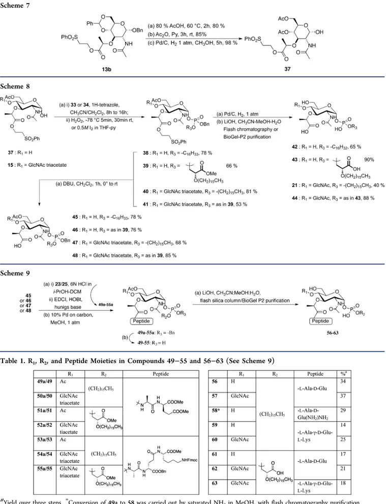
Bisdiisopropyl amine chlorophosphine was treated with benzyl alcohol to obtain benzyl diisopropylamine phosphor-amidite.37 This phosphoramidite reagent was used to prepare the phosphoramidite 33 and the phosphoglycerate 34 (Scheme 6). For the preparation of lipid I monophosphate analogues bearing a peptide moeity, MurNAc sugar 37 was obtained from compound 13b (Scheme 7). Acid mediated benzylidene acetal removal and reprotection of the two hydroxyl functionalities with acetates followed by hydrogenolysis of the anomeric Scheme 4
Scheme 6
dx.doi.org/10.1021/ja302099u | J. Am. Chem. Soc. XXXX, XXX, XXX−XXX
benzyl protecting group afforded 37 in 67% yield over three steps.
The synthesis of lipid I and lipid II monophosphate analogues without a peptide unit is depicted in Scheme 8. Scheme 7
Scheme 8
Scheme 9
Table 1. R1, R2, and Peptide Moieties in Compounds 49−55 and 56−63 (See Scheme 9)
#
Yield over three steps.*Conversion of 49a to 58 was carried out by saturated NH3in MeOH, with flash chromatography purification.
dx.doi.org/10.1021/ja302099u | J. Am. Chem. Soc. XXXX, XXX, XXX−XXX
The tricoordinated phosphorus cetyl compound 33 upon activation with 1H-tetrazole and treatment with MurNAc sugar 37 followed by oxidation with hydrogen peroxide afforded the α-phosphorylated compound 38 in 78% yield. Compound 38 on hydrogenolytic removal of the benzyl group and careful saponification with lithium hydroxide afforded the lipid I analogue 42 in 65% yield after flash column chromatographic purification. Peptidoglycan analogues 43, 21, and 44 were synthesized in the same way. β-Elimination of the ethyl phenylsulfonyl moiety in compound 38 by 1,8-diazabicyclo[5.4.0]undec-7-ene (DBU) gave compound 45 in 78% yield, affording the free lactoyl moiety for the peptide coupling reaction.
The synthesis of lipid I and lipid II peptidoglycan monophosphate analogues containing L-Ala-D-Glu and L -Ala-γ-D-Glu-L-Lys is shown in Scheme 9. Removal of the Boc group in dipeptide 23 and coupling of the resultant free amino group with the lactoyl carboxylic acid group of 45 were performed using EDCI and 1-hydroxybenzotriazole (HOBt) to afford 49a in 67% yield. Removal of benzyl and careful saponification of 49a afforded the lipid I monophosphate analogue 56 in 46% yield over two steps. In a similar manner, compounds 57−63 were obtained from their respective precursors as shown in Scheme 9 and Table 1. During hydrogenolysis of 53a, 54a, and 55a, apart from the debenzylation reaction (phosphate and glutamate), also partial removal of the Fmoc group on N6-Lys was observed. The remaining Fmoc groups were removed during the saponification reaction. Once the peptide is coupled to the sugar, the following reactions need to be carefully monitored by low-resolution mass spectroscopy,31P NMR, and thin layer chromatography, in order to obtain good yields. The purification of the final monophosphate diester compounds was found to be cumbersome. Flash column chromatography and BioGel-P2 purification yielded the final compounds in moderate yield. HPLC purification of these compounds on a reverse phase column under various conditions resulted in a complete loss of material (57, 58, 59, and 60) or the recuperation of 51 and 52 in very low yields.
All efforts to couple 35 with lipid I precursor 37 failed to give the corresponding monophosphate triester. The PMB protect-ing group for 35 was selected as it could have been easily removed using DDQ without affecting the double bond. Likewise the reaction between 37/15 and 36 failed. In both cases only starting material could be recovered. Also the reaction between the phosphoramidite of 37 and the glycerate geranyl alcohol 31 failed in our hands. Therefore, H-phosphonate chemistry was employed38 to synthesize the desired lipid I and lipid II phosphoglycerate geranyl analogues as shown in Scheme 10. Compound 37 upon treatment with 2-chloro-4H-1,3,2-benzodioxaphosphorin-4-one gave the H-phos-phonate which upon capping and activation with adamantane-carbonyl chloride39 and nucleophilic substitution with 31,
followed by mild oxidation in the presence of N-methylmor-pholine (NMM)−CCl4 and careful saponification, gave the desired analogue 64. In a similar way, the disaccharide monophosphate geranyl glycerate analogue 65 was obtained from 15.
GT Inhibition and Antibacterial Activities of Lipid I and Lipid II Analogues. Monophosphate Analogues. Monophosphate analogues of lipid I and lipid II carrying a saturated 16 carbon lipid chain without peptide stems or having a dipeptide or tripeptide (Schemes 8 and 9; 42, 21, 56, 57, 58, 59, and 60) have been evaluated for the inhibition of the GT activity and antibacterial effect. They were all able to inhibit the GT activity of PBP1b in vitro to some extent (residual activities between 7 and 84% at 500 μM compound). The disaccharide analogues (21, 57) are about 2-fold better inhibitors than their cognate monosaccharide forms (42, 56), and the inhibition of GT activity by lipid I and lipid II analogues was found to decrease in a similar manner as the length of the peptide increased (zero to three residues, 42, 56, and 59 or 21, 57, and 60; Table 2). The inhibitory activity of the mono- or disaccharide C16-phosphate without peptide was about 6- or 2.5-fold higher than that with tripeptide and dipeptide, Table 2. Peptidoglycan Glycosyltransferase Inhibition and Antibacterial Activities of the Peptidoglycan Precursor Analoguesa
compd RA (%) Ki(μM) MIC (μg/mL) cell shape
42 14 51.5 ± 10.5 >128 C 56 43 nd >128 N 58 41 nd >128 N 59 84 nd >128 N 43 3.5 21 ± 9 >128 C 61 15 46 ± 7.5 >128 N 21 7 26 ± 6 >128 C 57 16 48 ± 11 >128 C 60 40 nd >128 N 44 2.3 32.7 ± 5 >128 C 62 1.7 17.6 ± 2 128 L 63 6.5 36 ± 8 >128 N 64 102 nd >128 nd 65 89 nd >128 nd
aGT inhibition assays were performed with E. coli PBP1b. RA = GT
residual activity of PBP1b at 500 μM compounds. The data represent mean values of three independent experiments. Standard deviations were within ±10% of these mean values (RA). Minimum inhibitory concentration (MIC) values and cell shapes are for B. subtilis 168. Cell shapes: C, chaining cells; N, normal cells; L, cell Lysis. nd, not determined. Similar MIC values were obtained with S. aureus ATCC 25923. The MIC value of moenomycin disaccharide EF was >100 μg/ mL against several Gram-positive bacteria and IC500.24 μM using E. coli membrane.40
dx.doi.org/10.1021/ja302099u | J. Am. Chem. Soc. XXXX, XXX, XXX−XXX
respectively. These results show that GlcNAc plays a significant role in binding to the enzyme and that in contrast the di- or tripeptide has a negative effect. Note that the monosaccharide dipeptide with amidated glutamate (58) behaves as the free carboxyl form (56).
In terms of antibacterial activity, all the compounds (mono-and disaccharide C16-phosphate with or without peptide) showed MIC values higher than 128 μg/mL against Staph-ylococcus aureus ATCC 25923 and Bacillus subtilis 168 (Table 2). This activity is comparable to the MIC value (>100 μg/mL) of moenomycin disaccharide EFG derivative (close analogue of compound 21, Figure 2 and Scheme 8) for Gram-positive bacteria tested40despite the presence of a C16-phosphate lipid tail in compound 21 instead of C25-phosphoglycerate in moenomycin which is known to a play important role in its activity. Surprisingly, observation of the B. subtilis cells in the microscope after 16−18 h of incubation with compounds shows that the most active compounds (42, 21, and 57) induced cell chaining (Figure 3). Dead cells and curved filament have been also observed in some cases. The monosaccharide dipeptide (56) and monosaccharide tripeptide (59) (the less active) do not have any effect on cell morphology. In contrast, the cells treated with the disaccharide dipeptide (57) grow as long filaments of unseparated cells, while the disaccharide tripeptide (60) had no effect.
Membrane labeling of the cells with the fluorescent dye FM-1-43-FX shows that most of the cells are in an advanced stage of cytokinesis but remained attached to each other by their poles. Bright fluorescent spots and probably uncompleted septa have been also observed (Figure 3). These observations show that cell division is perturbed and that cell separation is blocked by these compounds. The compounds have been also tested
against E. coli and have no effect, probably because of the outer membrane barrier (data not shown).
Phosphoglycerate Analogues. The structures of E. coli PBP1b and other GTs in complexes with moenomycin show that the EF ring phosphoglycerate portion makes the major interactions with the active site residues of the GT domain.17It
is believed that the phosphoglycerate moiety of moenomycin could mimic the pyrophosphate group of the substrate. In order to improve the inhibitory activity of our compounds, we have prepared a new series of compounds with C16-phosphogly-cerate (Schemes 9 and 10) as a C-1 substituent.
The biological evaluation shows that all phosphoglycerate compounds (43, 44, 61, 62, and 63) had increased inhibitory activity of the GT (3−10-fold approximately) compared to their cognate monophosphate analogues (Table 2). Interest-ingly, the disaccharide dipeptide C16-phosphoglycerate (62) was about 2-fold more active than the peptide-free disaccharide C16-phosphoglycertae (44, Ki= 17.6 vs 32.7 μM) in contrast to the monophosphate forms where the peptide-free compound was more active. On the other hand, the antibacterial activity of all the compounds has virtually not changed (MIC > 128 μg/ mL) except that of the disaccharide dipeptide (62), which has a MIC value of 128 μg/mL in accordance with the in vitro activity of this compound. This result was further confirmed by looking at the cells under the microscope; the disaccharide dipeptide did not induce cell chaining but instead provoked cell lysis at 1−2 times the MIC (data not shown). The disaccharide tripeptide (63) had a minor or no effect on cell morphology, while the disaccharide phosphoglycerate without peptide (44) induced cell chaining (Figure 3). For the monosaccharide C16-phosphoglycerate compounds only the one without peptide Figure 3. Cellular effect of the compounds. The cells (OD600 nm= 0.1−0.2) were incubated with 256 μg/mL compounds and the samples were taken at 1 h interval over 3 h and stained with the membrane dye FM-1-43-FX. The images show the effect of the compounds indicated by their code number after 3 h on B. subtilis cells observed by using both phase contrast and fluorescence microscopy.
dx.doi.org/10.1021/ja302099u | J. Am. Chem. Soc. XXXX, XXX, XXX−XXX
with their cognate monophosphate C16 analogues (42, 21; Table 2). Interestingly, the disaccharide phosphoglycerate geranyl exhibits weak activity (11% of inhibition at 500 μM) while the geranyl monophosphate was completely inactive. This suggests that the phosphoglycerate improves binding by mimicking the pyrophosphate but that the length/substitution of the lipid is a more determinant factor.
Inactivation of all GTs (four class A PBPs) in B. subtilis was not lethal, and since the mutant was still able to produce peptidoglycan and does not have a monofunctional GT, it has been proposed that B. subtilis contains an unknown GT different in sequence from the GT51 family.41PG synthesis by the quadruple mutant was demonstrated in vitro and was shown to be sensitive to moenomycin, confirming the GT reaction as an antibacterial target.
Interestingly, the quadruple mutant cells have been shown to be filamentous and bent with irregularly spaced and uncompleted septa visualized by membrane staining and fluorescence microscopy. Similarly, the exposure of the wild-type strain to moenomycin resulted in a phenowild-type similar to that of the quadruple mutant. The similarities between our results and those of McPherson and Popham41 support the conclusion that the cellular defects we observed are the result of the GT inhibition. In the meantime we cannot exclude that, in addition to the GTs, these compounds also inhibit other enzymes of the peptidoglycan metabolism.
■
CONCLUDING REMARKSA systematic investigation of a GT enzyme inhibition by peptidoglycan precursor analogues with variations in the carbohydrate, lipid, pyrophosphate, and peptide moieties showed that saturated C16 phosphoglycerate linked to MurNAc-GlcNAc (44) and to the disaccharide dipeptide (L -Ala-D-Glu) (62) are good inhibitors of the GT activity in vitro and are able to induce growth defects or lysis of bacterial cells. The antibacterial activities observed with the precursor analogues in this work correlate with those of the inactivation of the GT or their inhibition by moenomycin in B. subtilis41and suggest that these compounds inhibit the GT activity in vivo. The disaccharide compounds (44 and 62) are lipid II substrate mimics and are expected to bind to both donor and acceptor sites of GT. They would have binding similar to that of the enzyme as natural substrates with the peptide (in compound 62) pointing away from the active site.15During the initiation of a glycan chain elongation by the GT, lipid II binds to both the donor and acceptor sites; subsequently, during the elongation phase and due to the processivity of the enzyme, chain elongation occurs in the donor site by successive additions of a disaccharide unit from the acceptor site bound lipid II. Therefore, the analogue compounds have to compete with lipid II for binding to the donor site and acceptor site at the initiation phase, and then during the elongation phase the compounds have to compete also with the elongating chain. This shows the complexity of the system and will require more
reduction of the lipid chain length, thus circumventing the negative properties of the long lipid chain observed for moenomycin.
■
MATERIALS AND METHODSReagents and Protein. Radiolabeled [14C-D-Ala]-lipid II (0.078 μCi nmol−1), [N-acetylglucosaminyl-N-acetylmuramoyl (L-Ala-γ-D -Glu-(L)-meso-A2pm-(L)-D-Ala-D-Ala)-pyrophosphate-undecaprenol] meso-diaminopimelic acid, meso-A2pm, was prepared essentially as previously described in ref 42. All compounds were solubilized in dimethyl sulfoxide (DMSO; 5−10 mM). Escherichia coli PBP1b was produced and purified as described in ref 43.
In Vitro Glycosyltransferase Activity and Inhibition Assays. In vitro peptidoglycan polymerization assay was performed by incubation of 2.5 μM D-[14C]Ala-D-[14C]Ala-labeled lipid II (0.078 μCi nmol−1) and purified PBP1b enzyme (100 nM) at 30 °C in 50 mM Tris·HCl, pH 7.5, 200 mM NaCl, 0.2% decylPEG, 10 mM CaCl2, and 20% DMSO. The reactions were stopped after 7 min with moenomycin (10 μM), and the products (unused lipid II substrate and polymerized peptidoglycan) were separated by TLC on silica gel plates (Fluka) using 2-propanol/ammonium hydroxide/water (6:3:1; v/v/v) as a mobile phase.44Lipid II moves with the solvent (Rf= 0.65), and all the polymerized material remains at the origin in one spot (Rf= 0). The TLC plates were exposed to a storage phosphor screen (GE Healthcare) for 16 h. The images were revealed using a Typhoon Trio + imager and analyzed using the ImageQuant TL software (GE Healthcare).
The initial GT inhibition assay was carried out by measuring the residual activity (RA) of E. coli PBP1b in the presence of 500 μM compounds. All assays were repeated at least three times. The IC50 values were determined when the RAs were <20% at 500 μM compounds. Using variable concentrations of inhibitor (0−500 μM), the concentration that caused 50% of reduction in the RA of an enzyme was considered as IC50. Kivalues were determined using the equation IC50= Ki(1+ [s]/Km) and a Kmvalue of 1.8 μM.42
MIC Determination and Microscopy. Minimum inhibitory concentration (MIC) determinations were performed by the broth microdilution method against S. aureus ATCC 25923 and B. subtilis 168 according to the EUCAST (European Committee on Anti-microbial Susceptibility Testing)/CLSI (Clinical and Laboratory Standard Institute) recommended procedures. The compounds were solubilized in 100% DMSO at a concentration of 5−10 mg/mL, and diluted in Mueller−Hinton broth (MHB), just before utilization.
Cells from the samples used for MIC determination were fixed on glass slabs coated with 1% agarose and examined in the micro-scope.45,46 This experiment was repeated by incubation of new cultured cells (DO600 nm= 0.1−0.2) with 256 μg/mL compounds, and the samples were taken at 1 h interval over 3 h and incubated with the fluorescent dye FM-1-43-FX (Invitrogen) to label the membranes. The cells were then fixed with formaldehyde and glutaraldehyde, washed with PBS, and observed under a fluorescent microscope (Zeiss Axio Imager.Z1). Photographs were taken with a cooled AxioCam MRm (Zeiss) mounted on the fluorescent microscope through an EC Plan-Neofluar 100 × 1.3 oil immersion objective in bright field and fluorescence using a filter set 37 (425−475-nm band-pass excitation and 485−535-nm band-pass emission; Zeiss). Images were analyzed using the AxioVision Rel. 4.5 (Zeiss) software as previously described.46
dx.doi.org/10.1021/ja302099u | J. Am. Chem. Soc. XXXX, XXX, XXX−XXX
■
ASSOCIATED CONTENT*
S Supporting InformationExperimental procedures and characterization data for all new compounds used in this study. This material is available free of charge via the Internet at http://pubs.acs.org.
■
AUTHOR INFORMATIONCorresponding Author
Piet.Herdewijn@rega.kuleuven.be
Notes
The authors declare no competing financial interest.
■
ACKNOWLEDGMENTSThis work was supported by the IAP 6/19 project, and the Fonds de la Recherche Fondamentale Collective (FRFC Nos. 2.4506.08 and 2.4543.12). M.T. is a Research Associate of the National Fund for Scientific Research (F.R.S_FNRS, Belgium). We thank Martine Nguyen-Distèche for helpful discussion and Marie Schloesser for technical assistance. We thank Prof. Jef Rozenski for mass spectrometric analysis and Luc Kerremans for technical assistance. Mass spectrometry was made possible by the support of the Hercules Foundation of the Flemish Government (Grant 20100225-7).
■
REFERENCES(1) Levy, S. B.; Marshall, B. Nat. Med. 2004, 10, S122.
(2) WHO, World Health Organization. Antimicrobial Resistance [Online]. 2011.http://www.who.int/mediacentre/factsheets/fs194/ en/index.html, No. 194.
(3) Zhang, Q.; Lambert, G.; Liao, D.; Kim, H.; Robin, K.; Tung, C. K.; Pourmand, N.; Austin, R. H. Science 2011, 333, 1764.
(4) Vollmer, W.; Blanot, D.; de Pedro, M. A. FEMS Microbiol. Rev. 2008, 32, 149.
(5) Mohammadi, T.; van Dam, V.; Sijbrandi, R.; Vernet, T.; Zapun, A.; Bouhss, A.; Diepeveen-de Bruin, M.; Nguyen-Disteche, M.; de Kruijff, B.; Breukink, E. EMBO J. 2011, 30, 1425.
(6) Bouhss, A.; Trunkfield, A. E.; Bugg, T. D.; Mengin-Lecreulx, D. FEMS Microbiol. Rev. 2008, 32, 208.
(7) Sauvage, E.; Kerff, F.; Terrak, M.; Ayala, J. A.; Charlier, P. FEMS Microbiol. Rev. 2008, 32, 234.
(8) Fuse, S.; Tsukamoto, H.; Yuan, Y.; Wang, T. S.; Zhang, Y.; Bolla, M.; Walker, S.; Sliz, P.; Kahne, D. ACS Chem. Biol. 2010, 5, 701.
(9) Ostash, B.; Walker, S. Nat. Prod. Rep. 2010, 27, 1594. (10) Welzel, P. Chem. Rev. 2005, 105, 4610.
(11) Ritzeler, O.; Hennig, L.; Findeisen, M.; Welzel, P.; Müller, D. Tetrahedron 1997, 53, 1665.
(12) Kahne, D. E.; Adachi, M.; Zhang, Y.; Leimkuhler, C.; Sun, B. Y.; LaTour, J. V. J. Am. Chem. Soc. 2006, 128, 14012.
(13) Sofia, M. J.; Allanson, N.; Hatzenbuhler, N. T.; Jain, R.; Kakarla, R.; Kogan, N.; Liang, R.; Liu, D. S.; Silva, D. J.; Wang, H. M.; Gange, D.; Anderson, J.; Chen, A.; Chi, F.; Dulina, R.; Huang, B. W.; Kamau, M.; Wang, C. W.; Baizman, E.; Branstrom, A.; Bristol, N.; Goldman, R.; Han, K. H.; Longley, C.; Midha, S.; Axelrod, H. R. J. Med. Chem. 1999, 42, 3193.
(14) Lovering, A. L.; de Castro, L. H.; Lim, D.; Strynadka, N. C. Science 2007, 315, 1402.
(15) Lovering, A. L.; Gretes, M.; Strynadka, N. C. Curr. Opin. Struct. Biol. 2008, 18, 534.
(16) Heaslet, H.; Shaw, B.; Mistry, A.; Miller, A. A. J. Struct. Biol. 2009, 167, 129.
(17) Wong, C. H.; Sung, M. T.; Lai, Y. T.; Huang, C. Y.; Chou, L. Y.; Shih, H. W.; Cheng, W. C.; Ma, C. Proc. Natl. Acad. Sci. U.S.A. 2009, 106, 8824.
(18) Yuan, Y.; Barrett, D.; Zhang, Y.; Kahne, D.; Sliz, P.; Walker, S. Proc. Natl. Acad. Sci. U.S.A. 2007, 104, 5348.
(19) Perlstein, D. L.; Andrew Wang, T.-S.; Doud, E. H.; Kahne, D.; Walker, S. J. Am. Chem. Soc. 2010, 132, 48.
(20) Ye, X. Y.; Lo, M. C.; Brunner, L.; Walker, D.; Kahne, D.; Walker, S. J. Am. Chem. Soc. 2001, 123, 3155.
(21) VanNieuwenhze, M. S.; Mauldin, S. C.; Zia-Ebrahimi, M.; Aikins, J. A.; Blaszczak, L. C. J. Am. Chem. Soc. 2001, 123, 6983.
(22) VanNieuwenhze, M. S.; Mauldin, S. C.; Zia-Ebrahimi, M.; Winger, B. E.; Hornback, W. J.; Saha, S. L.; Aikins, J. A.; Blaszczak, L. C. J. Am. Chem. Soc. 2002, 124, 3656.
(23) Hecker, S. J.; Minich, M. L.; Lackey, K. J. Org. Chem. 1990, 55, 4904.
(24) Qiao, L.; Vederas, J. C. J. Org. Chem. 1993, 58, 3480. (25) Cudic, P.; Behenna, D. C.; Yu, M. K.; Kruger, R. G.; Szewczuk, L. M.; McCafferty, D. G. Bioorg. Med. Chem. Lett. 2001, 11, 3107.
(26) Garneau, S.; Qiao, L.; Chen, L.; Walker, S.; Vederas, J. C. Bioorg. Med. Chem. 2004, 12, 6473.
(27) Mohan, H.; Vasella, A. Helv. Chim. Acta 2000, 83, 114. (28) Heidlas, J. E.; Lees, W. J.; Pale, P.; Whitesides, G. M. J. Org. Chem. 1992, 57, 146.
(29) Itakura, K.; Katagiri, N.; Bahl, C. P.; Wightman, R. H.; Narang, S. A. J. Am. Chem. Soc. 1975, 97, 7327.
(30) Davisson, V. J.; Woodside, A. B.; Neal, T. R.; Stremler, K. E.; Muehlbacher, M.; Poulter, C. D. J. Org. Chem. 1986, 51, 4768.
(31) Lioux, T.; Busson, R.; Rozenski, J.; Nguyen-Disteche, M.; Frere, J. M.; Herdewijn, P. Collect. Czech. Chem. Commun. 2005, 70, 1615.
(32) Fraipont, C.; Sapunaric, F.; Zervosen, A.; Auger, G.; Devreese, B.; Lioux, T.; Blanot, D.; Mengin-Lecreulx, D.; Herdewijn, P.; Van Beeumen, J.; Frere, J. M.; Nguyen-Disteche, M. Biochemistry 2006, 45, 4007.
(33) Schmidt, R. R.; Michel, J. Angew. Chem., Int. Ed. Engl. 1980, 19, 731.
(34) Li, J. B.; Sha, Y. W. Molecules 2008, 13, 1111.
(35) Thomas, B. N.; Lindemann, C. M.; Corcoran, R. C.; Cotant, C. L.; Kirsch, J. E.; Persichini, P. J. J. Am. Chem. Soc. 2002, 124, 1227.
(36) Michalski, J.; Dabkowski, W. Top. Curr. Chem. 2004, 232, 93. (37) Dreef-Tromp, C. M.; Lefeber, A. W. M.; van der Marel, G. A.; van Boom, J. H. Synthesis 1992, 1992, 1269.
(38) Stawinski, J.; Ströberg, R. Oligonucleotide Synthesis. Methods and Applications; Herdewijn, P., Ed.; Methods in Molecular Biology 288; Humana Press: Totowa, NJ, USA, 2005; p 81.
(39) Andrus, A.; Efcavitch, J. W.; McBride, L. J.; Giusti, B. Tetrahedron Lett. 1988, 29, 861.
(40) Goldman, R. C.; Baizman, E. R.; Branstrom, A. A.; Longley, C. B. Bioorg. Med. Chem. Lett. 2000, 10, 2251.
(41) McPherson, D. C.; Popham, D. L. J. Bacteriol. 2003, 185, 1423. (42) Terrak, M.; Ghosh, T. K.; Van Heijenoort, J.; Van Beeumen, J.; Lampilas, M.; Aszodi, J.; Ayala, J. A.; Ghuysen, J.-M.; Nguyen-Distèche, M. Mol. Microbiol. 1999, 34, 350.
(43) van Heijenoort, Y.; Gomez, M.; Derrien, M.; Ayala, J.; van Heijenoort, J. J. Bacteriol. 1992, 174, 3549.
(44) Derouaux, A.; Turk, S.; Olrichs, N. K.; Gobec, S.; Breukink, E.; Amoroso, A.; Offant, J.; Bostock, J.; Mariner, K.; Chopra, I.; Vernet, T.; Zervosen, A.; Joris, B.; Frere, J. M.; Nguyen-Disteche, M.; Terrak, M. Biochem. Pharmacol. 2011, 81, 1098.
(45) Koppelman, C.-M.; Aarsman, M. E. G.; Postmus, J.; Pas, E.; Muijsers, A. O.; Scheffers, D.-J.; Nanninga, N.; Den Blaauwen, T. Mol. Microbiol. 2004, 51, 645.
(46) Pastoret, S.; Fraipont, C.; den Blaauwen, T.; Wolf, B.; Aarsman, M. E.; Piette, A.; Thomas, A.; Brasseur, R.; Nguyen-Disteche, M. J. Bacteriol. 2004, 186, 8370.
dx.doi.org/10.1021/ja302099u | J. Am. Chem. Soc. XXXX, XXX, XXX−XXX