HAL Id: hal-02142955
https://hal.archives-ouvertes.fr/hal-02142955
Submitted on 29 May 2019
HAL is a multi-disciplinary open access
archive for the deposit and dissemination of sci-entific research documents, whether they are pub-lished or not. The documents may come from teaching and research institutions in France or abroad, or from public or private research centers.
L’archive ouverte pluridisciplinaire HAL, est destinée au dépôt et à la diffusion de documents scientifiques de niveau recherche, publiés ou non, émanant des établissements d’enseignement et de recherche français ou étrangers, des laboratoires publics ou privés.
Decision support systems as digital brain atlases
Bernard Gibaud, Serge Garlatti, Christian Barillot, Emmanuel Faure
To cite this version:
Bernard Gibaud, Serge Garlatti, Christian Barillot, Emmanuel Faure. Decision support systems as digital brain atlases. AI-ED’99 : Workshop on Medical Image Tutoring, July 19-23, Le Mans, France, Jul 1999, Le Mans, France. pp.10 - 22. �hal-02142955�
Decision Support Systems as digital brain
atlases
B. Gibaud1, S. Garlatti2, C. Barillot3, E. Faure2
1 Laboratoire SIM, Faculté de Médecine, Rennes , France 2 Laboratoire IASC, ENST Bretagne, Brest, France
3 IRISA/CNRS, Projet VISTA, Rennes, France
Abstract. This paper deals with the development of computerized brain atlases
addressing both research and clinical needs. The authors analyze in detail the potentialities of these systems and discuss the capabilities and limitations of the digital atlases currently being developed around the world. The authors propose to reconsider the concept of a brain atlas, regarding both its content, and the way it has to be used and managed in order to set up a more effective cooperation between the user and the system. Particular emphasis is put on the evolutivity and reuse issues, which are critical in this rapidly evolving field. These orientations result from both the authors’ experience and the analysis of current trends in the field of neuroimaging. The general methodology is illustrated with examples related to computer aided surgical planning.
1. Introduction
The role of medical imaging in the neurological and neurosurgical practice, as well as in neuroscience in general, is becoming increasingly important. Indeed, the interpretation of the images plays a prominent role in diagnostic and therapeutic decisions, and quantitative image analysis is a major research issue in neuroimaging. Brain atlases [e.g. 1, 2, 3, 4] aim at assisting the interpretation of brain images by providing a priori knowledge about brain anatomy by means of anatomical plates obtained from post-mortem brains or in vivo images. These plates provide the anatomical substrate, from which more details about brain features (e.g. morphology, relationships with surrounding anatomical structures, function, variability) are accessible in textual or graphical form.
From an historical point of view, atlases were developed to overcome the limitations of the in vivo imaging techniques. However, in spite of the availability of modern and powerful imaging techniques such as Magnetic Resonance Imaging (MRI), atlases are still necessary for three major reasons: (1) The spatial resolution and the contrast of the images are limited: for example the different nuclei composing the thalamus cannot be distinguished on MRI images; (2) Through the anatomical substrate, it is often the function of the different brain areas that is relevant to the surgeon in order to select the best surgical approach. In most cases this information is not available (functional imaging modalities are not so widespread) and this information has to be retrieved from an atlas; (3) Brain atlases are still commonly used in the neuroimaging community, especially by people working on the mapping of human brain functions [5, 6]. A first explanation is related to the low spatial resolution of functional imaging techniques like Positron Emission Tomography (PET): indeed, the registration with patient anatomy (through MRI) cannot be very precise, so it may be more
simple to relate functional information to anatomy by means of a reference atlas (e.g. Talairach) rather than the patient MRI data itself. A second explanation arises from the fact that PET experiments cannot be repeated many times on the same individual due to the use of radiopharmaceuticals. Therefore PET studies must be conducted onto several individuals in order to provide statistically significant results: registration to a common atlas is a simple way to merge results from different patients in a standardized way.
The digital nature of in vivo imaging techniques brings new capabilities to represent anatomical information in an atlas and to access to related knowledge. We have experienced some of them during the last decade. Our interest in the mid-80s was primarily focused on 3D representation of brain anatomy [7, 8], then on the registration of images between different patients [9, 10]. More recently we have studied how knowledge about the major anatomical features could be represented in a more explicit way (e.g. atlas plates, symbolic objects represented by frames, illustrations) [11, 12], and how access to this information could be managed in a way taking into account contextual information (domain of interest and task of the user) [13]. Finally some aspects of the evolutivity of such systems have been studied, especially regarding schema evolution capabilities in Object Oriented databases [14].
These works, as well as the analysis of general trends concerning the management of information in the neuroimaging community have led us to reconsider the concept of a brain atlas, regarding both its content, and the way it has to be used and managed. This is the major subject of this paper. Section 2 analyzes in more details new capabilities induced by the digital nature of the images, and discusses the major trends arising from the recent literature on brain atlases. Section 3 presents our approach to this problem, with particular emphasis on the evolutivity and reuse issues. These two constraints strongly influence both the content of the atlas and the way it is used and managed. Finally, section 4 summerizes this general approach, and underlines some difficulties in putting this methodology into practice.
2. Current Status of Digital Atlases
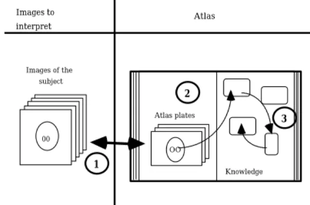
Before analyzing the potentialities and added value of digital brain atlases, and reviewing current works in this field, it is useful to briefly recall the basic function of an atlas. The utilization of an atlas involves 3 steps (Fig. 1): (1) spatial registration between the image data to interpret and the anatomy of one or several individuals displayed on atlas plates; (2)
identification of anatomical structures of the subject from the outlines and labels of the same
structures within the atlas plates; (3) access to the knowledge associated to the anatomical structures. Images to interpret Atlas Atlas plates OO Knowledge 3 1 2 Images of the subject 00
Atlases are usually printed and thereby have severe limitations. Digital atlases offer a wide range of capabilities, which overcome these limitations.
• The first concerns image display. Brain anatomy is 3D, so the understanding of 3D data sets makes it necessary to use both surface representation and volume representation (cut planes in the three major directions of space). Printed atlases obviously cannot offer this flexibility (limited to 2D display, no possibility to redefine viewing angles).
• The second concerns inter-individual variability. The matching between two brains assumes that some warping model can be applied. Printed atlases impose warping models to be very simple (for example Talairach [1]). Computer-based atlases allow much more complex models to be used (e.g. non-linear rather than linear), leading to simpler use and better accuracy.
• The third is related to extensibility. As opposed to printed atlases, a digital atlas can be extensible: i) the number of atlas plates can be increased (which helps to overcome the inter-individual variability problem); ii) new warping models can be added; iii) the corpus of accessible knowledge can be extended.
• The fourth concerns the flexibility of navigation within the associated knowledge corpus; this allows for taking into account the goals and tasks of the user in order to guide the navigation towards the more pertinent information.
• The last concerns the possible utilization of this knowledge by artificial cognitive
agents for decision-making. This is obviously not feasible with classical atlases and
permits to free the user from computing tasks that computers do more efficiently than humans.
The development of computer-based atlases is motivated by this potential added value; three major categories of systems have been or are being developed.
(1) The first category is basically a transposition of paper-atlases, and may be called
computerised maps. Some of these systems put emphasis on gathering encyclopaedic
knowledge from neuroscience sub-areas. For example Toga has developed a 3D anatomical and functional atlas of the rat brain [15]. Other systems propose the integration of data from various atlases, such as the system developed by Nowinski: this system provides atlas plates from the Talairach et Tournoux, Schaltenbrand/Wahren and Ono atlases [16]. Finally, some systems are organized around 3D display tools: for example the system called “Digital Anatomist”, proposed by Brinkley, provides 3D display of the anatomical features by means of both still and moving images, and offers designation facilities allowing their naming [17]. The “Voxel-Man” system developed by Höhne is based on the same concept with more enhanced facilities for editing the 3D model and retrieving related symbolic information [18]. These systems are more and more edited and distributed on CD-ROM; demonstrations are sometimes possible through Internet.
(2) The second category focuses on modeling the morphological inter-subject variability and on the design of warping models. The general principle consists of labelling precisely 3D data sets of one (or several) brains, and to estimate a displacement field between this reference brain and the particular brain to be matched, in order to assign the reference brain labels to the voxels of the unknown brain. The major issue is to make sure that this transformation guarantees the conservation of topological relationships between the labelled regions. Many methods have been developed, for example: [19, 20, 21, 22, 23, 24, 25].
(3) Finally, many atlas systems have been developed to support the automatic interpretation of anatomical images. They generally include symbolic knowledge about anatomy or imaging techniques in order to assist the delineation and labelling of these
features within morphological or functional images, for example: [26, 27, 28, 29, 30, 31, 32].
It is also important to mention collaborative efforts carried out in the context of the american Human Brain Project. Mazziota proposed the constitution of a multi-centric database, in order to build a probabilistic brain atlas [33, 34]. The “BrainMap” system developed by Fox at the University of Texas is accessible through the Internet and gathers many experimental results concerning cognitive studies from many laboratories (activation protocols, localization in the Talairach space, references of publications).
All these works have brought out significant contributions to the various aspects of brain atlas conception and use (more accurate models of the brain variability, 3D display and editing tools, computer-aided image interpretation techniques). Besides, projects like the Human Brain Project highlighted the interest of collaboration across disciplinary and geographic boundaries for brain research. Nevertheless, although claimed as important [34], evolutivity and reuse issues have not really been addressed yet. Finally, potentialities concerning the utilization of knowledge available from an atlas for automatic or assisted problem resolution (for example surgery planning) have not been really investigated yet. Projects like SAMMIE have tried to go into this direction, in particular for assisting the interpretation of Electroencephalography (EEG) or Magnetoencephalography (MEG), but achievements are still in their infancy [31].
This situation requires a more thorough analysis of the way knowledge is used by humans for the resolution of medical problems concerning the brain. Experiments carried out by Boshuizen and Schmidt [35] have shown that physicians make use of several kinds of knowledge, including both general knowledge (called biomedical knowledge) and situated knowledge derived from previous experiences. They also demonstrated that novices use more biomedical knowledge than experts do. However, in difficult cases where usual reasoning schemes may fail, experts use general knowledge as well. These works are very interesting with respect to our atlas problem. First, they reinforce the need of associating in an atlas several kinds of knowledge, namely general knowledge, and situated knowledge. Second, they suggest that the system should be designed in such a way that: (1) it provides the user with the most relevant knowledge, depending on the user's task and expertise level, and (2) it supports the emergence of new biomedical knowledge from the experience of past cases. This is the general orientation described hereafter.
3. Design and Use of Atlases
The general approach described here emphasizes the evolutivity and reuse issues. This concern tends to be more and more critical in the design of Information Systems in general, and are particularly relevant in the context of the management and use of brain related knowledge. Indeed, neuroscience is a very active research field [34], and many disciplines try to elucidate brain structure and functioning from different perspectives (e.g. anatomy, physiology, psychology, behaviour), which leads to an extreme fragmentation of knowledge. However it is generally admitted that multi-disciplinary research is very effective and fruitful, which assumes that knowledge and results must be shared in an appropriate way. Brain research is also very productive which means that this knowledge must be able to be
easily updated, which obliges to consider evolutivity as a major concern in the design of
brain knowledge management systems. The same arguments apply regarding clinical applications, since clinical processes and medical decisions oblige to integrate many components (patient specific anatomy, physiology, age, medical history, social condition) each of which referring to specific knowledge, and involving references to previous cases.
This approach articulates around three axes: (1) the first deals with the content of atlases (knowledge sources), (2) the second with the general organization of information processing (Decision-making and knowledge acquisition processes) and (3) the third with the way multi-disciplinary knowledge can be managed.
3.1 Knowledge Sources
Before describing in detail the different kinds of information a digital atlas should manage, it is necessary to introduce the terminology we are going to use in the following. The term
data refers to any signifying entity used by an information processing agent. The term information refers to the meaning attached to a particular datum, which thereby depends on
the cognitive background of the agent which interprets this datum [36]. The term knowledge is (as usual) more difficult to define: we will consider as knowledge any information allowing data to be interpreted and to which one wishes to attach a particular worth; this worth may arise from several origins: (1) abstraction level and capacity to synthesize a set of information; (2) level of consensus a piece of information is gathering; (3) applicability of a piece of information in a given context (solving power) and potential of reuse in other contexts.
These notions are very important with respect to evolutivity and reuse: the level of abstraction, applicability and degree of confidence play a major role in reuse. Moreover broadly accepted knowledge is likely to be more stable in time than uncertain one.
An atlas involves two fundamentally different kinds of information:
(1) biomedical knowledge concerning brain, characterizing the properties which are shared by most individuals, or within meaningful categories of individuals (such as right-handed people).
(2) Situated knowledge (cases): this information characterizes particular individuals, about which data have been recorded (e.g. images, physiological signals) and interpreted, in order to make their specific characters explicit.
One may feel appropriate to speak about these two kinds of information in terms of knowledge base and database. We prefer avoiding to do so in order to emphasize the conceptual difference between the two, rather than focusing on implementation issues.
3.1.1 Biomedical Knowledge
This general biomedical knowledge includes four major kinds of knowledge:
- Conceptual knowledge about one or more brain disciplines: it is usually organized in ontologies describing abstract brain entities (e.g. anatomical structures, functional systems, neurochemicals) or relationships between these entities (e.g. spatial relationships, neural connections, part-whole relationships). These entities are generally represented by means of object oriented models or frames.
- Illustrations (such as drawings or schemata) aim at making more explicit the meaning to be associated to the previous brain abstract entities. By definition they are indented to humans rather than artificial agents.
- Numerical data, functions of space or time: for example 3D probability maps represent the probability that a point belongs to a particular anatomical structure.
- Decision models include inference mechanisms or algorithms capable of deriving new information from existing one: for example a warping model which allows a brain to be
mapped onto another, or an image segmentation algorithm, consisting of successive image processing operations, aiming at delineating and labelling a particular structure.
3.1.2 Situated Knowledge
This information depicts various aspects of the brain of particular subjects: anatomy, physiology, behaviour of the subject, characteristics of the individual himself (for example sex, manual dominance, age, pathology, if any). It may have been obtained in vivo, by means of imaging techniques (e.g. CT scanner, MRI, functional MRI, PET) or neurophysiological techniques (e.g. EEG, Depth electrodes recordings, MEG), or from cadavers (photographs of brain cryosections, histological or histochemical data).
This information about individual brains can take several forms:
- numerical data, functions of space or time: e.g. images or physiological signals.
- instances of brain abstract entities, whose attributes describe the specific characters of each brain (e.g. length and depth of a cortical sulcus, volume of grey matter within a gyrus), or relationships between these entities for example spatial relationships between anatomical structures. These objects describe in an explicit way some properties which are shown by images or signals, as a result of a manual or automatic interpretation process.
- instances of application of a decision model, for example describing the successive steps of an image processing procedure resulting in the labelling of a particular anatomical structure (interpretation process).
This information can be considered as knowledge, because according to previous definitions, one assigns a worth to it, and therefore wishes to keep it, in order to refer to it and reuse it. This value arises from several factors:
- a particular brain feature may be typical or atypical;
- some information may be difficult to obtain for technical or medical reasons: for example depth electrodes recordings can be obtained in very few patients (e.g. patients suffering some form of epilepsy requiring surgery). This kind of information is very precious although a very small part of it can be understood yet: indeed it provides a view of what is really happening inside the brain.
- complexity level of the description: the delineation, identification and labelling of brain features on images or signals bring a significant added value to the data because it establishes explicit relationships between this data and abstract brain entities. This added value is manifold and brings potentialities of reuse: (1) the result of this delineation may help another user or expert to achieve a similar task; (2) the description itself provides many ways for accessing the images (indexation of the data); the more complex the description is, the more various and specific these ways are; (3) it allows multivariate analysis to be done in the future in order to put in light interesting correlations.
- level of consensus they gather: an interpretation may always be wrong or uncertain. Validation by several experts increases the confidence one may have, and thereby augments the chances of reuse.
A major issue is to be able to clearly distinguish, each time it is feasible, the raw data (which are relatively objective) and the interpretations one may wish to record (which are necessarily subjective). Digital atlases allow to do that, whereas it is much more difficult with classical printed ones (for example on paper, the delineation of a region on an image
generally hides the initial information, e.g. pixels values in an MRI image). This issue is also very important with respect to the system evolutivity: in effect, one may be able to interpret or re-interpret data, using some new knowledge appeared in the meantime: in a such case it is important to be able to use the original data, rather than processed ones.
3.1.3 Relationships Between Situated and Biomedical Knowledge
Biomedical knowledge and situated knowledge are complementary. In such an areas, it has been shown that experts acquire robust robust knowledge schemas that integrate general and situated knowledge:
* reflecting on situated knowledge (previous experiences) for a simple situation
* also, articulating the conceptual knowledge and actively making connections between situated and conceptual knowledge in case of a difficult situationAs previously mentioned, the descriptions of particular subjects’ brains refer to abstract brain entities, which have to be defined in a non-ambiguous way (notably to allow correct interpretation and reuse in other contexts). Conversely, situated knowledge explain general concepts by providing real world examples.
3.2 Decision and Knowledge Acquisition Processes
We are now presenting our general framework for managing information and knowledge in computerized brain atlases. It details the decision-making processes in which data are interpreted, leading to the production of new information, and the knowledge acquisition
processes by which information (situated knowledge) is transformed into new biomedical
knowledge. The analysis of decision-making processes will particularly focus on human-computer cooperation and on the management of multi-disciplinary knowledge.
3.2.1 Modeling of the General Approach
The general process of using and evolving an atlas is presented on Fig 2. It involves two major kinds of processes: decision processes which make use of current knowledge and produce new information, and knowledge acquisition processes which allow new knowledge to be created. New Case Decision process Access to info to interpret Acquisition Process Modeling process
New Knowledge / New Organization of Knowledge New Organization of Situated Knowledge
Decision Process Knowledge Acquisition
Access to previously interpreted cases Situated Knowledge: interpreted cases Biomedical Knowledge Access to relevant Biomedical Knowledge Access to Relevant Biomedical Knowledge Storage of interpreted cases
(1) Decision processes (Fig 2 - Left) usually consist of analyzing brain-related data acquired for clinical or research purposes, and interpreting them. These interpretations (for example in which anatomical structure a particular depth electrode is located) may be part of the resolution of a more global and more complex problem (in this case, locating an epileptogenic focus, in order to remove it by surgery). Such processes make use of:
- brain-related information concerning a particular individual (such as a 3D MRI dataset, neurophysiological data),
- general biomedical knowledge (brain abstract entities, decision models),
- information concerning previous cases (situated knowledge), which present similarities with the current problem.
These decision processes are active processes making connections between situated knowledge and the global framework and produces new information (interpretation) which becomes part of the information patrimony of the atlas.
(2) Knowledge Acquisition Processes (Fig 2 - Right) aim at formalising new biomedical knowledge from available information (more abstract or more synthetic models):
- classification into categories (for example cortical sulci having similar structural and morphological characteristics),
- numerical models as probability maps (probability that a point belongs to a particular anatomical structure),
- deformable models resuming the morphological properties of a particular anatomical structure.
Of course, the validity of this new information has to be assessed. In particular it should not be contradictory with previous knowledge. Eventual conflicts must be detected and solved in one way or another. This new knowledge may authorize or require to update previous knowledge (and concern both general biomedical knowledge and situated knowledge). For this reason, schema evolution capabilities must be provided in order to achieve these modifications.
3.2.2 Decision-making Processes and Human/Computer Cooperation
The decision-making processes requiring use of a medical atlas cannot be formalised, they are what Simon calls “unstructured decisions”. This means that the processes leading to the decision may be unknown to the user himself: they are usually very heuristic and progressive, and may require backtracking stages. Decision support systems address this kind of needs by providing multiple decision models (knowledge-based systems, image tools) and human computer interaction tools, allowing the most appropriate solution to be chosen interactively, case by case.
Tasks distribution between natural and artificial cognitive agents
Current digital atlases are purely reactive systems, limited to the supplying of the information requested by the user. A more fruitful approach is to design an active environment capable of augmenting his skills. Then, the interaction between the user and the system is based on a mixed approach. Users should be able to question the system and the
system can offer hints to the user as well. The tasks and the control are distributed among the user and the system in an adaptive manner.
This distribution must be flexible and evolutive. Let us take an example, in order to show how the system contribution can be smoothly augmented. In this example four stages of development of the system are considered, in order to assist the identification of a given anatomical structure in a 3D MRI data set:
1. the system allows for the retrieval of other individuals’ 3D MRI data sets ;
2. the system allows for the retrieval of those 3D MRI data sets in which the anatomical structure of interest has been delineated and labelled;
3. the system actually performs the superposition of a probability map concerning the anatomical structure of interest on the new 3D MRI data set;
4. finally, the system controls a segmentation algorithm which makes use of the probability map in order to achieve the delineation.
This example suggests how the boundary between automatic and human-controlled tasks can be shifted. The system architecture must be designed in such a way that this boundary can be moved very easily in order to enhance progressively the performances of the system (evolutivity).
Organization of cooperation
This cooperation should be organized in a way which is both flexible and natural to the user, allowing him to assess his reasoning, and validate his assumptions. It should allow him to interrupt the system’s resolution process, choose another decision model, or do the resolution himself. The system would primarily be used for executing calculations within well-known and well-formalized tasks. However, artificial agents may also be involved to provide the user with the information he needs or even play a part in determining the appropriate cooperation level, taking into account the distribution of skills in the context of a specific problem.
This imposes that the relevance of available information should be explicitly and continuously assessed, which can be achieved by means of a task model [37]. The precision and the sophistication of this model depend on the kind of support the system is supposed to offer. Supporting the retrieval of relevant general biomedical knowledge and relevant previous cases can be achieved in a relatively simple way, relating this information with the general concepts involved in the user’s task. If the system must contribute more effectively in solving sub-problems or be involved in the control of the cooperation, then it is necessary to develop much more complex models of the tasks and the skills of the (natural and artificial) agents involved (e.g. a priori distribution schemes, decomposition of tasks into sub-tasks) [38].
3.3 Managing Multi-disciplinary Knowledge
The necessity to manage multi-disciplinary knowledge has been underlined previously. A major issue is that knowledge production and maintenance is generally organized vertically (according to more and more specialized research fields), whereas utilization is primarily organized horizontally (i.e. multi-disciplinary). These two organization schemes lead to contradictory constraints: taking into account evolutivity constraints would lead to organize knowledge in separate ontologies, whereas multi-disciplinarity may orient to a more ad-hoc (specific problem driven) knowledge management.
Our approach to this problem takes into account the concern of evolutivity and reuse, and recommends to organize the knowledge base by means of multiple knowledge sources [39, 40] which can be partitioned by type (for instance, knowledge about tasks, methods and domains) and by level of abstraction (in our framework: neuroanatomy, neurology, neurophysiology, etc).
4. Discussion and Conclusion
We have proposed a general methodology for building and managing knowledge in digital brain atlases, in a way which guarantees evolutivity and facilitates reuse in different contexts: it can be summerized by a number of basic principles.
1. Information concerning particular brain instances is a form of knowledge, which is complementary to general knowledge (called biomedical knowledge). One should clearly distinguish several levels of interpretation of the data acquired about each brain instance, in order to allow these interpretations to be refined in the future, in the light of some new knowledge.
2. General biomedical knowledge should be organized in a vertical way (scientific disciplines). In effect, it is difficult (almost impossible) to design a single ontology that includes every aspect required to model the world. Dividing the world into distinct knowledge sources makes it easier to understand, to reuse and to update.
3. Knowledge based decision-making processes involve multi-disciplinary knowledge; it is therefore necessary to establish cross-speciality relationships.
4. Applications should as far as possible reuse existing knowledge rather redevelop it from scratch. This could save time and energy, and facilitate the maintenance and the evolution of this knowledge. Moreover it would allow the knowledge corpus to be built in an incremental way.
5. Cooperation between the user and the system should be user-centered because the decision-making processes are complex and not fully understood by the users themselves. The boundary between automatic and human-controlled tasks must be very flexible to upgrade progressively the system performance (evolutivity), and to achieve an optimal way of cooperation adapted to each situation (optimal reuse of available knowledge).
These principles provide general orientations. However putting them into practice raises many unsolved issues that one should not ignore. Evolving and reusing knowledge leads to face difficulties at four different levels: (1) at a geographical level, (2) at an organizational level, (3) at a semantic level, and finally (4) at a strategic level. Whereas the two first ones can be easily overcome, the two latter are far more fundamental.
• In the field of brain atlases, more and more information can be obtained in digital form, thus facilitating their communication between research labs or within the Healthcare System, through local and global networks (Internet).
• The second level is organizational. Indeed, in order to communicate or to share information, communicating parties have to share common objectives, in order to define the intention of communication and how it should take place. Regarding this issue, we can notice that many initiatives have been launched in the field of brain research to set up common frameworks between research labs (e.g. European projects such as SAMMIE, Human Brain Mapping project in the USA).
• The third obstacle arises from the various scientific disciplines concerned by brain research. Modeling brain related concepts within a given discipline is already a difficult task
(due to the brain intrinsic complexity). Establishing models which are understood and valid across several disciplines is probably not feasible. In practice, trying to describe explicitly those concepts by means of multiple forms of representation (symbolic objects, illustrations, cases) should facilitate the understanding and the reuse by both natural and artificial cognitive agents in various research or clinical environments, or at least facilitate the detection of contradictions and mismatches. Setting up explanation mechanisms may be a good way to satisfy both the needs for concision (e.g. referring to the concepts rather than detailing them when both parties share a common understanding) and semantical accuracy (e.g. control that the associated semantics are not contradictory).
• Finally, one must be able to assess and manage the value and relevance of knowledge with respect to the goals of each organization. It becomes more and more difficult, because global networks considerably increase the possibilities of accessing existing knowledge. The problem is to find efficient ways of appropriation of this knowledge, which is particularly challenging in a multi-disciplinary context like brain research.
5. Bibliography
[1] J. Talairach and P. Tournoux, Co-Planar Stereotactic Atlas Of The Human Brain. Stuttgart: Georg Thieme Verlag, 1988.
[2] G. Szikla, G. Bouvier, and T. Hori, Angiography of the human cortex. Berlin, Heidelberg, New York: Springer Verlag, 1977.
[3] G. Schaltenbrand and W. Wahren, Atlas for stereotaxy of the human brain, Thieme, Stuttgart 1977. [4] M. Ono, S. Kubik, and C. D. Abernathey, Atlas of the cerebral sulci. Stuttgart: Georg Thieme
Verlag, 1991.
5] R. J. Seitz, C. Bohm, T. Greitz, P. E. Roland, L. Eriksson, G. Blomqvist, G. Rosenqvist, and B. Nordell, “Accuracy and precision of the computerized brain atlas programme for localization and quantification in Positron Emission Tomography", Journal of cerebral blood flow and metabolism, pp. 443-457, 1990.
[6] A. C. Evans, T. S. Marrett, J. Torrescorzo, S. Ku, and D. L. Collins, “Mri-Pet Correlative Analysis Using A Volume Of Interest (Voi) Atlas", J. of Cerebral Blood Flow and Metabolism, vol. 11, pp. A69-A78, 1991.
[7] C. Barillot, B. Gibaud, J.M. Scarabin and J.L.Coatrieux, "Three dimensional reconstruction of cerebral blood vessels", IEEE computer graphics and applications , 5, 12, pp. 13-19, 1985.
[8] C. Barillot, B. Gibaud, O. Lis, L.M. Luo, A. Bouliou, G. Le Certen, R. Collorec and J.L. Coatrieux, "Computer graphics in medicine: a survey", CRC Critical Reviews in Biomedical Engineering, 15, 4, pp. 269-307, 1988.
[9] D. Lemoine, C. Barillot, B. Gibaud and E. Pasqualini, "An anatomical-based 3D registration system of multimodality and atlas data in neurosurgery", IPMI 1991, London, Information Processing in Medical Imaging, Lecture notes in computer science, Vol 511, A.C.F. Colchester and D.J. Hawkes (Eds), Springer Verlag, pp. 154-164, 1991.
[10] C. Barillot, D. Lemoine, L. L. briquer, F. Lachmann, and B. Gibaud, “Data fusion in medical imaging: merging multimodal and multipatient image, identification of structures and 3D Display aspects", European Journal of Radiology, vol. 17, pp. 22-27, 1993.
[11] E. Montabord, B. Gibaud, C. Barillot, S. Garlatti, I. Kanellos, B.S. Wu, A. Biraben and X. Morandi, "An Hypermedia System to Manage Anatomical Knowledge about Brain". in: Lemke HU, Inamura K., Jaffe CC, Felix R. Eds., Computer Assisted Radiology, Springer-Verlag, pp. 414-419, 1993. [12] C. Barillot, B. Gibaud, E. Montabord, S. Garlatti and I. Kanellos, "An Information System to Manage
Anatomical Knowledge and Image Data about Brain", SPIE Vol 2359, Visualization in Biomedical Computing, pp. 424-434, 1994.
[13] E. Montabord, B. Gibaud and C. Barillot, "HYPER-YAKA: Hypermedia et base de connaisances sensible au contexte". Congrès “Langages et Modèles à Objets”, Nancy, pp. 153-171, 1995.
[14] J.H. Yapi , A. Lasquellec and B. Gibaud, “Evolution de schéma et migration d'instances. Prise en compte des besoins d'une application médicale”. Congrès Bases de Données Avancées (BDA 96), Cassis (France), 1996.
[15] A. W. Toga, “A Three-Dimensional atlas of structure/function relationships", Journal of chemical neuroanatomy, pp. 313-318, 1991.
[16] W. L. Nowinski, A. Fang, and B. T. Nguyen, "Schaltenbrand-Wharen/Talairach-Tournoux brain atlas registration”, SPIE Vol 2431, pp. 126-136, 1995.
[17] J. F. Brinkley, K. Eno, and J. W. Sundsten, “Knowledge-based client-server approach to structural information retrieval: the digital Anatomist Browser", Computers Methods and Programs in Biomedecine, pp. 131-145, 1993.
[18] K. H. Höhne, M. Bomans, M. Riemer, R. Schubert, U. Tiede, and W. Lierse, “A volume based anatomical atlas", IEEE Comp. Graphics & Appl., vol. 12, pp. 72-78, 1992.
[19] R. Bajcsy and S. Kovacic, “Multiresolution Elastic Matching", CVGIP, vol. 46, pp. 1-21, 1989. [20] F. L. Bookstein, “Thin-Plate Splines And The Atlas Problem For Biomedical Images", in
Information Processing in Medical Imaging, Lecture Notes in Computer Sciences Vol.511, A. C. F. Colchester and D. J. Hawkes, Eds. Berlin: Springer-Verlag, pp. 326-342, 1991.
[21]. A. C. Evans, D. L. Collins, and B. Milner, “An MRI-based stereotactic atlas from 250 young normal subjects", Soc. Neurosci. Abstr, pp. 408, 1992.
[22] J. C. Gee, C. Barillot, L. Le Briquer, D. R. Haynor, and R. Bacjsy, “Matching Structural Images of the Human Brain using Statistical and Geometrical Image Features", presented at SPIE, Visualization in Biomedical Computing, SPIE Vol 2359, pp. 191-204, 1994.
[23] D. L. Collins, A. C. Evans, Holmes and T.M. Peters, “Automatic 3D segmentation of neuro-anatomical structures from MRI", in Computational Imaging and Vision: Information Processing in Medical Imaging, vol. 2432, Y. Bizais, C. Barillot, and R. Di Paola, Eds.: Kluwer Academic Publishers, pp. 139- 152, 1995.
[24] G.E. Christensen, R.D. Rabbitt, M.I. Miller, S.C. Joshi, U. Grenander, T.A. Coogan, and D.C. Van Essen , “Topological properties of smooth anatomic maps", in Computational Imaging and Vision: Information Processing in Medical Imaging, vol. 2432, Y. Bizais, C. Barillot, and R. Di Paola, Eds.: Kluwer Academic Publishers, pp. 101-112, 1995.
[25] G. Subsol, J.P. Thirion, and N. Ayache, “Une méthode générale pour construire automatiquement des atlas anatomiques morphométriques à partir d’images médicales tridimensionnelles : application à un atlas du crâne”, presented at RFIA, Rennes, pp. 159-168, 1996.
[26] E. Sokolowska and J. A. Newell, “Multi-Layered Image Representation: Structure and Application in Recognition of Parts of Brain Anatomy", Pattern Recognition Letters, pp. 223-230, 1986.
[27] H. H. Zachmann, “Interpretation of cranial MR images using a digital atlas of the human head", in: Lemke HU, Rhodes M.L., Jaffe CC, Felix R. Eds., Computer Assisted Radiology, Springer-Verlag Berlin Heidelberg, pp. 283-4287, 1991.
[28] I.C. Carlsen, M. Imme, M.H. Kuhn, K. Ottenberg and K.H. Schmidt, “Knowledge based Interpretation of cranial MR images”, in: Lemke HU, Inamura K., Jaffe CC, Felix R. Eds., Computer Assisted Radiology, Springer-Verlag Berlin Heidelberg, pp. 277-282, 1991.
[29] K. Natarajan, M. G. Cawley, and J. A. Newell, “A knowledge based system paradigm for automatic interpretation of CT scan", Medical informatics, vol. 16, pp. 167-181, 1991.
[30] E. D. Lehmann, D. J. Hawkes, D. L. G. Hill, C. F. Bird, G. P. Robinson, and A. C. F. Colchester, “Computer aided interpretation of SPECT images of the brain using an MRI derived 3D neuro-anatomical atlas", Medical Informatics, vol. 16, pp. 151-166, 1991.
[31] M. Staemmler, E. Claridge, and J. Cornelis, “SAMMIE - Software applied to multimodal images and education”, presented at IMAC 93, Berlin, pp. 91-98, 1993.
[32] G. P. Robinson, A. C. F. Colchester, and L. D. Griffin, “Model-Based Recognition of Anatomical Objects from Medical Images", in Lecture Notes in Comp. Science: Information Proc. in Med. Imag., vol. 687, H. Barrett and A. Gmitro, Eds.: Springer-Verlag, pp. 197-211, 1993.
[33] J. C. Mazziotta, A. W. Toga, A. C. Evans, and P. Fox, “A Probabilistic Reference System for the Human Brain", Application to the Human Brain Project: Phase I June 1993.
[34] J. C. Mazziotta, A. W. Toga, A. C. Evans, P. Fox, and J. L. Lancaster, “A Probabilistic Atlas of the Human Brain: Theory and Rationale for its development", Neuroimage, pp. 89-101, 1995.
[35] H. G. Schmidt and H. P. A. Boshuizen, “On Acquiring Expertise in Medecine", Education Psychological Review, pp. 205-221, 1993.
[36] V. Prince, Vers une informatique cognitive dans les organisations, le rôle central du langage, Editions Masson, 1996.
[37] V. O. Mittal and C. L. Paris, “Context: indentifying its elements from the communication point of view", presented at Workshop on Using Knowledge in its context, IJCAI, Chambery, 1993.
[38] J. Willamowski, “Modélisation de tâches pour la résolution de problèmes en coopération système-utilisateur", Université Joseph Fourier Grenoble 1, 1994.
[39] R. Simmons and R. Davis, “The Role of Knowledge and Representation in Problem Solving", in Second Generation Expert Systems, J. M. David, J. P. Krivine, and R. Simmons, Eds.: Springer Verlag, pp. 27-45, 1993.
[40] S. Garlatti, E. Montabord, B. Gibaud and C. Barillot, "A methodological approach for object knowledge bases", Expert Systems 95, Cambridge, Edited by M.A. Bramer, J.L. Nealon, R. Milne, Research and Developments in Expert Systems XII, pp. 201-214, 1995.