Varicella-zoster virus induces apoptosis in cell culture
Sadzot-Delvaux C, Thonard P, Schoonbroodt S, Piette J Rentier B.
Laboratory of Fundamental Virology, Department of Microbiology, Institute of Pathology B23, University of Liege, B-4000 Liege, Belgium
[This is an author manuscript that has been accepted for publication in Journal of General Virology, copyright Society for General Microbiology, but has not been copy-edited, formatted or proofed. Cite this article as appearing in Journal of General Virology. This version of the manuscript may not be duplicated or reproduced, other than for personal use or within the rule of ‘Fair Use of Copyrighted Materials’ (section 17, Title 17, US Code), without permission from the copyright owner, Society for General Microbiology. The Society for General Microbiology disclaims any responsibility or liability for errors or omissions in this version of the manuscript or in any version derived from it by any other parties. The final copy-edited, published article, which is the version of record, can be found at
http://vir.sgmjournals.org, and is freely available without a subscription.]
Abstract
Apoptosis is an active mechanism of cell death which can be initiated in response to various stimuli including virus infections. In this work, we demonstrate that lytic infection by varicella-zoster virus (VZV), a human herpesvirus, is characterized by nuclear fragmentation of DNA into oligonucleosomal fragments and by chromatin condensation. In vitro, VZV-induced cell death is actually mediated by apoptosis. The mechanisms developed by cells to protect themselves against apoptosis could be one of the parameters allowing the establishment of virus latency. In the case of VZV, which can remain latent in sensory ganglia, we have not yet identified a cellular or viral protein which could play this protective role, since the observed apoptosis mechanism seems to be independent from Bcl-2, the most frequently described inhibitor of apoptosis.
There are two major mechanisms of cell death, necrosis and apoptosis, which differ in their
morphological and biochemical characteristics (for review see Majno & Joris, 1995). Apoptosis, also called 'programmed cell death', is an active type of cell death which occurs during embryogenesis, ageing and tumour regression (Zhong et al., 1993; for review see Hockenbery, 1995). This type of cellular suicide is characterized by nuclear fragmentation, chromatin condensation and cleavage of DNA into nucleosomal oligomers (Schwartzman & Cidlowski, 1993). In many cases, apoptosis occurs in response to physiological stimuli such as trophic or osmotic modifications. More recently,
pathogenic agents have been shown to trigger programmed cell death: Sindbis virus (Levine et al., 1993), influenza A and B viruses (Takizawa et al, 1993; Hinshaw et al, 1994; Fesq et al, 1994), infectious bursal disease virus (Vasconcelos & Kenneth, 1994), chicken anaemia virus (Noteborn et al, 1994) and human immunodeficiency virus (Cameron et al, 1994; Meyaard et al., 1992) all induce apoptosis (for review see Shen & Shenk, 1995).
Varicella-zoster virus (VZV) is a human herpesvirus responsible for two eruptive diseases: chickenpox and shingles. As with all other herpesviruses, VZV can remain latent; after the usually benign primary infection (varicella), the virus reaches the sensory ganglia and remains quiescent for years before being reactivated and producing shingles (Hope Simpson, 1965). During this latent phase, only some regions of the virus genome are transcribed, infectious particles are not produced and the infected cells are not killed by the virus (for review see Hay & Ruyechan, 1994).
VZV can be grown in fibroblasts in vitro, where it develops a productive and lytic infection. However, a latent infection showing the same characteristics as in human dorsal root ganglia is observed in neuroblastoma cells (Bourdon-Wouters et al., 1990) and adult rat neurons (Merville-Louis et al., 1989). A very interesting hypothesis concerning virus latency is based on the fact that many viruses and cells have evolved mechanisms blocking the apoptotic process, and this would enable the virus to establish a
latent infection (Shen & Shenk, 1995). Latency could thus be the result of a protection against virus-induced programmed cell death.
We have looked for the characteristics of an apoptotic mechanism in semi-permissive Vero cells, where VZV infection is lytic. The most commonly used markers of programmed cell death are (i)
fragmentation of chromosomal DNA into oligonucleosomal fragments, as demonstrated by gel electrophoresis and quantified by ELISA, and (ii) morphological changes in the nucleus, where the chromatin appears condensed when observed by optical or electron microscopy.
First, we searched for fragmentation of chromosomal DNA (Panayiotidis et al, 1993): 8 x 10⁶ Vero cells were infected with VZV, harvested 72 or 96 h post-inoculation and lysed in 10 mM-Tris-HCl pH 80, 1 mM-EDTA and 0-2 % Triton X-100. The lysate was centrifuged (10000 g for 20 min) in order to pellet the intact genomic DNA. The fragmented DNA present in the supernatant was then extracted with phenol—chloroform, treated with RNase (50 µg/ml for 30 min at 37 °C) and precipitated overnight with isopropanol. DNA was then loaded onto a 2% (w/v) agarose gel (Fig. 1). Uninfected Vero cells, cultivated in the same conditions, were harvested after 72 or 96 h and treated by the same protocol. DNA extracted from 8 x 10⁶ thymocytes was used as both a negative and positive control for fragmentation (Fig. 1; lanes 5 and 6).
Fig. 1. DNA fragmentation analysis of uninfected Vero cells (harvested after 72 and 96 h of
cultivation; lanes 1 and 3) and of VZV-infected Vero cells (72 and 96 h after infection; lanes 2 and 4). Thymocytes recovered directly after dissection of a thymus from a 3 week-old mouse (lane 5) or cultivated for 12 h in the absence of trophic factors (lane 6) were used as negative and positive controls of the fragmentation, respectively. The total amount of DNA recovered from the cytoplasmic fraction of 8 x 10⁶ cells was loaded in each lane. A 100 bp ladder was used as a marker (lane 7).
DNA was extracted from thymocytes either directly after dissection of the thymus of a 3 week-old mouse, or after the cells were grown for 12 h in the absence of serum; the cells are known to die by apoptosis under these conditions. The cell lysis protocol used in this experiment allowed us to discard the intact genomic DNA and recover the fragmented DNA present in the cytoplasmic fraction. The amount of DNA loaded onto the agarose gel therefore varied, while corresponding in each lane to the material purified from 8 x 10⁶ cells. The DNA of uninfected Vero cells was not degraded and did not show any fragmentation (lanes 1 and 3). DNA from VZV-infected Vero cells harvested 96 h post-inoculation showed a laddering of bands in multiples of 180 bp, corresponding to oligonucleosomes present in the cytoplasm (lanes 2 and 4). The absorbance of the DNA purified from thymocytes or from VZV-infected cells, measured at 260 nm, indicated that the oligonucleosomic fragments recovered from cytoplasmic fractions represented up to 40 % of the total DNA in both cases.
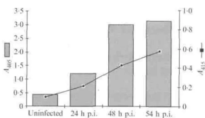
In order to quantify the increase in oligonucleosomal fragments found in infected cells, we performed a quantitative analysis of the fragmentation at different times after infection, using the Cell Death Detection ELISA kit (Boehringer Mannheim).
Fig. 2. Quantification of VZV antigens (curve) and oligonucleosomal fragments produced by apoptosis (histograms) in uninfected Vero cells or VZV-infected Vero cells at different times post-infection (p.i.).
Briefly, the cell monolayers (105 cells) and dying cells present in the culture medium were lysed and centrifuged in order to recover cytoplasmic fragmented DNA in the supernatant. The oligonucleosomes were then fixed in a microplate previously coated with anti-histone antibodies. The presence of fragmented DNA was revealed using peroxidase-conjugated anti-DNA antibodies. All reagents were used according to the manufacturer. This assay was performed on uninfected Vero cells (negative control) and on Vero cells infected for 24, 48 and 54 h. In parallel, viral antigens were quantified by ELISA using human anti-VZV antibodies. The amount of oligo-nucleosomes increased with time after infection and this increase was clearly correlated with the amount of viral antigens (Fig. 2).
One of the morphological characteristics of apoptosis is chromatin condensation, which can be visualized by DNA staining (Gregory et al, 1991). Cells were washed and incubated with acridine orange (1 µg/ml for 15 min at 37 °C), washed in PBS and observed. In the uninfected cells, the chromatin had a uniform distribution (Fig. 3 a), whereas staining in the infected cells was located on the periphery of the nucleus (Fig. 3b). These morphological observations were confirmed by electron microscopy. Infected cells were fixed 24 h post-infection with 2-5 % glutaraldehyde in 0-1 M-phosphate buffer pH 7-4 (30 min at 4 °C) and post-fixed with 2 % OsO4. After dehydration in ethanol and embedding in Epon, the samples were sectioned and stained with uranyl acetate and lead citrate before examination under a Jeol CX100 II electron microscope at 60 kV. Uninfected cells were used as controls. In the nucleus of uninfected cells, the chromatin was uniformly distributed (Fig. 3 c). In contrast, in infected cells the chromatin appeared disorganized, condensed and marginated in the nucleus, while the cytoplasm was well preserved (Fig. 3d).
Since Vero cells supporting a lytic infection showed all the characteristics of programmed cell death, we wondered whether protection against apoptosis could lead to the establishment of latency.
The most frequently described mechanism is the expression of the proto-oncogenic protein Bcl-2, a 26 kDa intracellular membrane-associated polypeptide, which prevents apoptosis induced by multiple agents in a variety of cells (Mah et al, 1993; Zhong et al, 1993). The overexpression of this protein can protect cells from apoptosis induced by viruses and extend their survival time (Alnemri et al, 1992; Hinshaw et ai, 1994; Takizawa et al, 1993; Ubol et ah, 1994). Other cellular proteins such as ced9 or Bax share some similarities with Bcl-2 and seem to protect cells against apoptosis. More recently, the role of a virus protein in such protection has been described (for review see Shen & Shenk, 1995; Wyllie, 1995). Moreover, it is interesting to note that different viruses have developed distinct mechanisms to block apoptosis. These mechanisms could be independent of Bcl-2, as described for baculovirus p35 (Radizadeh et at, 1993), or could modulate endogenous Bcl-2 expression, as with Epstem-Barr virus (EBV) LMP-1 (Henderson et al, 1991). Some virus proteins inducing protection against apoptosis are homologous or functionally similar to Bcl-2, as observed for LMW5-HL of African swine fever virus or BHRF-1 of EBV (Gregory et al, 1991; Tarodi & Chinnadurai, 1994). In the case of Sindbis virus, Bcl-2 can even convert a lytic infection into a persistent one (Levine et al, 1993).
Fig. 3. Visualization of chromatin condensation by acridine orange staining and fluorescence microscopic observations (a and b) and by electron microscopy (c and d). VZV-infected Vero cells were observed 48 h after infection (a and c). Uninfected Vero cells were used as controls (b and d). N, nucleus; nu, nucleolus; cy, cytoplasm; chr, chromatin; arrowhead indicates a virus particle.
In this context, we wondered whether there was a correlation between VZV latency in nerve cells and the expression of 2, the most frequent inhibitor of apoptosis. Using RT-PCR, we searched for Bcl-2 transcripts in Vero cells, where a lytic VZV infection induces apoptosis, and in murine
neuroblastoma (Neuro2A) and adult rat dorsal root ganglia cells, previously described as supporting VZV persistence. Briefly, RNA was extracted from Vero cells, Neuro2A cells and adult rat sensory
ganglia cells (Chomczynski & Sacchi, 1987). Samples were then treated for 20 min at 37 °C with DNase (10 mM-Tris-HCl pH 7-0, 10 niM-MgCl2, 5 U RNase-free DNase). After phenol extraction and a 10 min denaturation at 65 °C, reverse transcription was performed at 37 °C for 60 min using Moloney murine leukaemia virus reverse transcriptase according to the manufacturer (Boehringer Mannheim), with 2-5 µM random primers. In order to verify that the amplified nucleotide sequence was RNA and not DNA, each sample was incubated in the same conditions but in the absence of enzyme. The cDNA was then amplified by PCR [50mM-KCl, 10 mM-Tris-HCl pH 8-4, 1-5 mM-MgCl2, 200 µM of each
dNTP, 1 µM of each primer, 0-05 U Taq polymerase (Perkin Elmer)], using the oligonucleotides 5' GTCGCTACCGTCGTGACTT 3' and 5' CAGCCTCCGTTATCCTGGA 3' as primers. After 30 cycles in a Perkin Elmer Thermocycler (1 min at 95 °C, 1 min at 55 °C, 1 min at 72 °C), the PCR product was loaded onto a 4% (w/v) Nusieve agarose gel, stained with ethidium bromide and visualized by UV light. A 268 bp band corresponding to the Bcl-2 sequence was observed in all samples in which reverse transcription was performed (Fig. 4a).
Fig. 4. Detection by RT-PCR of the Bcl-2 transcript in Vero cells (lanes 2 and 3), Neuro2A cells (lanes 4 and 5) and adult rat sensory ganglia cells (lanes 6 and 7). The RNA were reverse-transcribed and amplified (lanes 3, 5 and 7), or directly amplified without previous reverse transcription (lanes 2, 4 and 6). The DNA marker (lane 1) was fxl74/HaeIIII
No band was observed in samples where reverse transcriptase was omitted. Bcl-2 was thus transcribed both in Vero cells, which support a productive and lytic infection, and in Neuro2A cells (data not shown) and adult rat sensory ganglia cells, in which a latent and non-lytic infection is observed. However, these observations are not quantitative and do not differentiate levels of expression of Bcl-2 in the different cell types. Bcl-2 expression in Vero cells was confirmed by immunocytochemistry (data not shown). In Vero cells, apoptosis is not prevented by Bcl-2. However, Bcl-2 is known to exert its protective function through heterodimerization with Bax, a protein of the Bcl-2 family, which can itself form homodimers that accelerate apoptosis (Yin et ah, 1994). The survival function thus appears to depend acutely on the stoi-chiometry of the interaction between Bcl-2 and Bax (Wyllie, 1994). Moreover, Allsopp et al. (1993) have demonstrated that sensory neurons depending on one or more members of the nerve growth factor family of neurotrophic factors for survival were rescued by Bcl-2, whereas ciliary neurotrophic factor-dependent ciliary neurons were not. This indicates that there are at least two death pathways in neurons with a different susceptibility to Bcl-2. Other proteins can protect cells against apoptosis. For example, LMP-1, BHRF1 and EBNA1, three proteins of EBV, and more recently the adenovirus E1B 19 kDa protein (Chiou et al, 1994) have been shown to play a role in protection against apoptosis. Finally, it is important to remember that apoptosis is associated with a modification of the cellular redox balance, and that there are many proteins which can protect cells by inhibiting peroxidation.
It would be interesting to search for a cellular or viral protein, expressed specifically in nerve cells during latency, which could inhibit apoptosis and allow for the establishment and maintenance of a quiescent state of the virus.
While performing this work, C.S.-D. and J.P. were Senior Research Assistant and Research Director, respectively, of the National Fund for Scientific Research (NFSR), Brussels, Belgium. This work was supported by grants from the NFSR, the Belgian National Lottery and the VZV Research Foundation, New York, USA.
References
ALLSOPP,T. E., WYATT,S., PATERSON,H. F. & DAVIES,A. M. (1993). The proto-oncogene Bcl-2 can selectively rescue neurotrophic factor-dependent neurons from apoptosis. Cell 73, 295-307.
ALNEMRI,A. S., ROBERTSON,N. M., FERNANDES,T. F., CROCE,C. M. & LITWACK,G. (1992). Overexpressed full-length human Bcl-2 extends the survival of baculovirus-infected Sf9 insect cells. Proceedings of the National Academy of Sciences, USA 89, 7295-7299.
BOURDON-WOUTERS,C, MERVILLE-LOUIS,M. P., SADZOT-DELVAUX,C, MARC,P., PIETTE,J.,
DELREE,P., MOONEN,G. & RENTIER,B. (1990). Acute and persistent varicella-zoster virus infection of human and murine neuroblastoma cell lines. Journal of Neuroscience Research 26, 90-97.
CAMERON,P. U., POPE,M., GEZELTER,S. & STEINMAN,M. (1994). Infection and apoptotic cell death of CD4+ T cells during an immune response to HIV-1-pulsed dendritic cells. Aids Research and Human Retroviruses 10, 61-71.
CHIOU,S. K., TSENG,C. C, RAO,L. & WHITE,E. (1994). Functional complementation of the adenovirus E1B 19-kilodalton protein with Bcl-2 in the inhibition of apoptosis in infected cells. Journal of Virology 68, 6553-6566.
CHOMCZYNSKI,P. & SACCHI,N. (1987). Single-step method of RNA isolation by acid guanidinium thiocyanate-phenol-chioroform extraction. Analytical Biochemistry 162, 156-159.
FESQ,H., BACHER,M., NAIN,M. & GEMSA,D. (1994). Programmed cell death (apoptosis) in human monocytes infected by influenza A virus. Immunobiology 190, 175—182.
GREGORY,C. D., DIVE,C, HENDERSON,S., SMITH,C. A., WILLIAMS,G. T., GORDON,J. & RICKINSON, A. B. (1991). Activation of Epstein-Barr virus latent genes protects human B cells from death by apoptosis. Nature 349, 612-614.
HAY,J. & RUYECHAN,W. T. (1994). Varicella-zoster virus - a different kind of herpesvirus latency? Seminars in Virology 5, 241-247.
HENDERSON,S., ROWE,M., GREGORY,C, CROOM-CARTER,D., WANG,F., LONGNECKER,R., KIEFF,E. & RICKINSON,A. (1991). Induction of Bcl-2 expression by Epstein-Barr virus latent membrane protein 1 protects infected cells from programmed cell death. Cell 65, 1107-1115.
HINSHAW,V. S., OLSEN,C. W., DYBDHL-SISSOKO,N. & EVANS,D. (1994). Apoptosis: a mechanism of cell killing by influenza A and B viruses. Journal of Virology 68, 3667-3673.
HOCKENBERY,D. (1995). Defining apoptosis. American Journal of Pathology 146, 16-19. HOPE SIMPSON,R. E. (1965). The nature of herpes zoster: a long term study and a new hypothesis. Proceedings of the Royal Society of Medicine 58, 9-20.
LEVINE,B., HUANG,Q., ISAACS,J. T., REED,J. C, GRIFFIN,D. E. & HARDWICK,J. M. (1993). Conversion of lytic to persistent alphavirus infection by the bcl-2 cellular oncogene. Nature 361,
739-742.
MAH,S. P., ZHONG,L. T., LIU,Y., ROGHANI,A., EDWARDS,R. H. & BREDESEN,D. E. (1993). The protooncogene Bcl-2 inhibits apoptosis in PC12 cells. Journal of Neurochemistry 60, 1183-1186. MANJO,G. & JORIS,I. (1995). Apoptosis, oncosis, and necrosis. An overview of cell death. American Journal of Pathology 146, 3-15.
MERVILLE-LOUIS,M. P., SADZOT-DELVAUX,C, DELREE,P., PIETTE,J., MOONEN,G. & RENTIER,B. (1989). Varicella-zoster virus infection of adult rat sensory neurons in vitro. Journal of Virology 63, 3155-3160.
MEYAARD,L., OTTO,S. A., JONKER,R. R., MIJNSTER,M. J., KEET,R. P. M. & MIEDEMA,F. (1992). Programmed death of T cells in HIV-1 infection. Science 257, 217-219.
NOTEBORN,M. H. M., TODD,D., VERSCHUEREN,C. A. J., DE GAUW,H. W. F. M., CURRAN,W. L., VELDKAMP,S., DOUGLAS,A. J., MCNULTY,M. S., VAN DER EB,A. J. & KOCH,G. (1994). A single chicken anemia virus protein induces apoptosis. Journal of Virology 68,346-351.
PANAYIOTIDIS,P., GANESHAGURU,K., JABBAR,S. A. B. & HOFFBRAND,A. V. (1993). Interleukin-4 inhibits apoptotic cell death and loss of the bcl-2 protein in B-chronic lymphocytic leukaemia cells in vitro. British Journal of Haematology 85, 439-445.
RADIZADEH,S., LACOUNT,D. J., FRIESEN,P. D. & BREDESEN,D. E. (1993). Expression of the
baculovirus p35 gene inhibits mammalian neural cell death. Journal of Neurochemistry 61, 2318-2321. SCHWARTZMAN,R. A. & CIDLOWSKI,J. A. (1993). Apoptosis: the biochemistry and molecular biology of programmed cell death. Endocrine Reviews 14, 133-151.
SHEN,Y. & SHENK,T. E. (1995). Viruses and apoptosis. Current Opinions in Genetics and Development 5, 105-111.
TAKIZAWA,T., MATSUKAWA,S., HIGUSHI,Y., NAKAMURA,S., NAKANISHI,Y. & FUKUDA,R. (1993). Induction of programmed cell death (apoptosis) by influenza virus infection in tissue culture cells. Journal of General Virology 74, 2347-2355.
TARODI,B.'S. & CHINNADURAI,G. (1994). Epstein-Barr virus BHRF1 protein protects against cell death induced by DNA-damaging agents and heterologous viral infection. Virology 201, 404-407. UBOL,S., TUCKER,P. C, GRIFFIN,D. E. & HARWICK,M. (1994). Neurovirulent strains of alphavirus induce apoptosis in bcl-2-expressing cells: role of a single amino acid change in the E2 glycoprotein. Proceedings of the National Academv of Sciences, USA 91, 5202-5206.
VASCONCELOS,A. C. & KENNETH,M. L. (1994). Apoptosis induced by infectious bursal disease virus. Journal of General Virology 75, 1803-1806.
WYLLIE,A. H. (1994). Death gets a brake. Nature 369, 272-273.
WYLLIE,A. H. (1995). The genetic regulation of apoptosis. Current Opinion in Genetics and Development 5, 97-104.
YIN,X.-M., OLTVAI,Z. N. & KORSMEYER,S. J. (1994). BH1 and BH2 domains of Bcl-2 are required for inhibition of apoptosis and heterodimerization with Bax. Nature 369, 321-323.
ZHONG,L., SARAFIAN,T., KANE,D. J., CHARLES,A. C, MAH,S. P., EDWARDS,R. H. & BREDESEN,D. E. (1993). Bcl-2 inhibits death of central neural cells induced by multiple agents. Proceedings of the National Academy of Sciences, USA 90, 4533-1537.