Université de Montréal
Natriuretic Peptide Receptor-C Activation Regulates Vascular Smooth Muscle Cell Hypertrophy: Molecular Mechanisms
Par Ashish Jain
Département de Pharmacologie et Physiologie Faculté de Médecine
Mémoire presenté à la Faculté de Médecine en vue de l’obtention du grade de
Maîtrise en Sciences (M.Sc.)
En physiologie moléculaire, cellulaire et intégrative
Décembre 2016 © Ashish Jain, 2016
Université de Montréal Faculté de Médecine
Ce mémoire intitulé:
Natriuretic Peptide Receptor-C Activation Regulates Vascular Smooth Muscle Cell Hypertrophy: Molecular Mechanisms
Presenté par
Ashish Jain
A été évalué par un jury composé des personnes suivantes:
Dr. Rémy Sauvé (président-rapporteur)
Dr. Madhu B. Anand-Srivastava (directrice de recherche) Dr. Puttaswamy Manjunath (membre du jury)
Résumé
L’hypertension est associée au remodelage vasculaire dû à l’hyperprolifération et l’hypertrophie des cellules musculaires lisses vasculaires (CMLVs). Nous avons démontré par le passé l’implication de l’expression élevée des protéines Gqα et PLCβ1 dans les CMLVs de rats spontanément hypertendus (RSH) âgés de 16 semaines. Le C-ANP4-23 est un agoniste du récepteur au peptide natriurétique de type C (NPR-C) qui possède la capacité d’inhiber la synthèse de protéines en réponse aux peptides vasoactifs dans les CMLVs. Cette étude a eu pour but d’examiner si le C-ANP4-23 pouvait atténuer l’hypertrophie dans un modèle de rat souffrant d’hypertrophie cardiaque et d’explorer les mécanismes responsables de cette inhibition. Pour ce faire, des CMLVs aortiques de RSH âgés de 16 semaines ont été utilisées. Le taux de synthèse de protéines, un marqueur d’hypertrophie, a été déterminé par l’incorporation de (3H)leucine et l’expression des protéines a été déterminée par la technique d’immunobuvardage de type Western. Le volume cellulaire a été estimé par imagerie confocale tridimensionnelle. Le taux de synthèse de protéines et le volume cellulaire étaient considérablement accrus dans les CMLVs des RSH comparativement aux rats WKY et ont été largement atténués par le traitement au C-ANP4-23. De plus, le traitement au C-ANP4-23 a normalisé l’expression élevée du récepteur AT1 et des protéines Gqα et PLCβ1, des niveaux intracellulaires d’anions superoxide (O2-), de l’activité de la NADPH (de l’anglais nicotinamide adenine dinucleotide phosphate) oxydase, ainsi que l’expression des protéines Nox4 et de p47phox dans les CMLVs des RSH. En outre, le C-ANP4-23 a réduit l’activation des récepteurs à L’EGF (de l’anglais epidermal growth factor), au PDGF (de l’anglais platelet-derived growth factor), et à l’IGF-1 (de l’anglais insulin-like growth factor 1). Le C-ANP4-23 a également atténué la phosphorylation des ERK1/2 (de l’anglais extracellular regulated kinase1/2), AKT et c-Src. Ces résultats indiquent que l’activation du NPR-C par C-ANP4-23 a atténué l’hypertrophie des CMLVs par sa capacité à diminuer la surexpression du récepteur AT1, l’expression élevée des protéines Gqα/PLCβ1, le stress oxydatif accru, l’activation augmentée des facteurs de croissance et l’augmentation de la phosphorylation des voies de signalisation MAPK/AKT. Ainsi, ces travaux suggèrent que le C-ANP4-23 peut être utilisé comme
agent thérapeutique pour le traitement des complications vasculaires associées à l’hypertension et à l’athérosclérose.
Mots-clés : hypertension, RSH, CMLV, NPR-C, stress oxydatif, récepteurs des facteurs de croissance, c-Src, AKT, MAPK, protéines Gqα.
Abstract
Hypertension is associated with vascular remodelling due to hyperproliferation and hypertrophy of vascular smooth muscle cells (VSMCs). We earlier showed the implication of enhanced expression of Gqα and PLCβ1 proteins in VSMCs from 16-week-old spontaneously hypertensive rats (SHR). The present study was undertaken to investigate whether C-ANP4-23, a natriuretic peptide receptor-C (NPR-C) agonist that has been shown to inhibit vasoactive peptide-induced enhanced protein synthesis in VSMCs, could attenuate VSMC hypertrophy in rat models of cardiac hypertrophy and to explore the underlying mechanisms contributing to this inhibition. For these studies, aortic VSMCs from 16-week-old SHR were used. The protein synthesis, a marker of hypertrophy, was determined by (3H)leucine incorporation and the expression of proteins was determined by Western blotting. Cell volume was determined by three-dimensional confocal imaging. The protein synthesis was significantly enhanced in VSMC from SHR as compared to WKY and C-ANP4-23 treatment attenuated the enhanced protein synthesis to WKY control levels. In addition, the enhanced expression of the AT1 receptor as well as Gqα and PLCβ1 proteins, enhanced levels of superoxide anion (O2-), nicotinamide adenine dinucleotide phosphate (NADPH) oxidase activity, as well as the increased expressions of NADPH oxidase 4 (Nox4) and p47phox exhibited by VSMC from SHR were all attenuated by C-ANP4-23 treatment. Furthermore, C-ANP4-23 also attenuated the enhanced activation of epidermal growth factor receptor (EGF-R), platelet-derived growth factor receptor (PDGF-R), insulin-like growth factor 1 receptor (IGF-1R) and the enhanced phosphorylation of extracellular signal-regulated kinases 1/2 (ERK1/2), AKT and c-Src. These results indicate that C-ANP4-23, via the activation of NPR-C, attenuates VSMC hypertrophy through its ability to decrease the overexpression of the AT1 receptor and Gqα/PLCβ1 proteins, the enhanced oxidative stress, the increased activation of growth factors and the enhanced phosphorylation of the MAPK/AKT signalling pathway. Thus, it can be suggested that C-ANP4-23, an activator of NPR-C, may be used as a therapeutic agent for the treatment of vascular complications associated with hypertension and atherosclerosis.
Key words: Hypertension, SHR, VSMC, NPR-C, oxidative stress, NO, growth factor receptors, c-Src, AKT, MAPK, Gqα proteins.
Table of Contents
Résumé ... III Abstract ... V Table of Contents ... VII List of Abbreviations ... XII Acknowledgements ... XVIII CHAPTER 1 Introduction and Literature Review ... XIX
1.1 The cardiovascular system ... 2
1.1.1 Vascular System ... 2
1.2 Blood Pressure ... 3
1.2.1 Blood Pressure Regulation ... 3
1.2.1.1 Blood Pressure Regulation: Short Term Mechanisms ... 4
1.2.1.2 Blood Pressure Regulation: Long Term Mechanisms ... 5
1.3 The Role of Vasoactive Peptides in the Regulation of Blood Pressure ... 6
1.3.1 G Protein-Coupled Receptors ... 6
1.3.2 Renin-Angiotensin System ... 7
1.3.2.1 Angiotensin II Receptors ... 7
1.3.2.1.1 Angiotensin II AT1 Receptor ... 8
1.3.2.1.2 Angiotensin II AT2 Receptor ... 9
1.3.3 The Endothelin System ... 10
1.3.3.1 Endothelin-1 Receptor ... 10
1.4 Transmembrane Signalling: Mechanisms ... 11
1.4.1 Guanine Nucleotide-Binding Proteins ... 11
1.4.2 The Adenylate Cyclase System ... 12
1.4.2.1 The Adenylate Cyclase System and Cyclic Adenosine Monophosphate ... 12
1.4.2.2 Giα ... 13
1.4.3 The Phosphoinositide System ... 13
1.4.3.1 Gqα ... 13
1.4.3.2 Phospholipase Cβ (PLCβ): Structure and Function ... 14
1.4.3.2.1 Activation of PLCβ by the Gαq Subunit ... 15
1.4.3.2.2 Activation of PLCβ by the Gβγ Complex ... 15
1.4.4.1 Formation of Diacylglycerol ... 16
1.4.4.2 Protein Kinase C ... 16
1.4.5 IP3 and Calcium ... 17
1.4.6 Growth Factors: Receptors ... 19
1.4.6.1 Epidermal Growth Factor Receptor (EGF-R) ... 19
1.4.6.2 Insulin-Like Growth Factor (IGF-R) ... 20
1.4.6.3 Platelet-Derived Growth Factor Receptor (PDGF-R) ... 20
1.4.7 c-Src Pathway ... 21
1.4.8 MAP Kinase Signalling ... 22
1.4.9 Phosphoinositide 3-Kinase Pathway ... 22
1.4.10 AKT Signalling Pathway ... 23
1.4.11 Reactive Oxygen Species and Oxidative Stress ... 23
1.4.11.1 ROS Metabolism ... 24
1.4.11.2 ROS: Sources of Cellular Production at the Vascular Level ... 24
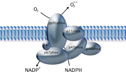
1.4.11.3 NAD(P)H Oxidase ... 25
1.4.11.3.1 NAD(P)H Oxidase Structure ... 25
1.4.11.3.2 NAD(P)H Oxidase: Vascular Characteristics ... 26
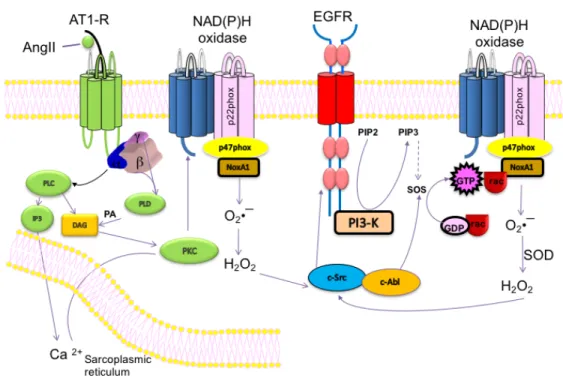
1.4.11.3.3 NAD(P)H Oxidase: Activation by G Protein Coupled Receptors ... 27
1.4.11.4 ROS: Elimination ... 29
1.4.11.5 ROS: Physiological Roles ... 29
1.5 Hypertension ... 29
1.5.1 Risk Factors ... 30
1.5.2 Consequences ... 30
1.5.3 Treatment ... 31
1.5.4 SHR: A model of hypertension ... 31
1.6 Cardiovascular Complications Linked to Hypertension ... 32
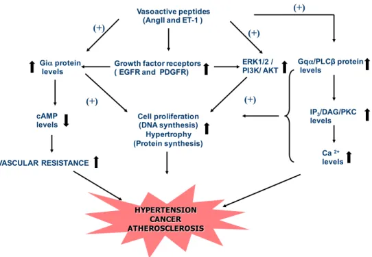
1.6.1 Hypertension and Vascular Remodeling: Molecular Mechanisms ... 32
1.6.1.1 VSMC Proliferation ... 33
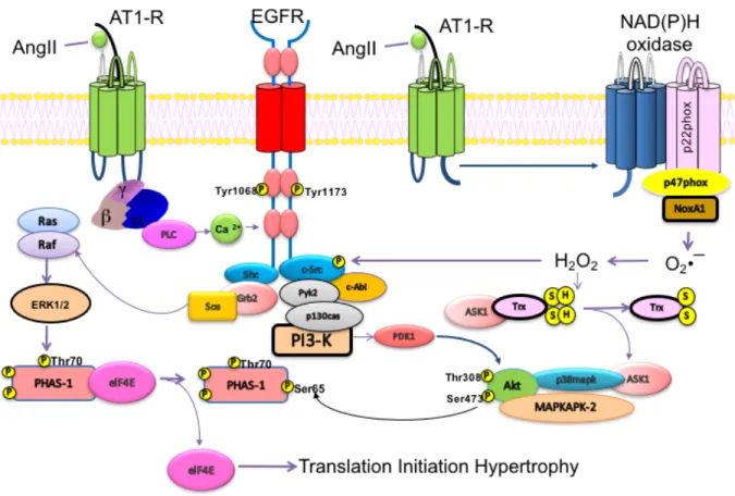
1.6.1.2 VSMC Hypertrophy ... 34
1.6.1.2.1 VSMC Hypertrophy: Implication of Gqα ... 34
1.6.1.2.2 VSMC Hypertrophy: Implication of ROS ... 34
1.6.1.2.3 VSMC Hypertrophy: Pressure Forces ... 36
1.6.1.3 Cardiac Hypertrophy: Molecular Mechanisms ... 36
1.7 Natriuretic Peptides ... 37
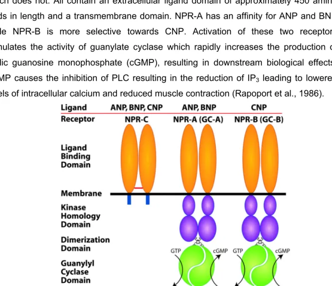
1.7.1 Natriuretic Peptide Receptors ... 38
1.7.2.1 Distribution of NPR-C ... 39
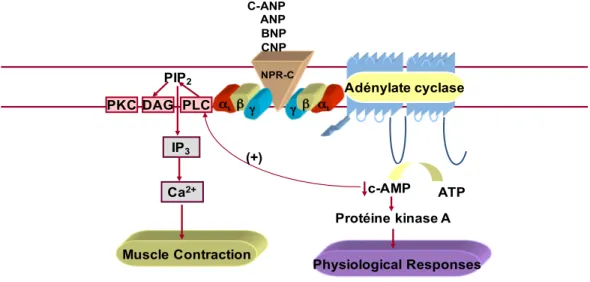
1.7.2.2 NPR-C Signalling ... 39
1.7.2.3 Physiological Roles of NPR-C ... 42
1.7.2.4 Regulation of NPR-C in Pathological Conditions ... 42
1.7.2.5 NPR-C and Vascular Remodeling ... 43
Hypothesis and Objectives ... 45
CHAPTER 2 Scientific Article ... 46
Abstract ... 49
Introduction ... 49
Materials and Methods ... 53
Results… ... 57
Discussion ... 61
References ... 66
Figures and Legends ... 71
CHAPTER 3 Discussion, Conclusions and Future Work ... 84
Discussion ... 84
Conclusions ... 90
Future Work ... 92
Table of Figures
Chapter 1
Figure 1: The structure of the arterial and venous vascular wall ... 3
Figure 2: Structure of a G Protein-Coupled Receptor ... 7
Figure 3: ANG II signalling mechanisms associated with Gqα and Giα proteins ... 9
Figure 4: Activation of G proteins. ... 12
Figure 5: Simplified structure of vascular NADPH oxidase ... 26
Figure 6: Activation of NAPDH Oxidase by ANG II and EGFR ... 28
Figure 7: Signalling mechanisms associated with hypertension ... 33
Figure 8: Signalling pathways implicated in hypertrophy ... 35
Figure 9: Natriuretic peptide receptors. ... 38
Figure 10: NPR-C signalling and the interaction between c-AMP and phospholipase C 41 Figure 11: Schematic diagram demonstrating the proposed mechanism for VSMC hypertrophy in SHR. ... 46
Chapter 2 Figure 1: Effect of C- ANP4-23 treatment on enhanced protein synthesis and cell volume in aortic VSMCs from 16-wk-old SHR and age-matched WKY rats…………...75
Figure 2: Effect of the inhibition of Gqα on enhanced protein synthesis in aortic VSMCs from 16-wk-old SHR and age-matched WKY rats. ... 76
Figure 3: Effect of the inhibition of Gqα on enhanced protein synthesis in aortic VSMCs from 16-wk-old SHR and age-matched WKY rats……….77
Figure 4: Effect of C-ANP4-23 treatment on enhanced AT1 receptor expression in aortic VSMCs from 16-wk-old SHR and age-matched WKY rats………..78
Figure 5: Effect of C-ANP4-23 treatment on enhanced O2– production and NADPH oxidase activity in aortic VSMCs from 16-wk-old SHR and age-matched WKY rats………79
Figure 6: Effect of C-ANP4-23 treatment on the enhanced expression of NADPH oxidase subunits p47phox and Nox4 in aortic VSMCs from 16-wk-old SHR and age-matched WKY rats………..80
Figure 7: Effect of C-ANP4-23 treatment on the enhanced phosphorylation of c-Src in aortic VSMCs from 16-wk-old SHR and age-matched WKY rats. ... 81 Figure 8: Effect of C-ANP4-23 treatment on the enhanced expression of epidermal
growth factor receptor (EGF-R) (A), insulin growth factor receptor (IGF-IR) (B) and platelet derived growth factor receptor (PDGF-R) (C) in aortic VSMCs from 16-wk-old SHR and age-matched WKY rats ... 82 Figure 9: Effect of C-ANP4-23 treatment on the enhanced phosphorylation of ERK1/2
(A) and AKT (B) in aortic VSMCs from 16-wk-old SHR and age-matched WKY rats. ... 83
Chapter 3
Figure 12: Schematic diagram demonstrating the proposed mechanism for the anti-hypertrophic effect of an NPR-C agonist in VSMCs from SHR…….…………90
List of Abbreviations
-A-
Aa Arachidonic acid
AC Adenylate cyclase
ACE Angiotensin-converting enzyme
ADH Antidiuretic hormone
Ang II Angiotensin II
ANP Atrial natriuretic peptide
ARBs Angiotensin receptor blockers
AT1 Angiotensin II type 1 receptor AT2 Angiotensin II type 2 receptor
AT2KO AT2 knockout mice
ATP Adenosine triphosphate
AVP Arginine vasopressin
-B-
BKc Big potassium channel
BNP Brain natriuretic peptide
BP Blood pressure
BW Body weight
-C-
Ca2+ Calcium
cAMP Cyclic adenosine monophosphate
C-ANP4-23 C-ANP4-23 (des(Gln18, Ser19, Gly20, Leu21, Gly22)ANP4-23-NH2) CDEA Comité de Déontologie de l’Experimentation sur les
Animeaux
cGMP Cyclic guanosine monophosphate
CNP C-type natriuretic peptide
Cdk Cyclin dependent kinase
Cki Cdk inhibitor
CO Cardiac output
-D-
DAG Diacylglycerol
DGAT Diacylglycerol acetyltransferase DIC Differential interference contrast
DNA Deoxyribonucleic acid
-E-
EDHF Endothelium-derived hyperpolarizing factor
EGF Epidermal growth factor
EGF-R Epidermal growth factor receptor
EGR Early growth response
ER Endoplasmic reticulum
ERK1/2 Extracellular signal-regulated kinase 1 and 2
ET-1 Endothelin-1
-F-
FGF Fibroblast growth factor
FSK Forskolin -G- GDP Guanosine diphosphate Gi Inhibitory G protein GPCR G protein-coupled receptor GPX Gluthathione peroxidase Gs Stimulatory G protein GTP Guanosine triphosphate -H- H202 Hydrogen peroxide HR Heart rate -I-
ICRAC Ca+2 release activated Ca+2 current
IGF Insulin-like growth factor
IP1 Inositol 4-phosphate
IP3 Inositol 1,4,5-triphosphate -J-
JAK Janus kinase
JNK c-Jun-N-terminal kinase -K- kDa Kilodalton(s) -L- Li+ Lithium -M-
MAP Mean arterial pressure
MAPK Mitogen-activated protein kinases
MLC Myosin light chain
mm Hg Millimeters mercury
mRNA Messenger ribonucleic acid
-N-
NADPH Nicotinamide adenine dinucleotide phosphate
NGF Nerve growth factor
NO Nitric Oxide
NOX4 NADPH oxidase 4
NP Natriuretic peptide
NPR Natriuretic peptide receptor
NPR-A Natriuretic peptide receptor-A NPR-B Natriuretic peptide receptor-B NPR-C Natriuretic peptide receptor-C -O- O2 Oxygen O2- Superoxide anion OH- Hydroxyl radical -P- PC Phosphatidylcholine
PGI2 Prostacyclin
Phox Phagocyte oxidase
PI Phosphatidyl inositol
PI3K Phosphoinositide-3 kinases
PI(4,5)P2 Phosphatidylinositol bisphosphate PIP2 Phosphatidylinositol-4,5-biphosphate
PKA Protein kinase A
PKB Protein kinase B
PKC Protein kinase C
PLC Phospholipase C
PLCβ Phospholipase Cβ
PLD Phospholipase D
PMA Phorbol 12-myristate 13-acetate
pRb Phosphorylated Rb
-R-
RAS Renin-angiotensin system
Rb Retinoblastoma protein
ROC Receptor operated channel
ROS Reactive oxygen species
RTK Receptor tyrosine kinase
-S-
SA Sinoatrial node
SBP Systolic blood pressure
SHR Spontaneously hypertensive rat
SHR-SP Stroke prone spontaneously hypertensive rats
SOC Store operated channel
SOD Superoxide dismutase
-T-
TAG Triacylglycerol
TGFβ Transforming growth factor β
TRP Transient receptor potential -V-
VSMC Vascular smooth muscle cell
-W-
To my dear mother, whose love knows no bounds
Acknowledgements
I would like to thank my supervisor, Dr. Madhu Anand-Srivastava, for accepting me into her lab and for believing in me. Her unwavering support and guidance will never be forgotten.
I would also like to thank the members of my jury, Dr. Rémy Sauvé and Dr. Puttaswamy Manjunath, for accepting to evaluate my graduate research work and for their valuable comments and suggestions.
I would also like to express my sincere gratitude to Dr. Yuan Li for all her teachings and advice.
To my lab members, Dr. Ekhtear Hossain, Dr. Mehdi Atef, Dr. Sara Almajdoob and Dr. Sofiane Rahali, it was a pleasure spending my days with you.
Finally, to all the faculty members, staff and students of the physiology department, thank you for the wonderful experience.
CHAPTER 1
1.1 The cardiovascular system
The cardiovascular system consisting of the heart and blood vessels circulates approximately 5 liters of blood throughout the body. Through this process, the system delivers oxygen, hormones and nutrients to the body and subsequently eliminates cellular waste products and carbon dioxide. Via the circulation of white blood cells, the cardiovascular system provides protection by removing cellular debris and combatting pathogens. Moreover, red blood cells and platelets generate scabs to block wounds as well as prevent foreign pathogens from entering the body, and fluids from exiting. Circulating antibodies provide an immune protection, while another function of the cardiovascular system is to maintain homeostasis. The system is comprised of two main circulatory loops: the pulmonary circulation loop and the systemic circulation loop. In the pulmonary circulation loop, deoxygenated blood is carried from the right side of the heart to the lungs, where it is oxygenated and returned to the left side. The systemic circulation then takes over, pumping highly oxygenated blood to the tissues and removing waste products. The systemic circulation terminates with the delivery of deoxygenated blood back to the right side of the heart. The arteries play the principal role carrying blood away from the heart (Boron & Boulpaep, 2003).
1.1.1 Vascular System
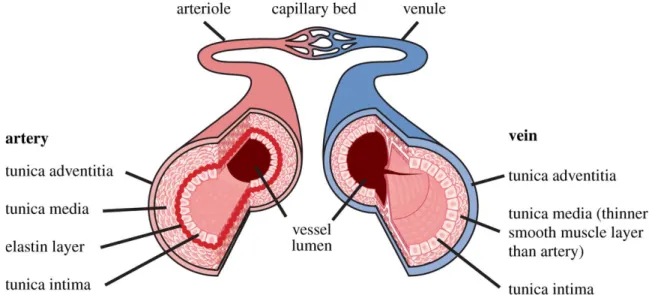
The vascular structure plays an integral role in blood pressure homeostasis. The wall of an artery consists of three distinct layers; from innermost to the outermost, we find the tunica intima, tunica media and tunica adventitia (Figure 1). The tunica intima secretes many vasoactive substances which control the diameter of the vessels. These include: endothelin-1 (ET-1), nitric oxide (NO) and C-type natriuretic peptide (CNP). The tunica media consists mainly of smooth muscle cells which allow for the vasoconstriction and vasodilation of blood vessels. The tunica adventitia consists of elastic fibers as well as collagen fibers which help anchor the structure to its surroundings. The walls of the aorta and other large diameter arteries contain a high amount of elastic tissue, primarily located in the internal and external elastic laminas. These stretch during systole and recoil during diastole. Arterioles, on the other hand, consist of a high percentage of smooth muscle, which constrict through the innervation by noradrenergic nerve fibers. Arterioles are the major site of resistance to blood flow, with minute changes in their
diameter causing significant changes in total peripheral resistance (Berne & Levy, 2001; Tortora & Derrickson, 2009).
Figure 1: The structure of the arterial and venous vascular wall. (Shaw, ter Haar, Rivens, Giussani, & Lees, 2014)
1.2 Blood Pressure
Blood pressure is the lateral force exerted by blood on the walls of blood vessels and is expressed as the systolic over the diastolic pressure. The systolic pressure is the maximum pressure exerted by circulating blood during contraction of the heart. The normal range in adults is 100-140 mmHg while the mean is 120 mmHg. The diastolic pressure, on the other hand, is the minimum pressure measured during relaxation and ventricular filling. The normal adult range is 60-90 mmHg with a mean of 80 mmHg. The pulse pressure is the difference between the systolic and diastolic pressure. The mean arterial pressure is the mean pressure exerted in the arterial compartment during a cardiac cycle and is calculated as the diastolic pressure plus 1/3 of pulse pressure. 1.2.1 Blood Pressure Regulation
Normal blood pressure is maintained through the regulation of several factors: cardiac output, peripheral resistance, blood volume, viscosity of blood and the elasticity of blood vessels. Cardiac output is defined as the volume of blood pumped out per ventricle per minute. Normal cardiac output is approximately 5L/ventricle. Cardiac output affects the systolic pressure. Peripheral resistance is the resistance offered by the wall
of the blood vessel on the blood flowing through. It is maximal in the arterioles; hence the arterioles are known as the seat of maximum peripheral resistance. Peripheral resistance affects the mean arterial pressure. Poiseuille’s law indicates that the resistance (R) is directly related to the length of the blood vessel (L) and the blood viscosity (η) and is inversely related to radius (r) to the power of 4:
R = 8 x η x L Π x r4
When the volume of blood increases, a greater venous return is facilitated. This, in turn, increases the cardiac output and hence, the systolic pressure. The viscosity of blood affects the peripheral resistance, which affects the diastolic pressure while the elasticity of blood vessels has an inverse relationship with blood pressure. Considering all of the above factors, the final determinants of blood pressure are cardiac output and peripheral resistance.
1.2.1.1 Blood Pressure Regulation: Short Term Mechanisms
Short term mechanisms try to regulate blood pressure within a few seconds of fluctuations, whereas other mechanisms begin acting after a few minutes to a few hours.
The vasomotor center alters the smooth muscle activity of blood vessels by activating the sympathetic nervous system originating in the reticular formation of the brain stem. The vasomotor center transmits continuous impulses to the lateral horn of the spinal cord through the reticulospinal tract thus exerting a constant stimulatory influence on the arteriolar smooth muscle and maintaining peripheral resistance. When the activity of the lateral horn is depressed, the loss of sympathetic influence on arteriolar smooth muscle will lead to a fall in blood pressure. Activation of the vasomotor center is regulated by baroreceptors, chemoreceptors and the CNS ischemic response (Boron & Boulpaep, 2003).
Baroreceptors are stretch receptors located in the walls of the heart and blood vessels, with the carotid sinus and aortic arch receptors monitoring arterial circulation. Baroreceptors are stimulated by the distention of the vessels in which they are located. The glossopharyngeal and vagus nerves transmit the afferent fibers from the carotid sinus and aortic arch respectively. Increased baroreceptor discharge will inhibit vasoconstriction and excite the vagal innervation of the heart, resulting in a decrease in
blood volume, bradycardia and a decrease in cardiac output. Below 60 mmHg, there will not be any stimulation of the baroreceptors, and above 180 mmHg stimulation will not increase any further. Hence, with a resting mean arterial pressure of 94 mmHg in a normal individual, there will be constant impulses transmitted from the baroreceptors. If there is a sustained increase in blood pressure for a prolonged duration, the baroreceptors become adapted to the new pressure, and thus fail to restore blood pressure to normal values (Guyton, 1961).
Chemoreceptors, present in the carotid and aortic bodies, exert their main effect on the respiratory system, however they also play a minor role in blood pressure regulation. Chemoreceptors are stimulated by a decrease in O2-, and an increase in H+ and pCO2. The cardiovascular response to chemoreceptor stimulation is the stimulation of the vasomotor and respiratory centers, as well as an inhibition of the cardioinhibitory center, resulting in peripheral vasoconstriction and bradycardia.
In the CNS ischemic response, a drop in blood pressure below 40 mmHg will result in a severe decrease in blood flow (ischemia) to the brain. This will decrease the pCO2, which directly stimulates the vasomotor center. The subsequent increase in activity of the sympathetic nervous system will act on the vascular smooth muscle and cardiac muscle to restore both systolic and diastolic blood pressures. This mechanism last only a few minutes and is a last resort mechanism to restore blood pressure and blood flow to the brain.
1.2.1.2 Blood Pressure Regulation: Long Term Mechanisms
Long term mechanisms control blood pressure by regulating blood flow through the renal system. These include: the fluid shift mechanism, the renin angiotensin-aldosterone system and anti-diuretic hormone. In the fluid shift mechanism, a decrease in blood pressure leads to the constriction of the precapillary sphincter thereby reducing blood flow. The hydrostatic pressure in the capillaries will be less than the colloidal osmotic pressure, resulting in an inward driving force, shifting fluids to the intravascular compartment. Blood volume, and hence venous return, will increase, resulting in an increase in cardiac output.
The renal system brings about appropriate alterations in the volume of urine formed thus altering blood volume and blood pressure. The renal system acts indirectly
through the renin-angiotensin system. Renin is secreted from the juxta glomerular apparatus and is stimulated by sympathetic stimulation, decreased blood flow through the kidneys as well as altered levels of sodium in the distal convoluted tubule. Renin acts on angiotensinogen and converts it to angiotensin I. Angiotensin I is acted upon by the angiotensin converting enzyme (ACE) to form angiotensin II. Angiotensin II aids in the restoration of blood pressure by acting on vascular smooth muscle to bring about vasoconstriction and thus increase peripheral resistance. It stimulates the secretion of aldosterone from the adrenal cortex thereby increasing blood flow and cardiac output. It stimulates the activity of the thirst center to increase blood volume. Finally, it elevates the release of anti-diuretic hormone (ADH), which increases blood volume. All of these mechanisms result in a rise in blood pressure. In cases of increased blood pressure, pressure diuresis and pressure natriuresis will aid in restoring it back to normal levels (Guyton, 1961).
1.3 The Role of Vasoactive Peptides in the Regulation of Blood Pressure
Vasoactive peptides are molecules which are capable of altering the diameter of blood vessels by either acting as vasodilators (ex. NO and prostacyclin) or as vasoconstrictors (ex. Ang II and ET-1). Hypertension is characterized by an increase in the concentration of vasoconstrictors, resulting in alterations in the vascular milieu, extending from small arteries to large conducting vessels. This increase in the local and/or systemic concentration of vasoconstrictors ultimately leads to an elevation in peripheral vascular resistance and thereby hypertension.
1.3.1 G Protein-Coupled Receptors
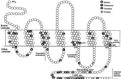
G protein-coupled receptors (GPCRs) are 7 passage transmembrane receptors belonging to the largest family of membrane-bound proteins. GPCRs are activated by glycoproteins, amino acids, phospholipids and peptides, among others. Once activated, GPCRs go on to activate various G proteins thus transmitting information from the extracellular to the intracellular milieu. GPCR’s consist of an extracellular glycosylated N-terminus containing the ligand-binding domain and disulfide bridges which serve to stabilize the structure. This is followed by seven transmembrane α-helices connected by three intracellular loops (IL-1 to IL-3) and three extracellular loops (EL-1 to EL-3), and lastly an intracellular C-terminus (Figure 2). Upon the binding of a ligand, the N-terminal
tail undergoes a conformational change which leads to its interaction with the extracellular loops and transmembrane domains. A change in the relative orientation of the transmembrane helices allows for the residues of the intracellular helices and transmembrane domains to be available for G-protein coupling (Gilman, 1987).
Figure 2: Structure of a G Protein-Coupled Receptor. Representation of amino acids and signalling domains in the rat AT1a receptor. (Berk & Corson, 1997)
1.3.2 Renin-Angiotensin System
Renin is synthesized by the myoepithelial cells of the afferent arteriole in the renal glomerulus and is the starting point of the renin-angiotensin system (Hackenthal, Paul, Ganten, & Taugner, 1990). Renin causes the cleavage of angiotensinogen, a substrate that is synthesized by the liver, into angiotensin I. Present in the pulmonary endothelium, ACE then converts angiotensin I into angiotensin II, the principal component of the renin-angiotensin system (Dorer, Kahn, Lentz, Levine, & Skeggs, 1972; Ng & Vane, 1967). Angiotensin II is a peptide implicated in hypertension, whose physiological effects are relayed through GPCRs.
1.3.2.1 Angiotensin II Receptors
Ang II exerts its effects through multiple signalling pathways via Ang II receptors (Berk 1997), namely AT1 and AT2 (Timmermans et al., 1993). The AT1 receptor is
predominantly found in the cardiovascular system and is responsible for transmitting the majority of the effects of Ang II. Goa and Wagstaff demonstrated in 1996 that treatment with Losartan, a selective antagonist of the AT1 receptor, reduced hypertensive effects (Goa & Wagstaff, 1996). Studies on the role of the lesser known AT2 receptor indicate that it inhibits the effects of the AT1 receptor under physiological conditions (Ciuffo, Alvarez, & Fuentes, 1998; T. Yamada et al., 1998).
1.3.2.1.1 Angiotensin II AT1 Receptor
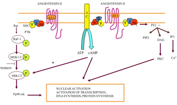
The AT1 receptor, having 7 transmembrane-spanning domains, is a G protein-coupled receptor linked to Gqα and Giα (Figure 3). Through Gqα, the AT1 receptor activates phospholipase C (Lassegue, Alexander, Clark, Akers, & Griendling, 1993; Ullian & Linas, 1990). Through Giα, the AT1 receptor is coupled to adenylate cylase inhibition (Anand-Srivastava, 1993a) as well as the activation of voltage-gated L-type and T-type calcium channels (Chiu, Roscoe, McCall, & Timmermans, 1991; Lu et al., 1996; Maturana, Burnay, Capponi, Vallotton, & Rossier, 1999). In 2001, Touyz and colleagues demonstrated that Ang II-induced activation of c-Src in hypertensive patients resulted in vascular smooth muscle cell growth (Touyz et al., 2001). c-Src exerts its effects on several downstream molecules, such as MAPK, PLC, JAK, PI3K and NAD(P)H oxidase (Touyz & Schiffrin, 2000). Through a different pathway, Ang II results in the activation of MAPK, ERK1/2, p38 kinase and JNK. Furthermore, Saito and Berk observed that the AT1 receptor activation resulted in the transactivation of epidermal growth factor (EGF), platelet-derived growth factor (PDGF) and insulin-like growth factor (IGF) (Saito & Berk, 2001). Evidence from more recent studies have indicated that the Ang II-mediated transactivation of the EGF receptor, through MAPK, plays a role in vascular smooth muscle cell hypertrophy and proliferation (Atef & Anand-Srivastava, 2016). Taken as a whole, activation of the AT1 receptor via Ang II contributes to the generation of hypertension and the complications that results from it.
Figure 3: ANG II signalling mechanisms associated with Gqα and Giα proteins. Through Gqα, the AT1 receptor
activates the phospholipase C signalling pathway, while through Giα, the AT1 receptor is coupled to adenylate cylase inhibition. Adapted from a lecture by Madhu B. Anand-Srivastava, PSL6090, 2015.
1.3.2.1.2 Angiotensin II AT2 Receptor
Like AT1, the AT2 receptor has 7 transmembrane-spanning domains and is largely expressed in the late gestational period of fetal development and quickly diminishes after birth. The AT2 receptor has been shown to play an important role in vasculogenesis by mediating the decline in vascular DNA synthesis occurring during the late stage of fetal development (H. Yamada et al., 1999). The implication of the AT2 receptor in vascular complications have produced controversial findings. A study on transgenic AT2 knockout mice (AT2KO) found them to be more susceptible to developing neointimal vascular inflammation as compared to control rats (Akishita et al., 2000). Conversely, the re-expression of the AT2 receptor was noted in pathological conditions characterized by inflammation (Ruiz-Ortega et al., 2003).
PD98059 ANGIOTENSIN II AT-1 AT-1 A C cAMP ATP αq β PTK SHC Ras RaF-1 P MEK1/2 ERK1/2 Pp90 rsk αi βαq PLC b IP3 DAG PKC NUCLEAR ACTIVATION ACTIVATION OF TRANSCRIPTION, DNA SYNTHESIS, PROTEIN SYNTHESIS
P P ANGIOTENSIN II Ca2+ PIP2 + γ βγ γ
1.3.3 The Endothelin System
Endothelin is a vasoactive peptide that is synthesized primarily by the endothelium. The four types of endothelin, ET-1, ET-2, ET-3 and ET-4, each consist of 21 amino acids containing 2 disulfide bridges (Cys1-Cys15 and Cys3-Cys11) (Rautureau & Schiffrin, 2012). Structurally, ET-1 and ET-2 are 90% homologous (Rautureau & Schiffrin, 2012). Each type of endothelin is coded by a single gene which gives rise to a precursor, prepro-endothelin, which is approximately 200 amino acids long. Following its cleavage, it forms pro-endothelin which is further cleaved by furine and other types of convertases to give rise to peptides which are 38 to 39 amino acids in length. At the vascular level, ET-1 is the most abundant. It is secreted by endothelin conversion enzymes and interacts in an autocrine and paracrine manner with the surrounding cells (Wagner et al., 1992). Playing a major role in blood pressure regulation, ET-1 mainly exerts its effects through 3 types of receptors: ETA, ETB1 and ETB2. The biosynthesis of ET-1 is regulated by several factors such as the shearing forces that stimulates NO release and which negatively influences ET-1 synthesis by the endothelium (Boulanger et al., 1992; Malek & Izumo, 1992). Vasoactive agonists such as Ang II, thrombin, leptin and adrenaline all positively affect the biosynthesis of ET-1 (Quehenberger et al., 2002) as well as ROS (Kahler et al., 2000). Pulmonary tissue constitutes an important site for ET-1 production indicating is important role in the development of pulmonary hypertension (Dupuis, Goresky, & Fournier, 1996; Wagner et al., 1992).
In several animal models of hypertension such as DOCA-salt, Goldblatt (1K1C) and the spontaneously hypertensive rat (SHR), an elevated systemic level of ET-1 is found (Kassab, Novak, Miller, Kirchner, & Granger, 1997; Schiffrin, 1995). An elevation in the concentration of ET-1 results in a hypertrophic vascular remodelling at the level of the arterioles by increasing peripheral vascular resistance (Intengan & Schiffrin, 2000). The elevation in the systemic concentration of ET-1 has also been linked to renal dysfunction (Hirai et al., 2004).
1.3.3.1 Endothelin-1 Receptor
ET-1 receptors are G protein-coupled 7 transmembrane-containing protein structures. VSMCs contain two types of endothelin receptors: ETA and ETB (Azuma et
al., 1995; Davie et al., 2002; Russell & Davenport, 1995; Sato & Amemiya, 1995). In the arteries, the vasoconstrictive effect of ET-1 is largely mediated by ETA (Moreland, Cilea, & Moreland, 1992). The ETB receptor is largely expressed in endothelial cells and plays an important role in NO synthesis as well as other endothelium-dependent vasorelaxants such as prostacyclin (PGI2) and endothelium-derived hyperpolarizing factor (EDHF), acting in a calcium-dependent manner (Triggle et al., 2012). The ETB receptor is also largely responsible for the clearance of ET-1, which has a half-life of approximately one minute (Gasic, Wagner, Vierhapper, Nowotny, & Waldhausl, 1992). Furthermore, ETB stimulates NO production in non-endothelial cells in the thick ascending loop of Henle and results in natriuretic effects following sodium and chloride inhibition. Moreover, the ETB receptor plays an important role in sodium and fluid homeostasis in the kidneys. This last role demonstrates the physiologic effects of endothelin in blood pressure regulation at the renal level (Ahn et al., 2004; Plato, Pollock, & Garvin, 2000).
1.4 Transmembrane Signalling: Mechanisms 1.4.1 Guanine Nucleotide-Binding Proteins
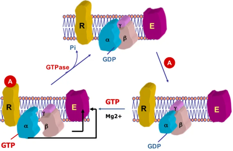
Guanine nucleotide-binding proteins, abbreviated as G proteins, are heterotrimeric, membrane-bound proteins that are coupled to GPCRs. There are four distinct families of G proteins: Gi/o, Gq/11, Gs and G12/13 (Kehrl, 1998). In its inactive state, G proteins are composed of three subunits: α, β and γ. The subunits as well as the GPCR are all linked through the N-terminal of the α subunit and the C-terminal of the γ subunit. The α subunit possesses GTPase activity which is regulated by the hydrolysis of guanosine triphosphate (GTP) to guanosine diphosphate (GDP) (Neer, 1995; Offermanns, Iida-Klein, Segre, & Simon, 1996) (Figure 4). The exchange of GDP for GTP activates the G protein and generates a cascade of signalling mechanisms through the use of second messengers such as inositol triphosphate, diacylglycerol and calcium (Birnbaumer, 1992; Neer, 1995).
Figure 4: Activation of G proteins. Binding of an agonist to the G protein-coupled receptor causes a dissociation of the α subunit from the βγ subunit following the enchange of GDP for GTP. Adapted from a lecture by Madhu B. Anand-Srivastava, PSL6090, 2015.
These second messengers transmit the signal to membrane-bound and intracellular targets such as adenylate cyclase (AC), ionic channels and the endoplasmic reticulum (ER) (Meij, 1996). In addition to the traditional G protein, there are also smaller G proteins, called small GTPases which are monomeric in structure and include proteins such as Ras and Raf.
1.4.2 The Adenylate Cyclase System
1.4.2.1 The Adenylate Cyclase System and Cyclic Adenosine Monophosphate AC is a ubiquitous enzyme made up of two transmembrane subunits. Its activity is regulated by the inhibitory G protein, Gi, and the stimulatory G protein, Gs. Activation of AC leads to the formation of cyclic adenosine monophosphate (cAMP) from a single molecule of ATP. In turn, cAMP possesses the ability to regulate its own activity through the activation of phosphodiesterases which signals its destruction (Furge, Winter, Meyers, & Furge, 2008). Functions of cAMP include the modulation of vascular tone as well as the regulation of vascular smooth muscle cell (VSMC) proliferation (Gusan & Anand-Srivastava, 2013). The antiproliferative effect of cAMP appears to be linked to EGFR-MAPK signalling pathway inhibition. Coupled to Gi, this pathway is generally stimulated by agonists such as EGF or through its transactivation by vasoactive peptides (Y. Li & Anand-Srivastava, 2002; D. Wu, Katz, & Simon, 1993), and its inhibition results
α γ β α β α γ β R R R E E E GDP A GTPase GDP Pi γ GTP Mg2+ A GTP
in the downregulation of transcription factors implicated in cell proliferation, such as c-Myc. Vasoactive peptides such as Ang II and ET-1 inhibit AC activity following the dissociation of the α subunit of Gi. Thus, an increase in the activity of the RAS or endothelin system, as noted under hypertensive conditions, is associated with a reduction in cAMP production at the vascular level. In the SHR rat, the basal activity of AC is reduced, and is associated with a decrease in sensitivity to its agonists (Anand-Srivastava, 1988). Moreover, an alteration in the expression of the G protein in the DOCA-Salt Hypertensive Rat was shown to be linked to a reduction in AC sensitivity (Anand-Srivastava, de Champlain, & Thibault, 1993b; Anand-Srivastava, Picard, & Thibault, 1991). In the L-NAME rat model of hypertension characterized by a pathological reduction in NO bioavailability, an alteration in Gi expression was also demonstrated (Di Fusco & Anand-Srivastava, 2000).
1.4.2.2 Giα
The inhibitory group of G proteins consists of 5 members: Giα1, Giα2, Giα3, GoαA and GoαB (Kehrl, 1998). These multimeric protein complexes are characterized by a sensitivity to pertussis toxin (Kehrl, 1998; Y. Li & Anand-Srivastava, 2002). Activation of the Gi protein results in the inhibition of adenylate cyclase and a subsequent reduction in the intracellular concentration of cAMP (Gilman, 1995). Numerous studies have demonstrated the important role of the Gi protein in the pathogenesis of hypertension in several animal models (Anand-Srivastava, 1993c, 2010; Anand-Srivastava et al., 1993b; Hashim & Anand-Srivastava, 2004). A reduction in the bioavailability of cAMP has been shown to be associated with a hyperproliferation of VSMCs (Gusan & Anand-Srivastava, 2013) as well as endothelial dysfunction (Shah & Singh, 2006).
1.4.3 The Phosphoinositide System 1.4.3.1 Gqα
The Gqα family of G proteins are multimeric protein structures which consists of four members: Gqα, G11α, G14α and G15/16α (Gilman, 1987). These proteins are characterized by their insensitivity towards pertussis toxin (Exton, 1996; Strathmann & Simon, 1990). The Gqα signalling pathway, also known as the phosphatidylinositol
signalling pathway, is composed of three elements: a receptor, a GTP-linked regulatory protein (known as a G protein), and an effector protein. Activation of Gqα results in the activation of phospholipase Cβ (PLCβ) (Alberts, 2002). PLCβ in turn hydrolyzes phosphatidylinositol bisphosphate (PI(4,5)P2) into 2 second messengers, namely inositol 1,4,5-triphosphate (IP3) and diacylglycerol (DAG). IP3 diffuses across the cytosol to join with its receptor in the endoplasmic reticulum which results in intracellular calcium release (Irvine, 1990). DAG, on the other hand, remains in the plasma membrane where it plays two different signalling roles. First, it can be hydrolyzed to release arachidonic acid (AA) which acts as a second messenger, or it can synthesize other small lipid messengers such as eicosanoids. Secondly, DAG can activate serine-threonine kinases, such as protein kinase C (PKC). Once PKC is activated, it can phosphorylate downstream target proteins (Alberts, 2002).
1.4.3.2 Phospholipase Cβ: Structure and Function
PLC is a calcium-dependent phosphoinositide enzyme. Thirteen isoforms of PLC exist in mammals which are divided into six families; β, γ (Patterson, van Rossum, Nikolaidis, Gill, & Snyder, 2005), δ, ε (Bunney & Katan, 2006), ζ (Swann, Saunders, Rogers, & Lai, 2006) and η (Zhou, Wing, Sondek, & Harden, 2005). Each family is comprised of multiple isoforms which are differentiated based on their structural organization, their regulation, their activation, as well as their distribution in tissues (Drin & Scarlata, 2007). Four types of PLCβ exist: PLCβ-1, PLCβ-2, PLCβ-3 and PLCβ-4 (Jalili et al., 1999), with their molecular weights varying between 120 and 155 kDa (Rhee, 2001). While all members of the Gqα family activate PLCβ, they do not activate PLCγ, PLCδ or PLCε (Smrcka & Sternweis, 1993; Taylor & Richardson, 1991). PLCβ-1 is expressed primarily in the heart (Schnabel, Gas, Nohr, Camps, & Bohm, 1996), while PLCβ-2 is found in smooth muscle, neuronal tissue as well as the liver (LaBelle & Polyak, 1996). PLCβ-3 is found in all tissues (Meij, 1996), while PLCβ-4 is expressed in the retina (C. W. Lee, Lee, Lee, Park, & Rhee, 1994) and in specific regions of the brain (Roustan et al., 1995).
Structurally, PLCβ contains two highly conserved regions, X and Y, which forms the catalytic subunit (Rhee & Bae, 1997; Rhee, Suh, Ryu, & Lee, 1989). It also
possesses an EF region which links with Ca+2 ions. There are also two phospholipid membrane regions: the N-terminal PH domain and the C2 domain (Suh et al., 2008). PLCβ distinguishes itself from the rest of the PLC family by the presence of a long C-terminal segment which constitutes approximately 450 residues and which contains multiple factors which are crucial for Gqα interaction as well as for membrane linking and nuclear localization (Drin & Scarlata, 2007; Rebecchi & Pentyala, 2000).
1.4.3.2.1 Activation of PLCβ by the Gαq Subunit
The Gqα family of proteins activates PLCβ in the following order of affinity: β1≥β3>β-2 (Jhon et al., 1993; Smrcka & Sternweis, 1993). The Gqα subunit interacts with the COOH terminal of PLCβ which contains the C2 domain located between residues 663-802 followed by a residue sequence 803-1216. Studies on deletion effects have pointed to residue 845 as being crucial for the association and stimulation by Gqα (Park, Jhon, Lee, Lee, & Rhee, 1993; D. Wu et al., 1993). Moreover, basic residues localized in residue sequences 663-802 and 1055-1075 are equally important for Gqα-mediated stimulation of PLCβ (Kim, Park, & Rhee, 1996).
1.4.3.2.2 Activation of PLCβ by the Gβγ Complex
All members of the PLCβ family, with the exception of PLCβ-4 are activated by the βγ dimer (Camps et al., 1992; Park et al., 1993; Smrcka & Sternweis, 1993). Studies have shown that while both PLCβ-1 and PLCβ-3 interact with the βγ complex, it interacts with PLCβ-2 with the greatest affinity (Runnels & Scarlata, 1999). Therefore, both the α subunit as well as the βγ dimer contribute towards the activation of PLCβ at the cellular level (Rhee, 2001).
1.4.4 Diacylglycerol and Protein Kinase C: Structure and Function
PLCβ hydrolyzes PIP2 to form IP3 and DAG. DAG is a simple lipid made up of a molecule of glycerol linked to a fatty acid chain at the Sn2 position, via an ester linkage, and to a second fatty acid chain at the Sn position, through an ester or ether alkenyl linkage (Cook, Briscoe, & Wakelam, 1991). A diverse array of DAG second messengers can be formed based on fatty chain profile which can be polyunsaturated, di-unsaturated, monounsaturated or saturated (Hodgkin et al., 1998; Wakelam, 1998). Saturated DAG is generally a weak activator of protein kinase C (PKC). Di-unsaturated
forms are more active, while polyunsaturated as well as DAG 1-stearolyl-2-arachidononyl are potent activators of PKC (Marignani, Epand, & Sebaldt, 1996; Schachter, Lester, & Alkon, 1996).
DAG interact with different proteins through their C1 domain. This domain consists of a conserved sequence of 50 amino acids with the motif HX11-12CX2CX12-14CX2CX4HX2CX6-7C (Yang, Ng, & Bikle, 2003). Two different type of C1 domains exist: typical and atypical (Hurley & Grobler, 1997). Among the proteins that contain the C1 domain, we find diacylglycerol kinase, DGK, an inhibitor of DAG, and PKC, an important agonist of DAG.
1.4.4.1 Formation of Diacylglycerol
DAG is principally generated through the hydrolysis of inositol bisphosphates by PLCβ, PLCγ, PLCε and PLCδ (Brose, Betz, & Wegmeyer, 2004). DAG can also be produced through the hydrolysis of phosphatidylcholine (PC) and by phospholipase D (PLD) (Timmers, Schrauwen, & de Vogel, 2008). In the case of insulin resistance, DAG is formed through the esterification of two long-chain acetyl-CoAs on glycerol-3-phosphate (Timmers et al., 2008). Through diacylglycerol acetyltransferase (DGAT), DAG can be transformed into triacylglycerol (TAG) (Carrasco & Merida, 2007). The hydrolysis of TAG by lipases results in an increase in the levels of DAG (Timmers et al., 2008).
1.4.4.2 Protein Kinase C
The major effector of DAG is protein kinase C. PKC belongs to a family of serine/threonine kinases which constitutes more than 12 different isoforms (Budhiraja & Singh, 2008). These isoforms are divided into three categories: conventional, new and atypical (Budhiraja & Singh, 2008). The different PKC isoforms each consist of a polypeptide chain containing an N-terminal regulatory region and a C-terminal catalytic region, both of which contain a conserved (C1-C4) and a variable (V1-V5) region (Budhiraja & Singh, 2008). The C1 site in the regulatory region contains two cysteine-rich domains which link with DAG, phosphatidylserine and a phorbol ester (Salamanca & Khalil, 2005). In certain isoforms of PKC, the C2 site is rich in acid residues, and links with calcium (Salamanca & Khalil, 2005). The C3 site links with ATP and is the principal
target of PKC inhibitors (Budhiraja & Singh, 2008), while the C4 site recognizes the PKC substrates. The catalytic domain is found on sites C3 and C4.
The conventional PKCs (α, β1, β2, γ) possess four conserved regions (C1-C4) and five variable regions (V1-V5) (Salamanca & Khalil, 2005). They are calcium dependent and are activated by phospholipids as well as by DAG (Budhiraja & Singh, 2008). New PKCs (δ, ε, η, θ) do not contain a C2 region (Salamanca & Khalil, 2005). They are stimulated by phospholipids and DAG, but are calcium independent. Atypical PKCs (ν, µ, 1/λ, ζ) possess a cysteine-rich zinc finger motif (Salamanca & Khalil, 2005). They are not activated by DAG, phorbol esther or calcium, but are dependent on phosphatidylserine (Salamanca & Khalil, 2005).
PKC has several biological targets. One of its targets is the phosphorylation of Giα causing the dissociation of the iα subunit from adenylate cyclase (Kanashiro & Khalil, 1998). PKC also targets plasma membrane-localized channels and pumps. In 2004, Baman et al. observed that PKC inhibited calcium-sensitive potassium channel (BKc) activity in lung VSMCs (Barman, Zhu, & White, 2004). Moreover, PKC phosphorylates cytoskeleton proteins and contractile myofilaments in VSMCs. PKC phosphorylates vinculin, a protein localized to focal adhesion plaques, which controls cellular structure and adhesion (Salamanca & Khalil, 2005). PKC also phosphorylates CPI-17, which in turn inhibits myosin light chain (MLC) phosphatase which increases the phosphorylation of MLC and elevates the force of VSMC contraction (Woodsome, Eto, Everett, Brautigan, & Kitazawa, 2001).
1.4.5 IP3 and Calcium
Aside from DAG, the hydrolysis of PIP2 by PLC also produces IP3. Activation of IP3 causes the release of calcium from intracellular stores by a specific class of calcium channels called IP3 receptors (IP3R) which are located in the sarcoplasmic or endoplasmic reticulum (Berridge, 1989). IP3 is transformed into inositol through three successive dephosphorylations which converts it into inositol 1-4-biphosphate (IP2), inositol 4-phosphate (IP1) and finally into free myo-inositol which is then incorporated into the new inositol phospholipid (Voet & Voet, 1998). IP1 phosphatase, the enzyme that catalyzes this last step, is inhibited by lithium (Li+) (Allison & Stewart, 1971). Free
inositol interacts with CTP-DAG to form phosphatidyl inositol (PI) which is phosphorylated into PIP2 by two phosphorylations (Voet & Voet, 1998).
Intracellular calcium release is often followed by a sustained period of extracellular calcium entry (Putney & McKay, 1999). In VSMCs, there are two principal types of calcium channels which are implicated in intracellular calcium entry following the activation of PLC: store-operated channels (SOC) and receptor operated channels (ROC) (Villereal, 2006; Y. Wang, Deng, Hewavitharana, Soboloff, & Gill, 2008).
Store-operated channels are ionic channels located in the plasma membrane (Albert, Saleh, Peppiatt-Wildman, & Large, 2007; Hoth & Penner, 1992). They are activated by a reduction in calcium concentration in the endoplasmic reticulum and not by a reduction in cytosolic calcium concentration (Parekh & Putney, 2005). In several types of cells, including VSMCs, store-operated channels generate a rectifier current called the “Ca+2 release activated Ca+2 current” (ICRAC) (Parekh, Fleig, & Penner, 1997; Parekh & Putney, 2005; Venkatachalam, van Rossum, Patterson, Ma, & Gill, 2002). Two proteins have been cited as being crucial for the proper functioning of SOC: Orai, which forms the pore of the SOC (Albert et al., 2007) and Stim1 (Liou et al., 2005; Roos et al., 2005; Y. Wang et al., 2008) an endoplasmic reticulum protein which functions as a calcium sensor (Liou et al., 2005). The reduction in the calcium concentration in the lumen of the endoplasmic reticulum results in the translocation of Stim1 towards regions of the endoplasmic reticulum which are in proximity to the plasma membrane (Y. Wang et al., 2008; Yeromin et al., 2006). This transfer allows for the interaction with Orai which causes the opening of the SOC and entry of calcium into the endoplasmic reticulum (Y. Wang et al., 2008; Yeromin et al., 2006).
The ROC are activated following a stimulation of the GPCR coupled to PLC which is independent of intracellular calcium depletion. The transient receptor potential (TRP) class of ionic channels is divided into two groups: those which are activated by DAG, TRPC3, TRPC6 and TRPC7, and those which are activated by DAG, TRPC1, TRPC4 and TRPC5 (Dietrich et al., 2005; Freichel, Philipp, Cavalie, & Flockerzi, 2004; Venkatachalam et al., 2002; R. Wang, Liu, Sauve, & Anand-Srivastava, 1998). TRP6 is strongly expressed in VSMCs (Inoue et al., 2001; Jung et al., 2003) and plays a role in receptor-mediated calcium signalling (Inoue et al., 2001; Soboloff et al., 2005). Soboloff
and colleagues utilized siRNA against TRP6 to demonstrate that TRP6-induced increase in intracellular calcium was due to a receptor coupled to PLC, and not due to the exhaustion of calcium reserves (Soboloff et al., 2005). However, other studies have demonstrated that TRPCs could also be activated through depleted calcium reserves. For example, TRPC1 is able to form a complex with TRPC5. This complex produces a current with a conductance that is similar to that of SOCs in VSMCs (Golovina et al., 2001; Trepakova et al., 2001).
The increase in intracellular calcium regulates a number of processes. In VSMCs and cardiomyocytes, it allows for the contraction of cells (Iino, Kasai, & Yamazawa, 1994). The contraction of VSMCs occurs due to the action of myosin light chain kinase which is only active when it associates with the Ca+2-calmoduline complex (Voet & Voet, 1998). In endothelial cells, calcium modulates the synthesis and release of the vasoactive and growth factors such as ET-1, Ang II and nitric oxide (Busse & Lamontagne, 1991; Inagami et al., 1995). Thus, calcium serves as an important second messenger which transmits signals from the extracellular milieu to the interior.
1.4.6 Growth Factors: Receptors
Growth factor receptors consist of a ligand-binding extracellular N-terminal domain, an intracellular C-terminal domain responsible for receptor tyrosine kinase (RTK) activity and an intermediary domain formed by a transmembrane helix. Through RTKs, a phosphate group is transferred from ATP to the tyrosine residue of a protein. The activation of these receptors results in a dimerization, followed by the autophosphorylation of the tyrosine residues. This allows for the activation of certain enzymes and adaptor proteins belonging to the SH2 domain, such as Src (Albert, 2004). RTK’s are implicated in the regulation of several biological processes including growth, differentiation, motility and apoptosis. An increase in the expression or activity of RTKs results in several pathological complications including cancer and cardiovascular disease.
1.4.6.1 Epidermal Growth Factor Receptor (EGF-R)
Epidermal growth factor receptor belongs to a family of ErB1 receptors which possess receptor tyrosine kinase activity (Prigent & Lemoine, 1992). The epidermal growth factor family is comprised of EGF, TGF-α and HB-EGF. EGF-R activation is
implicated in several biological processes such as proliferation, growth and cellular survival. In VSMCs, a defective EGF-R can result in spontaneous cell death, a reduced ERK1/2 sensitivity to GPCR and oxidative stress activity and plays an important role in the homeostasis of VSMCs (Schreier et al., 2011). Activation of EGF-R also forms an essential step in vasoconstriction (Griol-Charhbili et al., 2011) and vascular remodeling (Takayanagi et al., 2015).
Activation of the AT1 receptor coupled to Gqα in VSMCs is associated with the transactivation of EGF-R, which is a calcium dependent process. This is followed by the recruitment of the protein complex Grb2/Shc/Sos leading to the phosphorylation of N70 S6 kinase, a PI3k-Akt dependent signalling mechanism (Eguchi et al., 1999a), as well as p42/44MAPKs (Iwasaki & Ikeda, 1999) which results in enhanced proliferation and protein synthesis (Bahrami et al., 2014).
1.4.6.2 Insulin-Like Growth Factor Receptor (IGF-R)
Insulin-like growth factor receptor is a transmembrane receptor with tyrosine kinase activity. Its structure consists of two heterodimers α and β which are highly expressed in VSMCs (Arnqvist, Bornfeldt, Chen, & Lindstrom, 1995). Elevated concentrations of insulin triggers important biological phenomenons in VSMCs through the activation of IGF-R (Johansson & Arnqvist, 2006). IGF-R, in addition to being directly activated by its ligand, is also transactivated by second messengers such as ROS (Touyz et al., 2003), Ca+2 (Tu et al., 2010) and c-Src (Oligny-Longpre et al., 2012). This indicates IGF-R’s implication as an important mediator of vascular remodeling. Moreover, Wu et al. demonstrated that mechanical stress induced an elevation in the expression of early growth response (Egr-1), which has been shown to be involved in the activation of IGF-R (X. Wu et al., 2010). This also serves to explain the involvement of IGF-R in the formation of the neointima (X. Wu et al., 2010). Furthermore, mechanical stretching forces have been shown to result in an increase in the rate of IGF-R mRNA and protein expression. This suggests that IGF-R plays a pivotal role in the molecular mechanisms relating to vascular remodeling (Song et al., 2007).
1.4.6.3 Platelet-Derived Growth Factor Receptor (PDGF-R)
Platelet-derived growth factor receptors are membrane-bound heterodimer glycoproteins consisting of two chains, α and β, and an Ig-like extracellular domain.
There are two types of receptors for PDGF: PDGF-Rα and PDGF-Rβ (Andrae, Gallini, & Betsholtz, 2008). PDGF-Rα is activated by PDGF-A, PDGF-B and PDGF-C while PDGF-Rβ is activated by PDGF-AB and PDGF-BB (Andrae et al., 2008). Activation of PDGF-R results in a proliferation-inducing response in VSMCs and other types of cells (Kohler & Lipton, 1974). PDGF-R plays an important role in vascular remodeling through its transactivation, which is induced by factors such as Ang II (Touyz, 2005), mechanical shearing forces, and ET-1 (Gomez Sandoval & Anand-Srivastava, 2011). The activation, or transactivation of PDGF-R results in vascular remodeling through the MAPK/AKT signalling pathways (Andrae et al., 2008). Activation of PDGF-R can also result in the transactivation of EGF-R, a result of the ADAM17-independent mechanism (Mendelson, Swendeman, Saftig, & Blobel, 2010).
1.4.7 c-Src Pathway
Proto-oncogene tyrosine-protein kinase Src, known simply as c-Src, is an important member of the non-receptor tyrosine kinase protein family. Encoded by the SRC gene, includes an SH2 domain, an SH3 domain, and a tyrosine kinase domain. In humans, the C-terminal Tyr530 is phosphorylated and interacts with the SH2 and SH3 domains. This diminishes the access of the substrates to the kinase domain. Activation of c-Src is completed when the phosphotyrosine portion of the C-terminal is suppressed and the phosphorylation of Tyr419 occurs (Yeatman, 2004).
c-Src plays a role in numerous cellular functions including hypertrophy, proliferation, migration and cell survival by interacting with signalling cascades including PLC, MAPK, AKT and growth factor receptors (Bolen, Rowley, Spana, & Tsygankov, 1992; Leu & Maa, 2003; Prenzel, Zwick, Leserer, & Ullrich, 2000; Scaltriti & Baselga, 2006; Touyz et al., 2003). Through the use of PP1, a c-Src inhibitor, it was demonstrated that thrombin-induced COX-2 promoter activity, a regulator of cardiac hypertrophy, was attenuated (Chien, Lin, Hsiao, & Yang, 2015). Furthermore, in 2016, Peng and colleagues demonstrated that Ang-II-induced EGF-R activation was mediated by c-Src phosphorylation and may play an important role in Ang II-induced cardiac hypertrophy (Peng et al., 2016).
1.4.8 MAP Kinase Signalling
Mitogen-activated protein kinases (MAPKs) are a highly conserved family of serine/threonine proteins which are activated by mitogenic factors. MAP kinases are divided into 3 pathways: extracellular signal-regulated kinase 1 and 2 (ERK1/2) with isoform p42 and p44, c-Jun N-terminal kinases 1-3 (JNK1-3) and the p38 pathway (Cargnello & Roux, 2011). These pathways are involved in hypertrophy, proliferation, differentiation, stress response, motility, apoptosis and survival (Force & Bonventre, 1998). When an agonist attaches to the ERK1/2 receptor, it results in a phosphorylation cascade. Initiation of the receptor first activates Ras. Ras then goes on to phosphorylate Raf which in turn activates MEK. MEK finally activates ERK1/2, which activates a variety of transcription factors as well as protein kinases and phospholipids. Several stimuli, including vasoactive peptides (Ang II, ET-1), growth factors (EGF), and cytokines can activate ERK1/2 signalling cascades, and under pathological conditions, an increase in the concentrations of these stimuli can result in downstream complications. Moreover, Hashim and colleagues observed that the enhanced proliferation induced by Ang II is inhibited by the MEK1 antagonist, PD98059, in A10 cells (Hashim, Li, & Anand-Srivastava, 2006). Elevated phosphorylation of ERK1/2 is seen in VSMCs from SHR as compared to WKY rats, where the production of endogenous Ang II is greater (Lappas, Daou, & Anand-Srivastava, 2005). Thus, the ERK1/2 signalling pathway plays a critical role in Ang II-mediated signalling.
1.4.9 Phosphoinositide 3-Kinase Pathway
Activated by Ang II, the PI3K pathway constitutes a family of lipid kinases which are implicated in growth, proliferation, differentiation and cellular mobility. There are 3 classes of PI3K, namely class I, class II and class III, which are based on their primary structure, regulation and in vitro substrate. Found predominantly in the vascular system, class I consists of heterodimeric proteins which are composed of a catalytic subunit (p110) and a regulatory subunit (p85 and p101) (Leevers, Vanhaesebroeck, & Waterfield, 1999). This class is activated by receptor tyrosine kinases as well as by GPCRs. PI3Ks catalyze the formation of phosphatidylinositol (3,4,5)-trisphosphate which results in the activation of various protein kinases, including AKT (Oudit et al., 2004). Pharmacological inhibitors targeting PI3K have allowed for the observation of an
increase in intracellular calcium in VSMCs (Seki, Yokoshiki, Sunagawa, Nakamura, & Sperelakis, 1999). Furthermore, deletion of the p110γ subunit in transgenic rat models demonstrated a protective mechanism against Ang II-induced hypertension in vivo (Vecchione et al., 2005). Taken together, these studies highlight the importance of PI3K in the transduction of Ang II-mediated signalling.
1.4.10 AKT Signalling Pathway
AKT, otherwise known as protein kinase B, has been identified as an important target for PI3K in Ang II-mediated signalling in VSMCs (Takahashi, Ohba, & Kaneko, 2015). It has been shown that AKT is responsible for the activation of calcium channels leading to intracellular calcium release induced by Ang II (Seki et al., 1999). AKT also regulates protein synthesis by activating p70 S6-kinase (p70S6K) (Eguchi et al., 1999b). Moreover, AKT regulates c-Myc and Bcl-2 expression as well as inhibits caspases, thereby inhibiting apoptosis and stimulating VSMC survival (Coffer, Jin, & Woodgett, 1998). Taken together, these studies indicate that an imbalance between the mitogenic and apoptotic effects of AKT could contribute to the progression of hypertension.
1.4.11 Reactive Oxygen Species and Oxidative Stress
Reactive oxygen species (ROS) are a family of molecules comprised of oxygen and its derivatives. ROS are formed through a process of oxidation-reduction. Oxidation is defined as the loss of electrons of a molecule, while reduction is characterized by the gain of electrons by a molecule. The reduction of oxygen in the presence of a free electron (e-) results in the formation of superoxide anion (O2-), hydrogen peroxide (H2O2) and hydroxyl radical (OH-) (Touyz & Schiffrin, 2004), molecules which possess oxidizing properties. The harmful effects of ROS are mitigated through antioxidants which possess defense mechanisms which eliminate ROS when they are produced (Fridovich, 1997; Griendling & Ushio-Fukai, 2000). Oxidative stress is the result of an imbalance between the production of ROS and its elimination (Droge, 2002).
Evidence points to the fact that ROS creates toxic effects against cellular metabolism as well as in regulation and signalling (Chiarugi & Cirri, 2003; Griendling & Ushio-Fukai, 2000; Reth, 2002; Sauer, Wartenberg, & Hescheler, 2001). Under physiological conditions and at modest concentrations, ROS regulates VSMC contraction, relaxation, as well as growth (Cosentino, Sill, & Katusic, 1994; Rao & Berk,