Studying the beginning of the end: The roles of Tbf1 and Reb1 at
subtelomeres
Par Alexandra Krallis
Département de Microbiologie et d’Infectiologie
Mémoire présentée à la Faculté de Médecine et des Sciences de la Santé en vue de l’obtention du grade de maître ès sciences (M. Sc.) en Microbiologie
Sherbrooke, Québec, Canada Avril, 2020
Membres du jury d’évaluation
Pr Raymund J. Wellinger, Département de Microbiologie et d’Infectologie Pr Benoit Chabot, Département de Microbiologie et d’Infectologie
Résumé
Studying the beginning of the end: The roles of Tbf1 and Reb1 at subtelomeres Par Alexandra Krallis
Programme de Microbiologie
Mémoire présenté à la Faculté de médecine et des sciences de la santé en vue de l’obtention du diplôme de maitre en sciences (M. Sc.) en Microbiologie, Faculté de Médecine et des Sciences de la Santé, Université de Sherbrooke, Sherbrooke, Québec,
Canada, J1H 5N4
Les séquences télomèriques chez S. cerevisiae recrutent une multitude de protéines afin de remplir ses fonctions essentielles pour le maintien de l’integrité genomique : la réplication complète des chromosomes et la protection des mécanismes de réparation de l'ADN. Cependant, il y a des indices que les régions sous-télomèriques, qui se trouvent directement à l'intérieur des répétitions télomèriques, peuvent aussi affecter les fonctions des extrémités chromosomiques. Deux des principales protéines recrutées aux sous-télomères sont Tbf1 et Reb1. Bien qu'elles se lient aux régions promotrices d'une multitude de gènes, leurs fonctions aux extrémités chromosomiques ne sont pas encore comprises.
Des études précédentes suggèrent que Tbf1 et Reb1 pourraient jouer un rôle dans le maintien de la longueur des télomères et en empêcher la propagation de répression transcriptionnel des gènes près des télomères. Comme beaucoup de ces études ont été réalisées en l'absence de régions sous-télomèriques ou avec des allèles mutants, on ne sait pas si les phénotypes observés proviennent de changements dans les régions sous-télomèriques ou des altérations de la transcription des cibles Tbf1 ou Reb1. Afin d'éviter ces complications, un système a été conçu pour étudier les effets de l'absence de Tbf1 et Reb1 des sous- télomères avec des structures naturelles.
L'utilisation de ce système a permis de découvrir que, Tbf1 et Reb1 ne sont pas très importantes pour le maintien de la longueur des télomères ou pour limiter la
propagation de répression transcriptionnel. Cependant, il a été observé que Tbf1 et Reb1 ont un rôle dans la répression de TERRA, un ARN long non codant transcrit à partir des régions sous-télomèriques. Récemment, il a été suggéré que TERRA pourrait jouer un rôle dans le maintien des télomères. Toutefois, il est crucial de limiter la transcription du télomère, car elle pourrait mener à une cassure de l’ADN et l'instabilité génomique. Cette étude souligne l'importance de travailler avec des régions sous-télomèriques
non-modifiées ou non-modifiées en étudiant les télomères et offre un nouvel aperçu de la régulation de TERRA.
Summary
Telomeres protect the ends of linear chromosomes from being recognized as DNA breaks, helping to avoid events that could lead to genomic instability. The telomeric sequences in budding yeast recruit a multitude of proteins in order to carry out essential functions in end replication and protection from DNA repair machinery. However, there is evidence that subtelomeres, which lie directly interior to the telomeric repeats, may also affect the properties of the chromosomal ends. Two of the main proteins recruited to the subtelomeres are Tbf1 and Reb1. While they bind at promoter regions of a multitude of genes, their function at the chromosomal ends is still unclear.
TBF1 and REB1 are both essential genes, with some overlapping targets and functions in fine tuning transcription and creating nucleosome free regions. Past work suggests both Tbf1 and Reb1 could have roles in telomere length maintenance and limiting the spread of telomere silencing. As many of these studies were done in the absence of subtelomeric regions, or with mutant alleles, it is unclear if the telomere phenotypes observed stem from changes in the subtelomere regions or from alterations in transcription of Tbf1 or Reb1 targets. This is evidenced by different studies producing conflicting evidence pertaining to the functions of these proteins. In order to avoid such complications, a system was designed to study the effects of the absence of Tbf1 and Reb1 at subtelomeres with otherwise native structures.
Through the use of this system, it was found that Tbf1 and Reb1 may not be very important for telomere length maintenance or limiting the spread of telomere silencing. However, it was discovered that Tbf1 and Reb1 have a role in repressing TERRA, a long non-coding RNA transcribed from the subtelomere and telomeric repeats. Recent work suggests TERRA may have a role in telomere maintenance in the absence of telomerase. However, limiting transcription of the telomere is crucial, as it could lead to replication fork stalling, DNA breaks and genomic instability. This study underlines the importance of working with natural subtelomere regions when studying telomeres and offers a new insight into TERRA regulation.
Table of Contents
Introduction
Telomeres ... 1
Essential functions of telomeres: chromosome capping ... 3
Essential functions of telomeres: chromosome end replication ... 5
Subtelomeres ... 9
Telomeric properties: Telomere Position Effect ... 11
Telomeric Properties: Telomeric repeat containing RNA ... 13
Tbf1 ... 15
Reb1 ... 19
Objectives... 20
Materials and Methods Plasmid Cloning Methods ... 23
Construction of TEL01Lmod and TEL03Lmod plasmids ... 26
E.coli Transformation ... 32
Yeast Strains ... 33
Yeast Transformation ... 36
Tagging proteins in Yeast ... 37
Modification of TEL01L and TEL03L in S. cerevisiae ... 38
Site-Specific Recombination ... 39
Serial dilution growth tests on solid plates: “Spot Tests” ... 40
PCR mediated Gene Deletions ... 40
Mating, Sporulation and Microdissection ... 40
Yeast Genomic DNA extraction and quantification... 41
Yeast total RNA extraction ... 43
DNaseI Treatment of RNA ... 44
Yeast Rapid Protein TCA Extraction ... 44
Western Blot ... 45
Southern Blot ... 46
Northern Blot ... 47
Purification of His-Tev-Flag Tagged Proteins ... 47
Quantitative PCR (qPCR) ... 50
Reverse Transcription qPCR ... 52
Telo-PCR ... 53
Results Chapter I – Construction of system Preamble ... 59
Results ... 60
Results Chapter II – Roles of Tbf1 and Reb1 in telomere length maintenance Preamble ... 69
Results ... 69
Results Chapter III – Effects of Tbf1 and Reb1 on TPE Preamble ... 85
Results ... 85
Results Chapter IV – Effects of Tbf1 and Reb1 on telomeric transcription Preamble ... 92
Results ... 92
Results Chapter V – Purification of yKu70 and yKu80 Preamble ... 100
Results ... 101
Discussion of Chapters I-IV System constructed for the study of Tbf1 and Reb1 at subtelomeres ... 112
Weak roles of Tbf1 and Reb1 at native subtelomeres ... 114
Tbf1 and Reb1 regulate TERRA abundance ... 118
Telomeric properties vary at different chromosomal ends ... 122
Discussion of Chapter V Purification of yKu70 and yKu80 ... 125
Conclusions and Perspectives References
List of Figures
Figure 1: Schema of the telomere nucleoprotein structure in S. cerevisiae ... 3
Figure 2: End-replication problem. ... 6
Figure 3: Subtelomeric sequences and binding proteins. ... 10
Figure 4: Tbf1 and Reb1 binding sites in TEL01L XCR were mapped by YeTFasCo... 27
Figure 5: TEL01Lmod integrative plasmid... 28
Figure 6: Tbf1 and Reb1 binding sites in TEL03L XCR were mapped by YeTFasCo... 30
Figure 7: TEL03Lmod linearized plasmids. ... 30
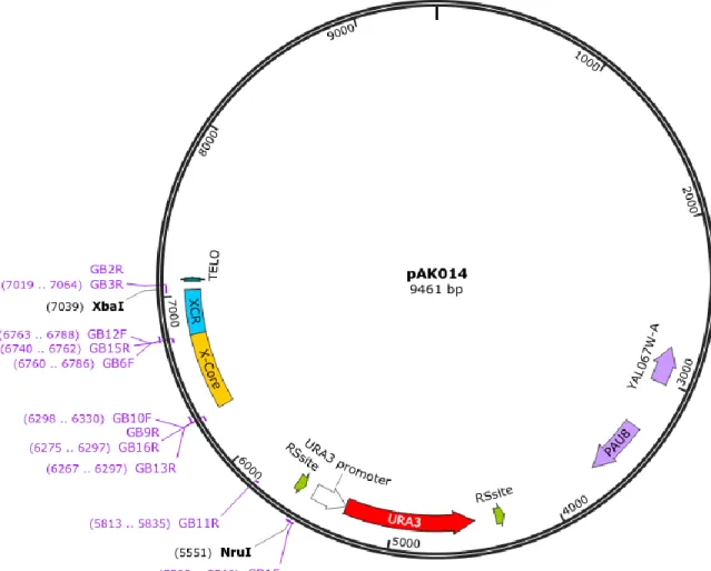
Figure 8: Primer placement on pAK014 ... 32
Figure 9: Schema of subtelomeric areas discussed. ... 61
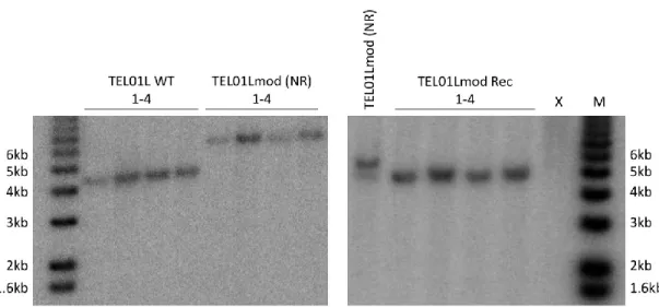
Figure 10: Analysis of integration and recombination of TEL01Lmod subtelomeres. ... 62
Figure 11: Primer pairs tested for TEL01Lmod ChIP qPCR. ... 63
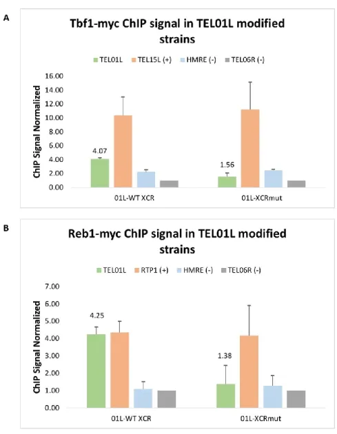
Figure 12: ChIP qPCR of Tbf1-myc and Reb1-myc in TEL01Lmod strains. ... 66
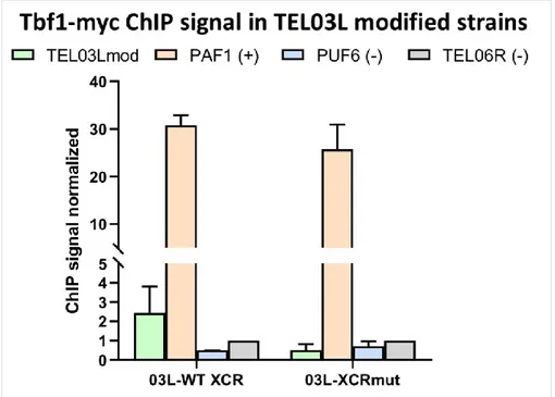
Figure 13: ChIP qPCR of Tbf1-myc in TEL03Lmod strains. ... 67
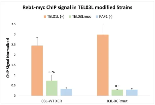
Figure 14: ChIP qPCR of Reb1-myc in TEL03Lmod strains. ... 68
Figure 15: XY’ telomere length in TEL03Lmod strains measured by Southern Blot. ... 70
Figure 16: TEL03L telomere length measured by southern blot. ... 72
Figure 17: TEL01L telomere length measured by southern blot. ... 73
Figure 18: Global telomere length of TEL03Lmod strains with tel1∆, measured by Southern Blot. ... 75
Figure 19: Global telomere length of TEL01Lmod strains with tel1∆, measured by Southern Blot. ... 76
Figure 20: TEL03L telomere length in tel1∆ background measured by southern blot... 77
Figure 21: Optimization to measure TEL01Lmod telomere length by southern blot. ... 79
Figure 22: TELO PCR of TEL01Lmod before and after digestion with XhoI. ... 80
Figure 23: TEL03Lmod in yku80∆ background measured by southern blot. ... 82
Figure 24: X-Only telomere length measured by Southern Blot. ... 84
Figure 25: Quantification of serial dilution growth tests of cells with URA3 at 01L-WT X-element, 01L X-element∆ and 01L-XCR∆. ... 87
Figure 26: Serial dilution growth test of cells with URA3 at 01L-WT XCR and 01L-XCRmut. ... 88
Figure 27: Quantification of serial dilution growth tests of cells with URA3 at 01L-X-Core∆ WT XCR, 01L-X-Core XCRmut and 01L-WTshort. ... 90
Figure 28: Serial dilution growth tests of cells expressing TBF1 or tbf1-82 and URA3 at 01L-WT XCR. ... 91
Figure 29: Probes developed to specifically detect TEL03Lmod ... 93
Figure 30: Northern Blots hybridized with TERRA pShort probe. ... 94
Figure 31: TERRA measured from X-only telomeres in sir4∆ strains ... 96
Figure 32: TERRA measured from X-only telomeres in sir4∆ tbf1-453 strains. ... 98
Figure 33: Testing yKu70-HTF and yKu80-HTF Flag tags by Western Blot. ... 103
Figure 34: Teloblot of yKu70 and yKu70-HTF strains... 104
Figure 35: Optimization of the IP of yKu70-HTF and yKu80-HTF ... 106 Figure 36: TEV cleavage of yKu70-HTF and yKu80-HTF from beads and in whole cell
List of Tables
Table 1: Plasmids referred to throughout this work. ... 23
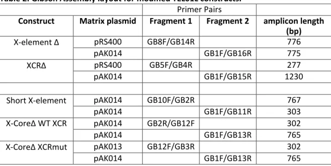
Table 2: Gibson Assembly layout for modified TEL01L constructs. ... 31
Table 3: S. cerevisiae strains referred to throughout this work. ... 33
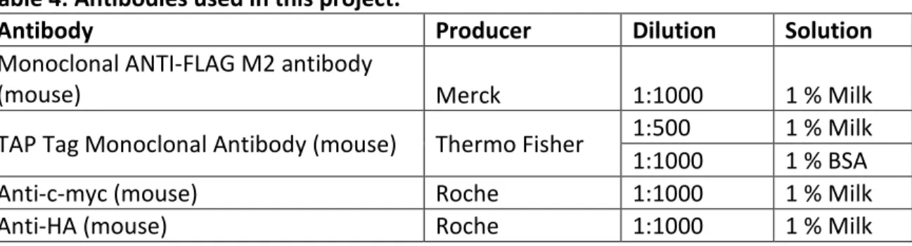
Table 4: Antibodies used in this project. ... 45
Table 5: Primer Pairs used for qPCR in this project ... 51
Table 6: Primers used in this project ... 54
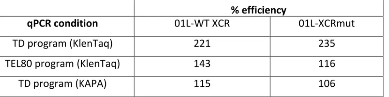
Table 7: The % efficiency of Ch2R/TXCRM primer pair measuring Tel01Lmod by qPCR. ... 64
Table 8: Average telomere length of TEL03Lmod. ... 72
Table 9: Average length of TEL01Lmod. ... 74
Table 10: Average telomere length of TEL03Lmod in tel1∆ strains ... 78
Table 11: Mean telomere length of TEL01Lmod strains measured by Southern Blot and TELO PCR. ... 81
Table 12: Mean telomere length of TEL03Lmod strains measured by Southern Blot ... 82
Table 13: Predicted Tbf1 and Reb1 binding sites at X-only telomeres. ... 99
Acknowledgements
I would like to first thank my research director, Professor Raymund Wellinger, for
welcoming me into his laboratory first as an intern and again as a masters student. I truly appreciate the enjoyable and friendly environment in the Wellinger Lab and would like to thank all Lab members, as well as Pr. Wellinger for their support, encouragement and positivity. I would like to especially thank Emeline Pasquier for mentoring me during my internship and continuing to offer so much invaluable advice and help and throughout my experience in the lab. I would also like to thank Erin Bonnell for the strains provided, as well as her support and advice on all things Tbf1. Finally, I would like to thank Pr. François Bachand and Pr. Benoit Chabot for agreeing to be part of my jury committee.
List of Abbreviations
∆: delta, identifies a deletion °C: degrees Celsius μg: microgram μl: microliter μM: micromolar
3’: 3’ extremity of a nucleotide, free OH group on the 3’ carbon of ribose ring
5’: 5’ extremity of a nucleotide, free OH group or phosphate ester on 5’ carbon of ribose ring
5-FOA: 5-Fluoroorotic acid A: adenosine
ALT: alternative lengthening of telomeres ARS: Autonomously replicating sequence b: base(s)
bp: base pair(s)
BIR: Break-induced replication BSA: bovine serum albumin C: cytosine
ChIP: Chromatin Immunoprecipitation
ChIP-seq: Chromatin Immunoprecipitation-sequencing CRAC: Cross-linking and analysis of cDNA sequencing C-terminal: carboxy-terminal
dCTP: deoxycytidine triphosphate dGTP: deoxyguanosine triphosphate DNA: Deoxyribonucleic acid
DSB: Double-strand break dsDNA: double-stranded DNA
dNTP: deoxynucleoside triphosphate DTT: Dithiothreitol
ECL: Electrochemiluminescence or electro generated chemiluminescence EDTA: Ethylenediaminetetraacetic acid
G: guanosine
GBD: Gal4 binding domain GRF: General regulatory factor
HEPES: 4-(2-hydroxyethyl)-1-piperazineethanesulfonic acid HO: Homothallic switching endonuclease
HR: Homologous recombination HDR: Homology directed repair HTF: 6xHis-TEV-3xFlag
ITS: interstitial telomeric repeat sequence IGR: Intergenic region
IgG: Immunoglobulin G IP: Immunoprecipitation kb: kilobase
LB: Luria Broth medium M: molar
m/v: mass per volume NEB: New England Biolabs
NDR: Nucleosome depleted region NHEJ: Non-Homologous End-Joining nM: nanomolar
NS: non-specific nt: nucleotide
N-terminal: amino terminal ORC: Origin Recognition Complex ORF: Open Reading Frame
PBS: phosphate-buffered saline
PBS-T: phosphate-buffered saline with 0.1% Tween-20 PCR: Polymerase Chain Reaction
pH: measure of acidity or basicity of a solution PMSF: phenylmethylsulfonyl fluoride
RNase: Ribonuclease
RS: sequence recognized by Recombinase R SDS: sodium dodecyl sulfate
SDS-PAGE: SDS-Polyacrylamide Gel Electrophoresis snoRNA: small nucleolar RNA
SPP: Service de Purification de Protéines at Université de Sherbrooke SSC: Saline sodium citrate
ssDNA: single-stranded DNA
STARs: SubTelomeric Anti-silencing Regions STEX: Single Telomere Extension assay STR: Subtelomeric repeated elements T: Thymidine
Tx: Time point X (T1 = Time point 1)
TAE: 40 mM Tris, 20 mM glacial acetic acid, 1 mM EDTA pH 8.0 TAP: Tandem Affinity Purification
Taq: DNA polymerase from Thermophilus aquaticus TBE: 40 mM Tris, 20 mM borate, 1 mM EDTA pH 8.0 TCA: Trichloroacetic acid
TEL01Lmod: modified TEL01L telomere TEL03Lmod: modified TEL03L telomere
TEL07Ltr: truncated TEL07L telomere, lacking subtelomere
Teloblots: Southern blots measuring telomere length TERRA: Telomeric repeat containing RNA
TEV: Tobacco etch virus protease
TdT: Terminal deoxynucleotidyl Transferase
TN150: 50 mM Tris-HCl pH 7.8, 150 mM NaCl, 0.1% NP-40 TN1000: 50 mM Tris-HCl pH 7.8, 1 M NaCl, 0.1 % NP-40
TPE: Telomere Position Effect TRF: Terminal Restriction Fragment ts: temperature sensitive
UAS: Upstream activation sequence UTR: Untranslated region
WCE: whole cell extract
XCR: X-element combinatorial repeats
XCRmut: XCR containing point mutations in Tbf1 and Reb1 binding sites v/v: volume per volume
V: Volt
YC: Yeast Complete synthetic media
Introduction
Telomeres
Eukaryotic genomes are arranged into multiple linear chromosomes beginning and ending with structures protecting them, named telomeres. These structures were first characterized by Herman Muller and Barbara McClintock (1939, 1941), who each noted that broken chromosomal ends were highly unstable and prone to fusion, while native chromosomal ends were resistant to chromosome fusion (McClintock, 1941; McClintock, 1939; Muller, 1938). The structures at the chromosomal ends were named by Herman Muller (1938) after the Greek “telos” (end) “meros” (part) and are known to be essential for the maintenance of genome stability due to their ability to inhibit
chromosome-chromosome fusions.
The structure of telomeres is conserved throughout eukaryotes, while the exact sequence varies across species. Telomeric DNA is comprised of non-coding G-rich repeat sequences, with a double stranded portion and 3’ single stranded overhang (Larrivée et al., 2004; McElligott and Wellinger, 1997; Wellinger et al., 1993). The lengths of double and single stranded sections vary across different species. The telomeres carry out their main function, maintenance of genome stability, by recruiting various protein complexes. For example, in human cells, the TTAGGG (T2AG3) telomeric repeats (de Lange et al., 1990)
form 5-15 kilobases (kb) of double stranded DNA and a 50-300 nucleotide (nt) 3’ single stranded overhang (McElligott and Wellinger, 1997). The T2AG3 sequence is conserved in
all mammalian species, while the length of double strand and single strand sections of the telomeres can vary. This telomeric DNA associates with the shelterin complex, consisting of 6 protein subunits (Chen, 2019; De Lange, 2005). The shelterin complex specifically binds telomeres by proteins TRF1, TRF2 and POT1, which directly bind DNA in a sequence specific manner. TRF1 and TRF2 bind double stranded T2AG3 repeats through their two
SANT/Myb type DNA binding domains, while POT1 binds the single stranded portion (Chen, 2019). Proteins Rap1, TIN2 and TPP1 bind and interconnect the DNA binding
proteins. Association with the shelterin complex remodels the telomeric DNA and recruits additional proteins to facilitate telomeric functions, such as inhibiting DNA repair
mechanisms and maintaining telomere length homeostasis (Chen, 2019).
Telomeres of S. cerevisiae (budding yeast), are structured similarly, although they do not interact with an analogous set of proteins. Telomeric DNA consists of 300 75 bp of double standed DNA with a TG1-3 degenerate repeat sequence (Wellinger and Zakian,
2012). The 3’-single stranded overhang is also G-rich, with the same TG1-3 repeats and is
12-15 nt long (Larrivée et al., 2004). Like the human telomere, budding yeast telomeres are bound by several protein factors. Although the proteins themselves vary, they too associate with telomeric DNA to allow the telomeres to carry out essential telomeric functions (Wellinger and Zakian, 2012). The yeast telomere and its binding factors are represented in Figure 1. Double stranded yeast telomeric DNA is covered with Rap1, an ortholog to human Rap1 (Li et al., 2000; Wellinger and Zakian, 2012). While the hRap1 does not directly bind DNA, scRap1 binds telomeric DNA via its double myb domain and recruits Sir3/Sir4 and Rif1/Rif2 (Buck and Shore, 1995; Graham et al., 1999; Hardy et al., 1992; Moretti and Shore, 2001; Moretti et al., 1994; Wotton and Shore, 1997). The Sir proteins form a complex involved in repressing transcription at the chromosomal ends and also interact with other proteins important for telomeric functions. In addition to this, Sir4 is involved in tethering the telomeres to the nuclear envelope, where they form foci in the G1-S-phase (Andrulis et al., 2002; Bupp et al., 2007; Taddei et al., 2004). Proteins Rif1 and Rif2 are important for telomere length homeostasis (Wellinger and Zakian, 2012).
As in human telomeres, the 3’ single strand overhang is bound by a protein that recognizes single stranded DNA. Cdc13 binds single stranded TG1-3 repeats and interacts
with different proteins, depending on the cell cycle phase (Mersaoui and Wellinger, 2018). Cdc13 is involved in a variety of functions such as telomere elongation, telomere replication and chromosomal capping, by interacting with various proteins at different stages of the cell cycle (Mersaoui and Wellinger, 2018; Wellinger and Zakian, 2012). Telomeres are also bound by yKu70 and yKu80 proteins, which form a ring shaped yKu complex. The complex can either directly bind DNA or be recruited through interactions
between yKu80 and Sir4 (Gravel et al., 1998; Larcher et al., 2016; Roy et al., 2004). The yKu complex plays a role in chromosomal end capping, repression of transcription near telomeres, clustering telomeres near the nuclear envelope and telomere length
maintenance (Boulton and Jackson, 1998; Fisher et al., 2004; Gallardo et al., 2011; Laroche et al., 1998; Polotnianka et al., 1998). However, it also has roles in DNA repair at double stranded breaks within the genome (Fell and Schild-Poulter, 2015). Many of the proteins involved in telomere functions also have roles in transcription or DNA repair pathways when bound at other genomic loci, some of which will be discussed below (Wellinger and Zakian, 2012).
Figure 1: Schema of the telomere nucleoprotein structure in S. cerevisiae
Double stranded DNA is bound by Rap1, which recruits proteins Sir2/3/4 to form the Sir complex. Rap1 also recruits Rif1 and Rif2 to negatively regulate telomere elongation. The yKu complex is formed by proteins yKu70 and yKu80 and binds at the double
strand/single strand junction to function in end capping. The CST complex, a complex containing Cdc13, Stn1 and Ten1, binds to single stranded TG1-3 repeats.
Essential functions of telomeres: chromosome capping
As Muller (1938) and McClintock (1939) described in their experiments observing stable chromosome ends, the telomeres prevent fusion between chromosomes, which is essential for genome stability in a healthy cell. When DNA breaks occur within a
chromosome, the cell must repair it through DNA repair mechanisms such as
the telomeres as double stranded breaks would lead to the fusion of chromosomes by DNA repair mechanisms and create dicentric chromosomes. This process is propagated in daughter cells in a cycle called the breakage-fusion-bridge cycle and results in gross genomic instability (McClintock, 1939). NHEJ is particularly hazardous for unprotected chromosomal ends, as this DNA repair mechanism recognizes double stranded breaks and joins the DNA together, regardless of sequence. To prevent these events, the telomere nucleoprotein structures protect the chromosomal ends from recognition by DNA repair machinery as a double stranded break. This function is called chromosome capping (Garvik et al., 1995; Wellinger and Zakian, 2012).
There are multiple proteins contributing to the capping functions of telomeres, however, many of these proteins have additional telomeric and non-telomeric functions. As is represented in Figure 1, Rap1 binds double stranded TG1-3 repeats (Conrad et al.,
1990; Gilson et al., 1993). In addition to Rap1 contributing to telomere capping by limiting resection of the C-rich strand, it recruits Rif2 and Sir4 proteins, which decrease
chromosomal fusion by 2 different pathways (Marcand et al., 2008; Vodenicharov et al., 2010). Rif2 is thought to inhibit telomere-telomere fusion by inhibiting the MRX complex required for NHEJ from functioning. Sir4 contributes to decreasing telomere-telomere fusion, possibly through interactions with yKu80. The yKu complex is a protein with functions at non-telomeric loci (Downs and Jackson, 2004). yKu facilitates NHEJ repair of double stranded breaks by binding to DNA at the breaks in a sequence independent manner and preventing MRX dependent 5’ end resection (Bonetti et al., 2010). When binding telomeres, for example after replication fork collapses, the yKu complex is
implicated in telomere capping by inhibiting 5’-end resection (Gravel et al., 1998; Larcher et al., 2016; Vodenicharov et al., 2010). The single stranded telomeric repeats are bound by Cdc13, which carries out its protective functions by recruiting essential proteins Stn1 and Ten1 to form the CST complex (Wellinger and Zakian, 2012). This complex protects the telomere from C-strand degradation and subsequent activation of DNA damage checkpoints (Garvik et al., 1995). The additional functions of Cdc13 in end-replication and telomere elongation events will be discussed below.
Essential functions of telomeres: chromosome end replication
In addition to linear chromosomes being vulnerable to degradation and chromosomal fusion, they also present a problem for conventional DNA replication machinery. Each round of DNA replication causes a slight loss of terminal sequences at one end of each chromosome, represented in Fig. 2 (Olovnikov, 1973; Soudet et al., 2014). Replication of the leading and lagging strands at the ends of the chromosomes produces two different types of DNA ends (Lingner et al., 1995). The lagging strand is replicated via short Okazaki fragments, using the G-rich strand as a template. The degradation of the last short RNA primer after DNA replication results in the 3’ single stranded overhang required for the functional telomere structure and does not result in a loss of terminal sequences (Soudet et al., 2014). Replication of the leading strand in the 5’ to 3’ direction uses the C-rich strand as a template and continues until the end of the template
(Olovnikov, 1973). The loss of terminal sequence occurs upon generation of the 3’ single stranded overhang. The newly replicated 5’ strand is resected in the 5’ to 3’ direction by an exonuclease and then filled in, leaving a 3’ overhang of 12-15 bp (Soudet et al., 2014). Thus, the newly synthesized G-rich leading strand is now 12-15 bp shorter than the original G-rich lagging strand.
Figure 2: End-replication problem.
Parental 5’ and 3’ strands are indicated in dark blue and dark red respectively. Daughter 5’ and 3’ strands are indicated in light blue and dark blue strands. After replication of the leading 5’ end, the 5’ strand is resected and subsequently filled in (light blue). RNA primers are shown by grey arrow. Sequence loss after replication of 5’ strand is indicated by dashed grey lines.
As the telomeres are made of non-coding sequences, the gradual loss of DNA does not lead to a loss of coding genetic material. However, telomeric proteins involved in capping require certain lengths of telomeric sequences in order to bind effectively and carry out capping functions. Thus, human cells have a limit of cell divisions (Hayflick Limit) due to the progressive shortening of telomeric sequences that comes with DNA
replication and cell division (Hayflick and Moorhead, 1961; Lundblad and Szostak, 1989). After somatic cells have reached this limit, they enter a G0 state and do not replicate
further. This also serves to limit the accumulation of mutations, which could eventually lead to genetic instability and cancer. However, mammalian germ and stem cells, as well as yeast cells, have unlimited dividing potential. This is due to the presence of a reverse transcriptase called telomerase, which is able to counteract the progressive shortening of chromosomal ends by elongating telomeric sequences (Greider and Blackburn, 1985, 1987; Morin, 1989).
Telomerase consists of an RNA scaffold that also encompasses a template for telomeric sequences, a catalytic protein subunit and several other proteins essential for in vivo function. In S. cerevisiae, the RNA moiety of the ribonucleoprotein, called TLC1, is a long non-coding RNA that possesses multiple distinct elements represented in Figure 3 (Singer and Gottschling, 1994). Near the 17 nt template region at the center of the RNA is a template boundary element and a pseudo-knot structure (Dandjinou et al., 2004). The RNA is folded into 3 stem loops such that the 3’ end is in proximity with the 5’ end. The catalytic subunit, Est2 (ever shorter telomere), binds to the central area containing the template for elongation of the G-rich 3’ overhang (Chappell and Lundblad, 2004; Livengood et al., 2002). The Est1 accessory protein binds to a bulge on the third stem loop, with the Est3 protein bridging Est1 and Est2 (Seto et al., 2002; Tucey and Lundblad, 2014). These proteins are all named for their “ever shorter telomere” phenotype, as their deletion causes progressive telomere shortening, eventually leading to genomic instability and cell death (Lendvay et al., 1996; Lundblad and Szostak, 1989). Proteins Pop1, Pop6 and Pop7 are thought to stabilize the complex by binding the same bulge as Est1,
potentially interacting with Est1 and Est2 (Lemieux et al., 2016; Laterreur et al., 2018). The 3’ end of the RNA is bound by the Sm7 complex, which also contributes to the stability of
the molecule (Seto et al., 1999). In order to import and retain the telomerase molecule in the nucleus, the yKU complex binds a 48 nt stem loop called the yKu binding stem
(Gallardo et al., 2008; Seto et al., 1999; Stellwagen et al., 2003).
Telomerase is only recruited to specifically short telomeres in the late S phase of the cell cycle (Teixeira et al., 2004). At long telomeres, more Rap1 is associated to the double stranded TG1-3 repeats, recruiting Rif1 and Rif2. Rif2 inhibits 5’ to 3’ end resection
by the MRX complex (Marcand et al., 2008). This limits the amount of 3’ single stranded overhang available for telomerase to associate with, thus limiting telomere elongation by telomerase. At short telomeres, less Rap1 is present, as there are less double stranded TG1-3 repeats, leading to a decreased presence of Rif2 (Levy and Blackburn, 2004;
Marcand et al., 1997). This allows MRX to be activated by the Tel1 kinase, leading to resection of the C-rich strand (Goudsouzian et al., 2006; Martina et al., 2012). The
3’ single strand overhang produced then binds Cdc13, which, at this stage in the cell cycle, is not associated with Stn1 and Ten1, but recruits telomerase to the telomere via
interactions with Est1 (Chen, 2019; Evans and Lundblad, 1999; Wu and Zakian, 2011). The 3’ single stranded overhang is elongated. In the G2 phase, Cdc13 begins to associate with Stn1 and Ten1 to form the CST complex and recruit DNA polymerase to fill in the telomeric C- strand, completing telomere elongation (Chandra et al., 2001; Grossi et al., 2004; Mersaoui and Wellinger, 2018).
In the absence of telomere elongation by telomerase, telomeres will gradually shorten, and cells will enter a permanent cell cycle arrest and senesce (Lundblad and Szostak, 1989; Singer and Gottschling, 1994). The same can happen due to the presence of a single, critically short telomere when it is unable to be repaired by telomerase (Abdallah et al., 2009; Hackett et al., 2001; Khadaroo et al., 2009). However, a certain subset of these cells can evade replicative senescence and regain replicative capacities (Lundblad and Blackburn, 1993; Teng and Zakian, 1999). These so-called “survivors” maintain their telomere length by break induced replication (BIR), a form of homologous recombination (Kass- Eisler and Greider, 2000; Lundblad and Szostak, 1989). BIR occurs at collapsed replication forks, eroded, uncapped telomeres and is also recruited at RNA-DNA hybrids called R- loops (Balk et al., 2013; Lydeard et al., 2007). This alternative form of telomere maintenance can cause two different DNA arrangements at the chromosomal ends, causing cells to be classified as type I and type II survivors (Lundblad and Blackburn, 1993; Teng and Zakian, 1999). Type I survivors are characterized by an amplification of Y’ elements and only short tracts of double stranded TG1-3 repeats. The formation of a type I
survivor requires RAD52, RAD51, RAD54, RAD55 and RAD57 (Larrivée and Wellinger, 2006; Lundblad and Blackburn, 1993). Type II survivors have only a slight amplification of the Y’ elements, but have an extremely variable extension of TG1-3 repeats, some arriving
to be over 12 kb in length (Teng and Zakian, 1999; Teng et al., 2000). The process to form type II survivors requires the MRX complex, TEL1, SGS1 and RAD59. While type I survivors often form first in a population, they grow slowly and are frequently outcompeted by type II survivors in liquid cultures (Lundblad and Blackburn, 1993; Teng et al., 2000).
Subtelomeres
The length of telomeres in S. cerevisiae can sometimes vary by approximately 150 bp from telomere to telomere. Even in humans, some chromosomes have been noted to have consistently longer or shorter tracts of telomeric repeats (Martens et al., 1998). This could be influenced by the subtelomeric regions, which vary across chromosomal ends (Gilson and Londoño-Vallejo, 2007). In S. cerevisiae, telomeres can be divided into two classes, depending on their subtelomeric areas (Figure 3). XY’ telomeres posess both X and Y’ subtelomeric elements, while X-only telomeres only have X-elements. Y’elements are present at approximately half of the chromosomal ends in 1-4 copies and exist in two forms; Y’ short (5.2 kb) and Y’ long (6.7 kb) (Chan et al., 1983; Chan and Tye, 1983). These elements are homogenous in sequence, only differing from each other in some insertions and deletions and undergo frequent mitotic recombination (Horowitz et al., 1984; Louis and Haber, 1992). Furthermore, Y’elements contain nucleosomes and are transcriptionally active, although many contain dubious or uncharacterized ORFs (Mak et al., 2009; Zhu and Gustafsson, 2009). At XY’ telomeres, the Y’ element is bordering the telomeric
repeats, with the X-element on the centromere proximal side of the Y’ element (Figure 3). X-elements are distinct from the Y’ elements in that they are present at every chromosomal end and are more heterogenous in size and sequence. In contrast to the Y’ elements, they have been reported to lack nucleosomes, but appear in a heterochromatic structure similar to telomeric repeats (Takahashi et al., 2011; Zhu and Gustafsson, 2009). The X-Core is a relatively homogenous sequence of approximately 475 bp, comprises an autonomously replicating sequence (ARS) and is present in all X-elements. Most
X-elements also have XCR (X-element combinatorial repeats) sequences, made of different combinations of four subtelomeric repeated elements (STR- A, STR-B, STR-C, STR-D). The sequences of the individual STRs are quite conserved in telomeres of the same strains (Louis et al., 1994). STR-A contains at least one TTAGGG sequence, along with several degenerate copies. STR-B, STR-C and STR-D are conserved in length, with STR-C containing a TG1-3-like sequence TGGTGGT (Louis et al., 1994). STRs A-C all contain a
G-rich strand. Although each of these elements are individually more conserved than the X-Core sequence, they are combined differently amongst the individual X-elements, such that the XCR regions are variable in sequence and in length. Thus, X-elements range from 0.5-4 kb in length. X-only telomeres have the XCR sequence directly bordering telomeric repeats, with the X-Core sequence on the centromere proximal side of the XCR (Figure 3). In XY’ telomeres, the X-element is separated from the telomeric repeats by the Y’
element. Some subtelomeres contain telomeric sequences between X and Y’ or Y’ and Y’ junctions (Walmsley et al., 1984). These are called interstitial telomeric repeat sequences (ITS).
Figure 3: Subtelomeric sequences and binding proteins.
The X-Core is approximately 475 bp long, is present at all chromosomal ends and has an ARS, bound by the Orc complex and Abf1 binding site. 15 of the 16 chromosomal ends contain the XCR, which contains binding motifs for Tbf1 and Reb1. 1-4 Y’ elements are present at approximately 50% of telomeres and exist in sizes of either 5.2 kb 6.7 kb. Y’ elements also contain binding sites for Tbf1 and Reb1 at the telomere proximal end. X-Y’ element junctions sometimes contain telomeric repeats, called interstitial telomeric repeat sequences (ITS).
One manner in which the subtelomeric sequences could influence the properties of the downstream telomeres is by recruiting different proteins in a sequence specific manner. The X-Core sequence, containing an ACS, can recruit ORC (origin recognition complex) and the Abf1 transcription factor, both of which influence chromatin silencing when bound at HM loci (Diffley and Stillman, 1989; Kurtz and Shore, 1991). Most telomeres also recruit essential proteins Tbf1 and Reb1 (Koering et al., 2000). Both proteins have roles in transcriptional regulation when bound at other genomic loci and
have similar binding motifs at subtelomeres (Bosio et al., 2017; Koering et al., 2000; Liu and Tye, 1991). The subtelomeric proteins Abf1, Tbf1, Reb1, as well as the telomere repeat binding protein Rap1, are a group of general regulatory factors (GRFs), as they bind at a multitude of promoters throughout the genome and have various functions in
transcriptional regulation at their targets (Bosio et al., 2017; Fourel et al., 2002; Koering et al., 2000). X-only telomeres contain Tbf1 and Reb1 consensus sequences in the XCR and one Reb1 binding site in the X-Core (Koering et al., 2000). The telomere of the right arm of chromosome VI (TEL06R), is the only exception, as the X-element of this telomere
comprises only the X-Core sequence. XY’ telomeres can recruit Tbf1 and Reb1 via the X-element and Y’ element. Binding sequences for Tbf1 and Reb1 are located in the
telomere proximal portion of Y’ elements, such that both XY’ and X-only telomeres have a cluster of Tbf1 and Reb1 at the telomere-subtelomere junction (Koering et al., 2000). The number of binding sites for these proteins can differ from subtelomere to subtelomere and may introduce differences in their individual properties. In addition to this, multiple transcription factors can bind to different subtelomeric areas in a variety of stress conditions, which could also influence telomeric behaviour (Mak et al., 2009).
Telomeric properties: Telomere Position Effect
The proteins recruited to the telomere repeats give the DNA in these areas’ unique properties. One of these properties is the telomere position effect (TPE), which describes the transcriptional silencing of DNA interior to the telomeric repeats (Gottschling et al., 1990). This was initially thought to be directly dependent only on the Sir proteins and on the yKu complex (Boulton and Jackson, 1998). Rap1 recruits Sir3 and Sir4 to the telomere, followed by the recruitment of Sir2, a histone deacetylase, by Sir4 (Moretti and Shore, 2001; Moretti et al., 1994). yKu is also able to recruit Sir2 via its interactions with Sir4 (Tsukamoto et al., 1997). The deacetylation of histone tails by Sir2 is propagated far from the telomeric repeats due to Sir3 and Sir4 interactions with histones H3 and H4 (Hecht et
al., 1995; Strahl-Bolsinger et al., 1997). TPE was discovered by inserting a URA3 gene adjacent to a truncated telomere TEL07Ltr (URA-tel), lacking its subtelomeric sequence
(Gottschling et al., 1990). Although cells expressing the URA3 gene are normally dead in the presence of 5-fluorotic acid (FOA), some cells with the URA-tel construct were resistant to FOA. This phenomenon was observed with various additional genes when localized near telomeric repeats (Gottschling et al., 1990).
It was initially proposed that TPE gradually diminished with increasing distance from the telomeric repeats (Renauld et al., 1993). However, at natural telomeres
possessing wild type X and Y’ elements there are large telomere dependent variations in TPE and its continuity, with some telomeres not exhibiting TPE at all (Pryde and Louis, 1999). This is thought to be due to different transcription factors binding to different subtelomeric areas and acting as boundary elements, inhibiting TPE spread, or
contributing to increased silencing (Fourel et al., 1999; Mak et al., 2009). The X-elements were found to have TPE increasing properties in the ACS of the X-Core, while sequences of the XCR were coined subtelomeric anti-silencing regions (STARs) (Fourel et al., 2001; Power et al., 2011). The discontinuity of TPE spreading was also observed in levels of the Sir proteins near telomeres (Ellahi et al., 2015; Zill et al., 2010). Sir proteins are found at telomeric repeats and spanning the X-elements, with the highest enrichments at the X-Core, potentially recruited by the ORC complex and Abf1 that bind there (Ellahi et al., 2015). However spreading and gradual dissipation was not observed. Although
transcription levels of genes within a distance of 20 kb were low, these areas are not transcriptionally silent (Ellahi et al., 2015; Wyrick et al., 1999). Furthermore, Sir proteins contribute to silencing of only 20 genes, most of which are located close to the telomere. (Wyrick et al., 1999). Thus, TPE may not be exclusively mediated by Sir proteins, as initially proposed. However, there seem to be other mechanisms in place to decrease
transcription near chromosome ends, as evidenced by low transcription levels in these areas. For example, histone deacetylase I (HdaI), is responsible for repressing
approximately 40 % of genes 10-25 kb from telomeres (Mak et al., 2009; Robyr et al., 2002). Silencing at chromosomal ends could be important for the repression of these
genes, as many of them are related to stress responses. In support of this idea,
subtelomeres have been found to recruit various transcription factors in different stress conditions (Mak et al., 2009).
Telomeric Properties: Telomeric repeat containing RNA
The telomeres of many eukaryotes are transcribed into long non-coding RNAs called telomeric repeat containing RNA (TERRA) (Azzalin et al., 2007; Feuerhahn et al., 2010). In mammalian cells, TERRA was found to be involved in the regulation of
telomerase, cellular differentiation and heterochromatinization of telomeres (Wang et al., 2015). In budding yeast, TERRA may also have a role in regulating telomere length
(Cusanelli and Chartrand, 2014). The transcription start site of TERRA has been mapped to the X-Core of telomere 1L and telomere proximal ends of some Y’ elements (Pfeiffer and Lingner, 2012). Thus, each TERRA RNA comprises sequences specific to the telomere it is transcribed from, as well as a G-rich sequence transcribed from telomeric repeats. TERRA is transcribed by RNA polymerase II to produce transcripts ranging in 100-1200 nt in length, some of which are poly-adenylated by poly(A) polymerase Pap1 (Luke et al., 2008). TERRA levels in yeast are extremely low, which is partially due to the degradation of these molecules by the Rat1 5’ to 3’ exonuclease (Luke et al., 2008). TERRA is also regulated on a transcriptional level via different pathways, depending on what telomere is being transcribed (Iglesias et al., 2011). Rap1 binds telomeric repeats and recruits Rif1/2 and Sir2/3/4 proteins, which have been found to regulate TERRA transcription. The deletion of any Sir proteins strongly derepresses TERRA transcription at X-only telomeres, but does not affect the transcription of XY’ telomeres. This is in accordance with findings indicating that Sir proteins are largely not localized in Y’ elements and would thus not be regulating transcription of these areas (Zhu and Gustafsson, 2009). Rif1 and, to a lesser extent, Rif2 contribute to transcriptional repression at all telomeres (Iglesias et al., 2011).
regulating telomere length. Using a short inducible telomere, it was shown that TERRA transcription is increased upon telomere shortening and that the levels of TERRA from this telomere decrease gradually as the telomere is elongated by telomerase (Cusanelli et al., 2013). By observing TERRA through live-cell imaging, it was found that TERRA forms foci along the nuclear periphery. Some of these foci colocalized with previously observed telomerase clusters (T-Recs). In addition, TERRA was found to associate with its telomere of origin in a manner dependent on Mre11, Tel1 and yKu70, which are factors also involved in telomerase recruitment. These observations suggest that TERRA transcribed from a short telomere recruits clusters of telomerase specifically for the elongation of the short telomere of origin. However, it was also observed that TERRA transcription may cause telomere shortening in cis, by impeding with the yKu complex’s function in blocking Exo1 resection activity at telomeres (Pfeiffer and Lingner, 2012).
As TERRA transcription is generally increased in telomerase negative (tlc1∆) cells (Cusanelli et al., 2013), it has been proposed by multiple groups that TERRA transcription plays a role in maintaining telomeres through recombination pathways. In humans, increased TERRA levels have been identified as one of the hallmarks of ALT (alternative lengthening of telomeres) cells, in which telomeres are maintained by homologous recombination pathways in the absence of telomerase (Cesare and Reddel, 2010; Episkopou et al., 2014; Schoeftner and Blasco, 2008). This has recently been observed in yeast as well (Graf et al., 2017; Misino et al., 2018). It was found that an increase of TERRA can delay senescence by aiding in the formation of type II survivors. In telomerase
negative yeast cells, telomeres are most likely maintained by break induced replication (BIR), which is triggered by the increased formation of DNA-RNA hybrids called R-loops (Balk et al., 2013; Lydeard et al., 2007). It was observed that TERRA forms such R-loops at the telomere it is transcribed from, by base pairing with subtelomeric and telomeric sequences (Graf et al., 2017). Degradation of “free” TERRA, not in R-loops, is mediated by Rat1 around the time of replication. An increase in TERRA and R-loops was observed in telomerase negative cells, particularly at critically short telomeres. The accumulation of TERRA originating specifically from short telomeres was attributed to a decrease in Rat1
mediated degradation, but not an increase in transcription, at these telomeres (Graf et al., 2017). Degradation of R-loops is mediated by RnaseH1 and RNaseH2, which are recruited by Rif2 (Graf et al., 2017; Misino et al., 2018). The increase in R-loops correlates with increased HDR events to elongate telomeres and delay senescence (Balk et al., 2013; Graf et al., 2017). HDR is presumably promoted by the DNA damage response due to the accumulation of Rad51 observed at very short telomeres with increased R-loops (Graf et al., 2017). Supporting this, the overexpression of RNase H1 and subsequent increase in R-loop degradation were found to slow the growth rates of type II survivors (Misino et al., 2018; Yu et al., 2014). The increase in TERRA levels and formation of these R-loops occur in the G1/S transition, preceding the passage of the replication fork through the telomere. At short telomeres, it was proposed that a decrease in RNase H2 recruitment by Rif2 allowed for the persistence of R-loops, leading to an increased possibility of a collision event with the replisome. This could lead to Rad51 inducing a DNA damage response, leading to HDR to elongate the telomere. The Rif2 mediated recruitment of RNase H2 was proposed to be the manner in which the cells inhibit R-loop accumulation at long
telomeres, as the presence of Rif2 is decreased at short telomers (Graf et al., 2017; McGee et al., 2010).
Tbf1
Although many experiments implicate this essential protein in both telomeric and non-telomeric processes, its exact role remains elusive. It has been implicated in DNA damage response, telomere maintenance, transcription and chromatin remodelling (Arnerić and Lingner, 2007; Berthiau et al., 2006; Bonetti et al., 2013; Preti et al., 2010; Ribaud et al., 2012). Tbf1 was first identified by DNase I footprinting as telomere binding factor (Tbf), due to its ability to bind TTAGGG sequences, which are mammalian telomere repeats (Liu and Tye, 1991). While Tbf1 is not able to bind yeast telomeric repeats, it was shown to associate to TAGGG consensus sequences located at the junction
of the telomere and subtelomeric repeats (Brigati et al., 1993; Koering et al., 2000; Liu and Tye, 1991). Tbf1 has similarities to the human proteins TRF1 and TRF2, as they all bind TTAGGG sequences conserved in many organisms (Brigati et al., 1993; Zhong et al., 1992). These proteins are able to specifically bind T2AG3 like motifs due to their telomeric DNA
binding motifs, named the telobox (Bilaud et al., 1996). The telobox is conserved from plants to yeast to animals and binds to a core TAGGG motif (Bilaud et al., 1996; Koering et al., 2000). The telobox is related to the Myb-binding domain, however it only contains one out of the three tandem repeats typically seen in Myb binding motifs and thus does not bind typical Myb DNA binding sites (Bilaud et al., 1996; Vassetzky, 1999).
Although Tbf1 binds subtelomeric repeats via a TAGGG motif found in both the X and Y’ elements, it is also found at a multitude of promoters in the genome (Koering et al., 2000; Lavoie et al., 2010; Preti et al., 2010). It is suspected that its essential role is related to transcriptional regulation at non-telomeric loci (Bilaud et al., 1996). Due to the
multitude of binding sites throughout the genome and its involvement in modulating chromatin structure and transcription, Tbf1 is considered a general regulatory factor (GRF) (Ko et al., 2008). In addition to binding upstream over 200 protein coding genes, such as ribosome biogenesis genes, Tbf1 also binds promoters of approximately 90 % of snoRNA genes (Bosio et al., 2017; Lavoie et al., 2010; Preti et al., 2010). Interestingly, Tbf1 may be involved in the regulation of its own expression, as it binds its own promoter (Lavoie et al., 2010). At snoRNA promoters, it is suspected that Tbf1 is important for fine tuning snoRNA transcription (Preti et al., 2010). Its role at protein coding genes is quite different. In these areas, it binds with Vid22 and Env11 to form nucleosome depleted regions (NDR), indicating a role in chromatin remodeling (Badis et al., 2008; Preti et al., 2010). The association of Tbf1 and Vid22 was also found to be important in DNA damage responses at double stranded breaks (Bonetti et al., 2013). Strains expressing loss of function Tbf1 and Vid22 alleles were shown to be more sensitive to DNA damage inducing drugs. This sensitivity is hypothesized to be connected to Tbf1’s role in generating an NDR, since the deletion of the histone deacetylase Rpd3 eliminated sensitivity to DSB inducing agents in tbf1 strains. Furthermore, Tbf1 and Vid22 were shown to be important
for the generation of 3’ single stranded DNA at HO-induced DSBs, which is necessary for DNA repair by homologous repair (HR). However, it is unknown how Tbf1 could be
recruited to these DSBs, as its DNA binding is sequence specific (Bonetti et al., 2013; Preti et al., 2010). In general, the Longhese Lab suggests that the role of Tbf1 in chromatin compaction could impact 3’ end processing, thus affecting DDR pathways (Bonetti et al., 2013).
Given that Tbf1 is associated with 15 of the 16 chromosomal ends in S. cerevisiae, it is expected to have an important telomeric function (Preti et al., 2010). Studies done thus far have implicated that Tbf1 could participate in a variety of telomeric functions (Berthiau et al., 2006; Fourel et al., 1999; Hediger et al., 2006; Ribaud et al., 2012). More than one study suggests Tbf1 could have a role as a back-up length regulator, when telomerase is not functioning properly (Arnerić and Lingner, 2007; Berthiau et al., 2006; Ribaud et al., 2012). Monitoring the extension of single telomeres by STEX (Single Telomere Extension) assay revealed that in tel1∆ strains with short telomeres, the presence of the subtelomere or of Tbf1 near TG1-3 telomeric repeats restores the
preferential elongation of short telomeres (Arnerić and Lingner, 2007). In tel1∆ strains, this property is lost in telomeres lacking a subtelomere. In these experiments, Tbf1 was expressed in fusion with a Gal4 binding domain (GBD) and tethered to truncated TEL07L (TEL07Ltr), lacking a subtelomeric area, by introducing UAS
G sites in the subtelomeric area
to recruit the GBD-Tbf1N. A different study suggests that tethering GBD-Tbf1N to TEL07Ltr
in tel1∆ backgrounds has a function in protecting this telomere from access to
telomerase, as telomeres became increasingly shorter as more GBD-Tbf1N was tethered to the chromosomal ends (Berthiau et al., 2006). The conflicting results of these two studies can be explained by an experiment investigating the potential capping functions of Tbf1 when bound to de novo formed T2AG3 telomere repeats (Ribaud et al., 2012). The
assay used to examine the formation of de novo telomere repeats involved integrating T2AG3 sequences of either 60 or 230 bp flanking an HO endonuclease recognition site in
opposing orientations (Diede and Gottschling, 1999; Ribaud et al., 2012). After a cleavage by the HO endonuclease at the HO recognition site between the vertebrate repeats, these
would mimic the behaviour of short (60 bp) or long (230 bp) telomeres. A functional Tbf1 was an important factor in regulating the length of the T2AG3 sequences. Telomerase was
preferentially recruited to rapidly lengthen short 60 bp tracts, while long tracts were not lengthened (Ribaud et al., 2012). The localization of telomerase and subsequent
lengthening of short telomeres, as well as the protection of “normal length”, 230 bp tracts, were dependent on a fully functional Tbf1 protein. Furthermore, in cells expressing a deficient tbf1∆i allele, long telomeres were recognized as double stranded breaks and caused a delay in the cell cycle. This was not the case in TBF1 strains. Thus, Tbf1 could be involved in recruiting telomerase to telomeres when present in small amounts, but increasing amounts lead to protection from over-elongation and the DDR (Arnerić and Lingner, 2007; Berthiau et al., 2006; Ribaud et al., 2012).
One of the initial roles proposed for Tbf1 at telomeres is that of an insulator to prevent the spread of TPE (Fourel et al., 1999). Regions called STARs (Subtelomeric Anti- silencing Regions) were identified in X and Y’ elements and characterized as sequences with anti-silencing properties. These sequences contain binding sites for both of the main telomeric binding proteins, Tbf1 and Reb1 (Koering et al., 2000). The experiments were done as explained above (see section on TPE), by evaluating the expression of a URA3 gene placed near telomeric repeats by monitoring resistance to growth on 5-FoA medium. These experiments also showed that when 1-3 Tbf1 binding sites were introduced into the subtelomere, there was a very slight decrease in TPE (Fourel et al., 1999). However, the insertion of 10 TTAGGG sequences in this area produced a stronger effect. The necessity to heavily alter the subtelomere to observe these functions, as well as the fact that the X-Core functions as a proto silencer overshadows the STARs anti-silencing effects and could reduce the importance of Tbf1 as an anti-silencer at the subtelomere (Fourel et al., 1999; Power et al., 2011).
Reb1
In addition to binding motifs for Tbf1, subtelomeres contain sequences that bind the essential general regulatory factor Reb1 (Chasman et al., 1990; Ju et al., 1990; Koering et al., 2000; Morrow et al., 1989). The REB1 gene was independently discovered several times and carried different names due to the multitude of targets it is associated with throughout the genome. It was first identified as Factor Y, binding to UASG, a stretch of
upstream activation sequences between GAL1 and GAL10 genes to create nucleosome free regions in flanking sequences (Fedor et al., 1988). The same group later renamed the protein GRF2, as they discovered that it binds a multitude of UASs and, through its effects on chromatin structure, has a synergistic effect on transcriptional activation when in proximity to thymidine rich regions (Chasman et al., 1990). Another study identified it as the Q-binding protein (QBP), necessary for the TATA-independent activation of GCN4 transcription (Brandl and Struhl, 1990). Around the same time, a different group found Reb1 as an rRNA enhancer binding protein, as it was found to bind enhancers and protect the bound sequences from access to other proteins and chemicals (Morrow et al., 1989). In addition to binding these enhancer regions, Reb1 binds a second site upstream the origin of transcription for rRNA and affects chromatin conformation. Additional studies found that Reb1 also binds sites in a number of promoters of genes transcribed by RNA polymerase II (Chasman et al., 1990; H. Wang et al., 1990).
We now know that Reb1 is an essential protein with a myb-related binding domain, recognizing a CCGGGTAA consensus sequence (Ju et al., 1990; Morrow et al., 1989). Indeed, it is found in the UASs of many genes involved in ribosome biogenesis (Ribi), along with Abf1, Rap1 and Tbf1, where it is important for the full expression of target promoters (Bosio et al., 2017). In terms of its role in establishing nucleosome free regions (NFRs), it has been shown that Reb1 is able to do so by recruiting the RSC
chromatin remodeling complex when bound at promoter regions (Hartley and Madhani, 2009). The same study showed that establishing NFRs is required for the recruitment of H2AZ variant nucleosomes that are characteristic of promoter regions. Furthermore, Reb1
bound to DNA forms a roadblock in order to induce termination of transcription by RNA polymerase II, yielding unstable transcripts that are degraded and impeding RNA pol II from transcribing into the next gene (Colin et al., 2014; Roy et al., 2016). The termination of transcription by Reb1 was found to be independent of NFRs. Reb1 also has a role in regulating the transcriptional start site (TSS) when bound at promoter regions (Challal et al., 2018). This study found that Reb1, as well as GRFs Abf1 and Rap1, are able to limit ectopic transcription by controlling nucleosome positioning such that transcription is initiated at the correct TSS.
Despite these important roles in transcriptional regulation, the function of Reb1 when bound at subtelomeres is not yet understood. Similar to Tbf1, the binding sites for Reb1 in the subtelomeric regions are variations of its consensus sequence, although they contain the core sequence GGGTAA (Koering et al., 2000). Tbf1 and Reb1 have been implicated in many of the same roles at subtelomeres. They have overlapping roles in anti-silencing, limiting the spread of TPE and both have binding sites in STARs (Fourel et al., 1999). Studies investigating the role of Tbf1 in telomere length maintenance have found Reb1 has similar roles in protecting telomeres from telomerase elongation (Berthiau et al., 2006). This study was also done by localizing Reb1 to truncated
telomeres, lacking a subtelomere, in tel1∆ backgrounds with a short telomere phenotype. However, contrary to Tbf1, Reb1 does not have the same capping abilities, as it does not protect arrays of its consensus sequence from degradation and does not recruit
telomerase to elongate these sequences (Ribaud et al., 2012). Thus, in the context of telomeric and non-telomeric functions, Tbf1 and Reb1 overlap in many ways, but also carry out distinct functions.
Objectives
There is clear evidence that both Tbf1 and Reb1 are important for cell function, as they are essential proteins, involved in transcriptional regulation and chromatin structure,
although it is still partially unclear how exactly they participate in these pathways. Despite these proteins being the main binding factors of the subtelomeric areas, their roles there are not yet well understood. Previous studies have shown that Tbf1 and Reb1 localized at chromosomal ends could be involved in telomere length maintenance and the telomere position effect. However, many experiments investigating the roles of Tbf1 and Reb1 at telomeres were done using heavily altered subtelomeres or expressing mutant protein alleles. A global reduction in DNA binding due to the expression of mutant Tbf1 and Reb1 alleles could lead to alterations in transcription at their targets, which could eventually lead to effects at the telomere. Thus, the use of mutant alleles may give an inaccurate picture of the roles of subtelomeric Tbf1 and Reb1. Although inferences can be made, working with heavily altered subtelomeres, or truncated telomeres lacking subtelomeres, also does not give a complete picture of the roles of Tbf1 and Reb1 at native telomeres. Thus, there is a lack of information concerning the function of Tbf1 and Reb1 at native subtelomeres.
The main objective of this project is to construct a system in which the roles of Tbf1 and Reb1 binding at a native subtelomere can be studied. This is achieved by introducing point mutations into Tbf1 and Reb1 binding sites of X-only telomeres TEL01L and TEL03L in order to decrease the binding efficiency at these sites. A further objective is to use this construct to evaluate the roles of Tbf1 and Reb1 in telomere length
maintenance and telomere position effect, as these roles have not yet been examined using native subtelomere structures. In addition, the roles of Tbf1 and Reb1 in
transcriptional regulation at telomeres is evaluated by monitoring TERRA transcription from wild type and mutated subtelomeres.
As a secondary project, the role of the yKu complex as an RNA binding protein is investigated. yKu also has dual functions, being involved in NHEJ when bound at double stranded breaks and binding at chromosomal ends to carry out functions in silencing, chromosomal end capping and potential telomerase recruitment. The yKu complex also binds the TLC1 RNA, with roles in shuttling and nuclear retention. Recent data from the Wellinger Laboratory suggests that the yKu complex binds two other non-coding RNAs
transcribed from intergenic regions. In order to investigate the RNA binding capacities of the yKu complex, we aim to perform a cross-linking and analysis of cDNA sequencing (CRAC-seq) in collaboration with the Granneman Laboratory. For this project, the
objective is to tag the yKu70 and yKu80 proteins with a 6xHis-TEV-3xFlag tag, used in the Granneman Laboratory for CRAC-seq and validate the compatibility of each aspect this tag with the proteins of interest.
Materials and Methods
Plasmid Cloning Methods
All plasmids used in this study are listed in table 1. The plasmids were constructed in multiple steps using various cloning strategies, which will be described in detail below. All oligonucleotides used for plasmid construction are described in table 6. All sequencing for this project was done by the Plateforme de séquençage et génotypage des génomes at the CHU de Québec, Université de Laval (http://www.sequences.crchul.ulaval.ca/).
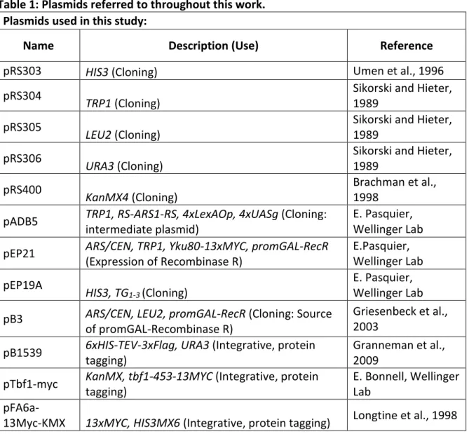
Table 1: Plasmids referred to throughout this work. Plasmids used in this study:
Name Description (Use) Reference
pRS303 HIS3 (Cloning) Umen et al., 1996
pRS304
TRP1 (Cloning)
Sikorski and Hieter, 1989
pRS305
LEU2 (Cloning)
Sikorski and Hieter, 1989
pRS306
URA3 (Cloning)
Sikorski and Hieter, 1989
pRS400
KanMX4 (Cloning)
Brachman et al., 1998
pADB5 TRP1, RS-ARS1-RS, 4xLexAOp, 4xUASg (Cloning:
intermediate plasmid)
E. Pasquier, Wellinger Lab
pEP21 ARS/CEN, TRP1, Yku80-13xMYC, promGAL-RecR
(Expression of Recombinase R) E.Pasquier, Wellinger Lab pEP19A HIS3, TG1-3 (Cloning) E. Pasquier, Wellinger Lab
pB3 ARS/CEN, LEU2, promGAL-RecR (Cloning: Source
of promGAL-Recombinase R)
Griesenbeck et al., 2003
pB1539 6xHIS-TEV-3xFlag, URA3 (Integrative, protein tagging)
Granneman et al., 2009
pTbf1-myc KanMX, tbf1-453-13MYC (Integrative, protein tagging)
E. Bonnell, Wellinger Lab
pFA6a-3HA-HIS3MX 3xHA, HIS3MX6 (Integrative, protein tagging) Longtine et al., 1998 pCT300 ~300 bp C1-3 repeats EcoRI fragment (Southern
Blot probe) Bourns et al., 1998
pAK003 RS-URA3-RS (Cloning, intermediate plasmid for
integrative plasmide in S. cerevisiae) This work
pAK002 TEL01Lmod WT XCR (Cloning, intermediate for
integrative plasmid) This work
pAK012 TEL01Lmod XCRmut (Cloning, intermediate for
integrative plasmid) This work
pT3F1 TEL03Lmod WT XCR RS-LEU2-RS (Cloning,
intermediate for integrative plasmid) This work
pRIM2D TEL03Lmod XCRmut RS-LEU2-RS (Cloning,
intermediate for integrative plasmid) This work
pAK007 TEL03Lmod XCRmut1 RS-LEU2-RS (Cloning,
intermediate for integrative plasmid) This work
pAK016
TEL03Lmod WT XCR RS-LEU2-RS TG1-3 repeats
(Integrative plasmid, linearization with NsiI/Eco53kI)
This work
pAK018
TEL03Lmod XCRmut RS-LEU2-RS TG1-3 repeats
(Integrative plasmid, linearization with NsiI/Eco53kI)
This work
pAK007T
TEL03Lmod XCRmut1 RS-LEU2-RS TG1-3 repeats
(Integrative plasmid, linearization with NsiI/Eco53kI)
This work
pAK013 TEL01Lmod XCRmut RS-URA-RS TG1-3 (Integrative
plasmid, linearization with BamHI/ NotI) This work pAK014 TEL01Lmod WT XCR RS-URA-RS TG1-3 (Integrative
plasmid, linearization with BamHI/ NotI) This work Traditional Cloning
Traditional cloning was carried out by digesting a vector was with one or more restriction enzymes followed by ligation with a DNA fragment with compatible ends. The ligation was done using a Rapid DNA ligation Kit (Thermo Fisher Scientific), following manufacturer’s directions. For each ligation reaction, approximately 50 ng of vector plasmid was linearized with 1 or more restriction enzymes. If only one restriction enzyme was used, plasmid re-circularization was prevented with a dephosphorylation reaction.
DNA ends were dephosphorylated using the Alkaline Phosphatase Kit (Roche), in a 10 µl reaction with 1x phosphatase reaction buffer and 1 U Alkaline Phosphatase and incubated for 1 hour at room temperature, following the manufacturer’s protocol. The reaction was stopped by the addition of EDTA to a 20 mM final concentration and incubation at 68 ˚C for 10 minutes. Insert DNA was digested with restriction enzymes leaving DNA ends compatible to the vector. Both vector and plasmid DNA were purified on a 0.45 % agarose gel run in TAE, using Spin-X ® (Sigma) centrifuge tube filters. Vector and insert DNAs were combined at a 5:1 molecular ratio for ligation reactions.
All Around the World PCR
To introduce point mutations into a plasmid, primers were designed to span the area to be mutated, with 1-2 nucleotides difference from the plasmid sequence. The ends of the forward and reverse primers were aligned such that the whole plasmid was
amplified. Q5 ® High Fidelity DNA polymerase (NEB) was used according to the
manufacturer’s instructions, using a temperature gradient spanning 5 ˚C of the melting temperatures (Tm) of the forward and reverse primers (as determined by Snapgene software). Template DNA was degraded using the KLD Enzyme Kit (NEB) containing enzyme DpnI to target the cell-derived methylated DNA, as well as kinase and ligase for plasmid circularization. 1 µl PCR reaction was treated in a 10 µl total volume reaction containing 1 µl of enzyme mix and 5 µl 2x reaction buffer for 20 minutes at room
temperature. 5 µl of the product was used for transformation into 50 µl One Shot ® Stbl3® chemically competent E. coli.
Gibson Assembly
The Gibson Assembly Kit® (NEB) was used to insert 2 or more DNA fragments into a vector plasmid linearized by restriction enzymes in a single step. The DNA fragments were amplified with primers designed to have overhanging flaps to add short regions with homology to the vector or neighbouring fragments. This homology ensures the fragments are assembled in the correct orientation and order. After PCR amplification of the primers from bacterial plasmids or yeast genomic DNA, the fragments were purified either by on a
0.45 % agarose gel in TAE using Spin-X® columns, or on columns from an EZ-10 Spin Column PCR Product Purification Kit (Biobasic). The Gibson Assembly reaction was carried out according to the manufacturer’s directions, in a 10 µl reaction. The products were diluted 1:5 in water before transforming 5 µl into One Shot ® Stbl3 ® chemically competent E. coli. (Thermo Fisher) (for plasmids containing telomeric repeats) or One Shot ® Top10 ® chemically competent E.coli (Thermo Fisher) bacteria as described below.
Construction of TEL01Lmod and TEL03Lmod plasmids
Mutation of TEL01L and TEL03L subtelomeres was achieved by cloning sequences interior to the telomeric DNA in bacterial plasmids and making desired changes using a combination of the cloning techniques described above. The steps used for the
modification of TEL01L and TEL03L subtelomeres will be outlined in the following section. The resulting constructs depicted below (Figures 4, 5), are referred to as TEL01Lmod and TEL03Lmod.
Construction of plasmids with modified TEL01L
A plasmid containing the X-element of the TEL01L telomere (pAK002) was constructed using 2 fragment Gibson Assembly. Both fragments were amplified from genomic yeast DNA from the W3749 strain (W303 background) by PCR and cloned by Gibson Assembly into the pRS303 vector, which was linearized by digestion with the NotI restriction enzyme. Fragment 1 (2011 bp), amplified by primers
TEL01L_GB_F/TEL01L_GB_RB, contained regions upstream of the subtelomere, including dubious ORFs PAU8 and YAL067W-A. Fragment 2 (primers: TEL01L_GB_R/TEL01L_GB_FB) contained the XCR and X-Core of the TEL01L X-element, as well as DNA upstream of the X-element (1404 bp). The Gibson Assembly reaction product was transformed into One Shot ® Stbl3 ® chemically competent E. coli (Thermo Fisher) as described below. The clones were screened by digestion with NsiI restriction enzyme and sent for sequencing with primers Xel-F and Xel-R.
Tbf1 and Reb1 Binding Site Mutation:
The DNA binding sites in the TEL01L XCR for Tbf1 and Reb1 were mapped using the Yeast Transcription Factor Specificity Compendium (YetFaScO,
http://yetfasco.ccbr.utoronto.ca/) (Figure 4). Tbf1 and Reb1 binding motifs both contain
CCC or GGG in their core binding sequences (Koering et al., 2000). Thus, point mutations were introduced into these sequences of each predicted binding site by “All Around the World PCR”. Primer pairs spanning each binding site were designed with one mismatching nucleotide to introduce a point mutation, changing the cytosine nucleotide (C) to a
guanosine nucleotide (G), or vice versa. All primer pairs used can be found in table 6 under “TEL01Lmod XCR Site directed PCR mutagenesis”.
Figure 4: Tbf1 and Reb1 binding sites in TEL01L XCR were mapped by YeTFasCo. The XCR sequence of TEL01L from the reference genome S288C is depicted, with Tbf1 (blue) and Reb1 (green) binding sites, as identified by the YeTFasCo database.
The PCR product was treated with the KLD Enzyme Mix (NEB) as described above and transformed into One Shot ® Stbl3 ® chemically competent E. coli cells. Plasmids were extracted using an EZ-10 Spin Column Plasmid DNA Miniprep Kit (Biobasic), screened by digestion with the XhoI restriction enzyme and sequenced to confirm. The resulting plasmid was subjected to another round of “All Around the World PCR” with primers introducing mutations to a second Tbf1 or Reb1 binding site, followed by the same steps of KLD treatment, transformation and screening. The process was repeated with each of the primer pairs in section “TEL01Lmod XCR Site directed PCR mutagenesis” (Table 6), resulting in the pAK012 plasmid containing point mutations in all Tbf1 and Reb1 binding sites.