1MRC Cognition and Brain Sciences
Unit, Cambridge CB2 7EF
2Coma Science Group, Cyclotron
Research Center and Neurology Department, Université de Liège, Bât B30 Allée du 6 août no 8, B-4000 Liège, Belgium Correspondence to: M M Monti [email protected] Cite this as: BMJ 2010;341:c3765 doi: 10.1136/bmj.c3765
The vegetative state
Martin M Monti,
1Steven Laureys,
2Adrian M Owen
1 The vegetative state may develop suddenly (as a conse-quence of traumatic or non-traumatic brain injury, such as hypoxia or anoxia; infection; or haemorrhage) or gradu-ally (in the course of a neurodegenerative disorder, such as Alzheimer’s disease). Although uncommon, the condi-tion is perplexing because there is an apparent dissocia-tion between the two cardinal elements of consciousness: awareness and wakefulness.1 Patients in a vegetative stateappear to be awake but lack any sign of awareness of them-selves or their environment.w1 Large retrospective clinical
audits have shown that as many as 40% of patients with a diagnosis of vegetative state may in fact retain some level of consciousness. Misdiagnosis has many implications for a patient’s care—such as day to day management, access to early interventions, and quality of life—and has ethical and legal ramifications pertaining to decisions on the dis-continuation of life supporting therapies.2w2-w4
Overall, our understanding of the vegetative state is incomplete. Although we know quite a lot about the neu-ropathology underlying the vegetative state, our ability to assess (un)consciousness and cognitive function in the clinic is extremely limited, as highlighted by the high rate of misdiagnosis.
What is the vegetative state and what is it not?
The 2003 guidance from the UK’s Royal College of Physi-cians on diagnosing and managing the permanent vegeta-tive state defines it as “a clinical condition of unawareness of self and environment in which the patient breathes spontaneously, has a stable circulation, and shows cycles of eye closure and opening which may simulate sleep and waking.”3 Three main clinical features define the
vegeta-tive state: (a) cycles of eye opening and closing, giving the appearance of sleep-wake cycles (whether the presence of eye opening and closing cycles actually reflects the pres-ence of circadian rhythms is unclearw5 w6); (b) complete
lack of awareness of the self or the environment; and (c) complete or partial preservation of hypothalamic and brain stem autonomic functions.3 4 The guidelines from
the Royal College of Physicians consider a vegetative state to be persistent when it lasts longer than a month and permanent when it lasts longer than six months for non-traumatic brain injuries and one year for non-traumatic brain injuries.3 Guidelines published in the United States,
how-ever, consider that for non-traumatic brain injury a per-manent vegetative state exists after only three months.4
Although both the persistent and the permanent veg-etative states are often abbreviated to “PVS,” authors of a letter in the BMJ in 2000 suggested that to avoid con-fusion the abbreviation should be used exclusively to indicate a permanent vegetative state.w7 The American
Congress of Rehabilitation Medicine suggested that the cause of injury (traumatic, anoxic) as well as the time elapsed since onset of the condition should be docu-mented, as both are important for prognosis.w8
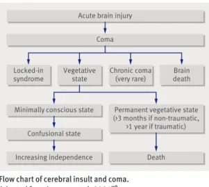
Experts have suggested that the vegetative state should be seen as part of a continuous spectrum of conditions, often referred to as disorders of consciousness, in which someone’s wakefulness and/or awareness are impaired after severe brain injury (figure, table 1).5 6 This
sugges-tion is consistent with the idea that awareness and una-wareness are part of a continuum, and it highlights the
SUMMARY POINTS
The vegetative state is a complex neurological condition in which patients appear to be awake but show no sign of awareness of themselves or their environment
Current clinical methods of diagnosis are limited in scope, evidenced by a high rate (about 40%) of misdiagnosis (that is, patients who are aware are considered to be unconscious)
The main causes of misdiagnosis are associated with a patient’s disability (such as blindness), confusion in terminology, and lack of experience of this relatively rare condition
Furthermore, standard behavioural assessments cannot distinguish an aware (that is, minimally conscious) but completely immobile patient from a non-aware patient (one with vegetative state)
In such behaviourally non-responsive patients, functional neuroimaging methods (such as magnetic resonance imaging or electroencephalography) can detect residual cognition and awareness and can even establish two way communication, without requiring any behavioural output from patients
Current guidelines should therefore be modified to include functional neuroimaging as an independent source of diagnostically relevant information
SOURCES AND SELECTION CRITERIA
This paper is largely based on a personal database of articles from all three authors, including the most recent published work in primary research journals as well as recent and influential reviews and chapters on the subject. We also searched PubMed using the keyword “vegetative state” and the limits “classical article, review and meta-analysis”
bmj.com archive
Previous articles in this
series
Ж
Management of
alopecia areata
(BMJ 2010;341:c3671)
Ж
Investigation and
management of
congestive heart failure
(BMJ 2010;341:c3657)
Ж
Obstetric anal
sphincter injury
(BMJ 2010;341:c3414)
Ж
Perioperative acute
kidney injury
(BMJ 2010;341:c3365)
Ж
Huntington’s disease
(BMJ 2010;340:c3109)
importance of differentiating the vegetative state from other related neurological conditions that may also fol-low catastrophic brain injury.
Coma
Coma is a condition of unresponsiveness in which patients lie with their eyes closed, do not respond to attempts to arouse them, and show no evidence of aware-ness of self or of their surroundings.7 Patients lack not
only signs of awareness (similar to vegetative state) but also wakefulness (unlike vegetative state) regardless of how intensely they are stimulated. Patients typically either recover or progress to a vegetative state (that is, they show signs of wakefulness) within four weeks.3
Irre-versible coma with absent brainstem reflexes indicates brain death, which is not the same as a vegetative state.8
Minimally conscious state
The minimally conscious state is a condition in which patients appear not only to be wakeful (like vegetative state patients) but also to exhibit inconsistent (fluctuat-ing) but reproducible signs of awareness (unlike patients with vegetative state).9 Like the vegetative state, the
minimally conscious state may be transitory and pre-cede recovery of communicative function or may last in definitely.
Locked-in syndrome
Locked-in syndrome (or pseudocoma), although not a
disorder of consciousness, may be confused with veg-etative state. Patients with locked-in syndrome are both awake and aware, yet they are entirely unable to produce any motor output or they have an extremely limited rep-ertoire of behaviours (usually vertical eye movement or blinking).w10 w11
What causes the vegetative state?
In terms of neuropathology, the vegetative state is mostly marked by cortical or white matter and thalamic, rather than brain stem, injury. A review of the evidence avail-able up until 1994 highlighted the fact that traumatic injury was found to be associated with diffuse damage to subcortical white matter (or diffuse axonal injury). Cases of non-traumatic injury, on the other hand, were found to have extensive necrosis in the cerebral cortex, almost always associated with thalamic damage.10
In a more recent survey of patients with brain injury (n=49), 35 (71%) patients had traumatic brain injury, of whom 25 (71%) had severe diffuse axonal injury and 7 (20%) had major injury to the cerebral cortex.11 Among
the 35 patients, the thalamus seemed to be abnormal in 28 (80%) and damage to the brain stem was present in only 5 (14%). In the 14 (29%) patients with non-trau-matic injury, 9 (64%) cases presented with diffuse neo-cortical damage; in all 14 cases a profound and diffuse neuronal loss was apparent in the thalamus and hippo-campus. Overall, these lesions effectively render a struc-turally intact cortex unable to function by destroying the connections between cortical areas via the thalamus, as well as afferent and efferent cerebral connections.
What affects prognosis in patients with a diagnosis of vegetative state?
Three major factors affect the prognosis of patients with vegetative state: time spent in the vegetative state, age, and type of brain injury.
Time spent in the vegetative state
A study of 140 patients showed that time spent in a veg-etative state is negatively correlated with the chances of recovering independence and consciousness and posi-tively correlated with the probability of remaining in a vegetative state.12 The role of time in prognosis was
con-firmed by a large review of 603 adult published cases,13
from which it was estimated that the chance of regaining independence at one year after injury steadily decreased with time from 18% (one month in the vegetative state), to 12% (three months), and 3% (six months). Similarly, the chance of recovering consciousness at one year also decreased, from 42% to 27% and 12% respectively. The chances of remaining in the vegetative state at one year after injury were estimated to increase from 19% to 35% and 57% respectively.
Age
Younger patients show better recovery rates.13 In one
report, for example, the rates of recovering independ-ence at one year decreased from 21% for patients below 20 years old to 9% for patients between 20 and 39 years old and 0% for patients above 40 years.12
Table 1 | Consciousness and motor behaviour characteristics in patients with disorders of consciousness and locked-in syndrome Condition Consciousness Motor behaviour characteristics Sleep-wake cycles Awareness
Coma No No No purposeful behaviour
Vegetative state Yes No No purposeful behaviour Minimally
conscious state Yes fluctuatingPartial, Inconsistent but reproducible purposeful behaviour
Locked-in
syndrome Yes Yes Yes, but limited to eye movements (depending on lesion)
Acute brain injury Coma
Permanent vegetative state (>3 months if non-traumatic,
>1 year if traumatic) Minimally conscious state
Chronic coma
(very rare) deathBrain Vegetative state Locked-in syndrome Confusional state Death Increasing independence
Flow chart of cerebral insult and coma. Adapted from Laureys et al, 2004w9
Type of brain injury
Traumatic brain injuries are associated with better out-comes at one year than non-traumatic injuries, in terms of recovery of independence (24% v 4%) and recovery of consciousness (52% v 13%).3 4 13 Once permanent
vegetative state is diagnosed, the chances of recovery are considered to be “extremely low,”4 with any further
recov-ery being “exceedingly rare, and almost always involving severe disability”13; and although cases of late recovery
have been reported,w12-w14 a precise estimate of the
likeli-hood of further recovery remains difficult to formulate. This is mainly because these cases are often difficult to verify, and when a set of 30 cases claiming late recovery were reassessed by the Multi-Society Task Force on PVS, evidence of conscious awareness could be detected in half of them well before the boundary for a diagnosis of permanent vegetative state.13 14
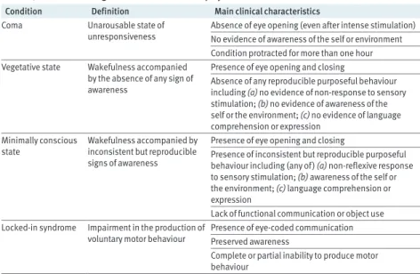
How is the vegetative state diagnosed?
No tool exists for quantifying the extent of conscious-ness. Differentiating between awareness and non-awareness ultimately relies on a pragmatic principle that someone is conscious if they can indicate so.
Cur-rently, the diagnosis of the vegetative state is based on two main sources of information: a detailed clinical history and careful (but subjective) observation of the patient’s spontaneous and elicited behaviour. Clinical assessments involve repeated examinations at different times of the day because patients who are not in a vegeta-tive state may have alternating periods of awareness and unawareness (and a single examination cannot exclude a state of minimal consciousnesses) as well as circadian oscillations in levels of wakefulness. Examinations aim to uncover evidence of (a) awareness of the self or the environment; (b) sustained, reproducible, purposeful, or voluntary response to visual, olfactory, auditory, tactile, or noxious stimuli; and (c) comprehension of language or expression. If evidence of these exists, the patient is con-sidered to be (minimally) aware. If meaningful “object use” (such as appropriate use of a spoon or comb) or con-sistent communication can also be established, then the patient is considered to have emerged from a minimally conscious state to a condition of severe disability (table 2).9 However, if no evidence of awareness can be found,
the patient is considered to be “not aware” and therefore in a vegetative state.
Although several protocols exist for conducting behav-ioural assessments (articles by Giacino et al and Majerus et al provide an overvieww15 w16), they differ greatly
in their ability to detect consciousness because of the number of domains (such as arousal and vision) assessed and the thoroughness of the assessment. Indeed, a recent study of 60 patients compared on three assessment tech-niques reported that the Glasgow coma scalew17 classified
as vegetative several patients who showed signs of con-sciousness according to other behavioural scales.15 The
Full Outline of UnResponsiveness (FOUR)w18 reclassified
13% of the supposedly vegetative patients as minimally conscious, and the coma recovery scale-revised (CRS-R)w19 reclassified an additional 28% of the patients as
minimally conscious. The main discrepancy between scales seems to relate to their different focus on oculomo-tor behaviour, with the FOUR and CRS-R protocols testing a greater variety of visual behaviours. For example, in all the patients reclassified by the CRS-R protocol, visual fixation was the key behaviour indicating awareness.
Does misdiagnosis of the vegetative state occur?
According to accumulating evidence from retrospec-tive clinical audits2 16 and comparisons of alternative
be havioural assessment techniques,17 18 misdiagnosis
of minimally conscious patients as being in a vegetative state is not uncommon. In particular, although some studies have reported relatively low rates of misdiagnosis (18% w20), most studies seem to converge, across time and
geographical location, on an approximate rate in excess of 40% (37%,16 41%,18 43%,2 45%19). Errors in diagnosis
may result from lack of skill or training in the assessment of patients with catastrophic brain injury, limited knowl-edge of this relatively rare condition, and confusion in terminology.16 20
Two main problems seem to underlie misdiagnosis. Firstly, behavioural assessments of awareness present many complexities. For example, patients with physical
ADDITIONAL EDUCATIONAL RESOURCES
• NHS Choices (www.nhs.uk/Conditions/Vegetative-state/ Pages/Introduction.aspx)—NHS information about the vegetative state
• Royal College of Physicians. The vegetative state: guidance on diagnosis and management. 2003. http:// bookshop.rcplondon.ac.uk/contents/47a262a7-350a-490a-b88d-6f58bbf076a3.pdf.
• Scholarpedia (www.scholarpedia.org/article/
Vegetative_state)—Web based encyclopaedia that gives a comprehensive, peer reviewed, overview of definitions, diagnostic criteria, and recent research on vegetative state and related disorders of consciousness • Headway (www.headway.org.uk/Core/DownloadDoc.
aspx?documentID=446)—Fact sheet on coma from the brain injury association Headway, a charity providing help and support to people affected by brain injury
Table 2 | Differential diagnosis in severe brain injury survivors
Condition Definition Main clinical characteristics
Coma Unarousable state of
unresponsiveness Absence of eye opening (even after intense stimulation)No evidence of awareness of the self or environment Condition protracted for more than one hour Vegetative state Wakefulness accompanied
by the absence of any sign of awareness
Presence of eye opening and closing
Absence of any reproducible purposeful behaviour including (a) no evidence of non-response to sensory stimulation; (b) no evidence of awareness of the self or the environment; (c) no evidence of language comprehension or expression
Minimally conscious
state Wakefulness accompanied by inconsistent but reproducible signs of awareness
Presence of eye opening and closing
Presence of inconsistent but reproducible purposeful behaviour including (any of) (a) non-reflexive response to sensory stimulation; (b) awareness of the self or the environment; (c) language comprehension or expression
Lack of functional communication or object use Locked-in syndrome Impairment in the production of
voluntary motor behaviour Presence of eye-coded communicationPreserved awareness Complete or partial inability to produce motor behaviour
disability may not be able to respond to stimulation— something that was true in all misdiagnosed cases in a large retrospective study of 97 patients with profound brain damage.2 Sensory impairments (particularly in
the visual domain) can also mask the presence of aware-ness,16 20 a factor that has been reported as underlying
as many as 65% of misdiagnoses.2 Other acquired
condi-tions, such as hydrocephaly,3w21 can also mask the
pres-ence of awareness. In addition, patients in a minimally conscious state may display inconsistent behaviour, mak-ing it difficult to interpret their responses, and they may be not aware for protracted intervals, making it difficult to interpret failure to respond.3
Secondly, there is a conceptual problem in the logic of establishing “lack of awareness”21 22: absence of evidence
(of awareness) is taken as evidence of absence (of aware-ness). Consequently, on the basis of the current clinical standards, patients who are aware but non-responsive cannot be distinguished from non-aware (vegetative) patients.23 24 Clinically, this flaw in logic introduces a
category of aware but non-responsive patients for whom a diagnosis of vegetative state is technically appropriate
(that is, they show no signs of awareness) but incorrect (in fact, they are aware).21 22
Is there a place for brain imaging as a diagnostic tool?
In recent years, techniques such as positron emission tomography, functional magnetic resonance imaging, and electroencephalography have been used to try to assess residual brain function and consciousness in vegetative patients without relying on motor behaviour. Neuroimag-ing studies in patients in a vegetative state have shown a consistent reduction in brain metabolism of as much as 50%w22 and reduced basal resting state activity.25 In
addition, unexpected levels of residual cognitive func-tion (such as processing of linguistic and self referential stimuli) are present in both minimally conscious patients and patients in a vegetative state.26 27 In some of these
cases, high level functions (such as learning and actively maintaining information through time) are present,28-30
as are awareness23 24 28 and the ability to communicate
solely by modulation of brain activity.23
The Multi-Society Task Force on PVS states, however, that “neurodiagnostic” tests, although recognised as “providing useful information when used in conjunc-tion with clinical evaluaconjunc-tion” are believed to be unable, alone, to “either confirm the diagnosis of vegetative state . . . or predict the potential for recovery of awareness.”4
Although we agree that functional neuroimaging cannot confirm a diagnosis of vegetative state, it is increasingly clear that functional neuroimaging can be used to rule out a diagnosis of vegetative state and may even yield information about prognosis. Indeed, limited data on prognosis show that quantitative measurements of brain activity—in particular, activations beyond primary sen-sory cortices—are positively correlated with recovery from the vegetative state.26 28w23
Conclusion
Disorders of consciousness remain challenging to man-age because of our superficial understanding of the phe-nomenon of consciousness and its neural mechanisms. Two main strategies seem promising for reducing the consistently high misdiagnosis rate. Firstly, behavioural assessments need to be conducted more thoroughly and by trained staff (a neurologist or another healthcare pro-fessional who has been trained to use the formalised assessments mentioned previously).2 16 18 20 Secondly,
we believe that the inclusion of recommendations for the use of functional neuroimaging techniques in revised guidelines will increase the detection of covert signs of awareness in the very circumstances susceptible to mis-diagnosis. In addition, these techniques can be used to explore the degree of mental life possible after severe brain injury,w24 thus tackling the medically and ethically
important question “what is it like to be in a vegetative state?” In a minority of cases, these techniques may even allow the patients to interact with their environment and to some extent let their voice be heard.23
Contributors: MMM researched the paper; all three authors contributed to the writing and are guarantors.
Competing interests: All authors have completed the Unified Competing Interest form at www.icmje.org/coi_disclosure.pdf (available on request
QUESTIONS FOR FUTURE RESEARCH
What proportion of patients with supposed vegetative state can show a state of consciousness by using functional neuroimaging methods?
What proportion of behaviourally non-responsive patients can convey yes/no answers by wilful modulation of brain activity?
Do patients with disorders of consciousness have a “stream of thoughts”? Do they suffer? Do they understand their circumstance? What is their quality of life?
Can more sophisticated brain computer interfaces be used to allow these patients to interact with their environment and regain some level of communication and autonomy? The ability of novel brain imaging technologies, such as functional magnetic resonance imaging, to covertly detect signs of consciousness and residual cognition can contribute to correctly diagnosing the vegetative state
M AR TI N M O N TI , M RC C O G N IT IO N & B RA IN S CI EN CE S UN IT
from the corresponding author) and declare: MMM had support from the Medical Research Council (U.1055.01.002.00001.01) and the European Commission (Deployment of Brain-Computer Interfaces for the Detection of Consciousness in Non-Responsive Patients) for the submitted work. AMO had support from the Medical Research Council (U.1055.01.002.00007.01 and U.1055.01.002.00001.01), the James S McDonnell Foundation and the European Commission (Deployment of Brain-Computer Interfaces for the Detection of Consciousness in Non-Responsive Patients) for the submitted work. SL had support from the James S McDonnell Foundation, the European Commission (Deployment of Brain-Computer Interfaces for the Detection of Consciousness in Non-Responsive Patients, Disorders and Coherence of the Embodied Self, Mindbridge, and Consciousness: A Transdisciplinary, Integrated Approach), Fonds de la Recherche Scientifique, the Mind Science Foundation, the Reine Elisabeth Medical Foundation, the Belgian French-Speaking Community Concerted Research Action, University Hospital of Liege, the University of Liege, and the National Institute for Health Research Biomedical research Centre (Neuroscience Theme); no financial relationships with any organisations that might have an interest in the submitted work in the previous 3 years; no other relationships or activities that could appear to have influenced the submitted work.
Provenance and peer review: Commissioned; externally peer reviewed. 1 Laureys S. The neural correlate of (un)awareness: lessons from the
vegetative state. Trends in Cognitive Sciences 2005;9:556-9. 2 Andrews K, Murphy L, Munday R, Littlewood C. Misdiagnosis of the
vegetative state: retrospective study in a rehabilitation unit. BMJ 1996;313:13-6.
3 Royal College of Physicians. The permanent vegetative state: guidance on diagnosis and management. Report of a working party. RCP, 2003. http://bookshop.rcplondon.ac.uk/contents/47a262a7-350a-490a-b88d-6f58bbf076a3.pdf
4 The Multi-Society Task Force on PVS. Medical aspects of the persistent vegetative state (1). N Engl J Med 1994;330:1499-508. 5 Andrews K. International Working Party on the Management of the
Vegetative State: summary report. Brain Injury 1996;10:797-806. 6 Laureys S, Boly M. The changing spectrum of coma. Nat Clin Pract
Neurol 2008;4:544-6.
7 Posner JB, Saper CB, Schiff ND, Plum F. The diagnosis of stupor and coma. 4th ed. Oxford University Press, 2007.
8 Laureys S. Science and society: death, unconsciousness and the brain. Nat Rev Neurosci 2005;6:899-909.
9 Giacino JT, Ashwal S, Childs N, Cranford R, Jennett B, Katz DI, et al. The minimally conscious state: definition and diagnostic criteria. Neurology 2002;58:349-53.
10 Kinney HC, Samuels MA. Neuropathology of the persistent vegetative state. A review. J Neuropathol Exp Neurol 1994;53:548-58. 11 Adams JH, Graham DI, Jennett B. The neuropathology of the vegetative
state after an acute brain insult. Brain 2000;123(part 7):1327-38. 12 Braakman R, Jennett WB, Minderhoud JM. Prognosis of the
posttraumatic vegetative state. Acta Neurochirurgica (Wien) 1988;95(1-2):49-52.
ANSWERS TO ENDGAMES, p 307. For long answers go to the Education channel on bmj.com
STATISTICAL QUESTION
Relative risks and confidence intervals
Answers a, b, and d, are true, whereas c is false.
CASE REPORT
Recurrent vomiting and lethargy in an infant—
just another viral illness?
1 The differential diagnoses are metabolic disorder, neglect, coeliac disease, viral gastroenteritis, hypothyroidism, and HIV infection. 2 Hyperammonaemia is responsible for this patient’s clinical
picture.
3 The raised plasma concentration of ammonia and respiratory alkalosis suggest a urea cycle defect caused by inherited defects of enzymes responsible for the metabolism of waste nitrogen. Further tests showed raised urinary orotate and plasma glutamine and low plasma citrulline, consistent with a diagnosis of ornithine transcarbamylase (OTC) deficiency.
4 Hyperammonaemia should be managed with nil by mouth; infusion of 10% dextrose and sodium benzoate; arginine supplementation; and regular monitoring of plasma ammonia, glucose, urea, and electrolytes in addition to blood gases.
ON EXAMINATION QUIZ
ICD-10 classification
False.
More questions on this topic are available from
www.onexamination.com/endgames until midnight on Wednesday.
ANATOMY QUIZ
T2 weighted axial magnetic resonance image
of the brain
A Right medial rectus muscle B Right side of the pons C Left lateral rectus muscle D Basilar artery
E 4th ventricle
13 The Multi-Society Task Force on PVS. Medical aspects of the persistent vegetative state (2). N Engl J Med 1994;330:1572-9.
14 Jennett B. The vegetative state. J Neurol Neurosurg Psychiatry 2002;73:355-7.
15 Schnakers C, Giacino J, Kalmar K, Piret S, Lopez E, Boly M, et al. Does the FOUR score correctly diagnose the vegetative and minimally conscious states? Ann Neurol 2006;60:744-5; author reply 745. 16 Childs NL, Mercer WN, Childs HW. Accuracy of diagnosis of persistent
vegetative state. Neurology 1993;43:1465-7.
17 Gill-Thwaites H. The sensory modality assessment rehabilitation technique—a tool for assessment and treatment of patients with severe brain injury in a vegetative state. Brain Injury 1997;11:723-34. 18 Schnakers C, Vanhaudenhuyse A, Giacino J, Ventura M, Boly M, Majerus
S, et al. Diagnostic accuracy of the vegetative and minimally conscious state: clinical consensus versus standardized neurobehavioral assessment. BMC Neurology 2009;9:35.
19 Gill-Thwaites H, Munday R. The sensory modality assessment and rehabilitation technique (SMART): a valid and reliable assessment for vegetative state and minimally conscious state patients. Brain Injury 2004;18:1255-69.
20 Gill-Thwaites H. Lotteries, loopholes and luck: misdiagnosis in the vegetative state patient. Brain Injury 2006;20:1321-8.
21 Monti MM, Coleman MR, Owen AM. Neuroimaging and the vegetative state: resolving the behavioural assessment dilemma? Disorders of Consciousness: Annals of the New York Academy of Sciences 2009;1157:81-9.
22 Owen AM, Coleman MR. Functional neuroimaging of the vegetative state. Nat Rev Neurosci 2008;9:235-43.
23 Monti MM, Vanhaudenhuyse A, Coleman MR, Boly M, Pickard JD, Tshibanda L, et al. Willful modulation of brain activity in disorders of consciousness. N Engl J Med 2010;362:579-89.
24 Owen AM, Coleman MR, Boly M, Davis MH, Laureys S, Pickard JD. Detecting awareness in the vegetative state. Science 2006;313:1402. 25 Boly M, Tshibanda L, Vanhaudenhuyse A, Noirhomme Q, Schnakers C, Ledoux D, et al. Functional connectivity in the default network during resting state is preserved in a vegetative but not in a brain dead patient. Hum Brain Mapp 2009;30:2393-400.
26 Coleman MR, Davis MH, Rodd JM, Robson T, Ali A, Owen AM, et al. Towards the routine use of brain imaging to aid the clinical diagnosis of disorders of consciousness. Brain 2009;132(part 9):2541-52. 27 Qin P, Di H, Liu Y, Yu S, Gong Q, Duncan N, et al. Anterior cingulate
activity and the self in disorders of consciousness. Human Brain Mapping [forthcoming].
28 Bekinschtein TA, Shalom DE, Forcato C, Herrera M, Coleman MR, Manes FF, et al. Classical conditioning in the vegetative and minimally conscious state. Nat Neurosci 2009;12:1343-9.
29 Monti MM, Coleman MR, Owen AM. Executive functions in the absence of behavior: functional imaging of the minimally conscious state. Prog Brain Res 2009;177:249-60.
30 Schnakers C, Perrin F, Schabus M, Majerus S, Ledoux D, Damas P, et al. Voluntary brain processing in disorders of consciousness. Neurology 2008;71:1614-20.