HAL Id: dumas-02491483
https://dumas.ccsd.cnrs.fr/dumas-02491483
Submitted on 26 Feb 2020HAL is a multi-disciplinary open access
archive for the deposit and dissemination of sci-entific research documents, whether they are pub-lished or not. The documents may come from teaching and research institutions in France or abroad, or from public or private research centers.
L’archive ouverte pluridisciplinaire HAL, est destinée au dépôt et à la diffusion de documents scientifiques de niveau recherche, publiés ou non, émanant des établissements d’enseignement et de recherche français ou étrangers, des laboratoires publics ou privés.
The diagnostic workup in children with arthrogryposis
multiplex congenita: description of practices through a
monocentric cohort and suggestion of recommendations
Pauline Le Tanno
To cite this version:
Pauline Le Tanno. The diagnostic workup in children with arthrogryposis multiplex congenita: de-scription of practices through a monocentric cohort and suggestion of recommendations. Human health and pathology. 2020. �dumas-02491483�
AVERTISSEMENT
Ce document est le fruit d'un long travail approuvé par le
jury de soutenance et mis à disposition de l'ensemble de la
communauté universitaire élargie.
Il n’a pas été réévalué depuis la date de soutenance.
Il est soumis à la propriété intellectuelle de l'auteur. Ceci
implique une obligation de citation et de référencement
lors de l’utilisation de ce document.
D’autre part, toute contrefaçon, plagiat, reproduction illicite
encourt une poursuite pénale.
Contact au SID de Grenoble :
bump-theses@univ-grenoble-alpes.fr
LIENS
LIENS
Code de la Propriété Intellectuelle. articles L 122. 4
Code de la Propriété Intellectuelle. articles L 335.2- L 335.10
http://www.cfcopies.com/juridique/droit-auteurUNIVERSITÉ GRENOBLE ALPES UFR DE MÉDECINE DE GRENOBLE
Année : 2020
THE DIAGNOSTIC WORKUP IN CHILDREN WITH ARTHROGRYPOSIS
MULTIPLEX CONGENITA : DESCRIPTION OF PRACTICES THROUGH A
MONOCENTRIC COHORT AND SUGGESTION OF RECOMMENDATIONS.
THÈSE
PRÉSENTÉE POUR L’OBTENTION DU TITRE DE DOCTEUR EN MÉDECINE
DIPLÔME D’ÉTAT
Pauline LE TANNO
THÈSE SOUTENUE PUBLIQUEMENT À LA FACULTÉ DE MÉDECINE DE GRENOBLE
Le : 10 février 2020
DEVANT LE JURY COMPOSÉ DE
Président du jury :
Monsieur le Professeur Pierre-Simon JOUK
Membres :
Monsieur le Professeur Dominic PÉRENNOU
Madame le Docteur Véronique BOURG
Monsieur le Docteur Julien FAURÉ
Monsieur le Docteur Klaus DIETERICH, Directeur de thèse
L’UFR de Médecine de Grenoble n’entend donner aucune approbation ni improbation aux opinions émises dans les thèses ; ces opinions sont considérées comme propres à leurs auteurs.
TABLE DES MATIÈRES
REMERCIEMENTS……… 7
LISTE DES ABRÉVIATIONS………. 9
LISTE DES TABLEAUX ET FIGURES……… 11
RÉSUMÉ………. 12
ABSTRACT………. 13
INTRODUCTION……… 14
1. Definition and Epidemiology………. 14
2. Causes and classification……….. 15
3. Context and purpose……….. 19
MATERIALS AND METHODS……… 21
1. Protocol registration……… 21
2. Cohort building……… 22
3. Data collection………. 23
4. Patients’ classification……… 23
5. Data analysis in the light of information provided by scientific literature………. 24
RESULTS………. 26
1. Cohort constitution………. 26
2. Etiologies of AMC………. 27
2.A/ Group 1 : Amyoplasia……… 27
3.B/ Group 2 : distal arthrogryposis..………. 27
3.C/ Group 3 : other patients..……… 30
3. Paraclinical investigations performed and results……… 31
3.A/ Other genetic investigations………. 31
3.B/ Other biological investigations……… 32
3.C/ Neuromuscular investigations………. 34
3.D/ Central Nervous System investigations……… 37
3.E/ Associated organ impairment assessment………. 39
DISCUSSION……… 42
1. Comparison of our cohort with literature of AMC patients………. 42
2. AMC diagnostic assessment : delineating indications for each investigation………. 45
2.A/ Widely performed and non-invasive investigations……… 45
2.B/ CNS explorations (cMRI, tcUS, EEG) are indicated for group 3C only……… 47
2.C/ Neuromuscular explorations (MB, ENMG) are invasive and of poor interest……….. 49
2.D/ Reflection about spinal cord explorations (scMRI, scUS)……… 50
2.E/ More rarely performed investigation that require specific indications……… 52
3. AMC diagnostic assessment : synthesis of recommendations……… 57
4. Limits of this study……….. 61
CONCLUSION……….... 64
REFERENCES……… 67
SUPPLEMENTAL DATA………. 75
REMERCIEMENTS
À Monsieur le Professeur Pierre-Simon JOUK. Je vous remercie de me faire l’honneur d’être le président de ce jury, afin de commenter ce travail centré sur une pathologie pour laquelle vous avez fait beaucoup. Je vous remercie également pour votre bienveillance et votre humanité qui ont accompagné le début de mon internat.
À Monsieur le Docteur Klaus DIETERICH. Merci de m’avoir proposé ce sujet, ambitieux mais essentiel. Merci pour ta patience, pour avoir répondu à mes nombreuses sollicitations y compris sur ton temps personnel.
À Monsieur le Professeur Julien FAURÉ. Merci d’accepter de relire ce travail. La qualité des interactions entre la génétique clinique et la biologie moléculaire, surtout dans le domaine de l’arthrogrypose, constitue une part essentielle de la prise en charge de nos patients.
À Madame le Docteur Véronique BOURG. Je te remercie également d’avoir accepté cette relecture. La MPR constitue en soi un pilier fondamental du bilan arthrogrypose, mais ton énergie et ton expérience le renforcent.
À Monsieur le Professeur Dominic PÉRENNOU. Merci d’accepter de relire ce travail. Merci également pour votre soutien dans la promotion des nombreux projets communs aux équipes de MPR et de génétique.
À mes parents, de m’avoir encouragée si tenacement, et de m’avoir hébergé pour mes longues heures de révisions qui ont émaillé mon cursus universitaire. Merci pour votre affection et votre fierté qui sont mes guides.
À ma sœur, mon frère : la petite dernière a enfin fini ses études !
À mes beaux-parents, pour m’accueillir avec autant de générosité et m’accepter comme leur fille.
À mes amis de médecine, le groupe grenoblois d’abord, David et Morgane et leur future mite, Flora et Olivier, Valérie et Grégoire, Floriane et Charlie, Claire, Pauline. Double merci à Floriane pour sa relecture !! Au groupe parisien aussi, qui m’a soutenue sur mes dernières semaines de travail intensif, Pauline, Alix et Reaksmei. Sans oublier les exilés bretons, Florie, ou bisontins, Océane et Clément. Je suis ravie d’avoir passé toutes ces années avec vous, un chemin semé d’épreuves (spéciale pensée au super groupe de conf) mais aussi de supers souvenirs (vive le MDB). Je suis super fière de voir les médecins et personnes que vous êtes devenus, vous assurez.
Il en va de même, bien sûr, pour mes amis anciens médecins, Armelle, éduc spé, Pinki, et pharmaciens, Momo et Natacha, dont l’amitié est d’une importance toute aussi capitale.
À mon ancien co-interne Brice, toujours là pour répondre à mes (nombreuses) questions, et mon seul repère dans cette aventure grenobloise.
À mes co-internes de génétique à travers la France, Pauline, Marine, Julian, Simon et tant d’autres ; plus largement à la SIGF, qui m’a été d’un soutien indispensable.
À mes autres co-internes croisés dans des stages hors génétiques ou inter-CHU, merci notamment à mes co-internes de Necker, dont la bonne humeur m’a aidé à traverser ces quelques semaines difficiles.
À Charles, Françoise, Véronique et Julien d’avoir encadré mes travaux tout au long de mon externat puis de mon internat. À Florence, Radu, plus récemment Isabelle. Merci à tous pour votre accueil, votre enseignement. Traverser cet internat à vos côtés a été un plaisir.
À l’équipe de biologie moléculaire, qui m’a accueillie 6 mois, merci notamment à Nathalie pour sa pédagogie et sa gentillesse, à John et Xenia pour leur implication dans les arthrogryposes.
À tout le service de génétique, des secrétaires aux techniciens en passant par les ingénieurs, les conseillères en génétique, ARC et psychologue. À Gipsy, Marjolaine et Emmanuelle, indispensables au bon fonctionnement du Centre de référence Arthrogrypose.
À tous les médecins de l’HCE, neuropédiatres et MPR aux premières loges. Aux rééducateurs de MPR, Claire et Véronique, équipe de choc.
À l’équipe du GETI, qui m’a accueillie pendant mon M2.
À tous les patients qui font l’objet de ce travail, et leur famille. A l’Association Alliance Arthrogrypose, leur énergie et leur force. Les week-ends de l’association sont pour moi des souvenirs précieux.
Et surtout à mon Hadri, futur mari et relecteur attentif de toutes les thèses, merci pour ton aide si précieuse, ta patience infinie, et ton amour tout aussi infini je l’espère. J’ai une chance inouïe.
LISTE DES ABRÉVIATIONS
AAA Association Alliance Arthrogrypose AChR Acetylcholine Receptor
AFM Active Foetal Movements
AMC Arthrogryposis Multiplex Congenita
AMC-NGS Next Generation Sequencing of genes involved in AMC C Central nervous system involvement
CBD Congenital Bone Disorders
CGH Comparative Genomic Hybridization CK Creatine Kinase
cMRI Cerebral Magnetic Resonance Imaging CNS Central Nervous System
CNV Copy Number Variant DA Distal Arthrogryposis EEG Electroencephalogram EKG Electrocardiogram ENMG Electroneuromyography
FADS Foetal Akinesia Deformation Sequence ID Intellectual Disability
IUFD Intra Uterine Foetal Death
LRP4 Low‐density lipoprotein Receptor‐related Protein 4 M Muscle
MB Muscle biopsy
MCC Multiple Congenital Contractures MD1 Myotonic Dystrophy type 1
MG Myasthenia Gravis
mMRI Muscle Magnetic Resonance Imaging MRC Mitochondrial Respiratory Chain
MRI Magnetic Resonance Imaging MuSK Muscle Specific Kinase
NMD Neuromuscular Diseases
NMD-NGS Next Generation sequencing of genes involved in neuromuscular diseases NMJ Neuromuscular Junction
O Other
PCH Ponto-Cerebellar Hypoplasia PN Peripheral Nerve
PWS Prader-Willi Syndrome
scMRI Spinal cord Magnetic Resonance Imaging scUS Spinal cord Ultrasonography
SMA Spinal Muscular Atrophy
SMALED SMA with Lower Extremity predominance TCS Tethered Cord Syndrome
tcUS Transcranial Ultrasonography
TNMG Transient Neonatal Myasthenia Gravis TOP Termination of Pregnancy
U Unknown
US Ultrasonography
VUS Variant of Unknown Significance WES Whole Exome Sequencing
WG Weeks of Gestation
LISTE DES TABLEAUX ET FIGURES
Table 1 Typical Amyoplasia criteria……….. 17
Table 2 Distal arthrogryposis classification………. 18
Table 3 Epidemiological information……….. 26
Table 4 Paraclinical investigations performed for diagnostic purposes ……… 33
Table 5 Anomalies observed on muscle biopsy examination………. 36
Table 6 Detailed anomalies observed on electroneuromyography……….. 36
Table 7 Details on anomalies reported on cMRI and/or tcUS……….. 38
Table 8 Details on anomalies observed on spinal cord imaging……….. 39
Table 9 Details on anomalies reported in cardiological explorations……….. 41
Table 10 Details on anomalies observed on abdominal ultrasonography……… 41
Figure 1 Simplified scheme of causes and consequences of decreased AFM……….. 16
Figure 2 Main steps of present study protocol……….. 21
Figure 3 Possible diagnosis in Group 3 with (C) or without (O) CNS involvement………. 25
Figure 4 Flowchart of the study and detailed molecular diagnoses obtained in group 2 and 3.. 28
Figure 5 Pictures of AMC patients of our cohort……….. 29
Figure 6 Proposition of recommendations in the aetiological assessment of AMC children……. 58
Figure S1 Study information note destined to patients………..……….. 75
Table S1 Features that led to the diagnosis of atypical Amyoplasia……… 77
Table S2 List of the 121 NMD-related genes sequenced in CHU Grenoble-Alpes laboratory…… 78
Table S3 Indications that had motivated prenatal karyotypes……… 79
Table S4 Detailed karyotype and array-CGH results (postnatal)………. 80
Table S5 Other Cytogenetic analyses performed in our cohort……….. 80
Table S6 Other targeted analyses performed in our cohort……….. 81
Table S7 Abnormal creatine kinase dosage results………. 81
Table S8 Details on metabolic investigations performed in our patients………. 82
Table S9 Details on anomalies observed on electroencephalogram……… 82
RÉSUMÉ
Titre : Le parcours diagnostique des enfants atteints d’Arthrogrypose Multiple Congénitale : description des pratiques actuelles à travers une cohorte monocentrique, et proposition de recommandations.
Introduction: L’Arthrogrypose Multiple Congénitale (AMC) correspond à des limitations articulaires touchant au moins deux niveaux. Sa prévalence est estimée entre 1/3000 et 1/12000. Le diagnostic étiologique est difficile car il en existe plus de 400 causes. L’objectif de ce travail est de décrire le parcours diagnostique des enfants atteints d’AMC et de proposer des recommandations afin d’optimiser les pratiques cliniques.
Matériel et méthodes: Nous avons mené une étude rétrospective observationnelle monocentrique, incluant les enfants évalués au Centre Hospitalier Universitaire de Grenoble de 2007 à 2019. Nous avons collecté les informations sur leur parcours diagnostique, puis avons mené une revue de la littérature pour chaque investigation paraclinique afin d’en évaluer la pertinence à la lumière du diagnostic final des patients, des connaissances scientifiques, de leur bénéfices, risques et coûts.
Résultats: Nous avons inclus un total de 125 patients, dont 43% cas d’Amyoplasie, 26% d’arthrogrypose distale et 31% d’autres formes. Un diagnostic étiologique était posé dans 63% des cas. Nous proposons une procédure diagnostique en deux temps: d’abord des investigations non invasives permettant d’orienter les patients vers l’un des trois groupes principaux, puis des investigations plus spécifiques avec des indications précises, en fonction de leur rendement attendu et de leur caractère invasif.
Conclusion: L’approche diagnostique des enfants avec AMC doit résulter d’un travail pluridisciplinaire. L’utilisation croissante du séquençage de nouvelle génération facilitera cette démarche, mais le phénotypage restera essentiel pour guider leur interprétation.
ABSTRACT
Title: The diagnostic workup in children with Arthrogryposis Multiplex Congenita: description of practices through a monocentric cohort and suggestion of recommendations.
Introduction: Arthrogryposis multiplex congenita (AMC) defines congenital contractures involving two or more body areas. The prevalence is estimated between 1/3000 and 1/12000. More than 400 conditions may lead to AMC through foetal hypo/a-kinesia, making the aetiological diagnosis challenging. The objective of this work was to describe the aetiological management of children with AMC and to propose recommendations in order to optimize clinical practices.
Material and methods: We conducted a retrospective single centre observational study. Patients had been evaluated at least once at a paediatric age in Grenoble University Hospital from 2007 to 2019. After determining the diagnostic status of these patients, data on their diagnostic procedure were gathered. A literature review was performed for each paraclinical investigation to discuss their relevance in the light of patients’ diagnoses, scientific knowledge, and benefit/risk or cost/benefit ratio.
Results: 125 patients were included, 43% had Amyoplasia, 26% had distal arthrogryposis, and 31% had other forms. A definitive aetiological diagnosis was available for 63% of cases. We propose a two-time diagnostic process : first, non-invasive investigations that aim at classifying patients into one of the three groups, and then specific investigations targeting a subset of patients according to the expected yield and invasiveness.
Conclusion: The diagnostic management of AMC patients has to result from a multidisciplinary approach. With the use of next generation sequencing, the aetiological assessment will be facilitated, but a relevant phenotyping will be paramount to guide their interpretation.
INTRODUCTION
1. Definitions and epidemiology
Etymologically, the term “Arthrogryposis” results from the association of the Greek words “arthron” (joints), “gryp” (curved) and “osis” (condition) and points to joint contractures with limited range of motion. The Latin term “Arthrogryposis Multiplex Congenita” (AMC) used alternatively with the term “Multiple Congenital Contractures” (MCC), more specifically defines a congenital arthrogryposis, involving at least two body areas (1–3). Contractures are typically symmetric and although they do not progress to unaffected joints, they may change in amplitude under the influence of growth and treatment (2).
Although AMC has been first defined as a condition per se, it is now admitted as a descriptive term to define a symptom, common to a group of heterogeneous congenital conditions (4). Depending on the cause, contractures location and severity may vary, as well as the primary involvement of spine and jaw, or the existence of muscle weakness in the involved body area. AMC can be either isolated or associated to other body systems impairment such as central nervous system (CNS) as well as respiratory, gastrointestinal and genitourinary systems. Prognostic factors like response to treatment, importance of deficits, activity limitation, participation restriction but also neurodevelopment and even life expectancy may differ widely according to the aetiology (2).
As true prevalence estimation remains challenging, it is thought to stand around 1/3000- 1/12000 (5–7). AMC is thus a rare affection, in contrast with isolated congenital contractures such as congenital clubfeet that affect 1/500 individuals (1).
2. Causes and classification
The common underlying cause of AMC is foetal hypokinesia or decreased active foetal movement (AFM) (4). AFM begin around 5 or 6 weeks of gestation (WG) and are essential for the further development of joints. Independently of the underlying cause, their absence may lead to connective tissue deposition around joints and stretching of tendons, increasing mobility reduction and resulting in contractures, whose severity depends on the earliness of hypokinesia (8,9). Foetal hypokinesia leads to a variety of other secondary deformations, that thus can be encountered in all forms of AMC: craniofacial changes, pulmonary hypoplasia, polyhydramnios, decreased gut mobility and shortened gut, short umbilical cord, growth restriction and skin changes (4) (Fig. 1). The whole set of symptoms constitute the most severe form of AMC called foetal akinesia deformation sequence (FADS) or Pena-Shokeir syndrome, usually perinatally lethal (10).
More than 400 conditions causing foetal hypokinesia and thus AMC have been described (4). They can involve any part of the anatomical structures implicated in movement and include all causes of lack or excess of muscle contraction as well as mechanical restriction. They can be divided into two categories (11) (Fig. 1) :
- Extrinsic causes : intrauterine foetal space restriction (severe oligohydramnios, tumoral process, multiple pregnancy, uterine malformation), intrauterine vascular compromise or environmental conditions (maternal illness including infections and myasthenia gravis, maternal exposure to toxics, treatment or trauma)
- Intrinsic causes (foetal dysfunction) : myopathic process (congenital myopathies, congenital muscular dystrophies, myositis...), neuropathic process (CNS impairment with or without structural anomalies, peripheral nerves impairment including anterior horn cells diseases, myelin anomalies…), neuromuscular end-plate process, connective joint tissue abnormalities (restrictive dermopathies or cartilage/bone tissue disturbances) and metabolic diseases.
Figure 1. Simplified scheme of causes and consequences of decreased AFM. Legends : AFM, Active foetal movements; AMC, Arthrogryposis Multiplex Congenita; CNS, central nervous system; M, muscle, NMJ, neuromuscular junction; PN, peripheral nerve.
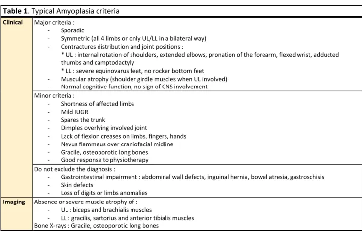
Although a genetic origin is involved in most cases, the first cause of AMC, involved in 1/3 cases with an estimated prevalence of 1/10 000 individuals, is sporadic : Amyoplasia congenita (5,12) (Table 1). This entity, first described in 1840, is recognizable by the characteristic position of upper and lower limbs joints, absence of CNS involvement, absence of primary spinal and jaw involvement although spinal deformities can be present at birth as the consequence of foetal hypomobility and clinical muscle atrophy (3). Associated signs are common and include digestive anomalies, skin defects, loss of digits or limbs, and classic nevus flammeus of the midforehead (12). Muscular MRI (mMRI) typically evidences a reproducible pattern (manuscript submitted). A four limbs AMC arise in 60% of cases, while upper or lower limbs only account for 15% each (12). Orthopaedic outcome is very favourable especially in comparison with other anterior horn cell diseases (13). Indeed, on the basis of the observation of clinical similarities with Erb-Duchenne obstetrical palsy and electroneuromyographic studies, this disorder is thought to result from a neurogenic process, i.e. the impairment of the anterior horn cells of the spinal cord in early embryonic and foetal life, responsible for the underdevelopment of muscle tissue and further replacement by fatty or fibrous deposits. While the exact cause of this impairment is still unclear, the nature of the associated signs and discordance in monozygotic twins suggest an ischemic event, impacting the spinal cord at the level of upper and lower limbs expansion, i.e. motor roots C5-C6 and L2-L4 (12,14).
Table 1. Typical Amyoplasia criteria Clinical Major criteria :
- Sporadic
- Symmetric (all 4 limbs or only UL/LL in a bilateral way)
- Contractures distribution and joint positions :
* UL : internal rotation of shoulders, extended elbows, pronation of the forearm, flexed wrist, adducted thumbs and camptodactyly
* LL : severe equinovarus feet, no rocker bottom feet
- Muscular atrophy (shoulder girdle muscles when UL involved)
- Normal cognitive function, no sign of CNS involvement
Minor criteria :
- Shortness of affected limbs
- Mild IUGR
- Spares the trunk
- Dimples overlying involved joint
- Lack of flexion creases on limbs, fingers, hands
- Nevus flammeus over craniofacial midline
- Gracile, osteoporotic long bones
- Good response to physiotherapy
Do not exclude the diagnosis :
- Gastrointestinal impairment : abdominal wall defects, inguinal hernia, bowel atresia, gastroschisis
- Skin defects
- Loss of digits or limbs anomalies
Imaging Absence or severe muscle atrophy of :
- UL : biceps and brachialis muscles
- LL : gracilis, sartorius and anterior tibialis muscles Bone X-rays : Gracile, osteoporotic long bones
Adapted from Hall et al., 2014 (12). Legends : CNS, central nervous system; IUGR, intra uterine growth retardation; LL, lower limbs; UL, upper limbs.
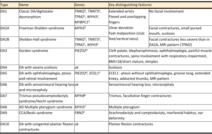
A group of affections called distal arthrogryposis (DA) represents another 1/5 to 1/3 of AMC individuals (15). First described in 1982, DA is a heterogenous group of syndromes where AMC involve mainly hands (overriding fingers, camptodactyly or pseudocamptodactyly with hypoplasia of interphalangeal creases, ulnar deviation and/or wrist extension) and feet (talipes equinovarus, calcaneovalgus deformities, vertical talus, and/or metatarsus varus), with varying involvement of proximal joints. This arises in the absence of any primary neurological and/or muscle disease affecting limb function, with thus conserved muscle mass and cognitive function although muscle weakness and fatigability can occur. Familial cases are common, with classically a wide intrafamilial variability or even incomplete penetrance. Transmission if most often autosomal dominant. The DA group comprises ten different entities, among which many are clinically recognizable (16–18) (Table 2). Genes responsible for DA were the first described in link with AMC, as soon as 1995 for FBN2 (19), and then for genes encoding sarcomeric muscle proteins like TNNI2, TNNT3 and TPM2, also involved in congenital myopathies (20). Mechanisms of AMC in DA are various, either muscle weakness in sarcomeric and ECEL1 forms, lack of relaxation in PIEZO2 patients, or connective tissues dysfunction in FBN2 (18,19,21,22).
Table 2. Distal arthrogryposis classification
Type Name Genes Key distinguishing features
DA1 Classic DA/digitotalar
dysmorphism
TNNI21, TNNT31,
TPM21, MYH32,
MYBPC13
Extended wrists Flexed and overlapping fingers
Ulnar deviation Feet malposition (club feet/vertical talus)
No facial involvement
DA2A Freeman-Sheldon syndrome MYH32 Facial contractures, small pursed
mouth, scoliosis
DA2B Sheldon-Hall syndrome TNNI21, TNNT31,
TPM21, MYH32
Facial contractures less severe than in DA2A, MRI pattern (TPM2)
DA3 Gordon syndrome PIEZO24 Cleft palate, blepharophimosis, ophthalmoplegia, painful muscle
contractures, spine involvement with respiratory impairment, BMI<18/short stature, dimples
DA4 DA with severe scoliosis uk Scoliosis
DA5 DA with ophthalmoplegia, ptosis
and retinal involvement
PIEZO24, ECEL15 ECEL1 : ptosis without ophthalmoplegia, groove tong, extended
knees, adducted thumbs, MRI pattern
DA6 DA with sensorineural hearing loss
and microcephaly
uk Sensorineural hearing loss, microcephaly
DA7 Trismus-pseudocamptodactyly
syndrome/Hecht syndrome
MYH86 Trismus, facultative finger contractures
DA8 AD Multiple pterygium syndrome MYH37 Multiple pterygium
DA9 CCA/Beals syndrome FBN28 Arachnodactyly and camptodactyly, marfanoid habitus, ear
deformity
DA10 DA with congenital plantar flexion
contractures
uk Plantar flexion contractures
From Bamshad et al., 2009 (16), Hall et al., 2017 (17) and Kimber et al., 2012 (18). Legends : AD, autosomal dominant; BMI, Body Mass index; CCA, congenital contractural arachnodactyly; DA, distal arthrogryposis; MRI, Magnetic Resonance Imaging; uk, unknown. 1Sung et al., 2003 (20); 2Toydemir
et al., 2006 (23), 3Gurnett et al., 2010 (24), 5Coste et al., 2013 (21),5Dieterich et al., 2013 (22), 6Toydemir et al. 2006 (25), 7Chong et al., 2015 (26), 7Putnam et al., 1995 (19).
Apart from DA genes, more than 400 genes have been reported in association with AMC. Attempts to classify AMC forms according to genes function have been made, distributing these genes into 29 different groups standing for different pathways. However, as a single gene may be involved in more than one pathway, it may be classified in several groups. In addition, many genes may lead to distinct phenotypes with or without AMC, and conversely a single phenotype may be caused by different genes (27). Classification is thus arduous. Other classification systems are based on the aetiological process of foetal akinesia as previously cited. However, clinical classifications are most useful in daily practice. We can count two of them, both dividing AMC patients in 3 groups :
- the first one is the most used in the literature and distinguishes three categories of patients each accounting for 30% : primarily limb involvement (group I), musculoskeletal involvement associated with other system anomalies (group II), and musculoskeletal involvement associated with CNS dysfunction, intellectual disability (ID) or lethality (group III) (28).
- the other is much more rarely used but is of great interest for clinical practice and for family communication. It distinguishes two first groups accounting for more of the half of cases, Amyoplasia congenita and DA, plus another one where all other aetiologies are gathered (17).
No fully satisfying classification system exist, they all have their advantages and drawbacks. However, they may be a useful way to stratify the diagnostic strategy at first meet of a patient presenting with AMC.
3. Context and purpose
The identification of the aetiological diagnosis is of major importance in AMC, which arises either sporadically or may be inherited with all transmission mode possible (12). As most forms are severe, and as AMC is diagnosed at birth with parents in age of expanding their family, questions arise about reoccurrence risk for future pregnancies, and often for prenatal diagnosis. In addition, while exhaustive correlations are not established yet, elements of prognosis, treatment and follow-up optimisation can be provided according to the underlying cause. Last but not least, as for all rare conditions, patients and their families have often been through a long and thorough medical evaluation with a long diagnostic wavering, that may have had a significant psychological impact on individuals and structural consequences in families. Getting a diagnosis thus helps families to work through their thoughts and feelings, by providing an answer to their questions about the origin of their child’s condition, by helping them to see more clearly their future, and by enabling them to get in touch with families who have a child affected with the same disease.
Considering that AMC remains rare and that causes are numerous and cover a wide spectrum of severity and clinical presentation, its aetiological diagnostic management is challenging. Together with the fact that clinicians in charge of AMC patients are from various medical specialties, their knowledge of diseases associated with AMC might be incomplete. Lastly, hitherto there is no uniform consensus guidelines regarding diagnostic check-up. All these elements may lead to heterogeneous medical practices and poor estimation of risk- benefit or cost-benefit balance of each diagnostic test, leading to a non-optimal diagnostic management of AMC patients.
Reference centres for rare diseases have been created to address this issue, common to all rare diseases. Grenoble University Hospital has been labelled in 2007 as one of the national reference centres for developmental anomalies and ID. In this context, our team has set up a dedicated consultation for AMC patients, first for children, then for adults. Paediatric dedicated workup comprises consultations with a clinical geneticist, a rehabilitation professional and an orthopaedic surgeon, full physical therapist and occupational therapist workup, psychological family evaluation and an imaging work-up when required. Thanks to the clinical experience acquired so far, the increasing recognition of this work by French clinicians, and due to the close relationship of our team with the national association of patient “Association Alliance Arthrogrypose” (AAA) created in 2005, this dedicated consultation attracts many patients across the country and abroad. As they first come to our centre with a medical diagnostic background, these patients nicely reflect diagnostic management of AMC in a national perspective. Adding to these patients those who have a local follow-up in Grenoble, we have now built a large cohort of AMC patients of various aetiologies.
Among authors that propose diagnostic management recommendations, only few drew their conclusions on the basis of a cohort description, a convenient way to adapt recommendations to existing practices. In addition, these publications are either too ancient, or their cohort present a selection bias, are of small size or include a large part of perinatally lethal cases (29,8,30,31).
The aim of this work was thus to describe our single centre cohort of AMC children patients gathered from 2007 to 2019, with the first objective of drawing a picture of the aetiologies found in our patients. In the light of this, the second goal was to redraw their genetic and paraclinical diagnostic work-up, in order to evaluate the relevance of each test with the help of information available in the literature, looking forward to proposing practices guidelines for clinicians to optimize AMC patients’ diagnostic management.
MATERIAL AND METHODS
We conducted a retrospective single centre observational study in a cohort of AMC paediatric patients, with the aim of redrawing their diagnostic process in order to evaluate each investigation in comparison with available information in the literature.
1/ Protocol registration
Standard protocol of the study (Fig. 2), was submitted and approved by the Grenoble- Alpes University hospital research section, who proceeded to an internal declaration of the study (No. 2205066v0). As it was a noninterventional retrospective study (MR-004), no CNIL (i.e. French data protection authority) declaration and no ethics committee approval were requested (JORF10/05/2017). There was no refusal to participate from any of the patients, and specific informed consent was obtained for photographs publication. The study information note destined for patients is provided in Supplemental data (Fig. S1).
Figure 2. Main steps of present study protocol. Legends : AMC, Arthrogryposis Multiplex Congenita.
2/ Cohort building
As it was a retrospective observational study, no theorical sample size was calculated. Cohort building was made using CEMARA, a national database designed for rare diseases instituted in 2004, whose completion is mandatory for all labelled rare disease reference or competence centre. Thus, every patient consulting in our reference centre for developmental anomalies and ID of the South-East, first labelled in 2007 and renewed every five years since then, has been registered in this application. The consultation purpose is one of the required information to fill in. Patients consulting for arthrogryposis can thus be easily identified using the application search tool. Recruitment is either local or national, with patients addressed by healthcare professionals from other centres, or coming on their own, most often on the advice of the national association of patients “AAA”.
Hence, we requested from our local CEMARA database the list of all patients for whom the French term “arthrogrypose” or “Amyoplasia” had been quoted as a consultation motive since 2007. We then selected patients meeting the following inclusion criteria :
- patients presenting with AMC, as previously defined, with or without associated signs - born alive
- whose first evaluation had been performed at a paediatric age (i.e. ≤17 years old) in Grenoble University Hospital
- and who had been evaluated at least once in our reference centre since its labelling in 2007 and until February 2019. This evaluation had to comprise at least a consultation with a medical geneticist, or a medical opinion based on numerous and sufficient clinical and paraclinical data.
Patients presenting with arthrogryposis at a late paediatric age evaluated by our adult team were excluded in order to homogenize gathered data and avoid a lack of information about the diagnostic work-up in infancy. Patients presenting with arthrogryposis involving a single joint level (e.g. isolated equinovarus feet) were also excluded to stick to the definition of AMC. Finally, we did not include foetal diagnosis of arthrogryposis leading to intra uterine death (IUFD), termination of pregnancy (TOP) for medical purposes or stillbirths since we wanted to focus on postnatal diagnostic management. To better reflect the recruitment of our reference centre for AMC patients, we chose not to select patients according to their home city or country.
3/ Data collection
Data were collected from patient’s paper and informatic files reading. As most patients had been evaluated in other cities health centres, information had been collected by parental questioning or written documents transmitted by the parents during medical appointments. Data were anonymized and an identification number was randomly assigned to each patient. Information collected were: family history and consanguinity, pregnancy history, birth, neonatal events and clinical presentation, joints spontaneous position and functional limitations assessed by the rehabilitation team, associated extra-articular signs, main treatments conducted and management, outcome of functional limitations, and age at first and each appointment with the clinical geneticist of the reference centre. More specifically, diagnostic investigations including biological, functional or imaging investigations performed pre or postnatally and their results were collected, as well as age at diagnosis when a molecular diagnosis was available.
4/ Patients’ classification
In order to facilitate data analysis, patients were classified into 3 groups. To do so, we used the conclusions of the last clinical evaluation in genetic consultation which already classified patients into one of these categories and checked it or updated it using clinical and biological collected data since then. In accordance with our clinical habits in the diagnostic query of our patients during and following the first evaluation, we chose to use an adaptation of the second clinical classification previously described in the INTRODUCTION section, as following:
Group 1: Amyoplasia congenita
Within this group we distinguished two subgroups :
- typical Amyoplasia (T) as defined in Table 1 if evidences were sufficient to be fully confident to raise this diagnosis
- atypical Amyoplasia (aT) if the diagnosis was suggestive of Amyoplasia but that atypical elements were found, making this diagnosis uncertain.
Group 2: Distal Arthrogryposis (DA)
DA was defined as explained in the INTRODUCTION section and in Table 2. To better reflect our ID process, we extended the definition of DA8 to include all other forms of multiple pterygia syndrome. Patients of group 2 were further subclassified as following:
- peripheral nerve involvement (PN) including patients bearing mutations in genes PIEZO2 and ECEL1
- neuromuscular end-plate impairment (NMJ): mutations in CHRNG
- muscle involvement (M): mutations in TPM2, TNNT3, TNNI2, MYH3, MYH8 and MYPBC1 - other (O): other genes previously associated with DA (such as FBN2)
- unknown (U): when clinical and paraclinical features were suggestive of DA but that no molecular diagnosis was available to categorize them into one of the previously defined group with strong confidence.
Group 3: other types of AMC
In this group we individualized patients presenting with CNS impairment (C) as defined by the presence of severe hypotonia, decreased alertness, psychomotor delay or ID, microcephaly, seizures, or other features suggestive often associated with CNS impairment such as dysmorphic features.
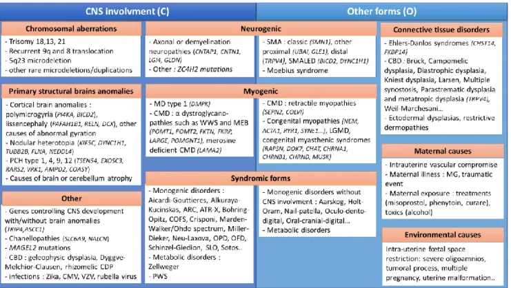
All other patients of group 3 (O) had various causes of AMC that could not be classified either as Amyoplasia, DA, or other types with central involvement. Possible diagnoses found in this group are gathered in Fig. 3.
5/ Data analysis in the light of information provided by the scientific literature
Each investigation performed for diagnostic purposes was listed. We determined the percentage of patients that had been submitted to it, reported the proportion of positive results and how it helped to guide the diagnosis. Each groups and subgroups of patients were analysed separately.
Then we compared our data to medical information available in the literature. We performed a rapid review for each paramedical investigation, using the search engine PubMed® (NCBI). Articles were selected from abstract analysis, and only relevant publications
were further studied. Additional papers, identified through the study of bibliography of firstly selected publications, if pertinent, were also considered. Review was performed between April and August 2019. Details on article selection and conclusion may be shared on request. For some paraclinical investigations, literature review had been previously performed in Dieterich et al, 2019 (32). In parallel, we completed our analysis by studying publications describing AMC cohorts or providing diagnostic management recommendations.
Combining conclusions obtained from the analysis of our data and results of our literature searches, we aimed at establishing clinical recommendations for AMC patients’ diagnostic management.
Figure 3. Possible diagnosis in Group 3 with (C) or without (O) CNS involvement.
From Dieterich et al., 2019 (32) and Hall et al., 2014 (4).
Legends : ARC, arthrogryposis Renal dysfunction and Cholestasis; ATR-X, Alpha Thalassemia/mental Retardation syndrome; CBD, Congenital Bone Disorders; CDP, Chondrodysplasia; CMD, congenital muscular dystrophies; CMV, cytomegalovirus; CNS, central nervous system; COFS, Cerebro-Facio-Oculo-Skeletal syndrome; LGMD, Limb Girdle Muscular Dystrophies; MD, Myotonic Dystrophy; MEB, Muscle Eye Brain; MG, myasthenia gravis; OFD, oro-facio-digital syndrome; OPD, oto-palato-digital syndrome; PCH, ponto-cerebellar hypoplasia; PWS, Prader-Willi Syndrome; SLO, Smith-Lemli Opitz syndrome; SMA, Spinal Muscular Atrophy; SMALED, SMA Lower Extremities predominant, autosomal dominant; VZV, varicella zona virus; WWS, Walker Warburg Syndrome.
RESULTS
1/ Cohort constitution
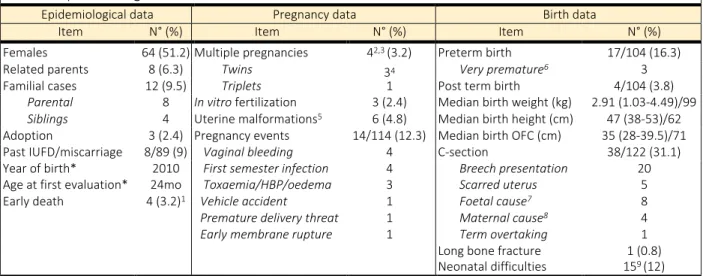
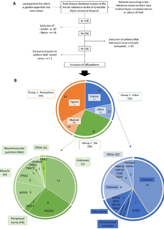
We gathered 123 paediatric AMC patients over 12 years. Additionally, two patients that had been evaluated first before 2007 but had been followed up in the reference centre after its certification were included as well, reaching a total of 125 patients (Fig. 4A). The full clinical workup proposed to AMC children had been performed for 118 of them. Twenty-five (20%) had a regular local follow-up, 90 (72%) were followed-up in secondary or tertiary level care centres in other French cities and 10 (8%) from other countries, mostly from Northern Africa (n=6). Epidemiological, pregnancy and birth data are summarized in Table 3.
54 patients were classified in group 1 (43.2%), 32 in group 2 (25.6%) and 39 in group 3 (31.2%). In group 1, 41 cases had typical Amyoplasia (75.9%) and in group 3 most patients had signs of CNS involvement (n=27, 69.2%) (Fig. 4B).
Table 3. Epidemiological information
Epidemiological data Pregnancy data Birth data
Item N° (%) Item N° (%) Item N° (%)
Females 64 (51.2) Multiple pregnancies 42,3 (3.2) Preterm birth 17/104 (16.3)
Related parents 8 (6.3) Twins 34 Very premature6 3
Familial cases 12 (9.5) Triplets 1 Post term birth 4/104 (3.8)
Parental 8 In vitro fertilization 3 (2.4) Median birth weight (kg) 2.91 (1.03-4.49)/99
Siblings 4 Uterine malformations5 6 (4.8) Median birth height (cm) 47 (38-53)/62
Adoption 3 (2.4) Pregnancy events 14/114 (12.3) Median birth OFC (cm) 35 (28-39.5)/71
Past IUFD/miscarriage 8/89 (9) Vaginal bleeding 4 C-section 38/122 (31.1)
Year of birth* 2010 First semester infection 4 Breech presentation 20
Age at first evaluation* 24mo Toxaemia/HBP/oedema 3 Scarred uterus 5
Early death 4 (3.2)1 Vehicle accident 1 Foetal cause7 8
Premature delivery threat 1 Maternal cause8 4
Early membrane rupture 1 Term overtaking 1
Long bone fracture 1 (0.8)
Neonatal difficulties 159 (12)
Legends : HBP, high blood pressure; IUFD, intra uterine foetal death; OFC, occipitofrontal circumference; mo, months old; N°, number of cases. *Median. 1group 3 only; 2and 2 additional early death of a twin;32 patients of group 1T, 2 of group 3O; 4 1 monochorial, 1 dichorial, 1 unknown; 5
unicornuate, bicornuate or septate uterus; 6before 32WG; 7foetal cardiac rhythm anomaly (n=5), laparoschis (n=2), twin-to-twin transfusion
2/ Aetiologies of AMC (Fig. 4B)
2.A/ Group 1: Amyoplasia congenita
Among patients with typical Amyoplasia, 66% had four limbs involvement (n=27), 22% only upper limbs (n=9) and 12% only lower limbs (n=5) (Fig. 5A). Thirteen cases were qualified as atypical Amyoplasia based on various clinical evidences (Table S1). Among these patients, nine had molecular investigations, negative in two and still ongoing in seven. For patient 98, clinical suspicion was strengthened by electroneuromyography (ENMG) results as we’ll discuss later (Fig. 5B).
2.B/ Group 2 : Distal arthrogryposis
In this group, 66% of patients had a molecular diagnosis, all obtained by targeted Sanger or next-generation sequencing (NGS) focused on AMC or neuromuscular diseases (NMD) related genes (AMC-NGS and NMD-NGS, respectively, whose list of sequenced genes is provided in Table S2) (Fig. 4B). Mutations in genes encoding proteins of the muscle sarcomere were the most frequent aetiology, among which half were MYH3 variants including two familial cases (Fig. 5C-D). Impairment of the peripheral nerve was nearly as frequent with a large majority of gain of function heterozygous PIEZO2 variants among which two were identified by specific research of the familial variation previously identified in their affected sibling; and one familial case of a homozygous ECEL1 variant (Fig. 5E-F). However, we had only one case of neuromuscular end-plate impairment due to a homozygous CHRNG variant and one case of Beals syndrome due to FBN2 mutation (Fig. 5G-H).
Among undiagnosed patients, eight NGS analyses were still ongoing. There was a clinical suspicion of DA2 in six patients including two cases with negative targeted Sanger sequencing and no further explorations ongoing. Multiple pterygium syndrome was suspected in three cases, with negative CHRNG sequencing in one. An ECEL1 mutation was negative in a case for which investigations were ongoing. Paraclinical examinations did not permit to get a formal diagnosis although it helped to better delineate the phenotype for two patients.
A
Figure 4. A. Flowchart of the study and B. Cohort distribution of the three groups, and detailed
definitive diagnoses in group 2 (green disk) and 3 (blue disk).
Legends : AAA, Association Alliance Arthrogrypose; AMC, Arthrogryposis multiplex congenita; C, CNS involvement; DA, Distal Arthrogryposis; M, muscle, NMJ, neuromuscular junction; O, other; PN, peripheral nerve; U, unknown.
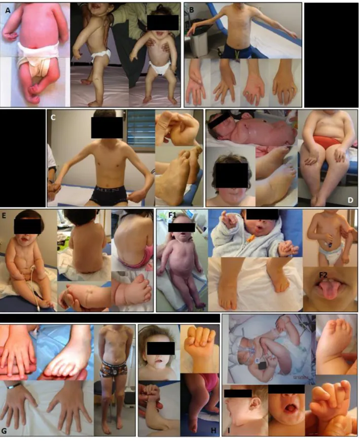
Figure 5. Pictures of AMC patients of our cohort. A. Typical Amyoplasia congenita involving four limbs (subject
87). B. Atypical Amyoplasia with asymmetric and caudal muscle impairment (subject 98). C. TNNI2 mutation in a patient with Sheldon-Hall syndrome (subject 105). D. Freeman-Sheldon syndrome due to MYH3 mutation (subject 108). E. Gain of function PIEZO2 mutation with early and progressive trunk involvement (subject 23).
F1. Confirmed ECEL1 homozygous variant (subject 39) and F2. characteristic groove tongue of a patient with
a strong clinical suspicion of ECEL1 mutation (subject 125). G. Discrete AMC of favourable outcome in a patient with Beals syndrome (subject 3). H. CHRNG homozygous mutations (subject 63). I. Subject with a ZC4H2 variant, with characteristic neck and extremities (subject 75).
2. C/ Group 3 : Other patients
In group 3, a definitive diagnosis was available for 41% and 50% of patients in group 3C and 3O, respectively (Fig. 4B).
In group 3C, ZC4H2 appears to be a frequent cause of AMC with CNS involvement, with four point mutations identified by targeted sequencing (n=3) or Whole Exome Sequencing (WES) (n=1), and one deletion evidenced by comparative genomic hybridization array (array- CGH). One case was familial (Fig. 5I). Chromosomal analysis identified two additional cases in this group, both first identified on karyotype: a 18p tetrasomy with an isochromosome and a 4p duplication due to an unbalanced translocation der(4;22)(p10;q10), further characterized by array-CGH. AMC-NGS identified a point mutation in TNNI2, a gene responsible for DA type 2B, in an individual that was initially suspected with DA but had been classified in group 3 because of a cognitive impairment. Looking back to his personal history, we hypothesised that his cognitive dysfunction could have a distinct cause, i.e. a car accident with head impact in childhood. Also, NMD-NGS pointed out a heterozygous NALCN variant in a child with cognitive dysfunction compatible with the diagnosis of CLIFAHDD syndrome (“Congenital contractures of the LImbs and FAce, Hypotonia and Developmental Delay”) (MIM#616266). Lastly, targeted sequencing of KAT6B and NSD1 were asked for suggestive presentations of SBBYSSS variant of Ohdo syndrome (MIM#603736) and Sotos syndrome (MIM#115750), respectively. Among patients without molecular diagnosis, there was a clinical suspicion of Muscle Eye Brain disease unexplored to date, and a case of hyperexplexia with negative GLRA1 sequencing.
Concerning patients of group 3O, WES and NMD-NGS identified variants in genes responsible for the kyphoscoliotic form of Ehlers-Danlos syndrome (MIM#614557) and for Larsen syndrome (MIM#150250), two conjunctive tissue disorders associated with AMC respectively due to FKBP14 and FLNB variants. Two other patients had mutations in genes responsible for syndromic forms of AMC, i.e. nail patella syndrome due to LMX1B variants (MIM#161200), and Aarskog due to a FGD1 mutation (MIM#305400). FKBP14 and FGD1 variants were identified in familial cases. Interestingly in this group we described two cases of environmental causes of AMC, one due to severe oligohydramnios secondary to a premature membrane rupture at 18WG and the other due to a complicated twin pregnancy (early cervical
cerclage, prolonged bleeding, premature rupture of membranes inducing extreme preterm birth).
More than half of these patients did not have any aetiological diagnosis. Among them, two were diagnosed with constitutional bone disorders (CBD) such as Diastrophic Dysplasia and Brück syndrome with so far negative analyses. Overall, NMD-NGS or WES analyses results were still pending in nine patients. As we’ll see, in group 3, paraclinical investigations could be useful to guide the diagnosis, although it did not allow to conclude formally at time of the study.
Overall, a definitive aetiological diagnosis of AMC was established for 79 patients, i.e. 63.2% of the cohort, comprising 43 clinical and 36 molecular diagnoses. Median age of patients at time of molecular diagnosis results was four years old. For the 46 undiagnosed patients, nine never had any analyses, 13 had previous negative investigations (including two NMD-NGS and one WES) but no ongoing explorations and 24 had pending NMD-related genes or untargeted NGS results.
3/ Paraclinical investigations performed and results (Table 4)
3. A/ Other genetic investigations
A karyotype was performed in less than half of our cohort, 48% antenatally (n=26), 41% postnatally (n=22) and 11% (n=6) both antenatally and postnatally.
In the antenatal period, karyotype was performed in a quarter of the cohort (25.6%, n=32) in equal proportions in the three groups (27.8%, 25% and 23.1% respectively). Half were performed because of bilateral equinovarus feet, isolated or associated with articular or extra- articular anomalies. Antenatal array-CGH were rarely performed (4.8%, n=6, 3 from group 3) and all were normal. Detailed indications are summed up in Table S3. Postnatal karyotypes (22.4%, n=28) were performed mostly in group 3 (41% (n=16), in contrast with 16.7 and 9.4% in group 1 and 2, respectively), as was array-CGH. Together, they contributed to three diagnoses, resulting in a yield of 2.4% in the whole cohort and 11% in group 3C. It also unravelled incidental findings in six cases, polymorphic copy number variants (CNVs) or
Variant of Unknown Significance (VUS) (Table S4). Other type of cytogenetic examinations had been performed in a limited number of patients (Table S5).
Targeted sequencing on a subset of genes involved in AMC was performed in almost half of the patients. Among the 48 tested patient, Sanger sequencing of AMC-genes had been performed in 33 and AMC-NGS in 17, both with a high yield (42 and 33%, respectively). Both were mostly performed in group 2 for which yield was the best. Finally, 22 patients had a NMD-NGS, mostly group 3 patients, with a global yield so far of 18.2%, with still a significant number of pending analyses.
Targeted sequencing of other genes was performed in 12.7% (n=16) of the cohort (Table S6). It enabled a diagnosis for five patients. Finally, genome-wide investigations, i.e. WES or whole genome sequencing (WGS), were performed in 11.1% of the cohort, mostly in group 3, and led to two diagnoses, resulting in a hitherto yield of 15.4%, here again with a significant number of pending results.
DMPK analysis, Prader Willi 15q11.2q13 locus analysis and SMN1 analysis were all performed in around 10% of patients. Some were performed prenatally (n=5, n=2 and n=4, respectively), mostly because of decreased foetal movements. DMPK triplet expansions were mostly investigated in group 3, SMN1 deletions mainly in group 1 patients, whereas Prader- Willi was questioned as much in the three groups. Those analyses never turned out positive.
3.B/ Other biological investigations
Creatine Kinase (CK) levels were determined in approximately one out of five individuals. CK levels were slightly elevated or subnormal only for four patients, especially in two local patients with ZC4H2 and NSD1 variants for which dosages were performed in particular circumstances, not mentioned in medical reports and without available control dosages, suggesting that they had been judged uninterpretable (Table S7). Moreover, elevated CK are not reported in these disorders and at least one other ZC4H2 mutated patients had normal values.
Table 4. Paraclinical investigations performed for aetiological purposes and their results
Investigations
Group 1 Group 2 Group 3
Total (%) tot (%) t at tot (%) PN NMJ M O+U tot (%) C O
54 41 13 32 9 1 10 12 39 27 12 G e n e ti c i n ve sti gati o n s* Karyotype tot 20 (37) 15 5 10 (31.3) 4 0 4 2 24 (61.5) 18 6 54 (43.2) + 0 0 2 2 0 2 Array-CGH tot 13 (24.1) 7 6 7 (21.9) 1 1 1 4 22 (56.4) 18 4 42 (33.6) + 0 0 1 1 0 1 AMC sequencing** tot 11 (20.4) 2 9 30 (93.8) 9 1 10 10 17 (43.6) 13 4 58 (46.4) + 0 21 9 1 10 1 3 3 0 24 pend 6 0 6 6 0 0 0 6 4 2 2 16 WES/WGS tot 2 (3.7) 1 1 2 (6.3) 0 0 0 2 9 (23.1) 6 3 13 (10.4) + 0 0 2 1 1 2 pend 2 1 1 1 0 0 0 1 6 4 2 9
Other targeted seq.1 tot 1 (1.9) 1 0 2 (6.3) 1 1 0 0 13 (33.3) 8 5 16 (12.8)
+ 0 0 5 2 3 5 DMPK analysis tot 7 (13) 6 1 3 (9.4) 1 0 1 1 6 (15.4) 5 1 16 (12.8) + 0 0 0 0 0 15q11.2q13 locus analysis tot 3 (5.6) 3 0 2 (6.3) 1 0 0 1 3 (7.7) 2 1 8 (6.4) + 0 0 0 0 SMN1 analysis tot 10 (18.5) 9 1 1 (3.1) 0 0 1 0 4 (10.3) 3 1 15 (12) + 0 0 0 0 Bi o lo gi cal in ve sti gati o n s CK dosage tot 13 (24.1) 9 4 2 (6.3) 0 0 0 2 8 (20.5) 6 2 23 (18.4) + 1 1 0 0 3 3 0 4
AChR/MusK antibodies tot 4 (7.4) 4 0 3 (9.4) 1 0 0 2 4 (10.3) 3 1 11 (8.8)
+ 0 0 0 0 Metabolic investigations tot 1 (1.9) 0 1 1 (3.1) 0 0 0 1 13 (33.3) 10 3 15 (12) + 0 0 1 0 1 1 N e u ro - mu sc .u lar MB tot 13 (24.1) 9 4 6 (18.8) 2 1 0 3 6 (15.4) 5 1 25 (20) + 7 6 1 1 0 0 0 1 3 3 0 11 ENMG tot 12 (22.2) 8 4 9 (28.1) 1 0 3 5 6 (15.4) 5 1 27 (21.6) + 5 3 2 1 0 0 1 0 3 3 0 9 mMRI tot 42 (77.8) 30 12 29 (91) 8 1 10 10 20 (51.3) 14 6 91 (72.8) + Ns Ns Ns Ns Ns Ns Ns Ns Ns Ns Ns Ns O rg an e xp lo rati o n s cMRI2 tot 20 (37) 17 3 7 (21.9) 0 1 1 5 21 (53.8) 19 2 48 (38.4) + 1 1 0 2 0 0 0 2 12 12 0 15 tcUS tot 15 (27.8) 13 2 4 (12.5) 1 0 0 3 21 (53.8) 16 5 40 (32) + 3 2 1 0 2 2 0 5 EEG tot 2 (3.7) 2 0 1 (3.1) 0 0 0 1 13 (33.3) 12 1 16 (12.8) + 0 0 7 6 1 7 scMRI tot 20 (37) 14 6 6 (18.8) 2 0 2 3 10 (25.6) 5 5 36 (28.8) + 2 0 2 0 2 1 1 4 scUS tot 5 (9.3) 3 2 1 (3.1) 1 0 0 0 8 (20.5) 5 3 14 (11.2) + 2 0 2 0 1 1 0 3 Ophtalmological evaluation tot 6 (11.1) 6 0 5 (15.6) 0 0 1 4 13 (33.3) 10 3 24 (19.2) + 0 0 0 0 Abdominal US tot 18 (33.3) 15 3 8 (25) 1 0 1 6 18 (46.2) 13 5 44 (35.2) + 3 2 1 2 0 0 1 1 1 1 0 6 Cardiological US tot 17 (31.5) 16 1 13 (40.6) 2 0 3 8 27 (69.2) 21 6 57 (45.2) + 3 3 0 3 1 0 0 2 9 8 1 15
Legends: AChR, acetylcholine receptor; AMC, arthrogryposis multiplex congenita; at, atypical; c, central nervous system involvement; CGH, comparative genomic hybridization; CK, Creatine Kinase; cMRI, cerebral MRI; EEG, electroencephalogram; ENMG, electroneuromyography; M, muscular; MB, muscle biopsy; mMRI, muscle magnetic resonance imaging; MusK, Muscle Specific Kinase; NMJ, neuromuscular junction; Ns, non-specified; O, other; pend, pending; PN, peripheral nerve; scMRI, spinal cord MRI; scUS, spinal cord ultrasonography; t, typical; tcUS, transcranial ultrasonography; tot, total; u, unknown; US, ultrasonography; WES, whole exome sequencing; WGS, whole genome sequencing.
* Investigations performed prenatally and postnatally are considered altogether
** Genes involved in AMC, includes Sanger sequencing of AMC genes, AMC-related genes and Neuromuscular disorders-related genes NGS (Table S2)
1Chromosomal or molecular investigations 2Postnatal
Investigation of maternal myasthenia antibodies targeting either Acetylcholine Receptor (AChR) and/or Muscle specific Kinase (MuSK) was even more rarely performed without differences in the 3 groups and was never found positive. For two patients, detection was performed during pregnancy because of foetal limb malposition and reduced limb movements.
Apart from hepatic, phospho-calcic or thyroid metabolism assessment, metabolic investigations were performed in one patient out of ten, as expected mostly in group 3C. Most screened patients had a basic checkup looking mainly for organic aciduria, urea cycle deficits or other aminoacidopathies, energetics disorders of citric acid cycle and fatty acid beta oxidation dysfunction. Only one patient had slightly abnormal ammonia levels whose cause remains unknown. Other investigations were performed in two or single patients (Table S8).
3.C/ Neuromuscular investigations
Muscle biopsy (MB) and electroneuromyography (ENMG) were both performed in about one out of five patients, with a slightly higher proportion in group 1 and 2.
MB was planned or discussed for 11 additional patients but had not yet been performed to our knowledge. Two patients were biopsied twice. MB was often performed in parallel of a surgical intervention. Many were interpreted as normal (n=6) or non-contributory because of lack of muscle tissue (n=5), and rarely results were not available (n=3). Histopathological examination reports described abnormalities in 11 cases (Table 5). Surprisingly, six had a clinical diagnosis of typical Amyoplasia and showed various types of anomalies. A neurogenic involvement was reported in two cases of typical and atypical Amyoplasia, in accordance with the hypothetic pathogenesis of this disorder, bringing another evidence toward Amyoplasia for the atypical case. However, two were suggestive of myogenic involvement, based on the association of poorly specific features, but did not question the diagnosis which was typical. One patient with a clinical diagnosis of DA had normal microscopic evaluation but a suspicion of multiminicore myopathy. In this patient (subject 12) for whom first clinical suspicion was a Freeman-Sheldon syndrome with normal Sanger sequencing of six DA genes and no other current investigations, this result may certainly help in the interpretation of future molecular investigations.
In group 3C, it appears that MB was mainly conducted to rule out a mitochondrial dysfunction. Eventually, the five tested individuals had a severe presentation with premature death in three of them. In the absence of evidence of such disorder on histology, investigations were completed by an analysis of mitochondrial respiratory chain (MRC) in three patients unravelling complex I borderline deficiencies and led to targeted genetic analysis in two individuals that turned out to be negative. Other anomalies were reported on histological examination of some of these patients but could not help to clarify the diagnosis. One had discordant results. In group 3O, MB was only performed in the patient with kyphoscoliotic Ehlers-Danlos syndrome and was non-contributory.
One third of ENMG were considered as abnormal (Table 6). Among patient with Amyoplasia, only one patient with a typical presentation had a neurogenic pattern, while the others had either normal ENMG, or a discordant myogenic pattern probably secondary to muscle disuse or atrophy. Interestingly, ENMG was of great value in an atypical Amyoplasia patient with unusual contracture location since it evidenced a neurogenic pattern which involved C7-C8-T1 motor roots instead of C5-C6, in accordance with his clinical presentation. In this patient it raised the hypothesis of a common pathophysiological mechanism with what is supposed in Amyoplasia, i.e. vascular compromise, but occurring in this case at a more caudal metameric location. Furthermore, this presentation is quite similar to Klumpke’s obstetrical palsy affecting motor roots C8-T1, a similarity that strengthened our hypothesis. This, together with the fact that this patient had unremarkable NMD-NGS results, lead to expect a low if ever null recurrence risk in this family, although it would be unwise to definitively and completely discard a genetic cause due to the lack of data about this presentation. Finally, ENMG completion helped to delineate the diagnosis of three patients of group 3 and one patient with DA (subject 20), adding for the latest new evidence toward the suspected diagnosis of Freeman-Sheldon though not confirmed on Sanger sequencing so far.
Whole-body T1-weighed mMRI was widely performed in this cohort and almost all patients with atypical Amyoplasia and DA had been investigated. It was planned in five additional patients, and failed in three others from group 3C. Eighteen individuals underwent several mMRI at different ages. Analysis of data delivered by this imaging is the purpose of other researches coordinated by our team and will not be detailed here. Overall, it mainly
Table 5. Anomalies observed on muscle biopsy examination
Ind. Group Sub group
Genetic
diagnosis Global conclusions MRC study
Mitochondrial genes/POLG1 sequencing
22* 1 T / Probable CMD merosine positive / /
104 1 T / Unspecific anomalies only, no congenital myopathy / /
94 1 T / Ns (Slight predominance of type 1 fibres, few mitochondrial aggregates, slight
increased lipid storage) / /
9 1 T / No myogenic involvement / /
26 1 T / Important myogenic involvement / /
8 1 T / Neurogenic involvement / /
52 1 aT uk Neurogenic involvement / /
12 2 U uk Probable multiminicore myopathy / /
34** 3 C uk No evidence for mitochondrial dysfunction Impairment of complex I function? Normal
17 3 C uk No evidence for mitochondrial dysfunction or myotubular myopathy. Discordant
results between IHC suggestive of a sarcoglycanopathy and normal WB
Normal (incomplete study of
complex IV) /
4** 3 C uk No evidence for mitochondrial dysfunction Normal but low complex I function MELAS mutation with
7% heteroplasmy
19** 3 C uk Non contributory Normal /
Legends : aT, atypical; C, CNS involvement; CMD, congenital muscular dystrophy ; Ind. individual; IHC, Immuno Histo Chemistry; MRC, mitochondrial respiratory chain; ns, not specified; T, typical; U, unknown type; uk, unknown genetic diagnosis; WB, Western Blot.* MB performed twice. ** premature death.
Table 6. Detailed anomalies observed on electroneuromyography Ind. Group group Sub diagnosis Genetic Conclusions
50 1 T / Possible neurogenic involvement suggestive of motor axonal neuropathy (microvoltage responses), with an atypical presentation.
Anomalies could either be secondary to orthopaedic deformations
26 1 T / Myogenic pattern and decrease voluntary activity in distal muscles of lower limbs (anterior tibial muscle and internal gemellus muscle)
66 1 T / Atypic myogenic pattern with rather rich pattern in location were muscle are atrophic
98 1 aT uk Neurogenic pattern with C7 to T1 motor root impairment (micro volted pattern with slight decreased conduction velocity in the right
median territory in motor conduction study, neurogenic contraction pattern in territory C7-C8-T1)
20 2 U uk
First ENMG normal, second with neuromuscular junction or muscle impairment (normal conduction velocity, but contraction pattern not as rich as expected on upper limbs, suggesting that a slight myogenic involvement cannot be discarded, although it could be secondary to muscle underuse.
17 3 C uk Myogenic pattern (low potential amplitude in right median territory)
60 3 C uk Moderate motor axonal neuropathy suggestive of anterior horn involvement
19 3 C uk Myogenic pattern (no detail available)