2/t33.3
2
UNIVERSITÉ DE MONTRÉAL
EFFECTS 0F CONCEALED CONDUCTION
ON AV NODAL FUNCTION
PAR
BOCHUNXU
DÉPARTEMENT DE PHYSIOLOGIE
FACULTÉ DE MÉDECINE
MÉMOIRE PRÉSENTÉ
À
LA FACULTÉ DES ÉTIJUES
SUPÉRIEURES
EN VUE
DE L’OBTENTION
DU GRADE DE MAÎTRISE EN PHYSIOLOGIE
w
kf
I
r
Université
de Montréal
Direction des bibliothèques
AVIS
L’auteur a autorisé l’Université de Montréal à reproduire et diffuser, en totalité ou en partie, par quelque moyen que ce soit et sur quelque support que ce soit, et exclusivement à des fins non lucratives d’enseignement et de recherche, des copies de ce mémoire ou de cette thèse.
L’auteur et les coauteurs le cas échéant conservent la propriété du droit d’auteur et des droits moraux qui protègent ce document. Ni la thèse ou le mémoire, ni des extraits substantiels de ce document, ne doivent être imprimés ou autrement reproduits sans l’autorisation de l’auteur.
Afin de se conformer à la Loi canadienne sur la protection des renseignements personnels, quelques formulaires secondaires, coordonnées ou signatures intégrées au texte ont pu être enlevés de ce document. Bien que cela ait pu affecter la pagination, il n’y a aucun contenu manquant.
NOTICE
The author of this thesis or dissertation has granted a nonexciusive license allowing Université de Montréal to reproduce and publish the document, in part or in whole, and in any format, solely for noncommercial educational and research purposes.
The author and co-authors if applicable retain copyright ownership and moral rights in this document. Neither the whole thesis or dissertation, nor substantial extracts from it, may be printed or otherwise reproduced without the author’s permission.
In compliance with the Canadian Privacy Act some supporting forms, contact information or signatures may have been removed from the document. While this may affect the document page count, it does not represent any loss of content from the document.
XU, Bochun -MSc Thesis
Université de Montréal Faculté des études supérieures
Mémoire intitulé
EFfECT$ 0F CONCEALED CONDUCTION ON AV NODAL FDNCTION
ou
EFFETS DE LA CONDUCTION CACHÉE SUR LA FONCTION DU NOEUD AV
Présenté par: Bochun Xu
Évalué par un jury composé des personnes suivantes: Dr Réjean Couture, président rapporteur Dr Jacques Billette, directeur de recherche
Dr René Cardinal, membre
XU, Bochun-MSc Thesis III
RÉSUMÉ
Bien-fondé: Les principes et le substrat soustendant la conduction cachée dans la double voie de conduction du noeud auriculoventriculaire (AV) demeurent mal compris.
Objectif Déterminer comment les propriétés de conduction cachée du noeud AV se développent dans ses voies lente et rapide.
Méthodes: La zone de conduction cachée et les effets de cycles choisis dans cette zone sur les courbes de fonction nodale ont été déterminés avec et sans un cycle conditionnant (10 ms plus long que la période réfractaire efficace et inséré avant le cycle caché) avantet après l’ablation de la voie lente dans 6 préparations de coeur de lapin.
Résutats: Étroite et inconsistante (22±12 ms, n=3) lorsque déterminée avec un battement caché seul, la zone de conduction cachée est devenue large et consistante (77±47 ms, n=6,
p<O.03)
quand un battement conditionnel conduit précédait le battement caché. La période réfractaire efficace et fonctionnelle du noeud AV s’allongèrent en proportionde la longueur du cycle caché qui déplaça toute la courbe de récupération (voies lente etrapide) vers la droite sans en changer la morphologie. En d’autres mots, tout battement caché réinitialise le cycle de récupération dans les deux voies. L’ablation de la voie lente a aussi élargi et rendu consistante la zone de conduction cachée sans altérer la réponse résultante de la voie rapide. Un battement caché dans une voie lente interrompue continue d’affecter la courbe de la voie rapide à toutes les longueurs de cycle.
Conclusions: Une large zone de conduction cachée est une propriété consistante du noeud AV. Le cycle conditionnant seul ou combiné à une ablation de la voie lente en facilite l’expression. La réinitialisation du cycle d’excitabilité dans les voies lente et rapide sont responsables pour la conduction cachée.
Mots clefs: Voie lente, voie rapide, conduction, état réfractaire, modulation électrotonique
XU, Bochun- MSc Thesis IV
ABSTRACT
Background: The mies and substrate underlying concealed conduction in atrioventricular (AV) nodal dual pathways remain unclear.
Objective: Determine how the concealed conduction properties ofthe AV node develop in its slow and fast pathway.
Methods: The nodal concealment zone and effects of increasing concealed cycle lengths chosen within the concealment zone on nodal function curves were determined with and without a conditioning cycle (10 ms longer than ERPN) before and afier slow pathway ablation in 6 rabbit heart preparations.
Resuits: from narrow and inconsistent (22± 12 ms, n=3) when determined with a blocked beat, the concealment zone became broad and consistent (77±47 ms, n=6, p<O.03) when determined with a blocked beat preceded by a conducted conditioning beat. The increase in the concealed cycle length resulted in proportional ERPN and fRPN (effective and functional refractory period of AV nod) prolongation and rightward shifi of the entire nodal recovery curve (slow and fast pathway) to a longer atrial cycle length range without affecting the curve shape. In other words, any concealed beat resets the whole recovery process in both pathways. The slow pathway ablation also broadened and made the concealment zone consistent but did flot alter the fast pathway responses to concealed conduction. A beat concealed in interrupted slow pathway still alters fast pathway curve at ah cycle lengths.
-Conclusions: A broad concealment zone is a consistent nodal feature. A conditioning cycle andlor slow pathway ablation favor its expression. Reset excitability cycle in the slow and fast pathway accounts for the effects of concealed AV nodal conduction.
Keywords: Slow pathway, fast pathway, conduction, refractoriness, electrotonic modulation
XU,
Bochun - MSc Thesis VTABLE 0F CONTENTS
INTRODUCTION
1CHAPTER I:
CURRENT STATE 0F
KNOWLEDGE ON THE AV NODE
31. Definition ofthe AV node 3
2. Anatomy ofthe AV node 4
2.1 Location aix! structure 4
2.2 Action potential based functional zones 6
2.3 Blood supply and innervation of AV node 7
3. Basic electrophysiology of AV node $
3.1 Excitability $
3.2 Refractoriness 9
3.3 Conductivity 12
3.3.1 Assessrnent of nodal conductivity with recovery curve 12
3.3.2 Location of the nodal conduction delay 13
3.3.3 Mechanisms of nodal delay 14
4. Rate- and tirne-dependent properties of the AV node 15
4.1 Complexity and unpredictability of nodal behavior 15
4.2 Definition and characteristics of AV nodal functional properties 17
4.2.1 Recoverytime 17
4.2.2 facilitation 18
4.2.3 f atigue 20
XU,
Bochun
-M$c Thesis VI6. Concealed conduction. 24
7. AV nodal function during atrial fibrillation 2$
OBJECTIVES 31
CHAPTER II: MANUSCRIPT
32ABSTRACT 34
INTRODUCTION 36
METHODS 36
Preparation, apparatus, and ablation 36
Protocols 37
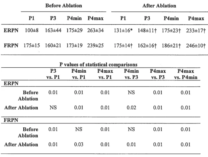
Interval measurements 3$
RESULTS 39
Concealment zone: effects of stimulation sequence and slow pathway ablation 39 Effects of concealed cycle length, conditioning cycle and slow pathway ablation on nodal
recovery curve 40
Effects of concealed cycle length, conditioning cycle, and slow pathway ablation on
nodal refractory curve 41
Comparison of concealed vs. conducted beats using two recovery indexes 43
DISCUSSION 44
Roles of slow and fast pathway in AV nodal concealed conduction 44 Does partial activation ofdual pathways accountfor concealed conduction? 44 Dual pathway excitability cycle and concealedA V nodal conduction 45
Effects ofa concealed beat on slow pathwayfunction 45
XU, Bochun- MSc Thesis VII Implications 4$ Conclusions 4$ REFERENCES 51 LEGENDS 55 TABLE II-1 58 TABLE II-2 59 FIGURE II-1 60 FIGURE II-2 61 FIGURE II-3 62 FIGURE II-4 63 FIGURE II-5 64 FIGURE II-6 65 FIGURE II-7 66
CHAPTER III:
GENERALDISCUSSION
67Main new findings 67
Meaning of concealed-conduction-induced changes in nodal recovery and refractory
curves 69
Site of nodal delayandaction potential characteristics 72
Implications 76
XV, Bochun-M$c Thesis VIII
LI$T 0F FIGURES
CHAPTER I
figure I-l: Structures and landrnarks ofrabbit AV node. Figure I-2: Relationship between action potentials and ECG.
Figure I-3: Methods used to characterize AV nodal conduction and refractoriness.
CHAPTER II
figure II-l : Preparation and stimulation protocols.
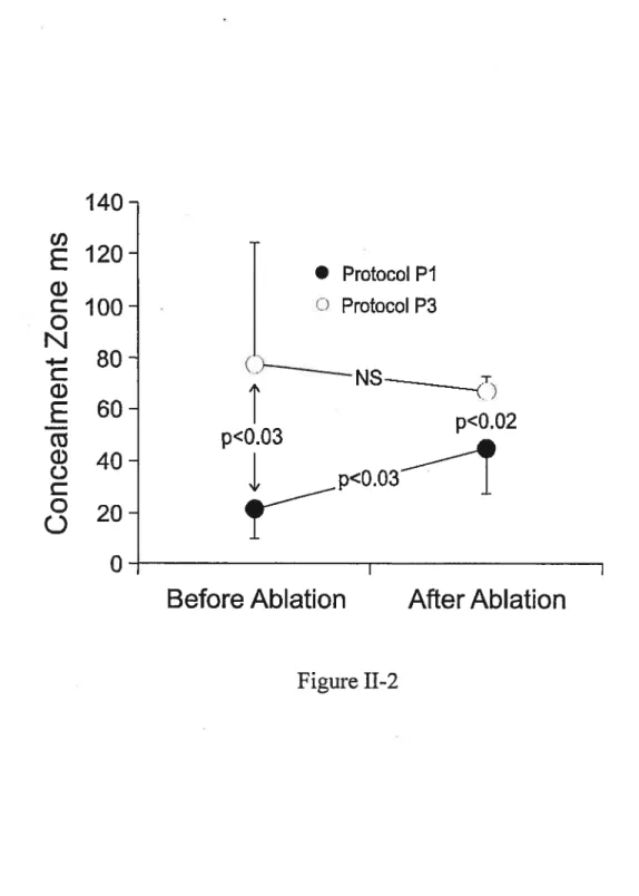
figure II-2 : Effects of a conditioning cycle and slow pathway ablation on the concealment zone.
figure II-3 : Effects of concealed conduction on AV nodal recovery curves obtained before and afier slow pathway ablation.
figure II-4 : Effects of enhanced concealed conduction induced with a conditioning cycle on AV nodal recovery curves obtained before and afier slow pathway ablation.
figure II-5 : Effects ofthe concealed cycle length, conditioning cycle (A1A1’), and slow pathway ablation on AV nodal refractory curves.
figure II-6 : Contribution of concealed cycle length and conditioning cycle in ERPN prolongation caused by concealed conduction.
Figure II-7 : Effects of concealed vs. barely conducted beats on the recovery curve as affected by chosen atrial cycle length variable.
CHAPTER III
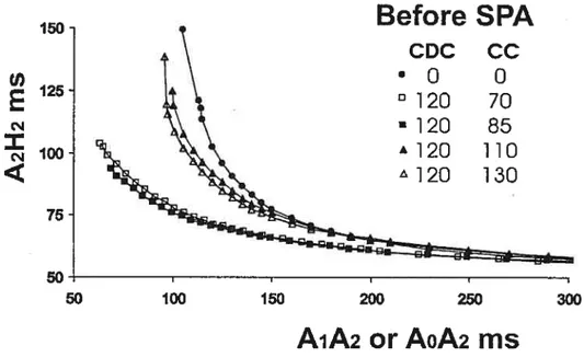
Figure III-l: Effects of concealed conduction, conditioning cycle and slow pathway ablation on A2H2 vs. A0A2 AV nodal recovery curve.
XU, Bochun-MSc Thesis DC
Figure III-2: Biphasic effects ofa conditioning and concealed cycle on A2H2 vs. A0A2 AV nodal recovery curves obtained before slow pathway ablation in one preparation.
figure III-3: Cycle-length dependent changes in transmembrane action potential characteristics at different AV nodal celis.
XU, Bochun- MSc Thesis X LIST 0F ABBREVIATIONS AV = atrioventricular AN = atrionodal N = nodal NH = nodal-Ris
RR = interval between two electrocardiographic R waves
ERPN = effective refractory period of AV node fRPN = functional refractory period of AV node
AA atrial interval HA = His-atrial interval
XV, Bochun- M$c Thesis
xi
ACKNOWLEDGMENTS
I would like to thankail peoples who contributed tomake this thesis possible and an enjoyable experience for me.
First, I wish to express my sincere gratitude to my thesis director, Dr. Jacques Billette, who guided this work and was easily available to help. His understanding, encouragement, support, and friendship help are especially appreciated. His scientific spirit andattitude of creativeness, rigors, as well as hard-worldng have impelled me to pursue my program in the past three years and will continuously influence my future career.
$incere thanks are also extended to:
Mr. Maurice Tremblay, for his expert assistance throughout my training, for his contributions to this work, and particularly, for providing us an enj oyable atmosphere.
Mrs. Lise Plamondon, for her skilful hands in assisting experiments and in preparing graphie illustrations, and for being so friendly ail which made it so pleasant to work with her.
Finally, I would like to express my deepest gratitude for the constant support,
understanding and love that I received from rny husband, Mr. Fang Guan and my parents during the past years.
)W, Bochun-MSc Thesis
INTRODUCTION
The AV node plays a strategic role in both normal and arrhythmic cardiac activation. It generates a delay between atrial and ventricular activation that favors ventricular fihling and effective blood pumping. It also filters impulses during supraventricular tachyarrhythmias to maintain a cycle length compatible with blood pumping. Since its discovery by Tawara,1 it has been the object ofmuch study by a number ofinvestigators both at the cellular and organ level. Despite important progresses made in ifie understanding of its anatomy, electrophysiology, and functional properties, the AV node remains puzzling in many respects.2 Zipes3 elegantly sumrnarized the problem by suggesting that “the AV node is a riddle wrapped in a mystery inside an enigma”. Despite important limitations in its understanding, the AV node remains a primary target of antiarrhytbmic therapy. Arnongst others, ablation therapy for the most frequent clinical arrhythmia called AV nodal reentrant tachycardia has greatly developed to become a success story in cardiology with a success rate ofnearly 1OO%.’
Paradoxically, the dual pathway substrate underlying this arrhythrnia remains highly debated both in terms of anatomy and physiology. The present study concems a problem related to this dual pathway substrate. To better understand this problem let us first recail briefly some basic notions of AV nodal physiology.
Unlike the other portions ofthe AV conduction system where the speed of
conduction is generalÏy constant from beat to beat, the conduction speed in the AV noUe is highly variable. The resulting AV nodal delay, referred ta as nodal conduction time in this thesis, can vary markedly in responses to changes in autonomic tone,6’7 intrinsic properties8’° or bath. The functional state ofthe AV node is particularly sensitive to
XV, Bochun -MSc Thesis 2
changes of heart rate that may resuit in a wide variety of responses even in the presence of a constant autonomic tone. This intrinsic rate-dependent behavior of AV node obeys 3 basic functional rules known as recovery, facilitation and fatigue.8’9”’4 Recovery refers to the increase in nodal conduction time with shortening ofpreceding cycle length. for a constant fast rate, the nodal conduction time increases with time and rate, a property called fatigue. for a constant recovery time and rate, nodal conduction time decreases in short recovery time range with the preceding cycle length, a property called facilitation. There is also increasing evidence that the normal AV node has a fast and a slow
pathway.15-19Recent studies suggest in fact that the slow and fast pathway have each their own set of rate-dependent properties so that the overali rate-dependent properties of the node would reflect their net sum.
Amongst communally observed rate-induced AV nodal responses is the one to atrial fibrillation, the most common sustained supraventricular tachyarrhythmias. In patient with atrial fibrillation, the ventricular response is typically made of irregular RR intervals, i.e., mark beat-to-beat variation in ventricular cycle length. This inegular ventricular rhythm arises from AV nodal filtering of high fibrillation rate through concealed conduction. During atrial pacing, an early atrial impulse may propagate some distance in the AV node before being blocked. The block, winch cannot be seen on the electrocardiogram, is therefore concealed. However, its occurrence can be detected by a prolongation ofthe conduction time at the next beat. Indirect evidence suggests that such a phenomenon occurs and repeat itself during atrial fibrillation.2023 However, the
physiology of concealed conduction and its specific role during atrial fibrillation remain to be factually established. The present study assesses how concealed conduction alters
XU, Bochun -M$c Thesis
j
nodal function in normal AV node with a slow and fast pathway, and in fast pathway alone obtained afier a slow pathway ablation.
CHAPTER I
CURRENT STATE 0F KNOWLEDGE ON THE AV NODE
1. Definition of the AV node
Clinicians, anatomists, and physiologists presently use the term AV node with different meanings. The term AV node variably stands for the compact node alone,
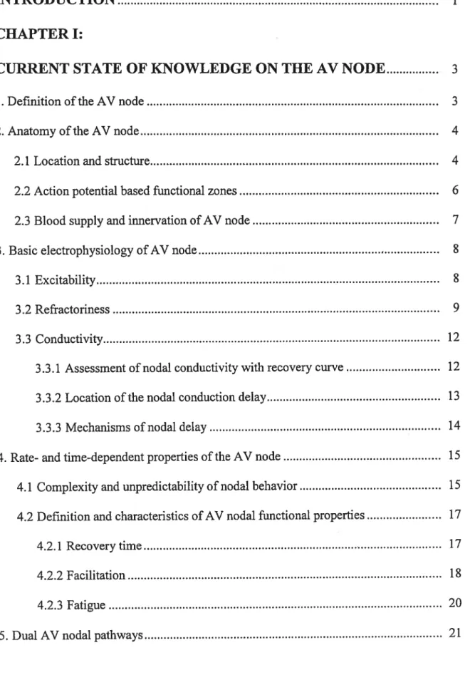
Figure I-l Structures and landrnarks ofrabbit AV node. Ris, Ris bundie; LNC, lower nodal bundle; CN, compact node; PNE, posterior extension; IC, transitional celis; C$, coronary sinus; TT, tendon ofTodaro; TV, tricuspid valve. from Medkour et colL15
compact node and immediate surroundings, or entire atrial-His junction (Figure I-l). Tawara provided the first definition of the AV node that was then limited to the compact node.’ Today, the specific role of the compact node in rate-dependent and dual pathway function remains unclear and very difficuit to study. Meanwhile, it has become clear that
T,
Iv
s
A
P
A
XU,Bochun-MScThesis 4
other structures, namely the transitional zone, posterior extension and lower bundie play critical role in nodal function. It has ffius become necessary to redefine the AV node to take into account these developments. Thus, the AV node was recently redefined as including ail structures (compact node, transitional zone, lower bundie and posterior extension) contributing to its recovery curve.15’24 This definition was aiso adopted in a recent prestigious book.25 In fact, a titie search for AV node shows that most
physiologists and clinicians adopted a similar broad definition of the AV node even though not explicitly expressed as such. In fact, of the numerous publications on the AV node, very few directly assess compact node function the role of which remains unclear.26 The definition that includes ah structures contributing to the nodal recovery curve is also used in the present study.24’25
2. Anatomy of the AV node
2.1
Location and structure
Working with Aschoff, Tawara’ first demonstrated that the AV node connects the atrium to the His bundie. Subsequently, many studies2729 confirmed that the AV node lies in the lower posterior portion of the interatrial septum beneath the endocardium and an overlap of atrial tissue, anterior to the ostium of the coronary sinus, and directly above the insertion of the septal leaflet of the tricuspid valve (Figure I-2). Its landmarks were clearly described by Koch in 1909 within a triangle, which now bears his name, delineated by the membranous septum at its apex, the inferior border at the septal tricuspid leaflet, and the superior border at a strand of fibrous tissue extending from the central fibrous body to the sinus septum above the ostium of the coronary sinus (the tendon of Todaro).28’30’3’ The compact node is close to the apex ofthe triangle ofKoch.
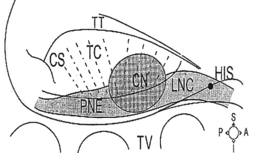
XU, Bochun- MSc Thesis 5 AVNodt’7’ ECG HBE V NodJ Vetrcuat
Figure 2. Relationship between action potentials and ECG. Lefi panel, typical action potentials from ceils ofvarious regions ofthe heart. Right panel, temporal correspondence between electrocardiogram (ECG), His bundie electrogram (HBE), and transmembrane action potentials recorded from atrial, AV nodal, and ventricular myocytes. From
http:Ilwww.hrsonhine.orqlprofessional educationhlearning categories/articles/shrbel/Default.asp
The compact AV node varies in size in different species. In the rabbit, it is approximately 0.3 to 0.5 mm in length parallel to the septal insertion of the tricuspid valve, 0.8 to 1 mm in the depth in the direction from interatrial to interventricular septum, and 0.15 to 0.25 mm in ffiickness in the direction from right to lefi atrium.32 In the aduit, it measures approximately 5 to 7 mm in length and 2 to 5 mm in width.
The AV node of the rabbit heart lias a complicated structure. Proxirnally, at the AV junctional region, the AV node has an extensive area in contact with the lower part ofthe
p
XV, Bochun -MSc Thesis 6
atrial septum. The transition from atrial fibers to transitional fibers occurs suddenly at the ultramicroscopic level.33 However, this sudden transition does flot occur at a detectable level macroscopicaÏly. $imilarly, the connection between transitional celis and compact node celis remains poorly defined from a macroscopic point of view but the two types of ceils have been shown to stain differently for connexins.30 The compact node exhibits a complex interlacing of specialized myocardial celis. In the distal portion of the node, fibers align in a parallel fashion to form the penetrating portion of His bundÏe.26’34 There are two main inputs to the AV node, an anterior and a posterior. The anterior input connects via the lower interatrial septum where the anterior and the middle internodal tracts descend and merge to form the superior margin of the AV node. The posterior input arrives at the node via the crista terminalis where the posterior internodal tract travels along right atrial endocardium and then into the interatrial septum above the coronary sinus, joining the posterior portion of the AV node.26’34
2.2
Action potential based functional zones
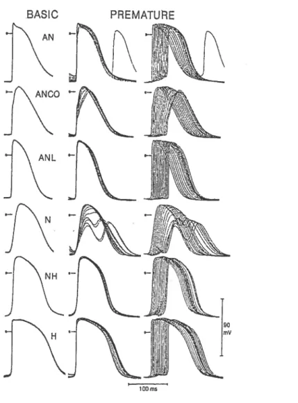
Based on electrophysiological characteristics (activation time and action potential configuration), Paes de Carvaiho and de Almeida35 divided the AV node into 3 zones: atrionodal (AN), nodal (N) and nodal-His (NH). The 3 functional zones form 3 layers that go from the I-lis bundle to ifie coronary sinus ostium. However, there are no anatomic structures corresponding to the 3 layers. The N zones is confined to a small central area, sunounded at its anterior, superior and posterior margins by the AN zone whule inferiorly making contact with the NH zone.34’36 In the rabbit, the AN zone corresponds to a
transitional ceil group in the proximal portion of the node and is made of elongated small cells.36’37 Moreover, a careful microelectrode-based mapping study conducted by Billette
XU, Bochun -MSc Thesis 7
showed that the AN ceils in fact occupied a large area in the proximal portion of the AV node and can be subdivided into AN, ANCO (AN with component) and ANL (late AN) cells.38The NH zone corresponds to the lower nodal ceil bundie that spreads from His bundie to coronary sinus. Initially considered as a dead-end pathway,39’4° recent studies has considered the posterior portion going from the center of the node to the coronary sinus as corresponding to the posterior extension (Figure I-1).15,41 The posterior extension
is made of nodal-like celis. It connects anteriorly wiffi the lower nodal bundle, which is made of elongated small ceils near the connection and larger celis near the His bundie.
2.3
Blood supply and innervation of AV node
The AV node receives a disproportionately large arterial blood supply relatively to its small size. In most individuals, this blood supply cornes from two main arteries.42 One is the AV node artery, which arises at the crux of the heart and ascends to enter the AV junctional area. It is a brandi ofthe right coronary artery. The second artery cornes from
the left anterior descending coronary artery as a septal branch.43
Nurnerous sympathetic and parasympathetic nerve branches richly inneiwate the AV node. The left and right cardiac sympathetic fibers, which corne from the paravertebral chain of either the stellate ganglion, the ventral limb of the ansa subclavian, or the caudal cervical ganglion, project onto the posterior surface ofthe heart, then cross the superior pulmonary vein, sending branches to penetrate the AV groove and into the epicardium overlying the AV nodal region. Cardiac vagal ganglia supplying AV nodal region are found within a srnaller fat pad overlying the epicardium at the junction of the inferior vena cava with lefi atrium, adjacent to the coronary sinus entrance. Recently, selective AV node vagal stimulation bas emerged as a novel strategy to control ventricular rate
Xii, Bochun -MSc Thesis 8
during atrial fibrillation.4448 When applied to the vagal ganglion, the stimulation achieves a desired average ventricular rate but unfortunately, the rate variability remains. More recently, Zhang et al’ used this strategy combined with ventricular on-demand pacing to obtain a satisfactory ventricular rate and regular rhythm in mongrel dogs. This novel approach resuits in a regular, slow ventricular rhythm during atrial fibrillation that does flot necessitate AV node ablation.
3. Basic electrophysiology of the AV node
3.1
Excitability
N and NH cells are hypoexcitable and recover slowly afier activation.9 their excitability is much lower ffian that of atrial and ventricular tissues. Microelectrode studies35’5° revealed that N ceils have a low resting membrane potential, low action potential amplitude, small or no overshoot, and slow rate of depolarization (10w Vmax) during phase O. With intracellular stimulation and recording, Merideth et al9 showed that the current requirement to reach threshold is higher in AV nodal area as compared to other cardiac tissues and that it is highest in N cells. They also demonstrated that the recovery of excitability lags beyond full repolarization in Ncells. This delayed recovery increases with rate. This rate-dependent reduction in nodal celi excitability was
demonstrated to be associated with a parallel depression of nodal conductivity.9 A study by Hoshino et ai5’ shows that, in single AV nodal myocytes, the recovery of excitability afier an action potential follows a very slow time course and greatly outlasts the action potential duration. $uch a slow recovery of excitability has been postulated to be responsible for the rate-dependent activation failure in these nodal myocytes.5’
XU, Bochun-MSc Thesis
9
3.2 Refractoriness
An important çharacteristic of excitable celis is their refractory period which provides a limit to the maximal frequency of activation. Refractoriness can be
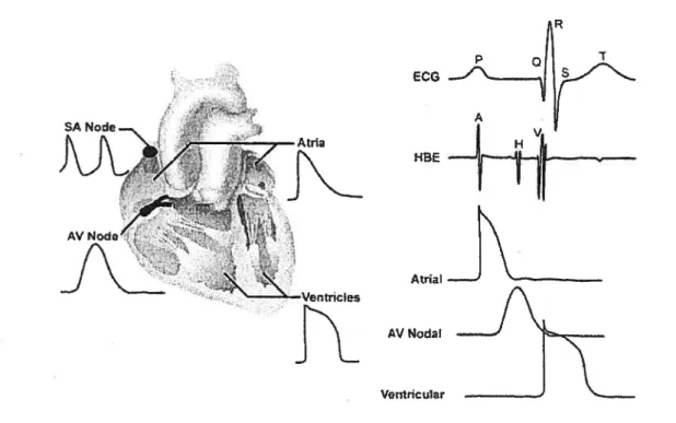
A EPN B - REFRACTORYCURVE RECOVERYCURVE 36 130 ___ 80 120 180 2C0 2’0 280 320 360 •0 80 120 180 200 20 250 32
AA1 INTERVAL(no) H,A2 INTERVAL(ms)
Figure 3 Methods used to characterize AV nodal conduction and refractoriness. A, superimposition, in reference to basic conduction time (A1H1), of prernature
conduction times (A2H2) obtained during a premature protocol in one rabbit heart preparation. B, refractory curve (H1H2 versus A1A2) with effective (ERPN) and functional (fRPN) refractory period. C, recovery curve obtained by plotting the A2H2 against corresponding H1A2. (Modified from Billette & Giles2)
characterized at the whole AV node level with a premature protocol (a test premature impulse introduced with a variable coupling interval during a constant basic rate).
XU, Bochun-M$c Thesis
The resuhing nodal responses are schematically superimposed in Figure I-3A andshow
that A2H2 increases with decreasing A1A2. These responses can be used to construct the nodal curve (Figure I-3C, A2H2 vs. H1A2 or A1A2) and obtain two indices of nodal refractoriness (figure I-33), which were first described by Krayer et aL52 They are obtained by plotting the different inter-Ris (R1H2) intervals against the corresponding interatrial (A1A2) intervals.6”4 The first index is the effective refractory period (ERPN), which is defined as the longest A1A2 interval that fails to generate a nodal response to A2 (Figure I-3B). When an atrial block develops before nodal block, ERPN is approximated from the shortest A1A2 interval thatresuits in a conducted A2. The shortest H1H2 interval achieved yields the second index, called the fiinctional reftactory period (FRPN).
Both ERPN and FRPN have been clearly dernonstrated to be rate dependent.’4’53’54 However, inconsistencies between the rate-dependent variations in FRPN and ERPN have been reported.5558 Cagin et al59 demonstrated that an increase in heart rate prolongs ERPN but shortens FRPN. Conversely, Young et a16° and Denes et al56 showed a parallel change of FRPN and ERPN, in which both parameters decreased with the increase of heart rate. It was also observed that the FRPN and ERPN change in the same direction under the effect of epinephrine or atropine.52’57’61’62 Moreover, differences in rate dependent changes of ERPN between chuidren and aduits were also reported.63 So far, there is stili no consensus on whether FRPN or ERPN can best represent nodal
refractorjness. ferrier and Dresel57 have reached the conclusion that there is a lack of relationship between FRPN and nodal refractoriness. Sirnson et al58 also reported that FRPN can vary independently of changes in nodal refractoriness as measured by ERPN. Thus, they concluded that the FRPN is a complex parameter and does flot reliably
XU, Bochun-MSclhesis 11
measure refractoriness. However, Young et a16° opposes the use of ERPN as an index of nodal refractoriness. Using Rosenblueth one-step delay hypothesis,55 Young et a16° assessed the effects of rate on nodal refractoriness and suggested that variations in ERPN corne from changes in recovery time due to variations in basic nodal conduction time which in turn cause a phase shifi of ffie recovery cycle rather than a real change in excitability. Thus, these authors considered that ERPN couïd flot reflex real changes in nodal refractoriness unless it is conected for the changes in basic nodal conduction tiine. Afler such correction, the rate-dependency of fRPN and the corrected ERPN were found to vary in the sarne direction.’4’60’64
Billette and Métayer’4 studied the factors involved in the intrinsic regulation ofthe rate-dependent changes offRPN in isolated rabbit heart preparation. These authors showed that, for a given fast rate, FRPN could vary markedly from beat-to-beat in a wide variety of pattems depending on the opposite effects of facilitation and fatigue. When present alone, facilitation and fatigue shortened and prolonged fRPN, respectively. Under cornbined effects of facilitation and fatigue, fRPN could be either shortened, unchanged, or prolonged depending on the state of interaction between these two properties.’4
More recently, Tadros et al65 studied AV nodal refractoriness with a protocol independently varying the effects of basic and pretest cycle, and reached the conclusion that fRPN consistently increases with fatigue and decreases with facilitation. ERPN also increases with fatigue and decreases with facilitation afier correction for the changes in basic nodal conduction tirne. The changes in basic nodal conduction tirne are an
XI], Bochun - MSc Thesis 12
differences with FRPN. Fatigue, facilitation and changes in basic nodal conduction time effects account for nodal responses to standard protocols but cannot be easily exposed by standard protocols alone.65
3.3
Conductivîty
As compared to other parts of specialized conducting system, the AV node Fias
a
low conductivity. Both anatomical and physiological factors contribute to this slow conduction. Two major anatomie factors are fiber diameter and geometric arrangement of fibers. Due to cable properties of fibers, the larger is the diameter of fibers, the easier and faster the impulses propagate. As to geometric arrangement of fibers, conduction velocity is greater when an impulse travels a fiber longitudinally than in a transverse direction.66 Two major physiological factors are the effectiveness of stimuli originating from
depolarized fiber and the excitability of responding fiber. An action potential with a greater amplitude and a faster rate of rise during phase O depolarization is a more effective stimulus and resuits in a greater conduction velocity than an action potential with a smaller amplitude and slow rate ofrise.67 The excitability cycle ofresponding fibers makes the time interval between two successive stimuli another important
detenninant of the impulse transmission.9 If a depolarization wave arrives in a fiber prior to its full repolarization from a preceding excitation (i.e., in a partial refractory state), the recovery of excitability of this celi is incomplete and the resulting premature discharges provides less effective stimuli to the responding fibers.
3.3.1 Assessment of nodal conductivity with recovery
curve The changes of nodal conduction time with slow recovery of excitabiïity can be characterized with a recovery curve (Figure I-3C). Such a curve is constructed from theXU, Bochun -MSc Thesis 13
data obtained with a periodic premature stimulation. There are two approaches used to constnict a recovery curve.68 One consists in plotting the nodal conduction time of premature beats (A2H2 interval) against the corresponding recovery times (H 1A2 interval).2”°’68’69 The second approach differs from the first by the use ofthe A1A2 interval as the index of recovery times.9’54’70’71
Since the early twenties, the recovery curve lias been widely used in describing the physiological or pathological characteristics of AV nodal conduction and in analyzing the changes produced by different atrial stimulation pattems and.for drugs. Mobitz72 in 1924 used the recovery curve to analyze the PR-RP relationship and to characterize the
Wenckebach periodicity. Lewis and Master also used the same approach one year later to study AV nodal function.8 With the development of experimental intracardiac
recording,7375 it became possible to dissociate ifie components ofifie PR inteiwal in which the AH interval specifically represents the nodal conduction tirne. Henceforth, the recovery curve relating PR-RP or PR-PP intervals was recasted as relating AH interval versus HA or AA interval. The A2H2 vs. A1A2 curve provides the scheme from whicli nodal frmnction is assessed in humans undergoing endocavitary investigations for cardiac arrhythniias.76’77
3.3.2 Location of the noda’ conduction delay
Diverging opinions on the origin of conduction delay in the node have been expressed since the very begiiming ofthis century. In 1913, Kure78 suggested that the delay takes place in the upper part ofthe AV node whule Zahn79 assurned that the middle portion ofthe node is responsible for most ofthe conduction delay. Much later in 1958, Matsuda et a18° achieved the first intracellular record from a nodal cell. Hoffrnan et al50’75
XV, Bochun -MSc Thesis 14
considered that the delay takes place at the atrial margin ofthe AV node. Paes de
Carvaiho and de Almeida who first proposed, based on thefr microelectrodes recordings from isolated rabbit heart, the AN, N and NH classification of AV nodal ceils located the greatest siowing of propagation in the N zone.35 However, changes of propagation
velocity in AN zone are gradual and smooth until a minimum is reached in N zone. In the NH zone, the conduction gradually accelerated as the Ris bundie is approached. Billette provided more information about the sites of AV nodal delay.38 This study demonstrated that the lowest upstroke velocities were found in N celis, wbich also had a greater
prernaturity-dependent increase in action potential duration. During premature atrial stimulation, action potential amplitude of N cdl decreased, and the response dissociated into two components. According to this study, the AN and NH zone accounted each for 25% ofthe basic conduction delay whereas the central node (N to NH zone) accounted for close to 50% ofthe delay. This study also showed that the prematurity-dependent increase in nodal conduction time develops also mainly between the N and NH zones.81
3.3.3 Mechanisms of nodal delay
Hoffinan and Cranefield82 suggested that the nodal delay is due to decremental conduction. These authors considered that, as an impulse travels in nodal non homogeneous tissues, it encounters tissues ofprogressively greater threshold, of decreasing space constant, of decreasing action potential amplitude, and of decreasing rate of depolarization, which combine to produce a progressive siowing of conduction. However, Rosenblueth55 suggested that the conduction delay and its rate-dependent increase are due to a “one step delay” which occurs at one point in the conduction path rather than to overali decremental conduction. Several lines of evidence showing
XU, Bochun- M$c Thesis 15 discontinuous propagation in non-homogeneous tissues support this explanation.69’83’84 Billette et al demonstrated that, with shortening of cycle length, action potential in the N zone dissociated progressively into two components that were synchronous with late AN and early NH activity, respectively.37 No action potential upstrokes occurred during the interval between two components. The increase in nodal conduction time with shortening of cycle length was largely due to the “stagnation” between N and M-I zone activation.81 The stagnation is probably caused by cessation of active transmission at the unexcitable elernent, which can be nevertheless crossed by electrotonic cunent, bringing distal excitable celis to threshold as described in other cardiac tissues.83’84 The ionic
mechanisms of nodal conduction delay are flot completely understood yet. It has been proposed that the slow recovery from inactivation of the slow inward calcium current and the slow deactivation of potassium current in ifie central AV node are important
determinants ofthe nodal delay.51’85
4. Rate- and time-dependent properties of the AV node
4.1
Complexity and unpredictability of nodal behavior
An increase in heart rate usually induces an increase in nodal conduction time. However, the relationship between heart rate and nodal conduction time can vary in a very complex mamier both with the rate itself and its duration. Disparate nodal conduction tirne may be obtained at identical heart rates reached with different
approaches.8”‘‘ Nodal responses can also vary with the stimulation pattem used. for
example, the addition of a single constant short cycle before the premature cycle result in a completely different set ofpremature nodal conduction time than those obtained
XTJ, Bochun - M$c Thesis 16
obtained by reducing the stimulus interval by 2Oms at every20th beat also have been
shown to differ entfrely from those induced by a similar incrernental pacing but with 20 control basic beats being inserted between subsequent reductions.’2 The controls basic beats then prevents the occurrence of cumulative effects of rate. The initial condition from which a stimulation is started is also an important factor affecting the nodal
behaviour.’3’86 Two identical periodic premature stimulations performed at the same fast rate, but one being started before and the other afier reaching a steady-state nodal
conduction time resuit in a very different nodal recovery responses.8’ Similarly, identical premature stimulation protocols performed at five different basic cycle lengths can resuit in five different nodal recovery curves.81
for a fixed heart rate, the nodal conduction time increases with the duration of the rate, an effect called fatigue.8”2”3’57 The faster is the rate the greater is the fatigue developed. However, part ofthe increase in the nodal conduction time observed afier a sudden increase in heart rate is not due to fatigue. Levy et al have demonstrated that there is then an inverse relationship between RP and PR interval.7 However, the contribution of this factor is ofien ignored thus leading to overestirnation of fatigue effects.58 When a fast rate is imposed with a constant RP or HA interval, which prevents RP-PR or HA-AH variations, the fatigue can be directly obtained and is much less than in the absence of this controL7”3 During transient responses such as those obtained during incremental pacing protocol recovery and fatigue effects occur together and cannot be easily dissociated.
To better understand the rnechanism responsible for the complex nodal behavior, it is important to identifyand characterize the underlying nodal functional properties
XV, Bochun - MSc Thesis 17
involved in the different steady-state and transient nodal responses. Lewis and Master8 identified 3 different components in the rate-induced changes of the nodal conduction time: a conduction siowing ofpremature atrial impulses, an abbreviation ofthe nodal conduction time and refractory period caused by a single cycle of a faster rhythm introduced before the premature cycle, and an impairing cumulative effect of frequency on the nodal conduction time. In a series of studies, Billette et al’214’8’ were able to dissociate and further characterize these 3 components which are now referred to as recovery time, facilitation and fatigue. Studiesalso indicate that the interactions between these 3 properties can account fora wide variety of nodal responses.’2’64’85’8792
4.2
Definition and characteristics of AV nodal functional
properties
4.2.1
Recovery time
The recovery time represents the time elapsed since last activation and reflects recovery of excitability before next activation.2 This time may be estimated by the Ris—
atrial (HA) interval, winch is the time from the beginning of a given His bundie
activation to the beginning of the next atrial activation.69 Recovery time is also frequently assessed by the atrial (AA) interval.9’57’93 Because the noUe consists ofmany different ceils which are activated in sequence (i.e., proximal nodal celis are activated and recover earlier than distal cells), there is no common recovery time for ah nodal celis. When the preceding AA interval is used to assess nodal recovery time, the recovery tirne of distal nodal ceils is overestimated when the nodal conduction time increases; the constant AA suggests that recovery time has flot changed whule in fact it has been shortened by the increased nodal conduction time. When the preceding HA interval is used to assess nodal
XU, Bochun-M$c Thesis 18
recovery time, the recovery time of proximal nodal ceils is underestimated.2’94’95 However, because the activation ofproximal nodal ceils is largely cycle length independent,8’ this bias may have a lesser impact than its overestimation with the AA interval.
Nodal recovery time is a major determinant ofthe variations of nodal conduction time in response to changed heart A shortened HA prolongs the next AH.69 This effect increases more rapidly in the short HA range. At a shortened HA interval, the atrial impulse reaches the node earlier in the nodal reftactory period. The shorter is the HA interval, the more refractory is the node, and the longer is the AH intervaÏ.”69 Nayebpour et al87 characterized the effects of vagal stimulation on nodal recovery
property. Tliey showed that under vagal stimulation, nodal recovery was incomplete for a longer period afier nodal activation, thus resulting in nodal block at longer recovery time, which could otherwise evoke a conducted beat.
Above studies suggest that the recovery time constitutes the major determinant of changes in nodal conduction time. However, the changes of recovery time alone cannot satisfactorily explain ah rate-induced changes in nodal conduction time which are also contributed to by nodal facilitation and fatigue.’2’8’
4.2.2
Facilitation
Nodal facilitation is defined as a shorter nodal conduction time than expected from recovery lime and fatigue.” It can be selectively characterized by simply adding a short cycle before the premature cycle during a protocol otherwise identical to that used for the determination ofthe recovery property. In addition, a conditioning intermediate cycle is also usually introduced before ffiis short cycle to allow for an induction of the maximal
XU, Bochun -MSc Thesis 19 facilitatory effect. The intermediate cycle shortens the minimum facilitating cycle ifiat can be tested. A facilitatory effect develops afier one short cycle, remains constant during a lasting fast rate and dissipates aller a single control long cycle.” Usually, one short cycle produces a maximum shortening in nodal conduction time and FRPN. Facilitatory effects are thus time-independent.”8’ Graphically, facilitatory effect is manifested by a leftward shifi and tilt ofthe HA-recovery curve in the short H1A2 range.
Nodal facilitation was first observed, though flot named as such, by Lewis and Master in 1925.8 However, its very existence remains debated. Several factors affect its manifestation. While facilitation is rnost easily seen on HA curve, it is also present and unchanged on AA curve but then hidden by greater effects of changes in basic nodal conduction tirne.13’68 Its identification then requires the correction ofAA curve for changes in basic nodal conduction time. Because flot spontaneously seen on AA curve, several investigators tend to simply ignored facilitation.71’94’95’97 However, careful examination ofthe published AA-recovery curves does indicate the presence of facilitation which appeared as the convergence andlor crossing of the recovery curve in the short AA interval range. The facilitatory effects were studied by Billette et al”2”4’8’ who successfiilly dissociated them from other rate-dependent effects. The presence of facilitatory effects has also been demonstrated during both transient and steady state nodal responses, and was found to occur concurrently with changes in recovery time and fatigue. In these circumstances, facilitation may be masked and difficult to identify. However, when changes in recovery time and fatigue are controlled or measures, it becomes possible to clearly identify the facilitatory effects.’2’4’85’88 Moreover, these studies indicate that the inclusion of facilitatory effects is a necessary condition to
XU, Bochun- MSc Thesis 20
quantitatively account for nodal conduction time observed in various rate-induced nodal responses.
4.2.3
Fatigue
Nodal fatigue is defined as flue rate- and time-dependent prolongation of nodal conduction time occurring for comparable recovery time and facilitation level. The fatigue effect develops slowly during a fast rate and dissipate in a reverse symmetric pattern afier the termination ofthe fast rate.12”3’81 The slow time course of fatigue
induction causes it to start affecting the nodal conduction time only several beats afier the begiiming of a fast rate and to take several minutes to reach a steady state. Similarly, the slow dissipation of fatigue may cause it to affect the nodal conduction time several minutes afier the cessation of a fast rate.
Nodal fatigue has been reported by several investigators.79’57 With sustained atrial pacing at a constant short cycle length, these authors observed cumulative increase of nodal conduction tirne which they called “exhaustion” or ‘fatigue”. However, these authors did not dissociate per se from the effects of uncontrolled changes in recovery time which accounted for an important fraction of the nodal conduction time prolongation that they observed. In later studies, Billette et a1’24’8’ selectively characterized nodal fatigue and distinguished it from other recovery-dependent effects. These induction and dissipation of fatigue were shown to folÏow an almost symmetric slow time course.13 During periodic premature stimulation performed at a constant fast basic rate but no pre established steady state, fatigue grew progressively with the duration of the rate needed to test the different premature beats.8’ Therefore, despite the same rate, the two approaches
XV, Bochun -MSc Thesis 21
then resulted in different nodal responses. Both the rate itself and its duration are important determinants of the level of fatigue reached.
5. Dual AV nodal pathways
Dual AV nodal pathway electrophysiology has long been accepted as the basis for AV nodal reentrant tachycardia (AVNRT).98’°3 The outstanding success of ablation therapy for AVNRT over last two decades has further focused the attention of cardiac electrophysiologists on this topic.5’104’°6 The basic underlying concept is that the AV node eau be ftmnctionally spiit into two pathways (a slow and a fast) with different conduction and refractory properties that favor the initiation of reentry.99 The fast
pathway conducts rapidly and is thereby dominant but has a long refractory period. When the coupling interval reaches a value shorter than the refractory period of the fast
pathway, the slow pathway can, due to its short refractory period, take over conduction. The transition from fast to slow conduction is frequently associated to a sudden increase in A2H2 and the initiation of echo beats in patients suffering from AVNRT. There is a general agreement that the slow pathway originates somewhere in the posterior portion of the node and is connected to the crista terminalis input while the fast pathway is located more anteriorly and connected to the interatrial septum. This is also supported by the fact that ablation therapy targeted at the posterior input intemipts the slow pathway whule an ablation at the interatrial septum interrupts the fast pathway.4”°7”°8 However, the exact anatornic and functional substrate underlying the dual pathways rernains debated.
Mendez and Moe initially proposed that the two pathways reflect a functional asymrnetry between the crista terminalis and interatrial septum inputs with their
XV, Bochun- MSc Thesis 22
portion ofthe node would form a common distal pathway. The fact that input-targeted ablation therapy does flot involve compact node legions indicates that the compact node may not be an important determinant of dual pathways, leaving the inputs as the primary substrate. There is evidence of some degree of fiinctional asymmetry between the two inputs.’°”°3”°9”4 $everal investigators have recorded double component action potentials in the input areas and some have attributed it to dual pathway physiology. However, this question was thoroughly reassessed recently by de Bakker et al”5 who concluded that that double potentials are unlikely due to dual pathway physiology.
In an effort to formerly establish the role of functional asymmetry between the inputs in dual pathways, Amellal et al”6 characterized them with local stimulation and recording techniques. They found that the two nodal inputs have equivalent conduction and refractory properties, and are equally effective in activating the rate-dependent portion of the node. Their findings predict that, even for symmetric functional properties ofthe inputs, an impulse entering the node from the low septum could be blocked at the anterior nodal input but propagated from the posterior input, thus resulting in an nodal conduction time prolongation when measured from His bundle lead. This prolongation is due to the extra traveling time of the impulse toward and in the low crista input rather than actual changes in input conduction velocity. Their results suggest that the proximal node acts as a matching gate that enables widely varying atrial wave fronts into equally effective activation signals for the rate-dependent portion of the node.
The search for alternative substrate led to the development of a new model in which the slow and fast pathway would be provided by the posterior extension and compact node, respectively.15’9 In this model, the two inputs forrn a common proximal pathway
XU, Bochun -M$c Thesis 23
while the lower bundie forms a common distal pathway. Mthough the characteristics of the demonstrated dual pathways differed from those ofAVNRT patients, the dual
pathways were found in ail rabbit hearts studied, thus establishing that dual pathways is a normal feature of rabbit AV node. The posterior extension is also consistently present in humans but its functional role remains to be established.4’ Recent dye mapping studies have provided further direct evidence for this model by demonstrating that it is
compatible with the characteristics of local activation during echo beats.25”7”9
The discontinuous AV nodal conduction curve has been used clinically as evidence for the existence of dual AV nodal pathways. The criterion used to identify such a
discontinuity is the occurrence of a 50-msec jump in AH for a 1 O-msec decrement of the atrial coupling interval during delivery of programmed premature atrial beats.’2° The two portions of the discontinuous conduction curve are thought to represent a shifi of the conduction from fast to slow pathway when the long effective refractory period of the fast pathway has been reached. While many AVNRT patients show such a discontinuity, flot ail patients do. Moreover, some patients without AVNRT do have ajump in AH intervals. Thus, the link between the jump in AH and AVNRT is a frequent but not a necessary one. In order to explain this phenomenon and the presence of residual AV nodal conduction afier ablations of slow and fast pathways, Hirao et al” suggested the existence of intermediate pathway(s). Anterograde conduction over the intermediate pathway would smooth the transition from the fast pathway to the slow pathway, resulting in a more continuous curve. $imilarly, anterograde conduction over the slow pathway without retrograde exit can cause a discontinuous curve with no inducible AVNRT. Recently, several investigators have shown that dual AV nodal physiology can
XU, Bochun MSc Thesis 24 be deduced in patients without discontinuous AV nodal conduction curves by the
following findings after radiofrequency ablation in the region of the slow pathway: (1) an increase in the anterograde AV nodal effective refractory period; (2) an increase in the anterograde Wenckebach cycle length; (3) shortening of the maximum AH in response to atrial extrastimuli and, (4) loss ofthe tau ofthe smooth AV nodal conduction curve.121’122
Recent mapping and ablation studies support several postulates. 1) The fast and slow pathway represent different atrionodal connections rather than a resuit of longitudinal dissociation within the compact node.5”°7” 2) These pathways are functional pathways instead ofcable-like structures.123 3) Multiple AV nodal input pathways likely exist as suggested by the presence of residual AV nodal conduction afier ablations of fast and slow pathways.”
6. Concealed conduction
In 1894, Engelmann’24 stated that every effective stimulus applied to the atrium, even if it is not followed by a ventricular response, lengthens the next AV interval. The rnechanism underlying this effect was investigated much later. In 1925, Ashrnan’25 studied the influence of blocked impulses on subsequent conduction in compressed atrial muscle of the turtie heart. He observed that the earlier the blocked impulse follows a transmitted one, the less is its effect upon the conduction time of a subsequent transmitted impulse. Conversely, the later is the blocked impulse after the transrnitted one, the greater is its effect upon subsequent conduction. Levis and Master8 in their classical observation upon AV conduction in the mammalian heart demonstrated clearly that, in 2:1 AV block, whether produced by raising the atrial rate, or by stimulating the vagus, the conduction intenïals are lengthened by the presence of the alternate atrial beats, that is, those to
XV, Bochun-M$c Thesis 25
which the ventricle fails to respond. In 1948, the term concealed conduction was first introduced by Langendorf’26 and used to describe such a phenomenon by which an atrial impulse activates a portion ofthe AV node before being blocked. This phenomenon is now considered a maj or determinant of nodal responses during supraventricular tachyarrhythrnias.21”27’29 In 1965, Langendorf et al’28 demonstrated successfully the existence of concealed AV conduction and its influence on subsequent conduction in the human heart. The prolongation of the P-R interval after interpolated ventricular
premature systoles is the most obvious example of this phenomenon. The retro grade impulse of the ventricular premature systole penetrates into the AV node and is stopped before reaching the atrium; this resuits in a prolonged A-V conduction time at the postextrasystolic beat. Even if this partial penetration does flot resuit in any specific electrocardiographic wave, its effect can be detected from the influence it has on the AV nodal conduction or PR interval at the next beat.
Although the supporting evidence is so far very limited, concealed conduction is considered to play a major role in the gross irregularity of ventricular response to atrial fibrillation. At the sanie time, summation and cancellation of atrial impulses entering the AV node at different inputs, rates and degrees ofregularity as well as autonomic tone may ah contribute to the high frregularity ofventricular response to atrial fibrillation. However, their sorting out may require that the mecianisms underlying concealed conduction be first better understood. Another difficulty in attributing the gross
irregularity of the ventricular response to concealed conduction is its puzzling link with the refractory period. If, as now generally accepted, the ventricular response to any type ofrapid atrial activity is primarily determined by the refractory period ofthe AV
XV, Bochun- M$c Thesis 26
junction, one would expect a more regular response ofthe ventricle than observed during fibrillation. Indeed, because an atrial impulse is aiways immediately available to the node at the termination of its refractory period, RR intervals should have values approximating this refractory period, which is obviously flot ifie case. The duration of the refractory period could be altered and the regularity of the ventricular rate disturbed, if one or more of the atrial impulses pefletrate into the AV junction without reaching the ventricles. Such concealed conduction of any impulses will affect conduction of the subsequent impulse by delaying it, blocking it entirely, or causing repetitive concealed conduction.20”3° Concealed propagation offfie atrial impulse may occur during various portions ofthe heart cycle and reach different levels within the AV junction. The varying depth of nodal penetration and its varying time of occurrence is likely to resuit in much variation in ceil refractoriness. These different effects at different AV junctional levels will favor
secondary concealment and repetition of long ventricular cycles. The above possibilities proposed by Langendorf13° in 1965 were further supported by later studies. Benefiting from technical progresses and a better understanding of anatorny, electrophysiology, and functional properties of AV node, several physiologists have used again these basic concepts that have become guiding principles in the study of concealed conduction. Whatsoever, current evidence indicates that the block is due to both increasing refractory period and decreasing excitability in temporally more distal portions ofthe AV node. These properties cause a prernature test impulse to be blocked increasingly more proximally when introduced with decreasing coupling interval.9’34’38 Potential sites of block are numerous but the junction between transitional and compact node tissue
XU, Bochun -M$c Thesis 27 constitutes a critical prevalent one.127 Whether the blocked beat affects only the activated portion of the AV node or also the distal inactivated part remains unclear.
There is evidence that a concealed beat postpones the recovery of nodal excitability as reflected on nodal curve (A2H2 vs. A1A2).97”3’ The blocked beat would prolong
refractoriness and postpone the recovery cycle in cardiac cycle. Recently, a study by Liu et al’32 stated that the concealed beat prolongs refractoriness ofproximal cells and prevents assessrnent of real refractoriness in more distal nodal elements that may or flot have changed. They also proposed new data and concept suggesting that events occurring in slow pathway may play a critical role in concealed conduction. They showed that a concealed beat arnputated the lefi portion of AV node curve (manifest portion of slow pathway) in both AVNRT and control non-AVNRT patients. The concealed beat
prolongs ERPN and reduces A2H2max in both groups. In AVNRT patients, the concealed beat reduces the incidence of discontinuous curve and AVNRT induced by premature stimulation. In other words, the concealed beat prevented slow pathway conduction in a similar mairner than did slow pathway ablation. Moreover, the concealment effects on fast pathway conduction were sirnilar before vs. afier slow pathway ablation. Thus, the understanding of AV nodal concealed conduction may require the sorting out of different individual effects induced in fast and slow pathway.
The finding that a concealed beat prevents manifest slow pathway conduction but shifis the fast pathway curve to the right is an intriguing one.132 The primary riddle is whether the concealed beat reduces excitability and prolongs refractoriness in slow pathwy. A primary objective ofthe present study was to assess the role of slow pathway in concealed AV nodal conduction.
XV, Bochun -M$c Thesis 2$
7. AV nodal function during atrial fibrillation
Concealed conduction is assurned to play a key role in the gross irregularity of the ventricular response to atrial fibrillation.20’82”33”34 Because it can repeatedly occur, concealed conduction would generate different ventricular cycle lengths during atrial fibrillation. The cycle length wouid then vary with the number of concealed beats. $hortest RR intervais wouid be irnposed by fRPN and would occur when there is no concealed beat.’35 Longer RR intervals wouid depend on the number ofconcealed beats.82”36 Tbree mechanisms have been proposed to explain concealed conduction during atrial fibrillation: (1) decremental conduction,35”37 (2) electrotonic modulation of automaticity’38 and, (3) electrotonic modulation of propagation.83”39
The idea of decrementai conduction cornes from the original studies of Roffinan et al’37 who proposed that the AV node was the site for slow but continuous conduction of electricai impulses from atrium to the Ris bundie. As the impulses travel across the center ofthe node, a progressively increasing threshold, a decreasing space constant, and a decreasing amplitude and rate of rise of the action potentiais would lead to a graduai decay ofthe effectiveness ofthe active regions to depolarize more distal tissues.
AccordingÏy, biock can occur if the area of decrernental conduction is sufficiently long. It is important to note that decrementai conduction can ànly occur in homogeneous and continuous excitable media. This is clearly flot the case for the AV conducting system, which is highiy heterogeneous and discontinuous.’4° Other studies’4’’43 have conciuded that it is difficuit to explain the complex activity of AV node during atrial fibrillation in terms of decremental conduction.
)W,Bochun-MScThesis 29
The pacemaker properties ofthe AV node are manifest during AV block or atrial arrest. This phenomenon arises from the intrinsic automaticity of the AV node and its potential pacemaker role. This was recognized by Lewis in 1925 on the basis ofthe similarity in structure with the sinus node.’44 The ftequency of such an oscillator can be rnodulated by extemal periodic electrical phenomena.’45 Cohen et al’46 later developed a theoretical model of an AV nodal pacemaker during atrial fibrillation. The model can explain several mathernatical characteristics of the ventricular rhythm during atrial fibrillation. Amongst others, it accurately predicts the effects of riglit ventricular pacing or of a ventricular extrasystole during atrial fibrillation. However, the AV node as a pacemaker cannot explain the absence of scaling of atrial rhythrn during atrial fibrillation.147Also, an AV nodal pacemaker might be suppressed by the overdrive
caused by the rapidÏy incoming atrial impulses. Thus, electrotonic modulation of AV nodal automaticity is unlikely responsible for random rhythm during atrial fibrillation.
Electrotonic modulation of AV nodal propagation may account for some characteristics of the ventricular response during atrial fibrillation. When an impulse initiated in the atria or ventricles is blocked in the AV node, ifie blocked depolarization is obviously subthreshold for celis distal to the site ofblock. This subthreshold event is associated to electrotonic cunent flowing from depolarized to nondepolarized celis. Such distal effect ofblocked beat ahead ofthe site ofblock can be dernonstrated using
microelectrode recordings of AV node action potentials during premature stimuli.99 It has been shown that electrotonic depolarization can have profound effects on the
electrophysiologic properties ofthe tissue distal to the block.’39”48 Antzelevitch and Moe’49 used two different models ofisolated cardiac Purkinje fibers to demonstrate that
XTJ, Bochun- MSc Thesis 30
electrotonic depolarization can produce delay or even blockade in ifie transmission of subsequent impulses, depending on their timing. They used the term electrotonic inhibition to describe this phenomenon and suggested that many published clinical
examples of concealed conduction may be explained in terms of electrotonic inhibition of excitability. More recently, Davidenico etaL’5° demonstrated electrotonic inhibition of
excitability in single ventricular myocytes under current clamp conditions. Liu et
studied the ionic mechanisms of electrotonic inhibition and determined the cellular basis of concealed AV nodal conduction. Resuits demonstrated that electrotonic inhibition was the result of partial inactivation of the transient calcium current (ICa,T). In addition, Liu et al’5’ demonstrated that the ability of the subthreshold response to prevent subsequent excitation of an AV nodal ceil was increased when the interval between the conditioning subthreshold pulse and the succeeding pulse was shortened, or when the amplitude ofthe subthreshold pulse was increased. Moreover, when a premature impulse fails to traverse the AV node, the subthreshold depolarization elicited downstrearn ofthe site ofblock led to a transient reduction of excitability, with consequent delay or block of the following impulse. Such resuits have provided the strongest evidence to date in support of the idea that at least some of the manifestations of concealed AV nodal conduction can be the resuit of electrotonic inhibition secondary to a transient decrease in‘Ca,T• The exact role
XU, Bochun -MSc Thesis 31
OBJECTIVES
The specific objectives ofthis project are as follows:
1) Characterize the concealment zone and concealed conduction effects in intact node (slow and fast pathway conduction) and afier a slow pathway ablation (fast pathway conduction).
2) Develop a model based upon the use of a conditioning cycle infroduced before the concealed cycle to induce a broad consistent concealment zone.
3) $tudy the effects ofthe conditioning cycle on the concealment zone and concealed conduction effects in intact node (slow and fast pathway conduction) and afier a slow pathway ablation (fast pathway conduction).
4) Determine the effects of the concealed cycle length concealed conduction effects. 5) Develop a functional model of concealed conduction that accounts for concealment
properties of bath intact node and fast pathway.
6) Apply this new knowledge to the understanding of ventricular response ta atrial fibrillation.
XU, Bochun- M$c Thesis 32
CHAPTER II
XU, Bochun- MSc Thesis 33
CONCEALED CONDUCTION IN NODAL DUAL PATHWAYS:
DEPRESSED CONDUCTION, PROLONGED REFRACTORINESS
OR
RESETEXCITABILITY CYCLE?
Short titie: Concealed Conduction and Dual Pathways
Bochun XU, Jacques BILLETTE and Michel LAVALLÉE
Département de physiologie, faculté de médecine, Université de Montréal, Montréal, Canada
Supported by Canadian Institutes of Health Research, and Heart and Stroke foundation of Quebec
Address for conespondence: Dr Jacques Billette Pavillon Desmarais # 2135 Départ. Physiologie (f ac. Médecine)
Université de Montréal CP 612$, $ucc CV Montréal (Québec) Canada, H3C3J7 Tel : 514-343-7953 fax: 514-343-2111 E-mail:
XU, Bochun- M$c Thesis 34
ABSTRACT
BackgrouncL Concealed conduction is recognized as a major determinant of
atrioventricular (AV) nodal filtering properties but littie is known about the underlying mechanisms.
Objective: To consistently elicit concealed conduction tbrough AV node and determine the involvement of slow and fast pathway in resulting changes in nodal function. Methods: The concealment zone (ERPN minus fRP of atrium) was determined in 6 rabbit heart preparations with and without a conditioning cycle (10 ms longer than ERPN). Nodal function curves were constructed for concealed cycle lengths seiected within the concealment zone. Experiments were repeated afier slow pathway ablation. Resuits: When assessed with a blocked beat alone, a narrow concealment zone (22±12 ms, n=3) was observed in 50 % ofthe preparations. In contrast, when assessed with a blocked beat preceded by a conducted conditioning beat, a wider concealment zone (77±47 ms, n=6, p<O.O3) was observed in ail preparations. Increases in the concealed cycle length resuited in graded increases in ERPN and fRPN, and graded rightward shifis ofthe recovery curve as a whole, consistent with a resetting ofthe excitability cycle in slow and fast pathway. These effects were analogous to those expected from a conducted beat. Slow pathway ablation widened the concealment zone but faiied to alter fast
pathway resetting.
Conclusions: Our approach reveais a wide concealment zone consistently displayed in ail preparations. Concealed conduction acts as a resetting mechanisrn of slow and fast
pathway excitabiiity cycle similar to that expected from a conducted beat. Keywords: Slow pathway, fast pathway, conduction, refractoriness, electrotonic modulation
XU, Bochun -M$c Thesis 35
List ofAbbreviations:
ERPN, nodal effective refractory period fRPN, nodal functional refractory period P, protocol
AV, atrioventricular
A1, basic atrial beat A2, test atrial beat Ao, concealed atrial beat A1’, conditioning atrial beat
XU, Bochun- M$c Thesis 36
INTRODUCTION
The AV node exhibits concealed conduction and dual pathway properties.
Concealed conduction refers to a partial activation ofthe AV node that fails to reach the Ris bundie. This leads to increases in conduction time and refractory period of the next beat, which presumably account for nodal filtering during supraventricular
tachyarrhythmias. Repeated concealments and varying depth of nodal penetration of concealed beats may both contribute to irregular RR intervals in atrial fibrillation.8” However, attempts to predict RR intervals during atrial fibrillation using concealed conduction indexes have yielded inconsistent results.’2”3 The poor predictive value of these indexes may reflect our limited knowledge of concealed conduction properties of slow and fast pathway.14-17 A better understanding of these properties may help explain why the slow and fast pathway can both conduct and block impulses during atrial
fibrillation,’8”9 yet only slow pathway ablation reduces mean ventricular rate.2022 In that coimection, a concealed beat may impair slow pathway conduction in patients in a way sirnilar to ablation.’5”6 The aim ofthe present study is to determine the role of slow and fast pathway in concealed conduction effects. b this end, we have developed a
stimulation approach that consistently reveals a wide concealment zone. This allowed us to more completely assess the effects of increasing concealed cycle lengths over the concealment zone before and after a slow pathway ablation. Our data suggest that a resetting of excitability cycle in slow and fast pathway may be the basis for concealed conduction.
METIIOD$
Preparation, Apparatus, and Ablation
Experiments were performed in 6 superftised isolated rabbit heart preparations. Animal care was according to guiding principles ofthe Declaration of Helsinki. The preparation, perfusion system, Tyrode solution, stimulation techniques, and recording