HAL Id: hal-00448988
https://hal.archives-ouvertes.fr/hal-00448988
Submitted on 20 Jun 2018HAL is a multi-disciplinary open access archive for the deposit and dissemination of sci-entific research documents, whether they are pub-lished or not. The documents may come from teaching and research institutions in France or abroad, or from public or private research centers.
L’archive ouverte pluridisciplinaire HAL, est destinée au dépôt et à la diffusion de documents scientifiques de niveau recherche, publiés ou non, émanant des établissements d’enseignement et de recherche français ou étrangers, des laboratoires publics ou privés.
A [3]Ferrocenophane polyphenol showing a remarkable
antiproliferative activity on breast and prostate cancer
cell lines
Damian Plazuk, Anne Vessières, Elisabeth A. Hillard, Olivier Buriez, Eric
Labbé, Pascal Pigeon, Marie-Aude Plamont, Christian Amatore, Janusz
Zakrzewski, Gérard Jaouen
To cite this version:
Damian Plazuk, Anne Vessières, Elisabeth A. Hillard, Olivier Buriez, Eric Labbé, et al.. A [3]Fer-rocenophane polyphenol showing a remarkable antiproliferative activity on breast and prostate can-cer cell lines. Journal of Medicinal Chemistry, American Chemical Society, 2009, 52, pp.4964-4967. �10.1021/jm900297x�. �hal-00448988�
A [3]Ferrocenophane Polyphenol Showing a Remarkable Antiproliferative
Activity on Breast and Prostate Cancer Cell Lines
Damian Plażuk,1,2 Anne Vessières,1 Elizabeth A. Hillard,1 Olivier Buriez,3 Eric Labbé,3 Pascal Pigeon,1 Marie-Aude Plamont,1 Christian Amatore,3 Janusz Zakrzewski,2 and Gérard Jaouen*,1
1
Ecole Nationale Supérieure de Chimie de Paris, Laboratoire Charles Friedel, UMR CNRS 7223, 11 rue Pierre et Marie Curie, 75231 Paris Cedex 05, France, 2 Department of Organic Chemistry, University of Łόdż, Narutowicza 68, 90-136 Łόdż, Poland, and 3 Ecole Normale Supérieure, Département de Chimie, UMR CNRS-ENS-UPMC 8640 “Pasteur”, 24 rue Lhomond, F-75231 Paris Cedex 05, France
*
To whom correspondence should be addressed. Phone: (+33) 1 43 26 95 55. Fax: (+33) 1 43 26 00 61. E-mail: [email protected].
a ER, estrogen receptor; RBA, relative binding affinity; CV, cyclic voltammogram;
QM, quinone methide; CD, partially methylated β-cyclodextrin; Fc, ferrocene.
We have previously shown that modification of polyphenols with a ferrocenyl group can dramatically enhance their cytotoxicity. We now present two new [3]ferrocenophane compounds, one of which has an antiproliferative effect seven times stronger than the corresponding noncyclic species, with IC50 values of 90 and 94 nM on hormone-independent MDA-MB-231 breast and PC-3 prostate cancer cell lines, respectively. Solubility studies in water using methylated β-cyclodextrin and electron transfer studies are also presented.
Introduction
Bioorganometallic chemistry is driving an increasing amount of growth in drug design.1-3 As part of an ongoing study of biologically active organometallic compounds, we have described a reasoned approach to preparing a new family of metallocene complexes with strong antitumor potential. This approach involves the substantial modification of the biological properties of polyphenols by the covalent addition of a ferrocenyl group to the organic skeleton.4-11 Compound 1 (Chart 1) is estrogenic on MCF-7 hormone-dependent breast cancer cells, promoting cell proliferation, while 2, bearing a ferrocenyl entity, produces a powerful antiproliferative effect on both MCF-7 cells and MDA-MB-231 hormone-independent breast cancer cells.4 Likewise, while the natural polyphenol resveratrol 3 has an IC50 value as high as 20 μM on MDA-MB-231 cells,12 the IC50 value for 2 is on the order of 0.5 μM,4 a 40-fold improvement. Thus, although organic polyphenols are not efficacious at a therapeutic level, modification with Fca vastly improves their antiproliferative properties in vitro.
It should be emphasized that the simple presence of a ferrocenyl group is not sufficient to generate cytotoxic compounds.11 For example, a proliferative effect was found on estrogen receptor (ER) positive cells for the molecule created by the attachment of a ferrocenyl group to the 17β position of the natural hormone estradiol.13 Both the position and the structural pattern in which Fc is inserted are important, and the motif [Fc]-[conjugated spacer]-[ p-phenol] seems to be crucial for strong cytotoxic effects; a change in the position of either the OH or ferrocenyl group resulted in a weakened cytotoxic effect.5-9
Our aim is now to seek related molecules that may have even higher efficacy than 2. We envisaged solutions based on new structures while preserving the key ferrocenyl-phenol motif, and it seemed to us that constrained ring structures were particularly appropriate. Rigid molecules can bind more strongly to a receptor than their flexible analogues if they have the proper geometry for entering the active site, while a flexible molecule must adopt such a geometry, causing entropy loss and weaker binding. Accordingly, molecules 4 and 5, based on the [3]ferrocenophane motif, were prepared and studied (Chart 2). Molecule 4 possesses a direct linkage between the cyclopentadienyl ring
and the double bond, while the other cyclopentadienyl is attached to the double bond by a two-carbon bridge. In 5, the ferrocenyl is symmetrically linked to the double bond by a single-carbon bridge.
Results
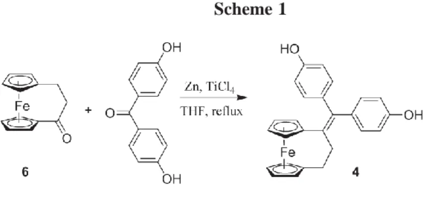
Compound 4 was obtained in 28% yield via a McMurry cross-coupling reaction with the ferrocenophanone 6 and 4,4’-dihydroxybenzophenone (Scheme 1). Compound 6 could be obtained via the reaction of Fc with acryloyl chloride in the presence of aluminum chloride, 14,15 or alternatively, via the acylation of Fc with ethyl mono-malonate,16 followed by Clemmensen reduction of the resulting ketoester and hydrolysis and cyclization of the ferrocenylpropionic acid formed. The synthesis of 5 required 1,1’-(2-ketotrimethylene)ferrocene, which has been previously reported.17 A McMurry cross-coupling reaction of this compound with 4,4’-dihydroxybenzophenone gave 5 as a yellow powder in 22% yield.
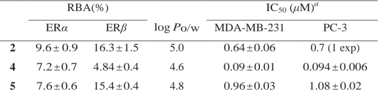
The relative binding affinity (RBA) to ER, lipophilicity, and proliferative/antiproliferative effects against the MCF-7 ER+ breast, the MDA-MB-231 ER- breast, and the PC-3 AR- prostate cancer cell lines are reported in Table 1, along with the values obtained for the noncyclic compound 2 for comparison. Molecules 4 and
5 recognize ERα and ERβ, present in varying degrees in MCF-7 cells, with overall values
on the same order of magnitude as that of 2. However, 4 and 5 exhibit different behavior on the growth of MCF-7 cells. After five days of culture, 4 is estrogenic at low concentration (10 nM; 124% vs control) and becomes cytotoxic at higher concentration (100 nM; 69%). At 10 nM, 5 is estrogenic after 4 days of culture (118%) but, unusually, becomes cytotoxic after 5 days of culture (80%). IC50 values of 4 μM for 4 and of 1 μM for
5 can be estimated for MCF-7 cells after five days of incubation. It is clear that there is a
competition on this cell line between the estrogenic (positive) and cytotoxic (negative) effects, and it seems from these results that 4 is more estrogenic than 5. Surprisingly, this could not be predicted from the RBA values, which are quite similar. Overall, the values for MCF-7 cells are on the same order of magnitude as that of the noncyclic diphenol 2.
MDA-MB-231 and PC-3 cells, with an IC50 value of 0.09 μM, an order of magnitude more cytotoxic than 5, which lacks conjugation between the phenol and the ferrocenyl groups. The cytotoxic effect of compound 2, lacking the ferrocenophane structure but possessing a conjugated system, falls intermediate between 4 and 5.
Table 1. RBA, log Po/w, and the Effect on Cancer Cell Growth of 2, 4, and 5
RBA(%) IC50 (μM)a
ERα ERβ log Po/w MDA-MB-231 PC-3
2 9.6 ± 0.9 16.3 ± 1.5 5.0 0.64 ± 0.06 0.7 (1 exp)
4 7.2 ± 0.7 4.84 ± 0.4 4.6 0.09 ± 0.01 0.094 ± 0.006
5 7.6 ± 0.6 15.4 ± 0.4 4.8 0.96 ± 0.03 1.08 ± 0.02
a
Measured after 5 days of culture (mean of two independent experiments).
Chart 1. Replacement of Ph with Fc on 1 Imparts Cytotoxicity
Scheme 1
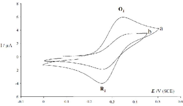
The presence of partially methylated β-cyclodextrin (CD) does not hinder the in vitro cytotoxic effects of 4. MDA-MB-231 incubation with the CD-4 complex (obtained by addition to an aqueous solution of 2 equiv of CD and allowed to dissolve overnight) gave identical results between the free and encapsulated forms (10% cell survival at 10-6 M). The complexation of 4 with CD in water was investigated by electrochemistry, as previously reported for 2.18 Qualitatively, the time required to dissolve 4 in H2O in the presence of CD was fast compared to that of 2 (20 min vs 1 h). The cyclic voltammogram (CV) of the Fc/Fc+ redox couple obtained with the solution of CD-2 (see Supporting Information) exhibited a plateau shape characteristic of a consecutive chemical-electrochemical mechanism, indicating that the oxidation process was limited by the dissociation step of the complex formed between the CD and compound 2. The peak shape and the faster electron transfer obtained with CD-4 are thus consistent with a weak complexation and a fast decomplexation dynamic.
Figure 1. Cyclic voltammograms of 4 (1 mM, scan rate 100 mV/s, left) and 5 (0.3
As observed in Figure 1, in the absence of imidazole, compounds 4 and 5 exhibit a monoelectronic oxidation process at 0.46 V (4) and 0.48 V (5), ascribed to the ferrocenyl oxidation. Upon addition of increasing amounts of base, the intensities of the oxidation waves of both compounds increase, indicating an electron transfer between the phenol and the ferricenium groups prior to the reverse sweep.
Discussion and Conclusions
Compound 4 represents a major advance in the study of antiproliferative organometallic compounds, with an IC50 value on ER- cells several times lower than our noncyclic lead compound 2. On ER+ cells, 4 displays a combination of estrogenicity and cytotoxicity, which suggests that it should be more efficacious against hormone-refractory tumors. However, to create a compound active against both types of cells, one could eliminate the estrogenic effect by replacing one hydroxyl group with the lateral chain -O(CH2)xN(CH3)2 (x=3-5), as we have previously shown for the hydroxyferrocifen series.19
Compound 4 is itself too hydrophobic to envision its intravenous administration, thus possibly limiting its bio-availability for clinical applications. The potency of 4 against MDA-MB-231 cells when encapsulated by partially methylated β-cyclodextrin was maintained. The ability of cyclodextrins to include part of an organometallic complex in their internal hydrophobic cavities has been amply demonstrated,20 and both the stoichiometry and the orientation of ferrocenyl depend on the size of the cyclodextrin. The fast complexation/decomplexation dynamic observed suggests that the ferrocenyl group is situated in an equatorial orientation21 in such a way that hydrophobic interactions between the phenyl and methoxy groups are minimized.
One of the possible mechanisms that we have identified to explain the cytotoxic effect of ferrocenyl phenols on various cancer cells is based on the in situ transformation to a quinone methide (QM), a process that is mediated by the ferrocenyl group and can be followed by electrochemistry.9 Electrochemical experiments suggest that transformation to a QM can be realized for 4 and 5. In this context, the intramolecular proton coupled electron transfer from the phenol to the ferricenium in 4 can be explained by the classical π-delocalized mechanism. For the unconjugated 5, one must consider either that the
electron transfer proceeds “through space”, or via the formation of an intermediate α-methylene radical (such α-α-methylene ferricenium molecules possess acidic protons),22 which can delocalize over the π system and undergo an additional oxidation step to yield the QM.
It has recently been shown that resveratrol can affect cancer cell growth and induce apoptosis in launching the synthesis of endogenous ceramide, a bioactive sphingolipid.12,25 Ceramide is a promising pharmacological target, particularly for cancer therapy. Many drugs that promote the production of ceramide have been proven to be more effective on malignant cells than on healthy tissue,12 and this is also the case with compound 2.23-25 The idea that compound 4, the most effective to date, could possess a high ceramide-mediated proapoptotic activity on human breast cancer cells is however just a working hypothesis and will be explored in the future. It remains that 4 shows a very strong antiproliferative effect against hormone independent cancer cell lines, and this next generation of cyclic ferrocenyl phenols is currently being expanded for future study.
Experimental Section
All reactions were performed under inert atmosphere, THF was distilled from Na/benzophenone, and commercial reagents were used without further purification. Purity of >95% was confirmed via elemental analysis for 4 and analytical HPLC for 5.
1 , 1 ’ - { 1 - [ 1 , 1 - B i s ( 4 - h y d r o x y p h e n y l ) m e t h y l i d e n e ] t r i m e t h y l e n e } f e r r o c e n e (4). To a suspension of 3.07 g (46.9 mmol) of Zn dust in 100 mL of THF
and 4.55 g (2.64 mL, 24 mmol) of TiCl4 was added at -10 °C. The cooling bath was removed, and the mixture was refluxed for 1 h. After cooling to rt, 3.79 g (3.87 mL, 47.9 mmol) of anhydrous pyridine was added and the mixture was stirred for 5 min. A solution of 1.92 g (8 mmol) of [3]ferrocenophan-1-one and 1.71 g (8 mmol) of 4,4’-dihydroxybenzophenone in 30 mL of THF was added and the mixture was refluxed for 90 min. After cooling to rt, the mixture was hydrolyzed with 100 mL of 8% aq K2CO3. The reaction mixture was extracted with several 100 mL portions of diethyl ether. The organic phase was washed with water (2x200 mL) and brine (200 mL), dried over MgSO4, and evaporated to dryness. Product 4 (2nd yellow fraction) was isolated by flash
-1
chromatography on silica gel (n-pentane:diethyl ether 2:1). Yield=1.57 g (47%, purity 90-95%). A second column using 60 mL of silica gel and n-pentane:diethyl ether 4:1 yielded a yellow powder, which was recrystallized from acetone or from a mixture of ethyl acetate (5 mL) and n-pentane (80 mL) at 4 °C. After 48 h, the organic solution was removed and the yellow crystals washed twice with n-pentane. Yield= 0.95 g (28%). 1H NMR (300.13 MHz, acetone-d6): 8.31 (s, 1H, OH), 8.10 (s, 1H, OH), 7.07 (d, J=8,7 Hz,
2H, Ar), 6.85 (d, J=8,7 Hz, 2H, Ar), 6.84 (d, J=8,7 Hz, 2H, Ar), 6.54 (d, J=8,7 Hz, 2H, Ar), 4.25 (t, J=1,7 Hz, 2H, C5H4), 3.98 (m, 4H, C5H4), 3.92 (t, J=1,7 Hz, 2H, C5H4), 2.70 (m, 2H, CH2), 2.34 (m, 2H, CH2). 13C NMR (75.48 MHz, acetone-d6): 157.0 (C), 156.4 (C),
141.5 (C), 136.0 (C), 135.9 (C), 133.4 (C), 132.4 (2CH), 131.2 (2CH), 115.7 (2CH), 114.8 (2CH), 87.6 (C C5H4), 84.9 (C C5H4); 71.0 (2CH C5H4), 70.8 (2CH C5H4), 69.1 (2CH C5H4), 68.8 (2CH C5H4), 41.5 (CH2), 29.1 (CH2). Analysis: calcd for C26H22FeO2: C 73.95%, H 5.25%; found: C 73.79%, H 5.34%. HRMS (CI – CH4): calcd for C26H22FeO2: 423.1047; found: 423.1040 (M+H)+.
1,1’-{2-[1,1-Bis (4-hydroxyphenyl) methylidene] trimethylene} ferrocene (5).
Compound 5 was synthesized as described for 4 but at 5% scale, with similar workup. Yield=0.038 g (0.090 mmol, 22%) of yellow powder. 1H NMR (300.13 MHz,
acetone-d6): 8.22 (s, 2H, OH), 7.16 (d, J=8.7 Hz, 4H, Ar): 6.80 (d, J=8.7 Hz, 4H, Ar), 4.11 (t,
J=1.9 Hz, 4H C5H4), 3.99 (t, J=1.9 Hz, 4H, C5H4), 2.81 (s, 4H, CH2). 13C NMR (75.48 MHz, acetone-d6): 156.7 (C), 141.7 (C), 138.6 (C), 135.8 (C), 131.0 (2 x 2CH), 115.7 (2 x2
CH), 83.5 (2 C C5H4), 70.3 (2x 2CH C5H4), 66.9 (2x2CH C5H4), 28,3 (2CH2). IR (KBr, cm-1): 1609, 1508, 1430, 1220, 1197, 831. HRMS (CI-CH4): calcd for C26H23FeO2: 423.1047 (M+H)+; found: 423.1034 (M+H)+.
Acknowledgment. We thank A. Cordaville for technical assistance and the Agence
Nationale de la Recherche for financial support (no. ANR-06-BLAN-0384-01, “FerVect”). D.P.’s postdoctoral stay was financed by the Foundation for Polish Science (FNP) within the framework of the KOLUMB Fellowship.
Supporting Information Available: Analytical instrumentation, modified synthesis of 4, biochemical testing conditions, and CV of CD-2 and CD-4. This material is available
free of charge via the Internet at http://pubs.acs.org. (or see the end of this file)
References
(1) Strohfeldt, K.; Tacke, M. Bioorganometallic fulvene-derived titanocene anti-cancer drugs. Chem. Soc. Rev. 2008, 37, 1174–1187.
(2) Hartinger, C. G.; Dyson, P. J. Bioorganometallic chemistry;from teaching paradigms to medicinal applications. Chem. Soc. Rev. 2008, 391–401.
(3) Metzler-Nolte, N.; Salmain, M.; Stepnicka, P. The bioorganometallic chemistry of ferrocene. Ferrocenes: Ligands, Mater. Biomol. 2008, 499–639.
(4) Vessières, A.; Top, S.; Pigeon, P.; Hillard, E. A.; Boubeker, L.; Spera, D.; Jaouen, G. Modification of the estrogenic properties of diphenols by the incorporation of ferrocene. Generation of antiproliferative effects in vitro. J. Med.
Chem. 2005, 48, 3937–3940.
(5) Hillard, E. A.; Vessières, A.; Le Bideau, F.; Plażuk, D.; Spera, D.; Huché, M.; Jaouen, G. A series of unconjugated ferrocenyl phenols: prospects as anticancer agents. ChemMedChem 2006, 1, 551–559.
(6) Hillard, E. A.; Pigeon, P.; Vessières, A.; Amatore, C.; Jaouen, G. The influence of phenolic hydroxy substitution on the electron transfer and anti-cancer properties of compounds based on the 2-ferrocenyl-1-phenyl-but-1-ene motif. J.
Chem. Soc., Dalton Trans. 2007, 5073–5081.
(7) Vessières, A.; Top, S.; Beck, W.; Hillard, E. A.; Jaouen, G. Metal complex SERMs (selective estrogen receptor modulators). The influence of different metal units on breast cancer cell antiproliferative effects. Dalton Trans. 2006, 4, 529–541.
Jaouen, G. Organometallic diphenols: The importance of the organometallic moiety on the expression of a cytotoxic effect on breast cancer cells. J. Organomet. Chem.
2007, 692, 1315–1326.
(9) Hillard, E. A.; Vessières, A.; Thouin, L.; Jaouen, G.; Amatore, C. Ferrocene-mediated proton-coupled electron transfer in a series of ferrocifen-type breast cancer drug candidates. Angew. Chem., Int. Ed. 2006, 45, 285–290.
(10) Jaouen, G.; Top, S.; Vessières, A.; Leclercq, G.; McGlinchey, M. J. The first organometallic selective estrogen receptor modulators (SERMs) and their relevance to breast cancer. Curr. Med. Chem. 2004, 11, 2505–2517.
(11) Nguyen, A.; Vessières, A.; Hillard, E. A.; Top, S.; Pigeon, P.; Jaouen, G. Ferrocifens and ferrocifenols as new potential weapons against breast cancer.
Chimia 2007, 61, 716–724.
(12) Minutolo, F.; Sala, G.; Bagnacani, A.; Bertini, S.; Carboni, I.; Placanica, G.; Prota, G.; Rapposelli, S.; Sacchi, N.; Macchia, M.; Ghidoni, R. Synthesis of a resveratrol analogue with high ceramide-mediated proapoptotic activity on human breast cancer cells. J. Med. Chem. 2005, 48, 6783–6786.
(13) Vessières, A.; Spera, D.; Top, S.; Misterkiewicz, B.; Heldt, J. M.; Hillard, E. A.; Huché, M.; Plamont, M. A.; Napolitano, E.; Fiaschi, R.; Jaouen, G. The presence of a ferrocenyl unit on an estrogenic molecule is not always sufficient to generate in vitro cytotoxicity. ChemMedChem 2006, 1, 1275–1281.
(14) Dogan, O.; Senol, V.; Zeytinci, S.; Koyuncu, H.; Bulut, A. Efficient synthesis of ferrocenylenones by Friedel-Crafts acylation with EtAlCl2-Me3Al. J. Organomet.
Chem. 2005, 690, 430–434.
(15) Turbitt, T. D.; Watts, W. E.; Bridged ferrocenes, X. I. I. The synthesis of [3] ferrocenophan-1-one from ferrocene by a novel one-step annelation reaction. J.
Organomet. Chem. 1972, 46, 109–117.
(16) Plażuk, D.; Zakrzewski, J. Acylation of ferrocene and a 1,1’-diphosphaferrocene with acyl trifluoroacetates in the presence of trifluoromethanesulfonic (triflic)
acid or some metal triflates. Synth. Commun. 2004, 34, 99–107.
(17) Mock, W.; Richards, J. H. The synthesis of bridged ferrocene derivatives with functional groups on the β-carbon of the bridge. J. Org. Chem. 1962, 27, 4050– 4051.
(18) Buriez, O.; Heldt, J. M.; Labbé, E.; Vessières, A.; Jaouen, G.; Amatore, C. Reactivity and antiproliferative activity of ferrocenyl-tamoxifen adducts with cyclodextrins against hormone-independent breast-cancer cell lines. Chem.;Eur. J.
2008, 14, 8195–8203.
(19) Top, S.; Dauer, B.; Vaissermann, J.; Jaouen, G. Facile route to ferrocifen, 1-[4-(2-dimethylaminoethoxy)]-1(phenyl-2-ferrocenyl-but-1-ene), first organometallic analogue of tamoxifen, by the McMurry reaction. J. Organomet. Chem. 1997,
541, 355–361.
(20) Hapiot, F.; Tilloy, S.; Monflier, E. Cyclodextrins as supramolecular hosts for organometallic complexes. Chem. Rev. 2006, 106, 767–781.
(21) Menger, F. M.; Sherrod, S. J. Docking calculations on ferrocene complexation with cyclodextrins. J. Am. Chem. Soc. 1988, 110, 8606–8611.
(22) Hitasome, M.; Watanabe, J.; Yamakawa, K. Selective oxidation of the α-methylene of the bridge in ferrocenophanes with silver oxide or silver perchlorate/sodium methoxide. Bull. Chem. Soc. Jpn. 1994, 67, 280–283.
(23) Signorelli, P.; Ghidoni, R. Resveratrol as an anticancer nutrient: molecular basis, open questions and promises. J. Nutr. Biochem. 2005, 16, 449–466.
(24) Allard, E.; Passirani, C.; Garcion, E.; Pigeon, P.; Vessières, A.; Jaouen, G.; Benoit, J. P. Lipid nanocapsules loaded with an organometallic tamoxifen derivative as a novel drug-carrier system for experimental malignant gliomas. J.
Controlled Release 2008, 130, 146–153.
(25) Michard, Q.; Jaouen, G.; Vessières, A.; Bernard, B. A. Evaluation of cytotoxic properties of organometallic ferrocifens on melanocytes, primary and metastatic melanoma cell lines. J. Inorg. Biochem. 2008, 102, 1980–1985.
Supporting Information
Analytical Instrumentation
Infrared spectra were obtained on an IRFT BOMEM Michelson-100 spectrometer equipped with a DTGS detector as a KBr plate. 1H and 13C NMR spectra were recorded on a 300 MHz Bruker spectrometer. Mass spectrometry was performed with a Nermag R 10-10C spectrometer. Melting points were measured with a Kofler device. Elemental analyses were performed by the microanalysis service of CNRS at Gif sur Yvette. Electrochemical experiments were performed Cyclic voltammograms were obtained utilizing a Princeton Applied Research potentiostat. Analyte solutions were 1 mM in DMF with 0.1 M Bu4NBF4
supporting electrolyte. Cyclic voltammetry experiments were performed at room temperature under an argon atmosphere in a three-electrode cell. The reference electrode was an SCE (Tacussel), which was separated from the solution by a bridge compartment filled with the same solvent/supporting electrolyte solution as used in the cell. The counter electrode was a platinum mesh (Goodfellow). The platinum working electrode was home-made (0.5 mm diameter; Goodfellow).
Synthesis of Intermediates
Ethyl ferrocenoylacetate, 6 was prepared by slightly modified procedure for acylation of
ferrocene with acyl trifluoroacetate.1 To a stirred solution of 13.2 g of ethyl mono-malonate and 21 g (14 ml) of trifluoroacetic anhydride in 100 ml of dichloromethane was added 18.6 g of ferrocene. After 5 min a solution of 30 ml of methanesulfonic acid in 50 ml of dichloromethane was added. The resulting violet mixture was stirred at room temperature. After 4 h the reaction mixture was poured onto ice (300g) and the resulting solution was neutralized by addition of solid NaHCO3. Products were extracted with DCM.
Chromatography on silica gel (DCM – ethyl acetate 95 - 5) gave 18 g of pure product. 1H, 13C NMR confirmed the structure as reported in the literature.2
Ethyl 3-ferrocenylpropionate, 7 was prepared as previously described3 for the synthesis of ferrocenylbutyric acid. To a mixture of 70 g of zinc dust and 10 g of Hg(OAc)2 in 150 ml of
water, 5 ml of 37% HCl was added and the resulting mixture was stirred for 10 min. The aqueous solution was decanted and the Zn/Hg was washed 4 times with 200 ml portions of water. The mixture of Zn/Hg, the solution of 10 g of FcCOCH2COOEt in 160 ml of toluene,
40 ml of water and 60 ml of 37% HCl was stirred and refluxed for 4 h (only one product on TLC – hexane). After cooling to room temperature the organic solution was separated and the inorganic residue extracted with toluene. The toluene solution was washed with sat. aq. NaHCO3 and dried with MgSO4. Evaporation of the solvents gave the product in quantitative
yield. The 1H and 13C NMR spectra confirmed the structure as reported in the literature.4
Ferrocenylpropionic acid, 8. A mixture of 9 g of 7, 25 ml of EtOH, and 100 ml of 20% aq.
solution of NaOH was refluxed. After 3 h the resulted solution was diluted with 200ml of water and extracted with several portion of diethyl ether and the aqueous solution was filtered and acidified with conc. HCl to pH 1. 8 was extracted twice with 100 ml portions of DCM, organic solution was washed twice with 200 ml portions of water and dried. Evaporation of solvent gave 5.3 g of pure 8. Crystallization from DCM – n-pentane gave product as a yellow powder. 1H, 13C NMR confirmed the structure as reported in the literature.5
Biochemical studies
Materials. Stock solutions (1 x 10⁻³ M) of the compounds to be tested were prepared in DMSO and were kept at 4 °C in the dark; under these conditions they are stable at least two months. Serial dilutions in DMSO were prepared just prior to use. Dulbecco’s modified eagle medium (DMEM) with phenol red/Glutamax I, Dulbecco’s modified eagle medium (DMEM) without phenol red, Glutamax I and fetal bovine serum (FBS) were purchased from Invitrogen; estradiol from Sigma. PC-3 and MDA-MB-231 cells were from American Type Culture Collection (ATCC, LGC Promochem). Sheep uteri weighing approximately 7 g were obtained from the slaughterhouse at Mantes-la-Jolie, France. They were immediately frozen and kept in liquid nitrogen prior to use.
Determination of the Relative Binding Affinity (RBA) of the compounds for ERα and ERβ. RBA values were measured on ERα from lamb uterine cytosol and on ERβ Panvera
purchased from Invitrogen ( France ). Sheep uterine cytosol prepared in buffer A (0.05 M Tris-HCL, 0.25 M sucrose, 0.1% ßmercaptoethanol, pH 7.4 at 25 °C) was used as a source of ERα. For ERβ, 10 μl of the solution containing 3500 pmol/ml were added to 16 ml of buffer B (10% glycerol, 50 mM Bis-Tris-Propane pH = 9, 400 mM KCl, 2 mM DTT, 1 mM EDTA, 0.1% BSA) in a silanized flask. Aliquots (200 μl) of ERα in glass tubes or ERβ in polypropylene tubes were incubated for 3 h at 0 °C with [6,7-3H]-estradiol (2 x 10⁻⁹ M, specific activity 1.62 TBq/mmol, NEN Life Science, Boston MA) in the presence of nine concentrations of the hormones to be tested. At the end of the incubation period, the free and bound fractions of the tracer were separated by protamine sulfate precipitation. The percentage reduction in binding of [3H]-estradiol (Y) was calculated using the logit transformation of Y (logitY: ln[y/1-Y] versus the log of the mass of the competing steroid. The concentration of unlabeled steroid required to displace 50% of the bound [3H]-estradiol was calculated for each steroid tested, and the results expressed as RBA. The RBA value of estradiol is by definition equal to 100%.
Culture conditions. Cells were maintained in a monolayer culture in DMEM with phenol
red/Glutamax I supplemented with 9% fetal bovine serum at 37 °C in a 5% CO2
/air-humidified incubator. For proliferation assays, PC-3 and MDAMB-231 cells were plated in 1 mL of DMEM without phenol red, supplemented with 9% decomplemented and hormone-depleted fetal bovine serum, 0.9% kanamycin, 0.9% Glutamax I and incubated. The following day (D0), 1 mL of the same medium containing the compounds to be tested was added to the plates. After 3 days (D3) the incubation medium was removed and 2 mL of the fresh medium containing the compounds was added. After 5 days the total protein content of the plate was analyzed as follows: cell monolayers were fixed for 1 h at room temperature with methylene blue (1mg mL-1 in 50:50 water/MeOH mixture), then washed with water. After addition of HCl (0.1 M, 2 mL), the plate was incubated for 1 h at 37 °C and then the absorbance of each well (6 wells for each concentration) was measured at 655 nm with a Biorad spectrophotometer. The results are expressed as the percentage of proteins versus the control.
Cyclic voltammogram of CD-4
Figure S1. Cyclic voltammogram of a) 4 and b) 2 (1 mM) in H2O + Na2SO4 (0.1 M) recorded
at a glassy carbon electrode (3 mm diameter) at 50 mV s-1, in the presence of 20 eq. Me-β-CD. O1 and R1 refer to the oxidation and reduction waves, respectively.
1
D. Plazuk, J. Zakrzewski Synthetic Communications 2004, 34(1), 99-107.
2
B. Floris, P. Galloni, R. Seraglia, P. Tagliatesta Journal of Organometallic Chemistry 2003 679, 202-207.
3
K. L . Rinehart Jr .,R. J. Curby Jr., D. H. Gustafson, K. G. Harrison, R. E. Bozak, D. E. Bublitz J. Amer. Chem. Soc. 1962, 84, 3263-3269.
4
C. M. Zakaria, C. A. Morrison, D. McAndrew, W. Bell, C. Glidewell Journal of
Organometallic Chemistry 1995, 485, 201-207.
5