Université de Montréal
Role of Sirtuin-1 in the Pathogenesis of
Hypertension in Spontaneously Hypertensive Rats:
Molecular Mechanisms
Par
MST NAHIDA ARIFEN
Département de pharmacologie et physiologie Faculté de médecine
Mémoire présenté en vue de l’obtention du grade de Maîtrise ès sciences (M.Sc.) en Physiologie moléculaire, cellulaire et intégrative
Mai 2020
Université de Montréal
Département de pharmacologie et physiologie Faculté de médecine
Ce mémoire intitulé
Rôle de la Sirtuine- 1 dans la pathogenèse de
l'hypertension chez les rats spontanément hypertendus:
Mécanismes moléculaires
Présenté par MST NAHIDA ARIFEN
A été évalué par un jury composé des personnes suivantes Dr. Réjean Couture Président-rapporteur Dr. Madhu B. Anand-Srivastava Directeur de recherche Dr. Ashok K. Srivastava Codirecteur Dr. Johanne Tremblay Membre du jury
iii
Résumé
Il a été démontré que la sirtuine 1 (Sirt-1), une histone désacétylase de classe III, est surexprimée dans le cœur des rats spontanément hypertendus (SHR). Nous avons récemment montré que les cellules musculaires lisses vasculaires (CMLV) des SHR présentent une expression accrue de Sirt-1 par rapport aux rats Wistar Kyoto (WKY) de même âge qui contribue à l’augmentation de la régulation de la protéine Giα impliquée dans la pathogenèse de l'hypertension. La présente étude a été effectuée pour étudier le rôle de l'augmentation de l'expression de la Sirt-1 dans la pathogenèse de l'hypertension chez les SHR et pour explorer les mécanismes moléculaires impliqués dans cette réponse. Dans cette étude, un inhibiteur sélectif de la Sirt-1, EX-527 (5 mg/kg de poids corporel), a été injecté par voie intrapéritonéale chez des rats SHR adultes de 8 semaines et des rats WKY de même âge, deux fois par semaine pendant 3 semaines. La pression artérielle (PA) et la fréquence cardiaque ont été mesurées deux fois par semaine par la méthode non invasive du brassard autour de la queue. Le traitement avec l’inhibiteur spécifique de la Sirt-1, l'EX-527, a atténuéles augmentations de PA (de 76 mmHg) et de fréquence cardiaque chez les rats SHR. La surexpression de Sirt-1 et des protéines Giα dans le cœur, les CMLV et l'aorte a été atténuée au niveau des contrôles par l'inhibiteur de la Sirt-1. L'inhibition de la Sirt-1 a également atténué les niveaux accrus des anions superoxydes, l’activité de la NADPH oxydase et la surexpression des sous-unités de la NADPH oxydase ; les protéines Nox2, Nox4 et P47phox dans les CMLV isolées des SHRtraités par l’EX-527. De plus, les niveaux réduits du monoxyde d'azote synthase endothélial (eNOS) et du monoxyde d'azote (NO) et les niveaux accrus de la peroxynitrite (ONOO-) dans les CMLV des SHR ont également
été rétablis à des niveaux contrôles par l'inhibiteur de la Sirt-1. Ces résultats suggèrent que l'inhibition de la surexpression de la Sirt-1, en diminuant les niveaux accrus des protéines Giα et du stress nitro-oxydant, atténue la PA élevée chez les rats SHR. Il est donc possible de suggérer que les inhibiteurs de la Sirt-1 puissent être utilisés comme des agents thérapeutiques dans le traitement des complications cardiovasculaires associées à l'hypertension.
iv
Abstract
Sirtuin-1 (Sirt-1), class III histone deacetylase, has been shown to be overexpressed in hearts from spontaneously hypertensive rats (SHR). We recently showed that vascular smooth muscle cells (VSMC) from SHR exhibit enhanced expression of Sirt-1 as compared to age-matched Wistar Kyoto (WKY) rats, which contributes to the upregulation of Giα protein implicated in the pathogenesis of hypertension. The present study was undertaken to investigate the role of upregulated Sirt-1 expression in the pathogenesis of hypertension in SHR and to explore the underlying molecular mechanisms involved in this response. For this study, a selective inhibitor of Sirt-1, EX-527 (5mg/kg of body weight),was injected intraperitoneally into 8-week-old adult SHR and age-matched WKY rats twice per week for 3 weeks. The blood pressure (BP) and heart rate was measured twice a week by the CODA™ non-invasive tail cuff method. Treatment of SHR with Sirt-1-specific inhibitor, EX-527, attenuated high BP by 76 mmHg and inhibited the augmented heart rate. The overexpression of Sirt-1 and Giα proteins in heart, VSMC and aorta was attenuated to the control levels by Sirt-1 inhibitor. Inhibition of Sirt-1 also attenuated the enhanced levels of superoxide anion, NADPH oxidase activity and the overexpression of NADPH oxidase subunits; Nox2, Nox4 and P47phox proteins in VSMC isolated from EX-527-treated SHR. Furthermore, the decreased levels of endothelial nitric oxide synthase (eNOS) and nitric oxide (NO) and increased levels of peroxynitrite (ONOO-) in VSMC from SHR were also restored to
control levels by Sirt-1 inhibitor. These results suggest that the inhibition of overexpression of Sirt-1 through decreasing the enhanced levels of Giα proteins and nitro-oxidative stress attenuates the high BP in SHR. It may thus be suggested that inhibitors of Sirt-1 may have the potential to be used as therapeutic agents in the treatment of cardiovascular complications associated with hypertension.
v
Table of Contents
Résumé
……… iii
Abstract
……….. iv
Table of Contents
……….……… v
List of Abbreviations
……… xi
Acknowledgements
……….. xiii
CHAPTER 1: Background and Literature Review……….
xiv
1. Hypertension………
1
1.1. Types of Hypertension………. 1
1.2. Genetic analysis of Hypertension………. 2
2. Blood Pressure
………2
2.1. Blood Pressure Regulation………... 3
2.2. Vascular Structure……….. 3
3. Vascular Remodeling……….. 4
3.1. Vascular Remodeling in Hypertension………..
..
54. Spontaneously Hypertensive Rats………....
5
5. Consequences of Hypertension……….. 6
6. Treatment of Hypertension………...
6
7. Molecular Mechanisms Implicated in Hypertension………
7
7.1. Role of Vasoactive Peptides in Hypertension
………
87.1.1. G-Protein Coupled Receptors (GPCR)
………....
87.1.2. Renin-Angiotensin System
………
9vi
7.1.2.2. Angiotensin II AT2 Receptor
……….
117.1.3. Endothelin System
………..
117.1.3.1. Endothelin-1 Receptors
………..
127.2. Role of Oxidative and Nitrosative Stress in Hypertension
………..
127.2.1. Major Reactive Oxygen Species Molecules
………..………
137.2.2. Source of ROS
………..…………..……….
157.2.2.1. NAPDH Oxidase: Structure, Mechanism and Function
…………
157.2.3. NO bioavailability and Hypertension
………
167.3. Transmembrane Signaling in Hypertension
………..
177.3.1. Guanin Nucleotide-Binding Proteins
………..
177.3.2. Adenylyl Cyclase/cAMP Signaling
……….
187.3.2.1. G-stimulatory (Gs) Protein
……….
207.3.2.2. G-inhibitory (Gi) Protein
………
207.3.2.2.1. Role of Giα Protein Overexpression in the Pathogenesis of Hypertension
………..…………..………
20 7.3.2.2.2. Role of Giα Protein Overexpression in the Pathogenesis of Tachycardia………..…………..………..
238. Histone deacetylase (HDAC)………..………. 23
8.1. HDAC Inhibitors
………..…………..…………..
248.2. HDAC in Vascular Remodeling, Emerging Therapeutic Targets for CVDs
…..
259. Sirtuins, Class III HDAC………..…………..………… 25
9.1. Sirtuins in Cardiovascular System
……….
2610. Sirtuin 1 (Sirt-1), Structure and Mechanism of Action……… 28
11. Sirt-1, as a therapeutic target
………..…………..
29
11.1. Sirt-1 in Nitroxidative Stress
………..……….
29vii
11.3. Sirt-1 in Hypertension
………..…………..……
3111.3.1 Sirt-1 Inhibitors………..…………..………. 32
11.3.2. Structure and Molecular basis of Selective Sirt-1 inhibitor EX-527
………..…………..…………..…………..…………..…………..……..
33 11.3.3. EX-527 as potential therapeutic approach………..…..
33Hypothesis and Aim….………..…………..…………..…………..…………..……….. 34
CHAPTER 2: Scientific Article…….…………..…………..…………..…………..
35
Abstract…….…………..…………..…………..…………..………..………. 37
Introduction…….…………..…………..…………..…………..………..……… 38
Materials and Methods……..…………..…………..…………..…………..……… 40
Results……..…………..…………..…………..…………..………..……….. 44
Discussion……..…………..…………..…………..…………..………..……….. 48
Perspectives..…………..…………..…………..…………..………..………. 51
References……..…………..…………..…………..…………..………..………. 53
Figures and Legends……..…………..…………..…………..…………..……….. 58
CHAPTER 3: Discussion, Conclusion and Future Work…..……….
67
Discussion……..…………..…………..…………..…………..………..……….. 68
Conclusion……..…………..…………..…………..…………..………..………. 74
Future Work……..…………..…………..…………..…………..………..……….. 76
viii
List of Tables
ix
List of Figures
Chapter 1
Figure 1 The Structure of the Arterial and Venous Vascular Wall……… 4
Figure 2 Several Factors Implicated in Hypertension Mechanism.………. 8
Figure 3 Schematic View of the Structure of G Protein-Coupled Receptor (GPCR)……….. 9
Figure 4 Sources and Formation of ROS in Mammalian Cells that are Associated with Hypertension……….……….……… 14
Figure 5 Reaction of the production of Superoxide (O2−) by NADPH Oxidase……… 15
Figure 6 Assembly and Activation of NADPH Oxidase Subunits……… 16
Figure 7 Activation of G-protein.……….……….……… 18
Figure 8 G-protein Mediated Adenylyl Cyclase/cAMP Signaling.……… 19
Figure 9 Schematic Diagram Summarizing the Effect of SNP/Resveratrol on Hypertension of SHR and the Implicated Molecular Mechanisms……… 22
Figure 10 Histone Deacetylases (HDAC) Classifications.……….……… 24
Figure 11 NAD+-dependent Enzymatic Deacetylation Reaction of Sirt-1..……… 29
Figure 12 Chemical Structure of EX-527 (6-Chloro-2,3,4,9-Tetrahydro-1H-Carbazole-1-Carboxamide) (Selisistat) ……….……….………. 33
Chapter 2
Figure 1 In vivo treatment with EX-527 attenuates high blood pressure (BP) and augmented heart rate in spontaneously hypertensive rats (SHR). 60 Figure 2 In vivo treatment with EX-527 inhibits overexpression of Sirtuin-1 (Sirt-1) in
vascular smooth muscle cells (VSMC) (A), aorta (B) and heart (C) from spontaneously hypertensive rats (SHR).
61
Figure 3 EX-527 mediated Sirtuin-1 (Sirt-1) inhibition decreases the overexpression of Giα-2 protein in vascular smooth muscle cells (VSMC) (A), aorta (B) and heart (C) from spontaneously hypertensive rats (SHR).
x
Figure 4 Sirtuin-1 (Sirt-1) inhibition attenuates superoxide (O2−) anion production
and NADPH oxidase activity in vascular smooth muscle cells (VSMC) from spontaneously hypertensive rats (SHR) and age matched Wister Kyoto (WKY) rats.
63
Figure 5 EX-527 treatment reduces overexpression of NADPH oxidase subunits in vascular smooth muscle cells (VSMC) from spontaneously hypertensive rats (SHR).
64
Figure 6 EX-527 mediated Sirtuin-1 (Sirt-1) inhibition restores the levels of eNOS protein expression in aorta (A) and heart (B) and increased the intracellular NO levels in aortic vascular smooth muscle cells (VSMC) from spontaneously hypertensive rats (SHR).
65
Figure 7 Effect of Sirtuin-1 (Sirt-1) inhibition on the levels of ONOO- in aortic vascular smooth muscle cells (VSMC) from spontaneously hypertensive rats (SHR).
66
Chapter 3
Figure 13 Schematic Diagram Summarizing the Effect of Sirt-1 Inhibitor, Ex-527 Treatment on Hypertension and Tachycardia in SHR. 75
xi
List of Abbreviations
AC Adenylyl Cyclase
ACE Angiotensin-converting Enzyme Ang II Angiotensin II
ANP Atrial Natriuretic Peptide ATP Adenosine Triphosphate BH4 Tetrahydrobiopterin
BP Blood Pressure
cAMP Cyclic Adenosine Monophosphate cGMP 3′,5′-Cyclic Guanosine Monophosphate CO Cardiac Output
CVDs Cardiovascular Diseases DOCA Deoxycorticosterone Acetate eNOS Endothelial Nitric Oxide Synthase EC Endothelial Cells
ET-1 Endothelin 1
ETA Endothelin receptor Type A
ETB Endothelin receptor Type B
GDP Guanosine diphosphate Gi Inhibitory G protein
GPCR G Protein-coupled Receptor
G protein Guanine Nucleotide-binding Protein Gs Stimulatory G protein
GTP Guanosine triphosphate H2O2 Hydrogen peroxide
HDAC Histone deacetylase JNK1 c-Jun N-terminal kinase 1
L-NAME Nω-Nitro-L-Arginine Methyl Ester MAP Mean Arterial Pressure
MAPKs Mitogen-activated Protein Kinases Mm Hg Millimeters Mercury
MnSOD Manganese Superoxide Dismutase NAD+ Nicotinamide Adenine Dinucleotide
NADPH Nicotinamide Adenine Dinucleotide Phosphate NF-κB Nuclear Factor-kappa B
NO Nitric oxide Nox NADPH oxidase
xii n-Tyr Nitrotyrosine
O2− Superoxide Anion
OH· Hydroxyl Radical ONOO− Peroxynitrite
PLC Phospholipase C
RAAS Renin-angiotensin-aldosterone System ROS Reactive oxygen species
SAHA Suberoyl Anilide Hydroxamic Acid SDS Sodium Dodecyl Sulfate
SHR Spontaneously Hypertensive Rats Sirt-1 Sirtuin 1
SNP Sodium Nitroprusside SODs Superoxide Dismutases TPR Total Peripheral Resistance VPA Valproic Acid
VSMC Vascular Smooth Muscle Cells WKY Wistar Kyoto
xiii
Acknowledgements
I would like to express special gratitude to my research director, Dr. Madhu B. Anand-Srivastava for accepting me as a Master’s student in her laboratory. She has been an excellent supervisor who has provided me exceptional guidance during this research project. I have learned a lot from her, and she was always there to help and motivate me. She has been very supportive, caring and encouraging throughout my studies. Her valuable feedback helped me in finalizing this project within the limited time frame.
I would also like to thank my co-director, Dr. Ashok K. Srivastava for his guidance, support and encouragement.
I am grateful to Dr. Yuan Li for her help, guidance and care throughout the past years which made this project more flexible.
I am also thankful to Sara Almajdoob and Stephanie, my colleagues for helping me during the project.
I wish to show my special gratitude to Vanessa Truong for the suggestions and guidelines during thesis writing.
Last, but not the least, I would like to cordially thank to the most special person of my life, my husband, Kh Arif Shahriar who was like a shadow throughout the study period. He was always there whenever I needed him despite his busy schedule of Ph.D. studies. His support and care made this journey easy and fruitful.
CHAPTER 1
1
1. Hypertension
Hypertension is one of the most common, multi-factorial chronic disorder affecting over 1 billion people worldwide (Bloch 2016; Frid et al. 2020; Schwartz et al. 2012; Fields et al. 2004). According to the World Health Organization (WHO), sustained raised blood pressure (BP) equal to or above 140 mm Hg (systolic BP) and/or equal to or above 90 mm Hg (diastolic BP) is called hypertension. High BP is associated with increased risks of coronary and cerebrovascular events (Kjeldsen 2018; Petrie, Guzik, and Touyz 2018). Whilst there has been a gradual improvement towards treatment of hypertension, a great number of patients with increased BP remains resistant with the currently available treatment (Chia, Pandey, and Vongpatanasin 2019). Majority of the patients need more than one group of drugs to control the BP suggesting the involvement of multiple pathways in BP regulation (Guerrero-García and Rubio-Guerra 2018). However, the underlying mechanisms contributing to hypertension are very complicated and still remain obscure. Therefore, better understanding of the molecular mechanisms of hypertension will open the pathways to explore the new therapeutic strategies and reduce the prevalence of this global burden.
1.1. Types of Hypertension
Almost 90-95% hypertensive patients that do not exhibit any clear etiology are classified as having primary or essential hypertension whereas, rest of the 5-10% of patients are grouped under secondary or non-essential hypertension with an underlying and potentially reversible cause (Gupta-Malhotra et al. 2015).
In essential hypertension, BP rises with undetermined cause that increases risk for cerebral, cardiac, and renal events (Staessen et al. 2003). Evidence suggest that essential hypertension magnifies cardiovascular risk factors and target end-organ damage such as left-ventricular hypertrophy and cognitive dysfunction which finally lead to the catastrophic events such as stroke, heart attack and renal failure (Bolívar 2013). This is mostly common in adulthood and elderly person.
The common causes of non-essential hypertension are renal parenchymal disease, coarctation of the aorta, hyperaldosteronism, Cushing syndrome, thyroid disease etc. The prevalence of
2
secondary hypertension is more common in younger person and may vary by age from 18 to 40 years (Charles, Triscott, and Dobbs 2017).
1.2. Genetic analysis of Hypertension
Hypertension is a heterogeneous disease. Environmental factor as well as genetic predisposition both have strong influence on the rise of BP. The influence of genetics to the BP regulation is of two types (Agarwal, Williams, and Fisher 2005). One is the rare familial forms of monogenic hypertension that is considered as secondary hypertension as it is caused by a single gene and can be identified by a few hundred genetic markers. However, for the primary or essential hypertension, genotyping of a large number of small-effect size genetic variants are involved (Russo et al. 2018). Through Genome-wide association studies (GWAS) 43 of loci have been identified to be associated with systolic, diastolic BP, and hypertension (Newton-Cheh et al. 2009; Azam and Azizan 2018). It is therefore, opening a perspective on the genetic architecture of BP and facilitating the better understanding of genes that are implicated in the regulation of BP (Ehret and Caulfield 2013; Patel, Masi, and Taddei 2017).
2. Blood Pressure
Blood pressure (BP) is the force exerted on the arterial system of the body by the circulating blood pumped by the heart (Magder 2018). BP values are measured in millimeters of mercury (mmHg) and major determinants of therapeutic decisions. It is conventionally separated into systolic and diastolic determinations. Systolic BP is the maximum BP during contraction of the ventricles and pumping oxygen-rich blood into the blood vessels. Normal range for systolic BP is 100-140 mmHg with an average of 120 mmHg. Diastolic BP is the minimum pressure on the blood vessels when the heart muscle relaxes. The diastolic pressure is always lower than the systolic pressure. Normal range for diastolic BP is 60-90 mmHg with an average of 80 mmHg. The difference between systolic and diastolic pressure is called pulse pressure that represents the force exerted by the heart during each contraction. Another important variable is mean arterial pressure (MAP), that is the average arterial pressure throughout one cardiac cycle, systole and diastole and is considered as a better indicator of perfusion pressure to supply oxygenated blood to the vital organs of the body (Guyton et al. 1981).
3 2.1. Blood Pressure Regulation
Regulation of the circulatory system is critical to maintain a constant arterial pressure in ensuring adequate perfusion. BP depends on cardiac output (CO) and total peripheral resistance (TPR). CO is the volume of blood that is pumped out of the left ventricle per minute. TPR is the sum of all the blood vessels in systemic circulation. Changing any one of these factors will change the BP regulation. BP regulation is a complex physiological process operating in short-term and long-term reflex responses mediated by hormones, local vascular factors, and neural mechanism (Guyton et al. 1981).
2.2. Vascular Structure
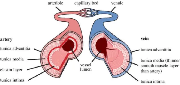
Vascular Structure is playing an indispensable role in maintaining BP homeostasis by providing adequate blood supply to the tissues, and then returns it to the heart. The vascular system of the human body is comprised of many vessels. Blood primarily moves through the body by the rhythmic movement of smooth muscles in the vessel wall and by the action of the skeletal muscle as the body moves. There are three major types of blood vessels: the arteries, the capillaries and the veins (Pugsley and Tabrizchi 2000). [Figure 1].
Arteries carry blood away from the heart. The wall of an artery consists of three layers (Burton 1954). The innermost layer, the tunica intima that is made with a layer of endothelial cells (EC) and connective tissue. The middle layer, the tunica media, that mainly consists of vascular smooth muscle cells (VSMC) and connective tissues full of collagen, elastin, and other elastic fibers (Rhodin 1967). VSMC control the caliber of the vessel and arterial tone. The outermost layer, tunica externa that is made up of fibrous tissue, which is mostly elastin and collagen fibers, and fibroblasts. BP homeostasis is highly influenced by the elasticity of arteries. The largest artery, aorta contains large amounts of elastin and persistent with the pressure fluctuations from the heart. Whereas, small arteries have the large proportion of tunica media making them more muscular and active in vasoconstriction, allowing changes in TPR in response to the BP regulation. The small arteries, lumen (diameters <400 μm) are called resistance arteries because they act as the major site of vascular resistance. Any change in lumen diameter of these resistance vessels has influence in normal BP (Intengan et al. 1999).
4
Figure 1: The Structure of the Arterial and Venous Vascular Wall. Adapted from (Shaw et al. 2014)
Capillaries are the smallest and most numerous of the blood vessels that mediate the connection between arteries and veins. Smooth muscle cells in the arteries help to regulate blood flow from the arterioles into the capillaries (Taylor, Moore, and Khimenko 1994).
Veins carry blood toward the heart. The walls of veins have the same three layers like the arteries but there is less smooth muscle and connective tissues. This makes the walls of veins thinner than those of arteries, because blood in the veins has less pressure than in the arteries (Klabunde 2011).
3. Vascular Remodeling
Vascular wall is an active organ composed of different types of cells including endothelial, VSMC and fibroblast cells that interact with each other to form an autocrine-paracrine complex. The vasculature undergoes structural and functional changes in response to long-term physiological alterations applied to the vessel walls such as increased transmural pressure and blood flow. These structural and functional alterations result in vascular remodeling (Gibbons and Dzau 1994). Vascular remodeling involves changes in at least four cellular processes: cell growth, cell death, cell migration, and the synthesis or degradation of extracellular matrix. Vascular remodeling is influenced by dynamic interactions between local growth factors, vasoactive substances, and hemodynamic stimuli (Intengan et al. 1999). Vascular remodeling is the hallmark
5
of the pathophysiology of vascular diseases and circulatory disorders including hypertension (Baumbach and Heistad 1989).
3.1. Vascular Remodeling in Hypertension
The histopathological change of hypertension is associated with vascular remodeling (Renna, de Las Heras, and Miatello 2013). The main characteristics of vascular remodeling are the thickening of the arterial wall including tunica intima, media, and externa. The thickening of arterial wall increases stiffness to reduce lumen diameter at a given pressure (Bund and Lee 2003). One of the major findings is that vascular tunica media thickening is caused by the abnormal proliferation and hypertrophy of VSMC (Inokuchi et al. 2001). More advanced findings indicated that several intracellular signaling pathways that regulate the expression of upstream and downstream target genes through cascade, are involved in the proliferation and hypertrophy of VSMC. Vasoactive peptides such as Ang II and ET-1 as well as growth factors receptors such as EGFR and PDGFR all contribute to VSMC hypertrophy and proliferation (Almajdoob, Hossain, and Anand-Srivastava 2018; Atef and Anand-Srivastava 2016; Inagami and Eguchi 2000; Gomez and Anand-Srivastava 2010; Li, Lévesque, and Anand-Srivastava 2010; Raines 2004; Ross et al. 1974). Therefore, understanding of cellular mechanisms involved in the proliferation, migration, and apoptosis of VSMC and the associated drug interventions may be a promising direction for the treatment of hypertension (Brown et al. 2018).
4. Spontaneously Hypertensive Rats
Spontaneously hypertensive rat (SHR) is the most common model of human hypertension that is genetically inherited hypertension. It has been widely used to define hypertension-induced changes in signaling mechanisms (Takata and Kato 1995) and to test new antihypertensive medication (Lerman et al. 2019; Doris 2017; Lund-Johansen 1990). This inbred strain was developed by Okamoto and colleagues during the 1960s, with the selective breeding of Wistar Kyoto (WKY) rats having hypertension (Okamoto and Aoki 1963). In SHR, development of hypertension begins at 4-week-old and increases with age (Li and Anand-Srivastava 2002). In adult (8-week- old) age, systolic BP reaches to around 180 mmHg (McGuire and Twietmeyer
6
1985). At the age of 40-weeks, SHR started to develop characteristics of cardiovascular disease, such as vascular and cardiac hypertrophy (Conrad et al. 1995). SHR demonstrates specific and uniform genetic predisposition that allow hypertension research including its causes, mechanisms and pathology, as well as possible therapeutic interventions (Folkow 1982). Like humans, male SHR shows to develop hypertension more rapidly and becomes more severe than female SHR (Iams and Wexler 1979). Moreover, increased media-to-lumen ratio observed in arteries from human hypertensive patients are identical to that observed in SHR (Heagerty et al. 1993). Thus, SHR provides a convenient approach of investigating hypertensive symptoms that are predictable and controllable without using life-threatening interventions for humans.
5. Consequences of Hypertension
Hypertension remains the leading cause of morbidity and mortality worldwide. High BP increases the risk of almost all the major cardiovascular events including stroke, sudden cardiac death, coronary heart disease, myocardial and cerebral infarction, abdominal aortic aneurysm, and peripheral vascular diseases (Kjeldsen 2018). Hypertension is also common among patients with diabetes mellitus (de Boer et al. 2017). The combined impacts of hypertension and diabetes can increase the risk of cardiovascular death. However, Oh et al. reported that hypertension is more strongly associated with all-cause and cardiovascular mortality than diabetes (Oh, Allison, and Barrett-Connor 2017). Hypertension is very frequent in patients with renal disease and its prevalence increases as renal failure progresses (Luo, Hu, and Jiang 2020). A considerable number of articles are devoted to pathophysiological and clinical aspects of hypertension-induced neurodegenerative and cognitive diseases. Hypertension leads to dementia and Alzheimer's disease that is a consequence of the damaging effects of high blood pressure on the cerebral vasculature (Carnevale et al. 2016; Gorelick et al. 2011).
6. Treatment of Hypertension
Hypertension associated risks of morbidity and mortality can be greatly reduced by treatment with antihypertensive drugs that lowerBP. A total of 69 antihypertensive drugs are approved by the US Food and Drug Administration (FDA) (Oparil and Schmieder 2015). Initially, groups of antihypertensive drugs to promote vascular health included ACE inhibitors, calcium-channel
7
blockers, α-adrenergic blockers and thiazide-type diuretics (Officers 2002). Some newer cardiovascular drugs, such as antagonists of the Ang II receptor (AT 1), vasopeptidase inhibitors, dual acting ARB–neprilysin inhibitors, and Endothelin-1 (ET1) receptor blockers are considered as second-line drugs with the potentiality of improving vascular functions (Prasad, Palaniswamy, and Frishman 2009; Correale et al. 2018). Despite the plethora of available drugs, optimal treatment remains a challenge. One of the important facts is that many of these drugs do not specifically target the vascular system to ameliorate hypertension-induced vascular damage. Moreover, most of the cases, patients are prescribed to take medications from more than one drug groups. Therefore, it has been challenging to follow-up patients to prevent potential adverse drug interactions with a multidrug regimen (Grossman and Messerli 2012). Besides that, 10% to 15% patients with hypertension are still resistant with this currently available treatments (Persell 2011; Sim et al. 2013). However, current evidence suggests that development of treatment-resistant hypertension is multifactorial (Hwang et al. 2017). Thus, advanced knowledge to the understanding of vascular mechanisms responsible for high BP will facilitate efficient drug discovery to reduce the enormous clinical and economic burden for this population.
7. Molecular Mechanisms Implicated in Hypertension
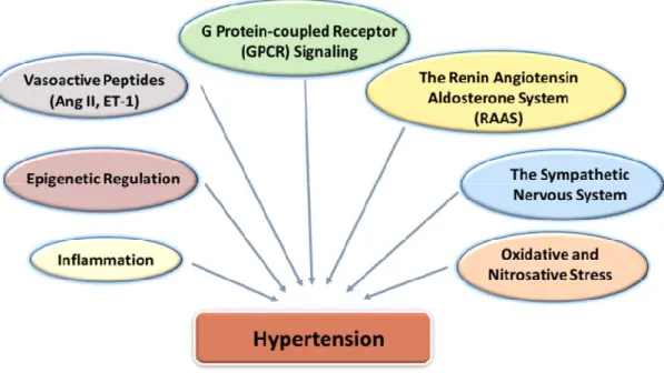
Hypertension is a multifactorial disease. Several factors including vasoactive peptides, the renin-angiotensin-aldosterone system (RAAS), activation of the sympathetic nervous system, abnormalities in G protein-coupled receptor (GPCR) signaling, oxidative and nitrosative stress and inflammation are implicated in the pathophysiology of hypertension [Figure 2]. However, the underlying mechanisms for development of hypertension are very complicated and remain still poorly understood.
8
Figure 2: Several Factors Implicated in Hypertension Mechanism.
7.1. Role of Vasoactive Peptides in Hypertension
Vasoactive peptides including angiotensin II (Ang II), endothelin 1 (ET-1), vasopressin (AVP) and natriuretic peptides are upregulated in hypertensive patients and animal models (Hynynen and Khalil 2006; Arendse et al. 2019). The regulation of the vasoactive peptides results in endothelial dysfunction, vascular remodeling and vascular inflammation, which are implicated in the hypertension-induced vascular damage. Ang II, ET-1 and natriuretic peptides mediate their physiologic effects via G-protein-coupled receptors (GPCR) mediated signal transmission.
7.1.1. G-Protein Coupled Receptors (GPCR)
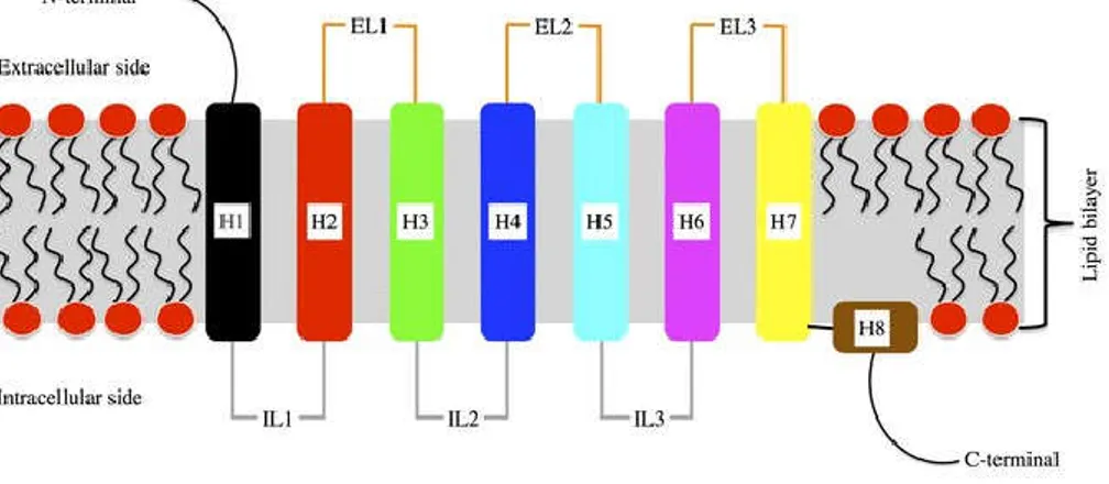
G-protein-coupled receptors (GPCR) are the most diverse family of membrane protein receptors. GPCR mediate most of our physiological responses to hormones, neurotransmitters and environmental stimulants, and possessed a great potential as therapeutic targets for a wide range of diseases (Rosenbaum, Rasmussen, and Kobilka 2009; Heng, Aubel, and Fussenegger 2013). Some examples of GPCR include beta-adrenergic receptors, which bind epinephrine; prostaglandin E2 receptors, which bind prostaglandin; angiotensin type I (AT1) receptor which binds Ang II (Fuxe et al. 2008). All GPCR are characterized by the presence of seven membrane-spanning α-helical segments interconnected by three extracellular loops (EL1, EL2, EL3)
9
containing the ligand binding domain and three alternating intracellular loops (IL1, IL2, IL3) provide binding sites for intracellular signaling proteins (Lomize, Pogozheva, and Mosberg 1999) [Figure 3]. GPCR bind with extracellular stimuli and transmit signals to the effectors adenylyl cyclase (AC) or phospholipase C (PLC) through the activation of guanine nucleotide-binding proteins (G protein) (Wess 1997) that eventually generates numerous cellular events, such as increased heart rate in response to epinephrine (Gether 2000). The adenylyl cyclase-cyclic adenosine monophosphate (cAMP) signal pathway and PLC-phosphatidylinositol signal pathway are the two principal signal transduction pathways of the GPCR
Figure 3: Schematic View of The Structure of G Protein-Coupled Receptor (GPCR), with depiction of the connectivity of the intracellular (IL) and extracellular (EL) loops between helices (H). Adapted from (Fossépré et al. 2014).
7.1.2. Renin-Angiotensin System
The renin-angiotensin system (RAS) is the major regulatory system of blood pressure. The RAS system is composed of different regulatory components and effector peptides that mediate the dynamic control of vascular function (Nakagawa et al. 2020; Carey and Padia 2018). Renin is a protease, released from juxtaglomerular cells of the kidney and is the starting point of the renin-angiotensin system (Kukida et al. 2020; Hackenthal et al. 1990). Renin causes the cleavage of angiotensinogen, a substrate that is synthesized by the liver, into angiotensin (Ang) I. Ang I is proteolytically cleaved by the dipeptide carboxypeptidase, angiotensin-converting enzyme (ACE) and produces angiotensin II (Ang II). Ang II is further cleaved by the carboxypeptidase, ACE2 and produces Ang (1–7) peptide (Donoghue et al. 2000). Ang (1-7) binds with the GPCR
10
and is involved in cardiovascular and neuronal regulation (Santos et al. 2003). Besides that, other Ang peptides, such as Ang III [Ang-(2-8)], Ang IV [Ang-(3-8)] are also produced from Ang II and may also have important biological activities (Hussain and Awan 2018).
Ang II is the dominant player of RAAS system. Ang II increases BP by vasoconstriction, sympathetic nervous stimulation, increased aldosterone biosynthesis and renal actions (Forrester et al. 2018). Ang II also contributes to high BP through the induction of growth and migration of VSMC, leading to thickening of the vascular wall and myocardium, and fibrosis (Touyz et al. 2018; Fyhrquist, Metsärinne, and Tikkanen 1995). Ang II mediates its effects through the activation of the two classes of GPCR: Ang II type 1 (AT1) receptor and Ang II type 2 (AT2) receptor (De Gasparo et al. 2000).
7.1.2.1. Angiotensin II AT1 Receptor
Most of the physiological actions of Ang II are mediated through AT1 receptor, which is widely expressed in the most cell types. The mechanism involves conformational changes of the receptor and coupling with the G protein, mainly Gqα and Giα (Shenoy and Lefkowitz 2005). Then, Gqα activates PLC and mediates downstream signaling (Lassègue et al. 1993; Kawai et al. 2017) . Giα is coupling to AC inhibition (Anand-Srivastava 1993) and activating of voltage-gated L-type and T-type calcium channels (Maturana et al. 1999). Ang II through the activation of AT1
receptor promotes growth and stimulates extracellular matrix production in cardiac fibroblasts, cardiomyocytes, and VSMC. Clear evidence suggests that an excessive activation of the AT1 receptor results in hypertension and vascular remodeling (Billet et al. 2008) and central application of an AT1 receptor blocker or AT1 receptor antisense approach lowered BP in rodent hypertensive models (Yang et al. 1992; Toney and Porter 1993; Gyurko, Wielbo, and Phillips 1993). Ang II has also been shown to increase the expression of Giα proteins and hyperproliferation of VSMC (Gomez Sandoval, Levesque, and Anand-Srivastava 2009). In addition, our laboratory has also demonstrated the role of the endogenous Ang II and overexpression of Giα proteins in high BP in SHR because captopril, an ACE inhibitor that decreases the levels of Ang II also resulted in the attenuation of high BP and enhanced expression of Giα proteins (Pandey and Anand-Srivastava 1996).
11 7.1.2.2. Angiotensin II AT2 Receptor
AT2 receptor is highly expressed in the neonatal tissues, but rarely expressed in the cardiovascular system of the normal adults (Shanmugam, Corvol, and Gasc 1996). The role of AT2 on vascular responses to Ang II in humans remains controversial. An increased expression of AT2 receptor has been reported under pathological conditions including hypertension (Cosentino et al. 2005; Savoia et al. 2006), myocardial infarction (Nio et al. 1995) and vascular injury (Nakajima et al. 1995). Whereas, a protective role of AT2 receptors has been observed by inducing vasodilation, antiproliferation, and apoptosis in cellular and animal models (Volpe et al. 2003).
7.1.3. Endothelin System
Endothelin (ET-1) is a powerful vasoconstrictor peptide (21-amino-acids) that is diversely expressed and has important role in the vascular system. Three distinct isoforms of endothelin family have been identified; ET-1, ET-2, and ET-3. Structurally ET-2 and ET-3 differ from ET-1 by two and six amino-acid positions, respectively. All three endothelin isoforms are synthesized by two proteases as preprohormones and then post-translationally processed to active peptides. Firstly, the ∼200-residue preproendothelins are proteolytically cleaved by endopeptidases to big ETs (37- to 41 amino acids called proETs). proETs is then cleaved by the endothelin converting enzyme (ECE) to the 21-amino acid mature active peptides (Inoue et al. 1989). However, ET-1 is most abundantly expressed. ET-1 is produced by VSMC, fibroblasts, cardiomyocytes, various brain neurons, but the predominant source of ET-1 is EC (Kisanuki et al. 2010; Kanse et al. 1991; MacCumber et al. 1989; Sakai et al. 1996; Yanagisawa et al. 1988). ET-2 is produced by intestinal epithelial cells, and ET-3 by neurons, renal tubular epithelial cells, and intestinal epithelial cells (Kedzierski and Yanagisawa 2001; Matsumoto et al. 1989). A wealth of evidence suggests that ET-1 is a key mediator in CVDs such as chronic heart failure and participates in the pathogenesis of the elevation of BP in both experimental animal models and human essential hypertension (Hynynen and Khalil 2006; Schiffrin 2001; Dhaun et al. 2008). Several animal models of hypertension such as DOCA-salt, Goldblatt (1K1C) and the SHR, showed an elevated systemic level of ET-1 (Kassab et al. 1997; Schiffrin 1995).
12 7.1.3.1. Endothelin-1 Receptors
There are two primary human endothelin receptors known; endothelin type A (ETA) and type B
(ETB). They are the members of the seven transmembrane GPCR superfamily. Both receptors
activate G proteins, leading to diverse responses such as activation of phospholipase C and increase in intracellular calcium (Inoue et al. 1989). ETA is located abundantly on VSMC
whereas, ETB is highly expressed in EC, but also present in VSMC (Sakurai et al. 1990; Arai et al.
1990). However, the expression level of ETB is increased in VSMC under vascular pathologic
conditions (Batra et al. 1993).
Interaction of ET-1 with ETA and ETB receptors on VSMC activates phospholipase C-inositol
triphosphate pathway and increases intracellular calcium level that causes VSMC contraction with the phosphorylation of myosin kinase (Miyauchi and Masaki 1999; Lüscher and Barton 2000). On the other hand, ET-1 interacts with ETB receptors on EC and activates endothelial nitric
oxide synthase (eNOS) which increases the release of nitric oxide (NO) and causes vasodilation (Lüscher and Barton 2000). Thus, activation of ET-1 leads to dual vasoregulatory effects. ETA
receptor inhibitors, BQ123 and BQ610 have been shown to lower BP in different animal models of hypertension including SHR suggesting the role of ETA receptor in the pathogenesis of
hypertension in SHR (Morand-Contant, Anand-Srivastava, and Couture 2010; Douglas et al. 1994; Cassinotti et al. 2018).
7.2. Role of Oxidative and Nitrosative Stress in Hypertension
Although mechanisms underlying hypertension are not yet fully elucidated, a considerable body of literature proposed that oxidative stress is one of the fundamental mechanisms responsible for the development of hypertension and other vascular diseases (Rodrigo, González, and Paoletto 2011; González et al. 2014; Baradaran, Nasri, and Rafieian-Kopaei 2014; Touyz 2004; Loperena and Harrison 2017).
Oxidative stress is an imbalanced state of systemic manifestation of reactive oxygen species (ROS), when the production of ROS exceeds antioxidant defense mechanism (Birben et al. 2012). ROS including superoxide anion (O2−), hydrogen peroxide (H2O2), hydroxyl radical (OH·)
and peroxynitrite (ONOO−) are generated within the vascular wall and play an active role in
13
biology. ROS are generally produced at a low concentration and function as signaling molecules regulating vascular contractility and cell growth. However, an enhanced production of ROS that is not counterbalanced by the endogenous antioxidant mechanisms and decreased NO levels causing nitro-oxidative stress contributes to the pathology of diseases including hypertension (Li et al. 2014; Poli et al. 2004; Saha et al. 2011; Schieber and Chandel 2014; Touyz and Briones 2011; Yin et al. 2013). Several studies have reported an excessive amount of ROS in essential hypertensive patients and various animal models of hypertension (González et al. 2014; Rodrigo et al. 2007; Lappas, Daou, and Anand-Srivastava 2005; Gusan and Anand-Srivastava 2013; Rahali, Li, and Anand-Srivastava 2018; Saha, Li, and Anand-Srivastava 2008). ROS production is also enhanced in cultured VSMC and isolated arteries from hypertensive rats and humans (Lappas, Daou, and Anand-Srivastava 2005; Atef and Anand-Srivastava 2016; Touyz and Schiffrin 2001). Our laboratory has also demonstrated that VSMC from SHR exhibit enhanced oxidative stress due to the enhanced production of O2-, increased activity of NADPH oxidase and the
overexpression of NADPH oxidase subunits, Nox2, Nox4, p47phox (Almajdoob, Hossain, and Anand-Srivastava 2018; Gusan and Anand-Srivastava 2013) and decreased levels of NO, eNOS and augmented levels of peroxinitrite (ONOO-) (Hossain et al. 2018; Li et al. 2014).
7.2.1. Major Reactive Oxygen Species Molecules
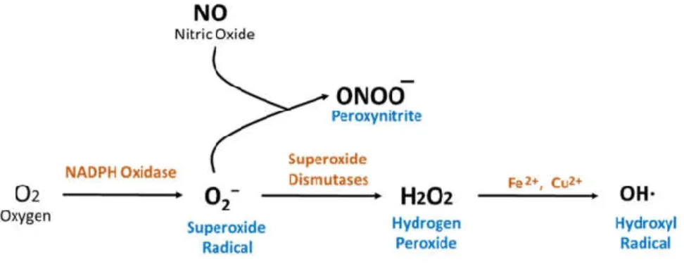
Superoxide anion (O2−): Superoxide (O2−) is a highly reactive anion radical, which is produced by
the one-electron reduction of molecular oxygen [Figure 4]. Superoxide can be generated from two major sources: the mitochondrial respiratory chain and nicotinamide adenine dinucleotide phosphate oxidase (NADPH oxidase) enzyme (Murphy 2009; Jones 1994). Production of O2− is
important because this acts as a progenitor for other ROS, including H2O2, OH· and ONOO−. O2−
can act as a reducing agent, donating its extra electron to form ONOO− with nitric oxide (NO), or
as an oxidizing agent, to produce H2O2. Thus, increased production of O2− can be harmful in
several ways including removing the beneficial effects of NO (Rubanyi and Vanhoutte 1986). In addition, several studies have demonstrated that O2− can act as a vasoconstrictor (Auch-Schwelk,
14
Hydrogen Peroxide (H2O2): Hydrogen peroxide (H2O2) is produced from O2− either by
spontaneously or catalyzed by the enzyme superoxide dismutases (SOD) [Figure 4]. H2O2 is
relatively more stable than O2−, as it doesn’t contain any free radicals. H2O2 can readily diffuse
across membranes and acts as a signaling molecule that is involved in vasodilatation, gene transcription, phosphatase activity, and activating other sources of ROS (Gough and Cotter 2011; Neill, Desikan, and Hancock 2002).
Figure 4: Sources and Formation of ROS in Mammalian Cells that are Associated with Hypertension
Hydroxyl Radical (OH·): Hydroxyl radical (OH·) is produced from the reaction between O2− and
H2O2 where O2− donates 1 electron to H2O2 (Lipinski 2011)[Figure 4]. The hydroxyl radical is a
highly reactive oxidant that can damage all types of macromolecules including carbohydrates, DNA, lipids (lipid peroxidation). The augmented level of OH· is reported to contribute to the increase contractions in the aorta of SHR (Auch-Schwelk, Katusic, and Vanhoutte 1989).
Peroxynitrite (ONOO−): Peroxynitrite (ONOO−) is produced by the spontaneous reaction
between O2− and NO [Figure 4]. ONOO− exerts exactly opposite of beneficial effects of NO. ONOO−
is a very strong oxidant and can react with lipids, DNA, and proteins and causing oxidative damage to these macromolecules. Peroxynitrite can turn into novel products such as nitrotyrosine (n-Tyr), nitrotryptophan, and nitrated lipids that serve as important biological markers for many diseases. The ability of producing n-Tyr by peroxynitrite can impair signaling processes by inhibiting phosphorylation of critical tyrosine residues (Gow et al. 1996; Kong et al. 1996). Peroxynitrite generation contributes to various cardiovascular pathologies such as myocardial
15
and vascular dysfunction during ischemia and reperfusion, myocarditis, chronic heart failure and blood pressure regulation (Pacher, Beckman, and Liaudet 2007).
7.2.2. Source of ROS
ROS are produced in several cellular systems localized in the plasma membrane, the cytosol, peroxisomes, mitochondria and endoplasmic reticulum (Phaniendra, Jestadi, and Periyasamy 2015). The nicotinamide adenine dinucleotide phosphate (NADPH) oxidases are principal enzymatic sources of vascular ROS production in mammalian cells (Brandes and Kreuzer 2005).
7.2.2.1. NADPH Oxidase: Structure, Mechanism and Function
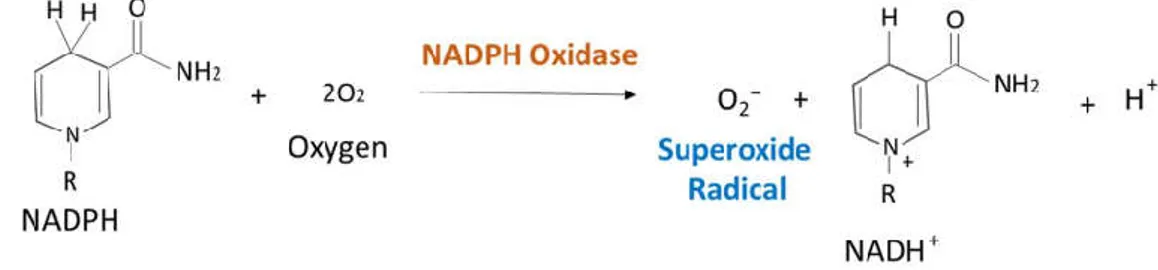
Vascular NADPH oxidases are by far the most researched topic amongst the sources of ROS in hypertension. NADPH oxidase is a membrane-bound, multi-subunit enzyme complex, that is distributed throughout the EC, VSMC and cardiac myocytes (Griendling et al. 2000; Xiao et al. 2002). It catalyzes the production of superoxide anion by transferring one electron to oxygen from NADPH [Figure 5].
Figure 5: Reaction of the Production of Superoxide (O2−) by NADPH Oxidase
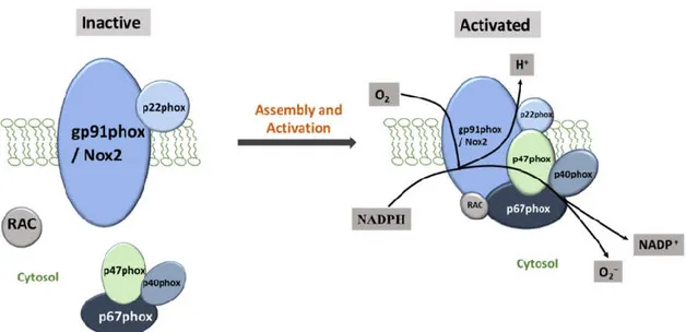
NAPDH Oxidase consists of two cytosolic subunits (p47phox and p67phox), a cytochrome b558 (gp91phox and p22phox) and a small G protein Rac. Gp91phox, also referred as Nox2 is an important Nox family member that is inactive until it binds to the membrane-anchored p22phox. The Nox2/p22phox complex requires phosphorylated p47phox, p67phox and p40phox for binding with the other cytosolic components. Upon complex assembly, the GTPase Rac then interacts with Nox2 and subsequently interacts with p67phox, resulting in an activated complex to produce superoxide through electron transfer from cytosolic NADPH to oxygen (Magnani and Mattevi 2019; Filip-Ciubotaru et al. 2016) [Figure 6].
16
Figure 6: Assembly and Activation of NADPH Oxidase Subunits. The vascular NADPH oxidase also contains Nox1 and Nox4 as substitutes for gp91phox or Nox2. NADPH Oxidase comprises cytosolic (p47phox, p67 phox, p40 phox and Rac) and membrane subunits (gp91 phox and p22 phox). During activation, cytosolic subunits comprise a multi-component enzyme and migrate to the plasma membrane to dock with the membrane subunits. This multi-subunit enzyme produces a superoxide anion (O2−).
The subcellular distribution of Nox subunits varies according to cell type and localization. Human possesses six additional Nox2 homologues: Nox1, Nox3, Nox4, Nox5, DUox1, and DUox2 and all can participate in the catalytic reaction of the reduction of molecular oxygen to superoxide (Rada and Leto 2008). Nox1 contains structurally and functionally most similarity with Nox2 but is expressed in a low concentration in the VMSCs, fibroblast and EC (Li and Shah 2002). Nox4 is highly expressed in all vascular cells (Hilenski et al. 2004). Nox5 is expressed in human, but not in rats (Touyz et al. 2019). VSMC from SHR showed a markedly increased level of oxidative stress with the overproduction of superoxide anion (O2-) and enhanced activity of NAPDH
oxidase as well as an over expression level of NAPDH oxidase subunits Nox1/Nox2/Nox4 and p47phox (Gusan and Anand-Srivastava 2013).
7.2.3. NO bioavailability and Hypertension
Accumulating evidence demonstrates that NO, produced by the endothelial nitric oxide synthase (eNOS) in the vascular endothelium, plays a critical role in regulating blood pressure (Hermann, Flammer, and Lüscher 2006; Demougeot et al. 2005). NO stimulates guanylyl cyclase to increase 3′,5′-cyclic guanosine monophosphate (cGMP) production, which promotes vasodilatation on
17
VSMC (Archer et al. 1994; Tanaka et al. 2006), prevents from platelet adhesion and aggregation, exerts antiproliferative and antimigratory effects on EC and VSMC (Lüscher et al. 2001; Sandoo et al. 2010). Reduction in NO bioavailability is the hallmark of endothelial dysfunction and contributes to the development of hypertension and other vascular diseases (Brunner et al. 2005; Lüscher and Vanhoutte 1986; Panza et al. 1990). eNOS knockout animal models as well as Nω-nitro-l-arginine methyl ester (L-NAME)-induced inhibition of NO synthesis are reported to develop arterial hypertension (Huang et al. 1995; Arnal et al. 1993; Di Fusco and Anand-Srivastava 2000). Several mechanisms have been implicated in reduced NO bioavailability in hypertension (Li, Yon, and Cai 2015). Destruction of NO by superoxide anion is one of the major causes for reduced NO bioavailability. In the absence or reduced levels of cofactor, tetrahydrobiopterin (BH4) required to activate eNOS to produce NO, eNOS generates superoxide anion instead of NO and thereby results in enhanced oxidative stress. This is referred to as the eNOS uncoupling (Luo et al. 2014; Yang et al. 2009) and is one of the crucial mechanisms contributing to hypertension (d'Uscio 2011; Karbach et al. 2014). Therefore, increasing the NO signaling via restoration of eNOS coupling activity may serve as an important therapeutic strategy for hypertension (Li et al. 2006).
7.3. Transmembrane Signaling in Hypertension 7.3.1. Guanine Nucleotide-Binding Proteins
Heterotrimeric guanine nucleotide-binding proteins (G protein) are the largest family of signaling proteins. These proteins take part in the signal transduction through their interaction with the GPCR and thus, modulates the function of many downstream effectors (Wess 1997). G proteins are involved in myriad number of cellular processes and have become the efficacious therapeutic targets for diseases like cancer, CVD including hypertension. These proteins derived the name due to their ability to bind with the guanine nucleotides guanosine triphosphate (GTP) and guanosine diphosphate (GDP) and to have intrinsic GTPase activity (Watson et al. 1996). They mediate a molecular switch between two interchangeable states through the activation and termination of a variety of downstream signaling mechanisms including adenylyl cyclase/cyclic AMP (cAMP), Phospholipase C and calcium, MAP kinase pathway etc. Based on the sequence homology, all vertebrate G proteins belong to 4 major classes: Gs, Gi, Gq, and G12/13 (Downes
18
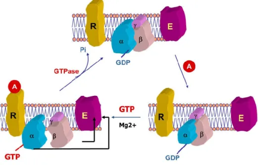
and Gautam 1999; Strathmann and Simon 1991). All G proteins are composed of three subunits: Gα, Gβ and Gγ (Neer and Clapham 1988). In the inactive state, Gα remains bound with Gβ/Gγ dimer and GDP molecule (Lambright et al. 1994) [Figure 7].
Figure 7: Activation of G-protein. Binding with a signaling molecule (A) to GPCR induces a conformation change that allows inactive G-protein to exchange of GDP for GTP on the α subunit of the heterotrimeric complex. Both GTP-bound Gα in the active form and the released Gβγ dimer can then go on to stimulate downstream effectors. After signaling mechanism, the GTP on Gα is hydrolyzed to GDP the original receptor is restored. (The diagram is adapted from a lecture by Dr Madhu B. Anand-Srivastava, PSL6090, 2015.)
When an agonist/signal molecule binds to GPCR, the GPCR undergoes a conformational change that activates the G proteins by promoting the exchange of Gα bound GDP to GTP. This leads to the dissociation of Gβ/Gγ dimer from Gα (Oldham and Hamm 2008). Now, activated Gα-GTP acts upon their downstream effectors and thereby initiate intracellular signaling responses. However, after the signaling mechanism, GTP from Gα-GTP is hydrolyzed to GDP and produces inactive state of G protein where Gα bound GDP (Gα-GDP) re-associates with Gβ/Gγ dimer to form the heterotrimeric structure (Tuteja 2009) [Figure 7].
7.3.2. Adenylyl Cyclase/cAMP Signaling
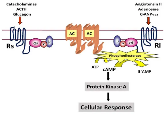
Adenylyl Cyclase/cAMP signal transduction system has been targeted for the treatment of CVDs from decades (Gold, Gonen, and Scott 2013). When a hormone binds to GPCR, activated Gα is released to transmit signal on the effector molecule called adenylyl cyclase (AC). AC then converts
19
adenosine triphosphate (ATP) to cyclic adenosine monophosphate (cAMP) and increases cAMP level. cAMP is a second messenger which has major implications in numerous cellular functions by activating enzyme protein kinase A [Figure 8].
Figure 8: G-protein Mediated Adenylyl Cyclase/cAMP Signaling. Where, AC is denoting as adenylyl cyclase. Rs and Ri are the stimulatory GPCR and inhibitory GPCR, respectively. (The diagram is adapted from a lecture by Dr Madhu B. Anand-Srivastava, PSL6090, 2020.)
Aberrant activation of cAMP can have pathophysiological consequences related to cardiac contractility, vascular tone and reactivity etc. (Asano, Masuzawa, and Matsuda 1988; Baumann et al. 1981). Basal activity of AC is reduced in SHR resulting in reduced production of cAMP associated with the hyperproliferation of VSMC and endothelial dysfunction in SHR (Gusan and Anand-Srivastava 2013; Shah and Singh 2006). However, activating or inhibiting AC signal propagation is regulated by G proteins (Gilman 1989). G protein, responsible for stimulating or activating AC and increased cAMP production is called G-stimulatory (Gs) protein, whereas, inhibition of AC and reduced cAMP production is regulated by G-inhibitory (Gi) protein.
20 7.3.2.1. G-stimulatory (Gs) Protein
The alpha subunit of G-stimulatory protein (Gsα) is positively coupled to AC and mediates the stimulatory responses of hormones upon activating AC, thereby increases cAMP messenger level. Molecular cloning of human cDNA has revealed four distinct types of Gsα resulting from differential splicing of an individual gene (Bray et al. 1986; Robishaw, Smigel, and Gilman 1986). Gs protein has been shown to regulate intracellular calcium homeostasis and induces phosphorylation of contractile filaments. A reduced stimulation of Gs protein is associated with the β-adrenoceptor downregulation, that contributes to cardiac complications in SHR (Asano, Masuzawa, and Matsuda 1988).
7.3.2.2. G-inhibitory (Gi) Protein
The alpha subunit of G-inhibitory protein (Giα) is negatively coupled to AC and inhibits AC activity from cAMP production. Based on the sequence identity of their alpha subunit, these proteins are classified into subfamilies: 1, 2, 3, GoαA, GoαB (Kehrl 1998). All three forms of Giα-1-3 take part in the AC inhibition and activation of atrial acetylcholine activated potassium channel. Dysfunction of Gi protein mediated signaling pathways has significant role in metabolic diseases like obesity and diabetes (Kimple et al. 2014). An absence of Giα-2 proteins has been shown to contribute in dilated cardiomyopathy and increased mortality in β1-adrenoceptor-overexpressing mice (Keller et al. 2015). An overexpression of Giα protein is thought to be one of the pathological factors contributing to hypertension and vascular remodeling in different hypertensive animal models (Ali El-Basyuni, Li, and Anand-Srivastava 2016; Anand-Srivastava, de Champlain, and Thibault 1993; Anand-Srivastava, Picard, and Thibault 1991; Ge, Garcia, and Srivastava 1999; Li and Srivastava 2002; Li et al. 2014; Sarkar, Li, and Anand-Srivastava 2019).
7.3.2.2.1. Role of Giα Protein Overexpression in the Pathogenesis of Hypertension
Gi proteins and associated signaling mechanisms are involved in maintaining vascular tone, contractility, proliferation and hypertrophy of VSMC (Almajdoob, Hossain, and Anand-Srivastava 2018; Atef and Anand-Srivastava 2014; Bou Daou, Li, and Anand-Srivastava 2016; Gomez
21
Sandoval et al. 2013; Rodbell et al. 1971). Thus, the impaired functions of Gi proteins results in developing hypertension and associated complications.
Our laboratory showed that Giα-2 and Giα-3 proteins and their mRNA but not of Goα and Gsα are overexpressed in heart and aorta from different hypertensive rat models including SHR, DOCA-salt, L-NAME-induced and 1 kidney-1clip hypertensive rats (Anand-Srivastava 1992; Anand-Srivastava, de Champlain, and Thibault 1993; Anand-Srivastava, Picard, and Thibault 1991; Di Fusco and Anand-Srivastava 2000; Ge, Garcia, and Anand-Srivastava 1999; Ge, Garcia, and Srivastava 2006; Lappas, Daou, and Srivastava 2005; Thibault and Anand-Srivastava 1992). Moreover, VSMC and lymphocytes from 12-week old SHR also showed an overexpression of Giα protein when compared to normotensive WKY rats (Lappas, Daou, and Anand-Srivastava 2005; Marcil and Anand-Srivastava 2001). Further studies confirmed that enhanced expression of Giα-2 and Giα-3 proteins in heart and aorta precedes the development of hypertension in SHR and DOCA-salt rats (Marcil, de Champlain, and Anand-Srivastava 1998; Marcil, Thibault, and Anand-Srivastava 1997). These studies suggest that enhanced expression of Giα proteins that results in decreased production of cAMP, could be one of the contributing factors in the pathogenesis of hypertension. This was further supported by the studies showing that inactivation or inhibition of Giα proteins by pertussis toxin or by antisense of Giα-2 protein attenuated the development of high BP in SHR (Ali El-Basyuni, Li, and Anand-Srivastava 2016; Li and Anand-Srivastava 2002; Triggle and Tabrizchi 1993). In addition, resveratrol, SNP, and C-ANP4-23, a natriuretic peptide receptor C agonist have also been shown to attenuate the
development of hypertension in SHR through the inhibition of overexpression of Giα proteins (Hossain et al. 2018; Li et al. 2014; Sarkar, Li, and Anand-Srivastava 2019).
The enhanced expression of Giα proteins in SHR was shown to be attributed to the augmented levels of endogenous Ang II, ET-1 and growth factors because the inhibition of AT1 receptor, ETA
receptor and growth factor receptors by pharmacological inhibitors or siRNA attenuated the overexpression of Giα proteins in VSMC from SHR (Sandoval Gomez, Li, and Anand-Srivastava 2011; Sandoval Gomez and Anand-Srivastava 2011). In addition, our laboratory by using the inhibitors or siRNA has also demonstrated that endogenous Ang II and ET-1 through the interaction with AT1 and ETA receptor respectively increased the oxidative stress which through
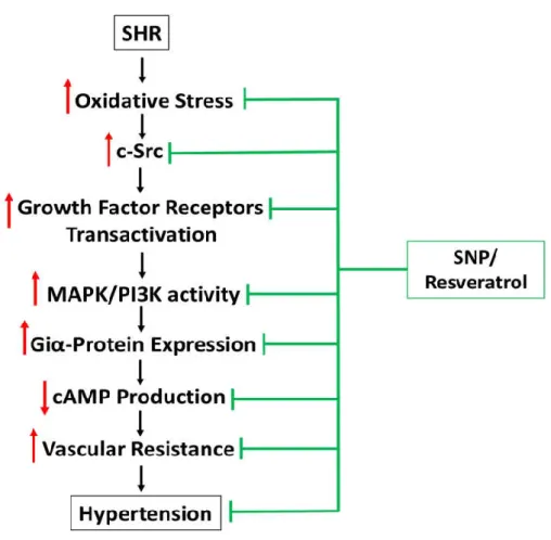
22
the activation of c-Src and growth factor receptor augments the activity of MAP kinase and contributes to the enhanced expression of Giα proteins (Sandoval Gomez and Anand-Srivastava 2011; Sandoval Gomez, Li, and Anand-Srivastava 2011). Enhanced expression of Giα proteins results in the decreased levels of intracellular cAMP and increased vascular resistance and thereby leads to high BP [Figure 9].
Figure 9: Schematic Diagram Summarizing the Effect of SNP/Resveratrol on Hypertension of SHR and the Implicated Molecular Mechanisms
In addition, SNP and resveratrol-induced attenuation of overexpression of Giα proteins and high BP in SHR was also shown to be attributed to the inhibition of oxidative stress and associated signaling mechanisms [Figure 9] (Hossain et al. 2018; Sarkar, Li, and Anand-Srivastava 2019). C-ANP4-23-induced attenuation of high BP in SHR was associated with the inhibition of
23
7.3.2.2.2. Role of Giα Protein Overexpression in the Pathogenesis of Tachycardia
Giα protein signaling is a critical mediator of the regulation of heart rate modulation and dynamics (Ang, Opel, and Tinker 2012; Sebastian et al. 2013). A mutated GTP binding domain of Giα-2 protein has been shown to be responsible for the development of idiopathic ventricular tachycardia in human (Lerman et al. 1998). Nagata et al showed that Giα-2 (but not Giα-1/Giα-3) deficient mice resulted in inhibition of β-adrenergic receptor–induced contractility and calcium currents in adult murine cardiomyocytes (Nagata et al. 2000). However, SHR typically showed an increased heart rate and inhibition of the overexpression of Giα-2 attenuates BP and tachycardia (Ali El-Basyuni, Li, and Anand-Srivastava 2016; Li et al. 2014). Our laboratory has also demonstrated the role of Giα-2 but not of Giα-3 in tachycardia in SHR (Ali El-Basyuni, Li, and Anand-Srivastava 2016).
8. Histone deacetylase (HDAC)
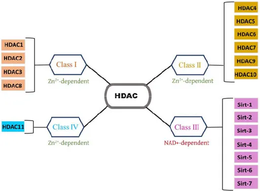
Histone modifications includes acetylation or deacetylation of lysine residues, that play a key role in the epigenetic regulation of gene transcription (Berger 2002). Histone deacetylases (HDAC) are a group of enzymes that remove acetyl moieties from an ε-N-acetyl lysine amino acid on a histone and some other non-histone proteins (Ito, Barnes, and Adcock 2000; Glozak et al. 2005). Thus, HDAC controls chromatin remodeling (Felisbino et al. 2013) and alters expression of genes implicated in regulating several cellular processes (Felisbino et al. 2013). Based on sequence homology and phylogenetic analysis, four diverse classes of HDACs have been identified and are numbered from I to IV. Class I includes HDAC1, HDAC2, HDAC3, and HDAC8; Class II are HDAC4, HDAC5, HDAC6, HDAC7, HDAC9, and HDAC10; Class III are known as Sirtuin that include Sirt-1, Sirt-2, Sirt-3, Sirt-4, Sirt-5, Sirt-6, and Sirt-7; and class IV has a solely member HDAC11 [Figure 10]. Class I, II and IV HDAC are Zn2+-dependent enzymes, which diverge
significantly from the Class III HDAC that are dependent on nicotinamide adenine dinucleotide (NAD+) for their catalytic activity (Holbert and Marmorstein 2005).
24
Figure 10. Histone Deacetylases (HDAC) Classifications. HDAC are divided into four classes, CLASS I (HDAC1, HDAC2, HDAC3, HDAC8); CLASS II (HDAC4, HDAC5, HDAC6, HDAC7, HDAC9, HDAC10); CLASS III (Sirt-1, Sirt-2, Sirt-3, Sirt-4, Sirt-5, Sirt-6, Sirt-7) and CLASS IV (HDAC11). Sirt- denotes as Sirtuin.
8.1. HDAC Inhibitors
A delicate balance between acetylation and deacetylation ensures an accurate gene regulatory event within the cell. Therefore, disturbance of HDAC activities may contribute to a varied range of diseases, from diabetes to cancer. For example, HDAC1 and HDAC2 show an overexpression in colon cancer (Yang et al. 2014), HDAC3 is upregulated in gastric cancers (Xu et al. 2018), HDAC7 exhibits an increased expression level in pancreatic islets from patient with type 2 diabetes (Daneshpajooh et al. 2017), HDAC6 has an higher expression in various neurodegenerative diseases (Simões-Pires et al. 2013). HDAC inhibitors exploit Zn2+-binding
sites and block the catalytic activity of HDAC. As a result, an increase acetylation of the histone promotes re-expression of silenced controlling genes, thus, emerging as potential therapeutic agents (Lombardi et al. 2011). Vorinostat, Romidepsin, Belinostat, and Panobinostat are the HDAC inhibitors which received FDA approval and are currently used as anti-tumor agents (Suraweera, O'Byrne, and Richard 2018).
25
8.2. HDAC in Vascular Remodeling, Emerging Therapeutic Targets for CVDs
Along with anti-tumor effect, HDAC inhibitors were recently shown to exhibit beneficial effects on vascular remodeling and cardiac function, thereby providing a novel cardiac therapeutic approach. Substantial studies showed that using various Zn2+-dependent HDAC inhibitors is
efficacious against several CVDs including cardiac arrhythmia, myocardial infarction, cardiac remodeling, hypertrophy, hypertension, and cardiac fibrosis (Yoon and Eom 2016). Increased expression of HDAC was shown to be implicated in increased proliferation, migration and hypertrophy of VSMC and associated diseases (Pietruczuk and Srivastava 2017). It was reported that overexpression of HDAC4 stimulated hyperproliferation and migration of VSMC and inhibition of HDAC4 effectively suppressed the proliferation (Zheng et al. 2019). Scriptaid, a broad spectrum HDAC inhibitor was also shown to attenuate mitogen-induced proliferation and neointimal hyperplasia in VSMC (Findeisen et al. 2011).
Inhibitory activity of class I HDAC has been shown to block Ang II-induced cardiac hypertrophy in mice or rats (Kee et al. 2006). Selective Inhibition of HDAC8 with PCI34051 also reduced the high BP through improving vascular remodeling in Ang II-induced hypertensive mice (Kee et al. 2019). Inhibition of Class I HDAC was also shown to attenuate hypertension and cardiac remodeling in SHRs (Cardinale et al. 2010). Taken together, it may be suggested that HDAC inhibitors are capable to reduce vascular remodeling, however, in depth-studies are needed to establish a potential therapy of CVDs.
9. Sirtuins, Class III HDAC
Sirtuins have garnered a remarkable attention in a short time since their discovery as key regulators of yeast replicative lifespan in 1955 (Kennedy et al. 1995). They are named after its founding member silent information regulator 2 gene (SIR2), that is vital for silencing heterochromatin in budding yeast, Saccharomyces cerevisiae (Rine and Herskowitz 1987). Mammalian sirtuins primarily catalyze the deacetylation of lysine residues on histones and various non histone proteins and some of them also possess ADP-ribosyl transferase activity (Liszt et al. 2005; Frye 1999). They are highly conserved, evolutionarily NAD+-dependent HDAC. There are seven known isoforms (Sirt-1–7) of sirtuins which participate in a wide range of cellular
26
processes and pathways with diversified cellular localization and molecular targets (Cen, Youn, and Sauve 2011). Each of them is comprised of a conserved 275 amino acid catalytic core and flanked by unique additional N-terminal and/or C-terminal region of variable length (Frye 2000). However, they vary in their sub-cellular localization: Sirt-1, Sirt-6 and Sirt-7 are located in the nucleus, Sirt-2 is prominent in the cytoplasm, and Sirt-3, Sirt-4 and Sirt-5 are found in mitochondria (Michishita et al. 2005; North et al. 2003) (Table 1). Function wise, 1 and Sirt-5 show deacetylase activity (Vaziri et al. 2001; Kumar and Lombard 2018), Sirt-4 and Sirt-6 have ADP-ribosyl transferase activity (Haigis et al. 2006; Liszt et al. 2005), whereas Sirt-2 and Sirt-3 exhibit both activities (Shi et al. 2005; North et al. 2003). Sirt-7 has not been shown to possess NAD-dependent deacetylase nor an ADP-ribosyl transferase activity (North et al. 2003).
Table 1: Functional and Localized Diversity of Mammalian Sirtuin Protein Family.
Sirtuin Functional Properties Localization
Sirt-1 Deacetylase activity Nucleus
Sirt-2 Deacetylase and ADP-ribosyl transferase activity Cytoplasm Sirt-3 Deacetylase and ADP-ribosyl transferase activity Mitochondria Sirt-4 ADP-ribosyl transferase activity Mitochondria
Sirt-5 Deacetylase activity Mitochondria
Sirt-6 ADP-ribosyl transferase activity Nucleus
Sirt-7 ? Nucleus
9.1. Sirtuins in Cardiovascular System
All sirtuins are broadly implicated in cellular aging and longevity (Grabowska, Sikora, and Bielak-Zmijewska 2017; Satoh, Imai, and Guarente 2017; Camins et al. 2010; Bonkowski and Sinclair 2016). Besides that, they play certain roles in the regulation of energy metabolism, DNA repair,