UNIVERSITÉ DE SHERBROOKE
Pathogenic Role of IL-15 in Non-Alcoholic
Fatty Liver Disease
Written by:
Yuneivy Cepero Donates
Department Pediatrics, Division of Immunology
Master’s thesis presented to the Faculty of Medicine and Health Sciences in
view of obtaining a Master of Science (MSc.) in Immunology
UNIVERSITÉ DE SHERBROOKE
RÔLE PATHOGÉNIQUE DE l’IL-15 DANS LA STÉATOSE HÉPATIQUE Yuneivy Cepero Donates
Département de pédiatrie, Service d’immunologie
Mémoire présenté en Juillet 2014 à la Faculté de Médecine en vue de l’obtention du grade de Maître en Sciences (MSc.) en Immunologie
RÉSUMÉ
Les cytokines pro-inflammatoires jouent un rôle important dans la pathogenèse de l’obésité et la stéatose hépatique. L'IL-15 est une cytokine pro-inflammatoire qui est trans-présentée par l'IL-15Rα aux chaines IL-2/IL-15Rβ et γc. La fonction de l'IL-15 a été largement décrite dans les cellules immunitaires, mais ses fonctions dans d'autres tissus sont moins connues. Le but de ce mémoire est d'élucider le rôle de l'IL-15 dans la stéatose hépatique.
Les souris C57BL/6 de type sauvage (WT) et Il15-/- ont été soumises à un régime hyperlipidique (HFD) ou à un régime normal. Après 16 semaines, le poids corporel, la masse hépatique, l'accumulation de lipides dans le foie, les taux de lipides sériques et l'expression des différents gènes reliés à l’inflammation et au métabolisme dans le foie ont été évalués. Les lymphocytes intra-hépatiques (IHL) ont été également étudiés. Des hépatocytes primaires ont été stimulés avec IL-15, et l'expression génique de chimiokines a été déterminée. Les populations de IHLs ont été également caractérisées chez les souris WT, Il15-/- et Il15ra-/-, ainsi que chez des souris dont la déficience dans
l’expression d’IL-15Rα est ciblée aux macrophages ou aux hépatocytes.
Nos résultats montrent que la déficience en IL-15 empêche l'accumulation de lipides dans le foie. Les taux de cholestérol et d’acides gras non estérifiés dans le sang étaient élevés chez les souris WT, mais pas chez les souris Il15-/- . L'expression hépatique des
chimiokines Ccl2, Ccl5, Cxcl10 et des marqueurs de macrophages était augmentée chez les souris WT sous HFD, mais pas chez les souris Il15-/-. La stimulation des hépatocytes
primaires avec l'IL-15 induit l'expression des gènes des chimiokines chez les hépatocytes WT, mais pas chez les Il15ra-/-. En outre, nous avons trouvé une infiltration réduite des
cellules NK et NKT dans le foie des souris déficientes en Il15ra-/- dans les hépatocytes, ce
qui suggère que l'expression d’IL15Rα chez les hépatocytes est nécessaire au recrutement des cellules NK, NKT et / ou à leur maintien.
En conclusion, nous proposons que l’IL-15 favorise l'accumulation de lipides dans le foie, et que ceci est associée à une réponse inflammatoire accrue. La disponibilité accrue de l'IL-15 dans l'obésité pourrait stimuler les hépatocytes à secréter des chimiokines ce qui favorise l'inflammation hépatique et conduirait à la stéatose hépatique. L’expression de l'IL-15Rα dans les hépatocytes semble jouer un rôle principal dans l’infiltration des cellules NK, NKT et iNKT dans le foie.
Mots clés : Stéatose hépatique non-alcoolique (NAFLD), lymphocytes intra-hépatiques
UNIVERSITÉ DE SHERBROOKE
PATHOGENIC ROLE OF IL-15 IN NON-ALCOHOLIC FATTY LIVER DISEASE Yuneivy Cepero Donates
Department Pediatrics, Division of Immunology
Master’s thesis presented to the Faculty of Medicine and Health Sciences in view of
obtaining a Master of Science (MSc.) in Immunology
ABSTRACT
Pro-inflammatory cytokines play a key role in pathogenesis of obesity and non-alcoholic fatty liver disease (NAFLD). IL-15 is a pro-inflammatory cytokine, which signals through a receptor complex composed of the IL-15 receptor (IL-15R) alpha chain, the 2/15R beta chain and the common gamma chain. The functions of IL-15 have been extensively described in immune cells but less is known about its functions in others tissues such as the liver. The aim of this thesis is to investigate the role of IL-15 in fatty liver disease.
C57BL/6 wildtype (WT) and IL-15 knockout (Il15-/-) mice were maintained on high fat
diet (HFD) or normal control diet (NCD). After 16 weeks, body weight, liver mass, fat accumulation in the liver, serum lipid levels and gene expression in the liver were evaluated. Intrahepatic lymphocytes (IHL) were also analysed. Primary hepatocytes were stimulated with IL-15 and chemokines gene expression was studied. IHLs were examined in WT, Il15-/- and Il15ra-/-, as well as in macrophage- and hepatocyte-specific Il15ra-/- mice.
We found that IL-15 deficiency prevents weight gain and accumulation of lipids in the liver. Circulating levels of cholesterol and non-esterified fatty acids were elevated in WT mice but not in Il15-/- mice. Hepatic expression of chemokines such as Ccl2, Ccl5
and Cxcl10 was increased in WT mice under HFD, but not in Il15-/- mice. The livers of Il15-/- and Il15ra-/- mice also showed decreased expression of Tnfa and iNOS, and
macrophage markers Cd68 and F4/80. Accordingly, stimulation of primary hepatocytes with IL-15 induced chemokine gene expression in WT but not in Il15ra-/- hepatocytes.
Furthermore, hepatocyte-specific ablation of IL-15Rα reduced infiltration of NK and NKT cells in the liver, suggesting that IL15Rα expression in the hepatocytes is needed for the recruitment and/or maintenance of the NK cell population in the liver.
In conclusion, IL-15 promotes fat accumulation in the liver, and this is associated with increased inflammatory response in the liver. Increased availability of IL-15 in obesity may stimulate hepatocytes to secrete chemokines that promote hepatic inflammation resulting in fatty liver disease. IL-15Rα expression in hepatocytes appears to play a role in the maintenance of NK, NKT and iNKT cells.
Keywords: non-alcoholic fatty liver disease (NAFLD), intra-hepatic lymphocytes (IHL),
iv
RÉSUMÉ ... ii
ABSTRACT ... iii
LIST OF FIGURES AND TABLES ... vi
LIST OF ABBREVIATIONS... viii
1. INTRODUCTION ... 1
1.1. Structure and functions of the liver ... 1
1.1.1. Structure of the liver ... 1
1.1.2. Metabolic functions of the liver ... 2
1.1.3. Liver as part of the immune system... 4
1.2. Obesity ... 5
1.2.1. Obesity-associated inflammation ... 6
1.3. Fatty liver diseases ... 7
1.3.4. Mouse models of NAFLD ...12
1.4. Cytokines that signal via the common γ-chain (γc; CD132) receptor ...12
1.5. Interleukin-15 (IL-15) ...15
1.5.1. Structure ...15
1.5.2. IL-15 receptor ...16
1.5.3. IL-15 signalling ...17
1.5.4. Mechanisms mediating IL-15 responses ...18
1.5.5. Function of IL-15 in T cells ...21
1.5.6. IL-15 in NK cell biology ...21
1.5.7. The role of IL-15 in iNKT cells ...23
1.5.8. IL-15 functions in non-immune cells ...24
1.5.9. Functional dichotomy between IL-2 and IL-15 ...26
1.6. The thesis premises ...27
1.7. Hypothesis ...28
1.8 Objectives ...28
2. MATERIALS and METHODS ... 29
2.1. Mice ...29
2.1.1 Induction of NAFLD in mice ...29
2.2. Isolation of intrahepatic lymophocytes (IHL) ...30
v
2.4. FACS analyses...31
2.5. Isolation of primary hepatocytes ...31
2.6. Stimulation of hepatocytes ...32
2.7. Assessment of mitochondrial respiration in hepatocytes ...32
2.7.1 Fatty acids oxidation test ...33
2.8. Tissues processing for histology ...34
2.9. Histology and lipids detection ...35
2.10. Immunofluorescence microscopy ...35
2.11. RNA isolation ...36
2.12. Quantitative PCR ...36
2.13. Graphs and statistical analysis ...37
3. RESULTS ... 38
3.1. IL-15 promotes weight gain and fat accumulation in the liver ...38
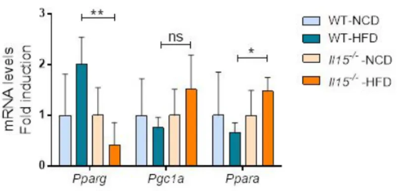
3.2. IL-15 deficiency reduces Pparg and increases Ppara induction after HFD ...40
3.3. IL-15 regulates β-oxidation ...41
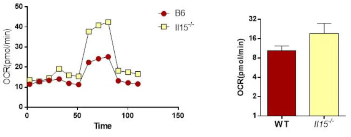
3.4. IL-15 suppresses mitochondrial respiration in mice primary hepatocytes ...43
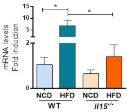
3.5 HFD induces Il15 expression in the liver ...46
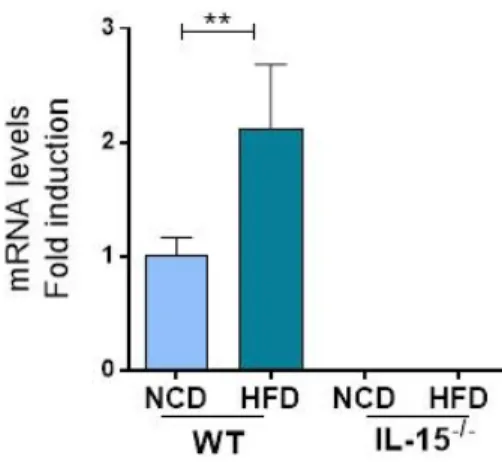
3.6. HFD promotes the expression of pro-inflammatory mediators in the liver. ...46
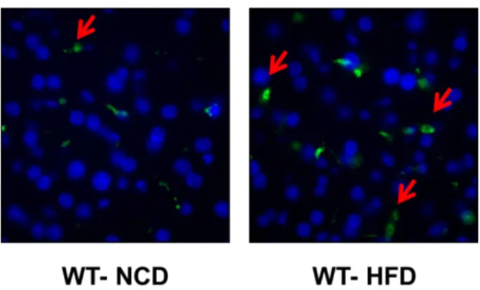
3.7. HFD-induced macrophage infiltration in the liver is reduced in the absence of IL-15 ...47
3.8. HFD increases immune cells infiltration in mice liver ...48
3.9. HFD induced chemokine gene expression in the liver is mediated by IL-15. ...51
3.10. IL-15 induces chemokines expression in mice primary hepatocytes ...53
3.11. IL-15 and IL-5Rα are required for the maintenance of NK populations in the liver ...54
3.12. IL-15Rα expression in both macrophages and hepatocytes contribute to NK cell maintenance in the liver ...56
3.13 Summary of results ...59
4. DISCUSSION ... 60
5. CONCLUSIONS ... 75
6. ACKNOWLEDGEMENTS ... 77
vi
LIST OF FIGURES AND TABLES
FIGURES Page
Figure 1.1 Figure 2.1:
Principals mechanism for IL-15 delivery XF Cell Mito Stress Test Profile
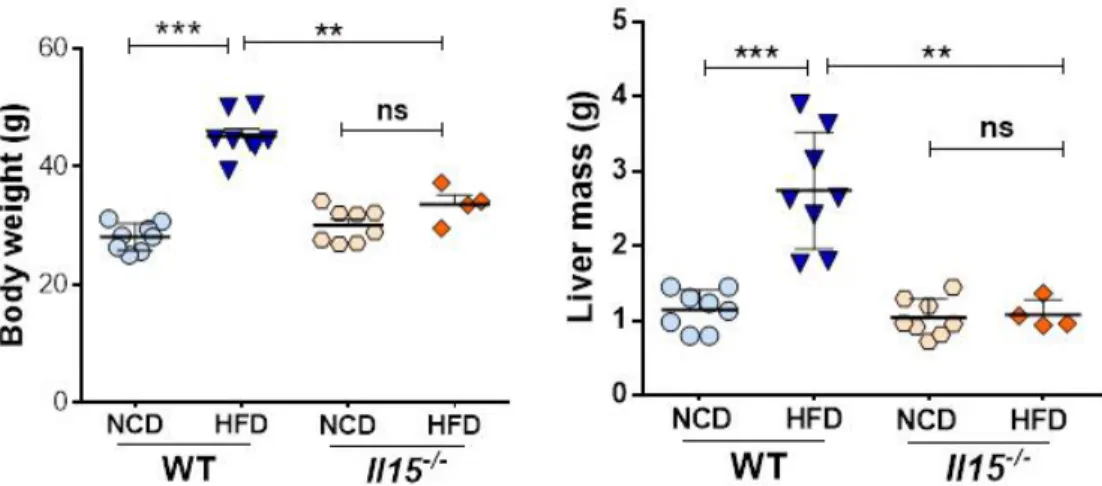
20 33 Figure 3.1: A) IL-15 deficiency prevents weight gain in the liver and hepatic fat
accumulation.
39
B) HFD results in increased circulating levels of cholesterol and NEFA in wild type mice, but not in Il15 deficient mice
39
C) IL-15 deficiency prevents hepatic fat accumulation 40 Figure 3.2: IL-15 deficiency reduces Pparg and increases Ppara induction after
HFD
41
Figure 3.3: A) HFD induces the expression of Cd36 in the liver 42 B) Cpt1a levels are increased in Il15-/- mice in NCD 42
C) HFD induces Acadm expression in the liver 43
Figure 3.4: A) IL-15 deficiency does not affect cytochrome c oxidase levels 44 B) IL-15 deficiency increases mitochondrial respiration 45 C) IL-15 deficiency enhances fatty acid oxidation in hepatocytes 45 Figure 3.5: Il15 mRNA levels are increased in WT mice in HFD 46 Figure 3.6: HFD induces Tnfα and iNOS expression in the liver 47 Figure 3.7: A) HFD increases macrophages infiltration in the liver 48
B) Expression of macrophages markers is increased in WT mice maintained on HFD
48
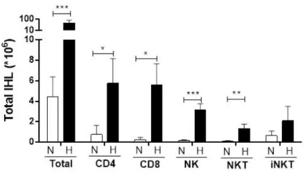
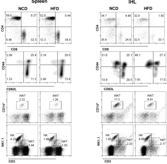
Figure 3.8: A) HFD induce lymphocytes infiltration in the liver 50 B) HFD induces CD8+ T cell activation and NK cell infiltration in the
liver
51
Figure 3.9: HFD induced chemokine mRNA expression in the liver requires IL-15 52 Figure 3.10: IL-15 induces Ccl5 and Cxcl10 expression in mouse primary
hepatocytes
vii Figure 3.11: A) Splenic but not liver CD8+ T cells are dependent on IL-15 signaling 54
B) NK and NKT cells are reduced in the absence of IL-15 or IL-15Rα 55 C) IL-15 and IL-15Rα are needed for iNKT cell maintenance in the liver 55 Figure 3.12: A) IL-15Rα expression in macrophages is required for NKT cells
maintenance in the liver
57
B) IL-15Rα expression in the hepatocytes is required for the maintenance of NK, NKT and iNKT cells in the liver
58
Figure 5.1: IL-15 regulates immune cells recruitment and homeostasis in the liver 76
TABLES
viii
LIST OF ABBREVIATIONS
Acadm Acyl-Coenzyme A dehydrogenase, C-4 to C-12 straight chain
acetyl-CoA Acetylcoenzyme A
ACK Ammonium-chloride-potassium, lysing buffer
AICD Activation-induced cell death
APPs Acute-phase proteins
Bcl2 B-cell lymphoma 2
BM Bone marrow
BMI Body mass index
BSA Bovine serum albumin
Cepba CCAAT/enhancer binding protein
Cox4i1 Cytochrome c oxidase
Cpt1a Carnytyl palmitoyl transferase
DCs Dendritic cells
DIO Diet-induced obesity
DMEM-F12 Dulbecco’s modified eagle medium: nutrient mixture f-12
ER Endoplasmic reticulum
FA Fatty acids
FACS Fluorescence-activated cell sorting
FCCP Carbonyl cyanide p-[trifluoromethoxy]-phenyl-hydrazone
FCS Fetal calf serum
GFP Green fluorescent protein
HBSS Hank’s buffered salt solution
HCC Hepatocellular carcinoma
HDL High-density lipoprotein cholesterol
HFD High fat diet
HGF Hepatocyte growth factor
ix
IELs Intraepithelial lymphocytes
IFN-γ Interferon gamma
IGF-1 Insulin growth factor-1
IHL Intrahepatic lymphocytes
IL- Interleukin
IL-15Rα IL-15 receptor alpha
iNKT Invariant Natural killers T cells
iNOS Inducible nitric oxide synthase
IP-10 Interferon gamma-induced protein 10
IRS1 Insulin receptor substrate 1
JAK Janus kinase
JNK1 C-Jun N-terminal kinase 1
KCs Kupffer cells
KHB Krebs-Henseleit buffer
KO Knockout
KRB Krebs-ringer-buffer
Lck Lymphocyte-specific protein tyrosine kinase
LPL Lipoprotein lipase
LSEC Liver sinusoidal endothelial cells
LSP Long signal peptide
M1 Pro-inflammatory macrophages
M2 Anti-inflammatory macrophages
MCP-1 Monocyte chemotactic protein-1
MRC Maximal respiratory capacity
mRNA Messenger ribonucleic acid
mTECs Medullary thymic epithelial cells NAFLD Non-alcoholic fatty liver disease
NASH Non-alcoholic steatohepatitis
x
NEFA Non-esterified fatty acids
NK Natural killer cells
NKp NK precursors
NKT Natural killer T cells
ns Not significant
PBMCs Peripheral blood mononuclear cells
PBS Phosphate buffered saline
PCD Programmed cell death
PFA Paraformaldehyde
Pgc1a PPARγ coactivator 1-alpha
PPAR Peroxisome proliferator-activated receptor
RA Rheumatoid arthritis
RNA Ribonucleic acid
ROS Reactive oxygen species
SRC Spare respiratory capacity
SREBP1c Sterol regulatory element-binding protein 1c Srebpf1 Sterol-regulatory-element-binding protein
SSP Short signal peptide
STAT Signal Transducer and Activator of Transcription
Syk Spleen Tyrosine kinase
TCR T-cell receptor
TECs Tubular epithelial cells
TG Triacylglycerol
TGF Transforming growth factor
Th1 T helper 1
Th2 T helper 2
TLRs Toll-like receptors
TM Transmembrane region
xi
Treg Regulatory T cells
TSLPR Thymic stromal lymphopoietin receptor
Tw Tween®20
VLDL Very-low-density lipoprotein
VSV Vesicular stomatitis virus
WHO World health organization
WT Wild type
1
1. INTRODUCTION
1.1. Structure and functions of the liver 1.1.1. Structure of the liver
Liver is one of the most important metabolic organs in the body and plays a central role in the synthesis of many plasma proteins and carbohydrates, storage of minerals and vitamins and detoxifying toxins that enter portal vein circulation (Fausto, N. et al., 2006). Liver cells are classified into two main groups: epithelial cells and mesenchymal cells. In the epithelial cells group, the major subtype is hepatocytes, which represents more than 90% of the liver parenchyma (Papoulas, M. and Theocharis, S., 2009). Cholangiocytes also belong to this group, and are located in the intra-hepatic and extra-hepatic biliary duct system. Moreover, cholangiocytes release bicarbonate and water, which modifies the bile produced by hepatocytes (O'Hara, S.P. et al., 2013). Hepatic non-parenchymal cells include endothelial cells, Kupffer cells (KCs), lymphocytes, hepatic stellate cells (HSCs) and biliary ductal cells (Lemoinne, S. et al., 2013). KCs reside in liver sinusoids and are derived from circulating monocytes. They constitute approximately 20% of hepatic non-parenchymal cells (Ruck, P. and Xiao, J.C., 2002)
Hepatic stellate cells produce hepatocyte growth factor (HGF), store retinoids and serve as precursors of liver myofibroblasts (Friedman, S.L., 2008). Myofibroblasts are the main fibrogenic effector cells and are absent in the healthy liver. However, following liver damage, HSCs differentiate and start producing extracellular matrix and collagen (Lemoinne, S. et al., 2013). Liver sinusoidal endothelial cells (LSEC) line the capillaries and sinusoids and differ from the endothelium of big vessels in lacking basement membrane and containing the fenestrae structures. Moreover, a subset of LSECs is derived from the bone marrow and, like HSCs, is one of the main sources of HGF (DeLeve, L.D., 2013). Liver lymphocytes are primarily located around the portal tracts.
2 This distribution of lymphocytes in the liver aids in the rapid removal of gastrointestinal antigens from the circulation (Zhan, Y.T. and An, W., 2010).
1.1.2. Metabolic functions of the liver
Liver is a major metabolic organ in the body and plays important roles in protein, carbohydrate and lipid metabolic pathways.
In proteins metabolism, liver participates in amino acids degradation to supply both, carbohydrates and fatty acids (FAs) metabolic pathways, and protein synthesis. In the human serum, the most abundant protein is albumin, which is synthetized by hepatocytes and represents the 15 percent of the total hepatic protein synthesis. Albumin’s half-life of is around 20 days, so that a decrease in serum albumin is unlikely to occur within a short time of acute liver injury. In patients with ascites and chronic liver disease the serum level of albumin is often low (Rothschild, M.A. et al., 1969). Various coagulation factors such as II, VII, IX, X, V, XI, XII, and XIII and fibrinogen are synthesized in the liver. As coagulation factors have short life-span, defects in coagulation quickly become apparent in acute liver damage. Some inhibitors of coagulation and fibrinolysis are also synthesized in the liver. Moreover, in liver disease, the prolongation of prothrombin time results from a deficiency in factors II, V, VII, and X; and can be used as a predictive factor of acute liver failure (Aranha, G.V. and Greenlee, H.B., 1986). Transamination and oxidative deamination of amino acids takes place in the hepatocytes and leads to nitrogenous excretory products, which enter in the Krebs-Henseleit cycle producing urea (Zieve, L., 1979).
In carbohydrates metabolism, liver regulates glucose levels in the blood by controlling glucose uptake after meals and releasing glucose during fasting. Carbohydrate metabolism in the liver is proportional to the degree of hypoglycemia or hyperglycemia. In hypoglycemic condition, glucose synthesis from endogenous sources is increased, for
3 example, endogenous glycogen is catabolized into glucose that enters the blood (Stanley, J.C., 1981). This process is controlled by several hormones, such as catecholamines that stimulate the rate of gluconeogenesis via cyclic adenosine monophosphate (Fausto, N. et al.)-dependent (beta-mimetic) and cAMP-independent (alpha-mimetic) mechanisms (Hems, D.A. and Whitton, P.D., 1980). On the other hand, insulin antagonizes the action of both glucagon and catecholamines on hepatic gluconeogenesis.
Fat containing chylomicrons reach the liver via lymph and the blood. Degradation of fat to acetyl coenzyme A (acetyl-CoA), a main step in the metabolic integration of different pathways, occurs in the liver. Acetyl-CoA can enter into the tricarboxylic acid cycle or participate in the synthesis of triglyceride phospholipid, cholesterol and lipoproteins. Fatty acids in the liver can be metabolized by esterification or oxidation. Glucagon markedly stimulates the rate of FA oxidation, whereas insulin inhibits it. The beta oxidation of FAs results in the production of acetyl-CoA in the mitochondria, where the acetyl-CoA is further oxidized to carbon dioxide and water or is converted to ketone bodies (Voet, J.G. and Voet, D., 2000).
FA synthesis from excess glucose also occurs in the liver. FAs are esterified with glycerol in the liver to form triglycerides, which are then incorporated into lipoproteins, principally very-low-density lipoprotein (VLDL), that are secreted by the liver. The principal factor affecting VLDL production is the amount of free FAs reaching the liver. Secretion of VLDL is also stimulated by insulin (Kamagate, A. and Dong, H.H., 2008). Hepatic lipid synthesis is stimulated by insulin after food intake. Interestingly, in metabolic disease (insulin resistant state), liver and plasma triglycerides are increased. Different studies propose a model where distinct insulin signalling pathways independently modulate glucose and lipid metabolism (Brown, M.S. and Goldstein, J.L., 2008). In insulin-resistant subjects, whereas insulin fails to adequately augment hepatic glucose uptake or suppress hepatic glucose production, hepatic lipogenesis and triglycerides accumulation remain elevated and
4 contribute to hypertriglyceridemia. The major metabolic pathways disrupted in insulin resistance condition are: hepatic glucose uptake and glycogen deposition (impaired); gluconeogenesis and de novo lipogenesis (active) and FA delivery and triglyceride esterification and secretion (accelerated). The collective effects of these dysregulated pathways in multiple tissues precipitates the metabolic syndrome phenotype (Otero, Y.F. et al., 2014).
1.1.3. Liver as part of the immune system
Recent evidence suggests that liver can also be considered as part of the immune system as it plays a major role in innate immunity (Dong, Z. et al., 2007; Gao, B. et al., 2008). The innate immune system is the first barrier in the host defense during an infection. Innate immunity consists of innate lymphocytes, phagocytic cells, physical and chemical barriers and humoral factors. Between 80%-90% of innate protein biosynthesis takes place in the liver, for example acute-phase proteins (APPs), complement factors and secreted pattern recognition receptors. Complement system components are synthesized in the liver and represent about 5% of the globulin fraction of blood plasma. The complement system is involved in the development of many liver disorders, including liver fibrosis and alcoholic liver disease (Zhan, Y.T. and An, W., 2010).
The proportion of innate immune cells is much higher in the liver compared to other organs such as spleen. Due to its anatomical location, liver immune cells are exposed to large amounts as well as a wide variety of antigens and toxins. KCs are the main phagocytic cells in the liver and constitute almost the 80%-90% of the total tissues macrophages in the body (Doherty, D.G. and O'Farrelly, C., 2000). As liver macrophages, KCs possess scavenger receptors that eliminate blood-borne pathogens and bacteria from blood stream (Lemoinne, S. et al., 2013). They also secrete pro-inflammatory
5 cytokines and reactive oxygen species (ROS) (Nagy, L.E., 2003). The immune response induced by these mediators promotes hepatocytes injury. Natural killers (NK) and natural killers T cells (NKT) cells are the major subsets of intra-hepatic lymphocytes and they account for 10%-30% in mouse and between 30%-50% in rat and human livers (Exley, M.A. and Koziel, M.J., 2004).
The adaptive immune cells of the liver have been also studied, mostly in the context of viral infections. CD8+ T cells are the main effector cells that control viral infections via cytotoxic activity and cytokine secretion, as observed in acute hepatitis C virus infection (Sung, P.S. et al., 2014). It has also been reported that liver-resident memory CD8+ T cells are required for protection against liver-stage Plasmodium infection (Van Braeckel-Budimir, N. and Harty, J.T., 2014).
1.2. Obesity
Sedentary lifestyle and increased energy intake are the major causes of obesity and the associated metabolic syndrome (Otero, Y.F. et al., 2014). Other contributing factors of obesity include genetic susceptibility, endocrine disorders, medications and psychiatric illnesses. The excessive accumulation of body fat also lead to decreased mobility and other obesity-associated pathologies that reduce life expectancy (Haslam, D.W. and James, W.P., 2005). Obesity is expressed as body mass index (BMI), a measurement obtained by dividing a person's weight by the square of the person's height. Values greater than 30 kg/m2 are considered overweight (WHO 2000, p.9).
Obesity is characterized by altered glucose homeostasis, hyperinsulinemia and hypertriglyceridemia. Hyperinsulinemia is thought to be driven by the accompanying insulin resistance in multiple tissues including liver, muscle, adipose tissue, vasculature and the brain (Carey, M. et al., 2013). Furthermore, obesity leads to many co-morbidities,
6 particularly heart disease, hypertension, type 2 diabetes, obstructive sleep apnea, certain types of cancer, and osteoarthritis (Haslam, D.W. and James, W.P., 2005). For example, hypertension incidence is increased five-fold in obese patients compared to those with normal weight (Haslam, D.W. and James, W.P., 2005). Adipocytes from obese individuals secrete an angiotensin precursor, which increases blood pressure. Profibrinogen and plasminogen activator inhibitor 1 are also secreted by adipocytes and are responsible for the change in blood viscosity (Haslam, D.W. and James, W.P., 2005).
The most common consequence of obesity is type 2 diabetes. Almost 90% of individuals who develop type 2 diabetes have an elevated BMI. There are others factors that increase the susceptibility of obese patients to develop diabetes, such as family history of diabetes, mothers who had gestational diabetes, age, etc. (Haslam, D.W. and James, W.P., 2005). Fat accumulation in different tissues leads to the development of tissue specific obesity-associated pathologies.
1.2.1. Obesity-associated inflammation
Fat deposition in obesity is also characterized by increased infiltration of pro-inflammatory immune cells into adipose tissues causing chronic, low-grade inflammation (Sell, H. et al., 2012). Adipose tissue dysfunction during obesity is a consequence of inflammatory response in the visceral compartment (Despres, J.P. and Lemieux, I., 2006). The link between obesity and inflammation remains unclear, and different mechanisms have been proposed. One of first hypotheses proposed is that macrophage infiltration is initiated following death of adipocytes as a consequence of increased production of chemokines and adipokines. During obesity, adipocyte hypertrophy results in necrosis-like death leading to increased macrophage recruitment. Cinti et al. report that these infiltrating macrophages form crown-like structures around necrotic adipocytes (Cinti, S. et al., 2005). On the other hand, apoptosis, a physiological process during normal adipocyte
7 turnover leads to accumulation of M2-type macrophages, which are anti-inflammatory. (Sell, H. et al., 2012). Thus, the inflammation in the adipose tissues is determined by the fate of adipocytes.
It has been suggested that the phenotype switching of the adipose tissue-associated macrophages from anti-inflammatory M2 type to pro-inflammatory M1 type plays a central role in obesity-associated inflammation (Sell, H. et al., 2012). The M2 macrophage population, predominantly anti-inflammatory, decreases during obesity with a concomitant increase in the pro-inflammatory M1 macrophages. The presence of M1 macrophages correlates with insulin resistance and states of over-nutrition (Lumeng, C.N. et al., 2007). A complex crosstalk between adipocytes, macrophages and other immune cells from both innate and adaptive immune systems results in adipose tissue inflammation (Sun, K. et al., 2011).
1.3. Fatty liver diseases
In developed countries, the increase in obesity is also related to the increased prevalence of non-alcoholic fatty liver disease (NAFLD). Fatty liver diseases progress from benign fatty changes to cirrhosis, portal hypertension, and hepatocellular carcinoma (Haslam, D.W. and James, W.P., 2005). Among fatty liver patients, 50% develop fibrosis, 30% cirrhosis, and 3% will develop liver failure and with a requirement for transplantation. Fatty liver is one of the most common causes of end-stage liver failure. The main predisposing factors to NAFLD are obesity, diabetes, hyperlipidemia, and hypertension. The disease is mainly asymptomatic. The first clinical indication is the increase in the concentration of γ-glutamyl transpeptidase and alanine aminotransferase, and to a lesser extent, aspartate aminotransferase and alkaline phosphatase (Haslam, D.W. and James, W.P., 2005). The
8 main treatment for NAFLD is weight reduction by regulated diet and physical activity, insulin sensitizers and medications that reduce oxidative stress (Tapia, N.C. et al., 2006). In 1998, Day et al., describe the pathogenesis of NAFLD as two-step pathology. Fat accumulation is considered the first one and makes the liver susceptible to the injurious effects of others factors, while the second one promotes the development of inflammation (NASH: non-alcoholic steatohepatitis) and fibrosis (Day, C.P. and James, O.F., 1998). These factors include cytokine overproduction, lipid peroxidation, hepatocyte organelle (particularly mitochondria) malfunction, ROS and peroxisome proliferator-activated receptor (PPAR) dysfunction in the nucleus (Zhan, Y.T. and An, W., 2010).
In the context of insulin resistance, the development of NAFLD depends on the functional crosstalk between the liver and peripheral tissues, including the skeletal muscle and adipose tissues, but the underlying molecular mechanisms have not been fully elucidated. In insulin-resistant subjects, increased lipolysis in the adipose tissues increases the circulating free-FAs that are incorporated into hepatic triglyceride (Adams, L.A. et al., 2005). De novo hepatic lipogenesis is also upregulated by the activation of several lipogenic transcription factors, including sterol regulatory element-binding protein 1c (SREBP1c) and carbohydrate response element binding protein (also known as MLXIPL). Insulin-mediated activation of SREBP1c promotes malonyl-CoA increase, which also inhibits FA oxidation (Browning, J.D. and Horton, J.D., 2004).
Free-fatty acid toxicity has been partially explained by the endoplasmic reticulum (ER) stress and apoptosis, induced by metabolites, including ceramides and diacyglycerols. ER stress as well as serum-free FAs, cytokines, etc., can activate c-Jun N-terminal kinase 1 (JNK1) (also known as MAPK8), which phosphorylates insulin receptor substrate 1 (IRS1) resulting in its inhibition and induces pro-inflammatory cytokines in target cells such as macrophages leading to insulin resistance (Smith, B.W. and Adams, L.A., 2011).
9 NASH and insulin resistance are extensively related to a cytokine imbalance towards pro-inflammatory microenvironment in the liver. Even if NASH is not classically considered to be a T helper 1(Th1)-polarized disease, recent studies show that the increase in pro-inflammatory Th1 cytokines and a decrease in anti-pro-inflammatory cytokines can affect fatty liver disease progression (Li, Z. and Diehl, A.M., 2003; Maher, J.J. et al., 2008).
FAs from the diet, adipose tissue lipolysis and intestinal bacteria activate toll-like receptors (TLRs) expressed on immune cells in the liver, resulting in the activation of innate immune system (Wolowczuk, I. et al., 2008). Moreover, the adipose tissue-derived cytokines can also activate immune cells in the liver. In NAFLD, KCs become activated by proinflammatory cytokines such as tumor necrosis factor-α (TNFα) (Fan, J. et al., 2001; Su, G.L., 2002), and their inactivation could prevent the development of alcoholic fatty liver and NAFLD (Zhan, Y.T. and An, W., 2010). Moreover, different cell types within the liver can promote each other in perpetuating the inflammatory response. For instance, KCs stimulate NK activity by direct interaction or indirectly through interleukin (IL)-12, IL-18 and TNF-α (Hou, X. et al., 2009).
Similarly to macrophages, KCs can switch from anti-inflammatory state (M2) to pro-inflammatory activated (M1) phenotype. Activated KCs play an important role in the development of NAFLD by producing TNF-α, IL-12, IL-6, and ROS. Particularly, TNF-α exerts a principal role in the development of NAFLD. Thus anti-TNF-α treatment improves high fat diet-induced NAFLD (Li, Z. et al., 2003). Besides TNF-α, several other pro-inflammatory cytokines have been studies in the context of NAFLD and insulin resistance. IL-6 is also implicated in promoting hepatic inflammation and fibrosis. Moreover, in the liver, IL-6 mediates insulin resistance while in the muscle it promotes the insulin-regulated glucose metabolism (Zhan, Y.T. and An, W., 2010). Another study showed that IL-6 treatment is beneficial in NAFLD in mice, even though the mechanism is not clear (Hong, F. et al., 2004). In addition to the proinflammatory cytokines, activated KCs also generate
10 ROS that promote insulin resistance and are believed to play an important role in the conversion of simple hepatic steatosis to NASH. However, the molecular mechanisms involved in the production of ROS by KCs is not clear (Maher, J.J. et al., 2008). Transforming growth factor (TGF)-β1 secreted by KCs, hepatic stellate cells and sinusoidal endothelial cells, is one of the main fibrogenic factors in NASH (Zhan, Y.T. and An, W., 2010).
Natural killer (NK) and NK cells expressing TCR (NKT) are implicated in the pathogenesis of fatty liver diseases. NK cells originate from the bone marrow and undergo a complex maturation process, resulting in the acquisition of their effector functions. They redistribute from the bone marrow and lymph nodes to blood, spleen, liver and lung (Gregoire, C. et al., 2007). NK cells are abundant in the liver sinusoids. NK cells have been shown to contribute to the pathogenesis of liver injury, fibrosis and regeneration (Sun, R. and Gao, B., 2004; Melhem, A. et al., 2006; Chen, Y. et al., 2007). NK cells are also implicated and in the development of NAFLD (Lamas, O. et al., 2004). In contrast, the cytotoxic activity of NK cells was observed to be significantly decreased in rats maintained on high fat diet when compared to control rats (Lamas, O. et al., 2004). Also, obese patients had significantly lower circulating NK (CD56+CD3-) cells (7.6% of all lymphocytes) when compared with lean healthy controls (16.6% of all lymphocytes) (O'Shea, D. et al., 2010). Another study reported an increase in hepatic NK cells in patients with NASH, but not in patients with NAFLD, while they are barely detectable in healthy controls (Kahraman, A. et al., 2010).
Two different roles have been proposed for hepatic NK cells in the context of liver injury. First, NK cells exert an anti-fibrotic function by inducing cell cycle arrest and apoptosis in HSCs. Second, interferon-γ (IFN-γ) secreted by NK cells can induce the apoptosis of hepatocytes, which is one of the prominent feature of liver injury during the pathogenesis of NASH (Zhan, Y.T. and An, W., 2010). NK cell-regulating cytokines are involved in
11 NASH; for example, IL-18 as well as IL-12 are the most important KC-derived cytokines, which promote NK cell activation (Diehl, A.M., 2002). Another important cytokine in NK cell homeostasis and proliferation is IL-15, which will be discussing in later sections.
NKT cells, co-express T-cell receptor (TCR) and NK cells markers (Balato, A. et al., 2009). In the fatty liver of leptin-deficient ob/ob mice, NKT cells were selectively reduced (Guebre-Xabier, M. et al., 2000). However, recent studies show that NKT cells accumulate in progressive fatty liver disease. For example hepatic CD3+CD56+ NKT cells increased during progression of NAFLD (Tajiri, K. et al., 2009). In patients with chronic liver disease, NKT cell accumulation coincides with the development of cirrhosis (Syn, W.K. et al., 2010). However, the role of NKT cells in NAFLD is not clear. Recently Lynch, L. et al., showed that invariant NKT (iNKT) cells are decreased in mice maintained on HFD. The same group also reported a reduction in circulating iNKT population in obese patients (Lynch, L. et al., 2012). The iNKT cells (also known as classical, conventional or class-I NKT cells) express a restricted TCR repertoire composed of Va14–Ja18 and Vb8.2/Vb7/Vb2 chains in mice and homologous Va24–Ja18 and Vb11 chains in humans, and recognize glycolipid antigens in the context of the MHC-I-related protein CD1d (Bendelac, A. et al., 2007).
The reported differences in the NKT cells infiltration in the fatty liver in several studies may be explained by the different fatty liver models used and the variety of markers that have been used to identify NKT cells. The role of the different subsets of NKT cells in fatty liver disease is not clear at present. Activation of NKT results in the upregulation of FAS ligand on their cell surface. FAS ligand can interact with FAS that is expressed on hepatocytes inducing their apoptosis (Swain, M.G., 2008). Based on this observation, it has been proposed that, therapies aimed at reducing the accumulation of NKT cells in the liver could be a therapeutic strategy to minimize liver damage in patients with NASH.
12 1.3.4. Mouse models of NAFLD
The published models for fatty liver disease can be divided into two main groups: those caused by genetic mutations and those with an acquired phenotype produced by dietary modifications or pharmacological treatment. These models reproduce some of the features of human fatty liver disease, but not the complete phenotype (Anstee, Q.M. and Goldin, R.D., 2006). In the first group, leptin deficient mice (ob/ob) are is the most widely used mouse model. These mice became hyperphagic, inactive, obese and severely diabetic with marked hyperglycemia. Young ob/ob mice develop steatosis and become obese even on normal chow. Histological analysis of the liver tissues revealed fat deposition. Similar phenotype is also observed in mice and rats carrying a genetic invalidating mutations in the leptin receptor (db/db mouse and fa/fa rat) (Anstee, Q.M. and Goldin, R.D., 2006).
In diet-induced models, the mice are maintained on diet that is high on carbohydrate (65% sucrose) or fat (high fat diet, HFD). By 8 weeks into these diets, mice develop hepatic steatosis with obesity, insulin resistance and macrovesicular steatosis (Anstee, Q.M. and Goldin, R.D., 2006). Mice on HFD gradually develop visceral adiposity, hyperglycemia, insulin and leptin resistance, as well as hepatic steatosis. On the other hand, mice fed with low fat diet do not gain weight, are euglycemic, have normal insulin and leptin levels, and do not develop hepatic steatosis (Collins, S. et al., 2004). As obesity is the main cause of fatty liver in the western countries, diet induced obesity (DIO) is a good model to study NAFLD.
1.4. Cytokines that signal via the common γ-chain (γc; CD132) receptor
Cytokines are a diverse group of soluble proteins, peptides, and glycoproteins that act as hormonal regulators of the immune system. They regulate proliferation, differentiation and
13 survival of immune cells. Type I cytokines include many interleukins, as well as some growth and hematopoietic factors. They have a common structure that contains four α-helical bundles (Rochman, Y. et al., 2009). The cytokines that signal via the common γ-chain (γc; also known as IL-2Rγ and CD132) constitute an important family cytokines. These include interleukin-2 (IL-2), IL-4, IL-7, IL-9, IL-15 and IL-21 (Leonard, W.J., 2001). The γ-chain was first described as a part of IL-2 receptor, which is the prototypic member of this family (Takeshita, T. et al., 1992). In human patients the deficiency in the gene encoding this receptor subunit leads to X-linked severe combined immunodeficiency (XSCID). Phenotypic characterization of immune cells from those patients revealed a lack of T cells and NK cells (Noguchi, M. et al., 1993). Moreover, the immune defect in those patients is much more severe than in humans or mice lacking IL-2 only, suggesting that this receptor can be shared by others cytokines, as described later (Leonard, W.J., 2001). IL-2, also known as T cell growth factor, has an important role in the activation of NK and B cells (Kim, H.P. et al., 2006). It also promotes peripheral T cell tolerance by controlling the development of regulatory T cells (Treg), as well as regulating the proliferation and apoptosis of activated T cells (Lenardo, M.J., 1991; D'Souza, W.N. and Lefrancois, L., 2003). The high-affinity receptor for IL-2 is composed of three chains (IL-2Rα, IL-2Rβ and γc) (Takeshita, T. et al., 1992).
IL-4 is a classical T helper 2 (Th2) cytokine that is required for the development and function of TH2 cells. In allergy and asthma, as well as in immunoglobulin class switching, IL-4 has been reported to play a critical role (Holgate, S.T. and Polosa, R., 2008). IL-7, another member of the IL-2 family of cytokines, is required during the early stages of T cell development in the thymus (Mazzucchelli, R. and Durum, S.K., 2007). It is also required for the survival of mature T and B cells. However, NK cell development and acquisition of effector functions is perfectly normal in the absence of IL-7 (Surh, C.D. and Sprent, J., 2008). It should be noted that IL-7Rα is used in combination with the Cytokine
14 Receptor-like factor 2 to form the thymic stromal lymphopoietin receptor (TSLPR). IL-7 is also required for the development of B cells in mice but it is not necessary for B cell development in humans (Rochman, Y. et al., 2009).
IL-9 production was first associated with the Th2 phenotype, and many of the preliminary functions of IL-9 were tested in models of Th2-associated immunity (Gessner, A. et al., 1993). IL-9 is produced by a subset of activated CD4+ T cells and it induces the activation of epithelial cells, B cells, eosinophils and mast cells (Hauber, H.P. et al., 2004). Other Th subsets also appear to have the potential to produce IL-9. Th17 cells, which are characterized by the secretion of IL-17A and IL-17F, may also secrete IL-9 in vitro and ex vivo (Elyaman, W. et al., 2009). IL-9R has two subunits: the α-chain (IL-9Rα) and the common γ-chain receptor (Renauld, J.C. et al., 1992), and is expressed on T cell lines and effector T cells but not naive T cells (Cosmi, L. et al., 2004).
Interleukin-21 (IL-21), the most recently discovered IL-2 family member, is believed to be a key factor in the transition between innate and acquired immunity. IL-21 is secreted by activated NKT cells, CD4+, but not CD8+ T cells, CD19+ B cells, CD14+ monocytes and dendritic cells (Brandt, K. et al., 2003). IL-21 is preferentially expressed in Th2 cells in murine models, whereas in humans IL-21 mRNA was detected in Th1 cells and in follicular helper T cells (Pelletier, M. and Girard, D., 2007). A more detailed study showed that human IL-21 is mainly expressed by activated CD4+ central and effector memory T cells, some activated Th1-polarized cells, but not Th2-polarized cells (Onoda, T. et al., 2007). IL-15 is essential for the development of NK cells the homeostasis of CD8+ T cells (Rochman, Y. et al., 2009). Given that this cytokine is the main subject of this thesis, it is discussed in detail in the following section.
15 1.5. Interleukin-15 (IL-15)
1.5.1. Structure
IL-15 is a 14–15kDa cytokine containing 114 amino acids and is located in human chromosome 4q31, and the central region of mouse chromosome 8 (Waldmann, T.A., 2006). The genomic structure of human IL-15 contains 9 exons (7 coding exons), with a similar intron/exon structure (Anderson, D.M. et al., 1995a). The overall intron/exon structure of IL-15 is similar to that of the IL-2 gene and other 4 α-helix bundle cytokines. However, at the nucleotide or protein level, there is minimal homology between IL-2 and IL-15 (Fehniger, T.A. and Caligiuri, M.A., 2001).
IL-15 precursor protein contains a long 48–amino acid leader peptide and a 114-AA mature protein (Nagarajan, S. et al.). Identification of human, simian, and murine IL-15 indicated that this cytokine was conserved between species (97% identity between human and simian; 73% identity between human and murine) (Grabstein, K.H. et al., 1994). An alternative IL-15 precursor protein expresses a 21-AA short signal peptide (SSP) compared to the originally discovered IL-15 precursor with 48-AA long signal peptide (LSP).However, both IL-15 isoforms encode an identical mature IL-15 protein in human and mouse (Fehniger, T.A. and Caligiuri, M.A., 2001). Both LSP–IL-15 and SSP–IL-15 appear to have 2 to 3 log-fold less secretion than IL-2. Studies with IL-15–green fluorescent protein (GFP) fusion protein, show that LSP–IL-15 was targeted to the secretory pathway (ER/ Golgi apparatus), whereas SSP–IL-15 appeared to be restricted to the cytoplasm and nucleus (Gaggero, A. et al., 1999). SSP–IL-15 mRNA is expressed in the heart, thymus, appendix, and testis, whereas LSP–IL-15 is in skeletal muscle, placenta, heart, lung, liver, thymus, and kidney. The biologic significance of these different IL-15 isoforms is not clear (Gaggero, A. et al., 1999).
16 1.5.2. IL-15 receptor
The structure of the IL-15/IL-15R quaternary complex bears similarity to that of the two other γc-containing cytokine-receptor complexes reported so far, IL-2 and IL-4 (Wang, X. et al., 2005; LaPorte, S.L. et al., 2008). The IL-15 quaternary complex, containing its own α-receptor subunit and the shared signalling receptors IL-2Rβ (CD122) and γc, assembles in a way nearly identical to that of the IL-2 quaternary complex (Protein Data Bank accession code, 2B5I), with IL-2Rβ binding to site I on the cytokine and γc binding to site II (Ring, A.M. et al., 2012). The IL-2R/15Rβ receptor contain a 214 amino acid extracellular segment, a 25 amino acids transmembrane region (Balato, A. et al.), and a 286-amino acid cytoplasmic domain (Hatakeyama, M. et al., 1989). The human γc consists of a 233-amino acid extracellular domain, a 28-233-amino acid TM domain, and an 86-233-amino acid cytoplasmic region (Takeshita, T. et al., 1992). IL-15Rα is structurally similar to IL-2Rα with a conserved extracellular protein-binding Sushi domain. IL-15Rα has a 173-amino acid extracellular domain, a single 21-amino acid TM region, and a 37 amino acid cytoplasmic domain (Takeshita, T. et al., 1992).
The gene coding for IL-15Rα (Il15ra) is located on human chromosome 10 and contains seven exons. Alternative splicing leads to eight different isoforms of IL-15Rα (Dubois, S. et al., 1999). In humans there are three types of splicing events: (i) alternative usage of exon 7 or 7’; (ii) deletion of exon 3 encoding the linker region, and (iii) deletion of exon 2, which encodes the Sushi domain and thus cannot bind 15 (Dubois, S. et al., 1999). IL-15Rα alone is sufficient for high-affinity (Kd greater than or equal to 10-11 M) binding of IL-15, but, like IL-2Rα, it plays no role in signal transduction. Thus, the signal transduction can happen only in the presence of the IL-2/15Rβ and γc, even if IL-15Rα binds IL-15 with high affinity. 15, like 2, may also bind and signal through the heterodimeric IL-2/15Rβγc with intermediate affinity (Kd approximately 10-9 M) in the absence of IL-15Rα (Armitage, R.J. et al., 1995).
17 Transcript levels of full-length IL-15Rα are found in numerous tissues and cell lines (Steel, J.C. et al., 2010). In certain tissues such as brain, intestine, liver, peripheral blood mononuclear cells [PBMCs], the expression of all 8 IL-15Rα isoforms was observed; however, the relative expression of each isoform varied (Anguille, S. et al., 2009). IL-15Rα mediates proliferative and homing functions that are essential for the homeostasis of mature lymphocytes. Lodolce et al. (Lodolce, J.P. et al., 1998), show that Il15-/-, Il15ra-/-, Il2rb-/- and Il2rg-/- mice are deficient in NK, NKT, and TCRγδ intraepithelial lymphocytes
(IELs). Thus, they proposed that all the 3 subunits of IL15R are required for IL-15-mediated signals during the development of these cell types. The dependence of NKT lymphocytes and TCRγδ IELs on IL-15Rα appears to be intermediate between NK cells and CD4+ T lymphocytes, suggesting that other cytokines (e.g., IL-7) may partly compensate for IL-15 in the differentiation of NKT lymphocytes and TCRγδ IELs (Lodolce, J.P. et al., 1998). CD8 single positive (SP) thymocytes were reduced in IL-15RαKO mice indicating that intrathymic T cell differentiation of CD8 SP, but not CD4 SP thymocytes is dependent on IL15Rα (Lodolce, J.P. et al., 1998).
1.5.3. IL-15 signalling
IL-15R signalling leads to activation of the Janus kinase (Sabatti, C. et al.) and Signal Transducer and Activator of Transcription (STAT) pathway. CD122 recruits JAK1, which leads to the phosphorylation of STAT3. The γc recruits and phosphorylates JAK3, which leads to the phosphorylation of STAT5. Once phosphorylated, STAT3 and STAT5 form homo- or hetero-dimers and translocate to the nucleus where they are responsible for the activation of certain genes (Johnston, J.A. et al., 1995). The IL-15 signalling pathway can also induce phosphorylation of Lck and spleen Tyr kinase (Syk) kinase (Ratthe, C. and Girard, D., 2004; Uhlin, M. et al., 2005).
18 1.5.4. Mechanisms mediating IL-15 responses
As IL-15 was initially identified through its ability to mimic IL-2–induced T-cell proliferation, the biochemical and functional relationship between these two cytokines were examined in detail. IL-2 and IL-15 signalling starts when the cytokines bind IL-2Rα or IL-15Rα, respectively, and are presented to IL-2Rβ and γc. IL-15 can also be presented in trans, by cells expressing IL-15Rα, to IL-15-responsive cells expressing IL-2Rβ and γc (Ring, A.M. et al., 2012). In 2001, it was reported that 15 response in T cells did not required IL-15Rα expression in the lymphocytes, but was absolutely dependent on the IL-IL-15Rα expression by the non-hematopoietic cells (Lodolce, J.P. et al., 2001). Given that IL-15 was previously shown to have direct effect on T cells, other investigations were performed in order to elucidate the role of IL-15Rα expression in others cells. In 2002, Dubois et al. (Dubois, S. et al., 2002) proposed the theory of trans-presentation based on the effect of IL-15R component on responding T cells and in monocytic cell lines. In this study, they showed that the effect of IL-15 on T cells was longer than that of IL-2 since IL-15R allowed for the continued presence of IL-15 on the cell surface of monocytes. Moreover, IL-15 and its α-receptor associate in the endoplasmic reticulum (ER) (Dubois, S. et al., 2002). The finding that IL-15R shuttles IL-15 to the cell surface suggest that IL-15 is not secreted, providing an explanation for the inability to detect IL-15 in biological fluids. Overall, trans-presentation was proposed as a mechanism to explain how the expression of IL-15Rα by neighbouring cells was crucial for IL-15 to signal through the βγc (Dubois, S. et al., 2002).
The theory of IL-15 trans-presentation was postulated based on in vitro experiments, but subsequent in vivo results provided additional evidence. The generation of antigen-specific memory CD8+ T cells and their homeostatic proliferation in vivo was independent of IL-15Rα expression in T cells, but the response required IL-15Rα expression by the host cells (Schluns, K.S. et al., 2004a). Moreover, similar results were found in NK cells
19 development and homeostasis. Using specific combinations of cell transfers and bone marrow (BM) chimeras, Ma, A. et al., proved that NK cells require IL-15R expression by the cells in their environment but did not need self-expression (Koka, R. et al., 2004). IL-15 trans-presentation is not the result of surface-capture effect mediated by IL-15 binding to IL-15Rα in the same cell, as is the case for IL-2. Nevertheless, trans-presentation has proven to be a major mechanism of IL-15 action in vivo, which suggests that IL-15Rα may have other IL-15-sensitizing functions in addition to surface capture. For example, Aaron et al., suggested that IL-15Rα might stabilize a conformation of IL-15 that is more able to bind IL-2Rβ, akin to the effect of IL-2Rα for IL-2 (Ring, A.M. et al., 2012). IL-15Rα binding increased the affinity of IL-15 for IL-2Rβ approximately 150-fold (Ring, A.M. et al., 2012). IL-15Rα is widely expressed in the tissues, IL-15 is believed to exist in the body mainly in a complex with IL-15Rα and is therefore primed for trans-presentation to cells that express IL-2Rβ and γc (Stonier, S.W. and Schluns, K.S., 2010). The cytoplasmic domain of IL-15Rα, like that of IL-2Rα, appears to be dispensable for signalling. Indeed, it has been reported that high concentration of IL-15 can bind and transduce a signal in cells expressing only the IL-2Rβ and γ chains, as can IL-2 (Anderson, D.M. et al., 1995b).
Other groups have proposed alternative mechanisms for IL-15 responses and delivery. Following Vesicular stomatitis virus (VSV) infection, it was found that CD8+ T cell expansion was partially defective in the absence of IL-15 but was not defective in the absence of IL-15Rα (Schluns, K.S. et al., 2002). This suggested that in an inflammatory context, an abundance of sIL-15 (define soluble or surface, as sIL-15 can mean both) may be available to act through the βγc complex and bypass IL-15Rα trans-presentation. In this scenario, IL-15Rα is completely dispensable. As described above, numerous studies postulate that the expression of IL-15Rα is not required on IL-15-dependent cells (i.e.
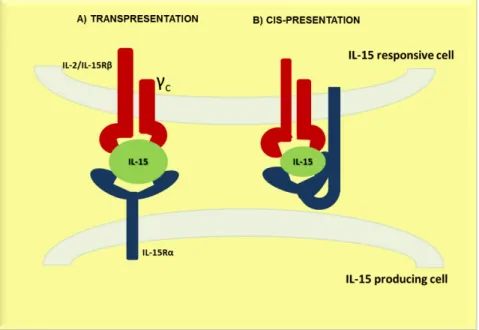
20 CD8 T cells, NK cells, and IELs). However, lymphocytes express some of the highest levels of IL-15Rα, the significance of which is not known (Schluns, K.S. et al., 2004b). Crystal structure of IL-15Rα revealed a threonine/proline-rich region between the trans-membrane and the 15 binding domain, that confers flexibility. This suggests that IL-15Rα can also present IL-15 to βγc in the same cell, in a process called as cis-presentation (Olsen, S.K. et al., 2007). The physiological relevance of this process has not been yet elucidated. Transfection experiments with IL-15Rα in naïve CD8+ T cells showed that the response to IL-15 is enhanced in transfected cells versus non-transfected counterparts (Rowley, J. et al., 2009). Another group reported that IL-15Rα knock-in in T cells had slightly increased levels of homeostatic proliferation (Wu, Z. et al., 2008). Taken together, these observations reveal that cis-presentation can also mediate IL-15-induced responses. In the Fig 1.1 we summarise the main mechanisms for IL-15 delivery to the responsive cells.
Figure 1.1: Principals mechanism for IL-15 delivery. A) Trans-presentation: IL-15Rα and IL-15 are synthetized in the same cell and transported to the cell surface where the cell surface complex can stimulate neighboring cells through the IL-15R βγc. B) Cis-presentation: IL-15 is presented by IL-15Rα on the same cell; these mechanisms may utilize IL-15 derived from autocrine or paracrine sources (Stonier, S.W. and Schluns, K.S., 2010).
21 1.5.5. Function of IL-15 in T cells
IL-15 induces proliferation of naïve CD8+ and memory CD4+ and CD8+ T cells (Kanegane, H. and Tosato, G., 1996; Stoklasek, T.A. et al., 2006). In the presence of IL-15, CD4+ and CD8+ T cells resist the regulatory effects of Tregs (Ben Ahmed, M. et al., 2009). IL-15 also has demonstrated chemotactic activity towards CD4+ and CD8+ T cells (Wilkinson, P.C. and Liew, F.Y., 1995).
The anti-apoptotic effects of IL-15 in lymphocytes are mediated by the upregulation of anti-apoptotic proteins of the Bcl-2 family. Stimulation with IL-15 increases the levels of anti-apoptotic proteins BCL-2, BCL-xL, MCL-1 (Lodolce, J.P. et al., 1998; Becker, T.C. et al., 2002) and downregulates pro-apoptotic proteins BAX, BID, BIM, NOXA and PUMA (Van Belle, T. and Grooten, J., 2005; Huntington, N.D. et al., 2007). IL-15 can also prevent apoptosis by activating NF-kB (Hoontrakoon, R. et al., 2002), and inhibiting caspase-3 and -8 (Bouchard, A. et al., 2004). Long-term survival, proliferation and renewal of memory CD8+ T cells by IL-15 are mediated by the upregulation of Bcl-2 (Lodolce, J.P. et al., 1998; Becker, T.C. et al., 2002). As a result, Il15-/- and Il15ra-/- mice
lack memory CD8+ T cells (Lodolce, J.P. et al., 1998; Kennedy, M.K. et al., 2000).
1.5.6. IL-15 in NK cell biology
Natural killer cells (also known as NK cells, K cells, and killer cells) are a type of lymphocytes that play a major role in elimination of both tumours and viral infected cells. Their functions do not require priming. While the earliest stages of NK cells development is confined to the BM, the later stages of development is a continuous process that occurs in both the BM and peripheral tissues, such as the spleen and liver. In the BM, the common lymphoid progenitor differentiates into NK precursors (NKp), which express IL-15Rβ (Schluns, K.S. et al., 2004a). In mice, transition from NKp to immature NK cells is
22 marked by the expression of NKG2D, NK1.1 and the CD94/NKG2 heterodimeric complex. This is followed by the increased expression of both activating and inhibitory Ly49 receptors (Ly49R) (Kim, S. et al., 2002). Maturation of NK cells is characterized by increased killing capacity and pro-inflammatory cytokine secretion, as well as the expression of CD49b (DX5) (Yajima, T. et al., 2001). These functional attributes become enhanced with the upregulation of CD11b and CD43, which identifies the second stage of maturation (Kim, S. et al., 2002). There are many transcription factors involved in NK cell differentiation and functional maturation, such as Id2, Id3, Ikaros, Runx3, E4bp4, Gata-3, T-bet and Eomesodermin (Boggs, S.S. et al., 1998; Gordon, S.M. et al., 2012).
IL-15 is indispensable for NK cell development. The absence of IL15 or its receptor components results in a decrease of this cell population (DiSanto, J.P. et al., 1995; Lodolce, J.P. et al., 1998; Kennedy, M.K. et al., 2000). IL-15-mediated upregulation of the expression of anti-apoptotic molecules Bcl-2 and Mcl-1 and the downregulation of pro-apoptotic molecules Bim and Noxa also mediate the survival of NK cells (Huntington, N.D. et al., 2007; Nakazato, K. et al., 2007). Activation of NK cells is also dependent on IL-15. Accumulating evidence suggests that trans-presentation of IL-15 is essential for the activation of NK cells. In lymphopenic recipients, adoptively transferred IL-15Rα-deficient NK cells can survive, but this response requires IL-15Rα expression by the recipients (Koka, R. et al., 2003). Interestingly, the lack of IL-15Rα in non-hematopoietic cells does not affect NK cell numbers. However, trans-presentation of IL-15 by hematopoietic cells is more efficient than that by non-hematopoietic cells. Limiting IL-15Rα expression to hematopoietic cells alone is sufficient to generate normal NK cell numbers in the BM, while their numbers are only slightly reduced in the periphery (Schluns, K.S. et al., 2004b).
23 1.5.7. The role of IL-15 in iNKT cells
iNKT cells are an NKT cell subset that expresses an invariant TCR that recognizes galactoceramide and do not require antigen priming for activation. Development of iNKT cells in the thymus and their homeostasis in the periphery are dependent on IL-15 (Kennedy, M.K. et al., 2000; Matsuda, J.L. et al., 2002). Il15ra-/- as well as Il15-/- mice
present similar defects in iNKT cells (Matsuda, J.L. et al., 2002; Matsuda, J.L. et al., 2006). In the thymus, IL-15 enhances the survival of developing iNKT cells (Castillo, E.F. et al., 2009; Chang, C.L. et al., 2011; Gordy, L.E. et al., 2011). Moreover, it has been shown that IL-15 upregulates anti-apoptotic factors, such as Bcl-2, BclxL and Mcl-1 and downregulates proapoptotic factors like Bim (Huntington, N.D. et al., 2007). Conversely, in the periphery, IL-15 regulates both proliferation and survival of iNKT cells. IFN-γ production by iNKT cells following a-galactosylceramide stimulation is deficient in Il15ra
-/-mice suggesting that IL-15 signalling also regulates their activation (Chang, C.L. et al., 2011; Gordy, L.E. et al., 2011). Unlike NK cells, which require IL-15Rα expression on hematopoietic cells, IL-15Ra expressed on non-hematopoietic cells is sufficient for thymic iNKT development (Castillo, E.F. et al., 2010; Chang, C.L. et al., 2011).
In the thymus, selective expression of IL-15Rα in dendritic cells (DCs) had no impact on iNKT development whereas IL-15 trans-presentation by medullary thymic epithelial cells (mTECs) seems to play a major role in the development and functional maturation of iNKT cells (Castillo, E.F. et al., 2010). In the periphery, studies using BM chimeras and transgenic models suggest that IL-15Rα expression is equally important in both hematopoietic and non-hematopoietic cells (Castillo, E.F. et al., 2010). IL-15Rα expression only by hematopoietic cells or DCs lead to a defective thymic iNKT development while differentiation and expansion of hepatic iNKT cells is relative normal, demonstrating the existence of IL-15-mediated extrathymic iNKT cell development (Castillo, E.F. et al., 2010).
24 In the liver, IL-15 is produced by liver resident DCs, macrophages (KCs) and hepatic stellate cells (non-hematopoietic origin). DC-mediated IL-15 trans-presentation generates functionally mature iNKT cells (Castillo, E.F. et al., 2010). As the role of KCs and HSCs cells in IL-15 trans-presentation is less clear, future studies using different IL-15Rα-conditional KO mice are needed to elucidate their role. Overall these studies suggest that IL-15 trans-presentation by the tissue microenvironment is absolutely required for the maintenance of iNKT cells in peripheral tissues.
1.5.8. IL-15 functions in non-immune cells
Mesenchymal stem cells, osteoblasts, adipocytes, endothelial cells and myoblasts express high amounts of IL-15 mRNA (Nilsen, E.M. et al., 1998; Satoh, J. et al., 1998; Silva, W.A., Jr. et al., 2003), suggesting that IL-15 could have some effects on these non-immune cells. For example, fibroblasts are the main source of increased levels of IL-15 in the synovium of rheumatoid arthritis (RA) patients (Miranda-Carus, M.E. et al., 2004). Moreover, apoptosis of synovial fibroblasts as well as TNFα-induced apoptosis in mouse L929 fibroblasts is inhibited by 15 (Yang, L. et al., 2002; Budagian, V. et al., 2005). IL-15 stimulates the formation of osteoclast-like cells in rat bone-marrow cultures in vitro. Also, stimulation with IL-15 induces the expression of calcitonin receptor mRNA in preosteoclasts (Ogata, Y. et al., 1999). In RA patients IL-15 aggravates bone destruction by stimulating excessive bone resorption by osteoclasts (Ogata, Y. et al., 1999; Miranda-Carus, M.E. et al., 2006).
Epithelial cells lines such as human keratinocytes and immortalized HaCaT keratinocytes express both IL-15 and IL-15Rα (Ruckert, R. et al., 2000). IL-15 inhibits apoptosis in keratinocytes (Ruckert, R. et al., 2000; Yano, S. et al., 2003). Additionally, IL-15 induces epithelial cells proliferation through the activation of ERK1/2, PI3K and Akt pathways
25 (Yano, S. et al., 2003). Tubular epithelial cells (TECs) are a rich source of IL-15 that stimulates intratubular CD8+ T cells (Robertson, H. and Kirby, J.A., 2003). Also IL-15 is an autocrine survival factor for TECs, protecting them from apoptosis and inhibiting manifestation of nephrotoxic serum-induced glomerulonephritis (Shinozaki, M. et al., 2002).
Extravasation of activated T cells is stimulated by IL-15 since this cytokine induces hyaluronan expression by endothelial cells. Interaction between cell-surface glycoprotein, CD44, and hyaluronan is important for T cells adhesion and recruitment to the blood vessel wall (Estess, P. et al., 1999). Endothelial cell-derived IL-15 induces transendothelial migration of T cells by activating the binding capacity of LFA-1 integrin, and increases T-cell motility (Oppenheimer-Marks, N. et al., 1998).
Muscles express high levels of IL-15 mRNA (Grabstein, K.H. et al., 1994). IL-15 has anabolic effects in the muscle in vitro, as described for insulin growth factor-1 (IGF-1) (Quinn, L.S. et al., 1995). Overexpression of IL-15 induces skeletal muscle hypertrophy in vitro (Quinn, L.S. et al., 2002). The general reduction of proteolysis induced by IL-15 could be the main mechanism involved in its anabolic effects (Busquets, S. et al., 2005). IL-15 has been also described as a myokine (cytokines which are secreted by muscle cells) with effects on adipose tissue. Muscle-to-fat endocrine axis is responsible for fat body composition and insulin sensitivity (Carbo, N. et al., 2001; Quinn, L.S. et al., 2005). Interesting, IL-15 sensitivity differ in different adipocyte subpopulations and there are also species- and developmental stage- dependent differences. For example in 3T3-L1 preadipocytes, IL-15 inhibits lipid deposition while it has no effect on lipid deposition in fully differentiated 3T3-L1 cells (Quinn, L.S. et al., 2005).
The wide range of expression of IL-15 and its receptor also occurs in the central nervous system. IL-15 mRNA is detected in microglia, astrocytes and neuronal cell lines. Low
26 doses of IL-15 support microglial cell growth, attenuats their nitric oxide production and induce JAK1 phosphorylation (Hanisch, U.K. et al., 1997; Kurowska, M. et al., 2002).
1.5.9. Functional dichotomy between IL-2 and IL-15
IL-15 and IL-2 have similar biologic properties in vitro, consistent with their shared receptor signalling components (IL-2/15Rβγc). Like IL-2, IL-15 stimulates lymphocyte activation and proliferation in vitro (Bamford, R.N. et al., 1994; Armitage, R.J. et al., 1995). This stimulation occurs via both promotion of cell proliferation and protection against apoptosis. Despite the apparent similarities, in vivo experiments with IL-2 and IL-2RαKO mice indicate that IL-2R and IL-15R are not redundant proteins (Lodolce, J.P. et al., 1998). IL-2 and IL-2Rα mice suffer severe lymphadenopathy and autoimmunity suggesting the critical role of IL-2 in the regulation of immune response (Sadlack, B. et al., 1993). IL-2R functions cannot be compensated by IL-15R or by other cytokines of the Ɣc receptors family. Thus, IL-15R likely performs distinct immune functions from IL-2R in vivo. Accordingly, IL15Rα-deficient mice show lymphopenia and innate immune deficiency (Lodolce, J.P. et al., 1998). Moreover, the cellular distribution of the distinct IL-15Rα and 2Rα chains might also contribute to the temporal and spatial distinction between the IL-2 and IL-15 induced activation of the via βγc-dependent signalling pathways (Fehniger, T.A. and Caligiuri, M.A., 2001).
27 1.6. The thesis premises
Obesity is considered as a major problem in this decade. This condition is frequently associated with the increased prevalence of fatty liver disease. Liver diseases can evolve from benign non-alcoholic fatty changes to NASH, and in the long-run to hepatocellular carcinoma (Haslam, D.W. and James, W.P., 2005). Chronic low-grade inflammation is a common feature of obesity and NAFLD. Under obese conditions, inflammatory cells infiltrate the adipose tissues and liver. In addition to activation of resident immune cells, inflammatory cells are also recruited from the circulation. In the normal liver, immune cells are mainly represented by NK, NKT and CD8+ T cells, whereas in fatty liver disease there is an increase in the infiltration and activation of these cells, in addition to the activation of liver-specific macrophages (KCs) and Th1 cells (Li, Z. and Diehl, A.M., 2003; Zhan, Y.T. and An, W., 2010). NASH and NAFLD are extensively related to an imbalance in the cytokines in the liver towards pro-inflammatory microenvironment. KCs, stellate cells and other cells can produce the inflammatory cytokines TNF-α, IL-12 and IL-6 upon activation (Li, Z. et al., 2003). IL-15 is another pro-inflammatory cytokine, which is indispensable for the development and homeostasis of CD8+ T cells, NK, NKT and iNKT cells (Kennedy, M.K. et al., 2000). As these cells are implicated in perpetuation of inflammation in obesity, and the influence of IL-15 on these cells in the liver under conditions of obesity is unclear, I have addressed these issues using IL-15 knockout mice under conditions of diet-induced obesity.
28 1.7. Hypothesis
Based on the above premises, we hypothesize that IL-15 signalling in liver cells promotes the development of NAFLD by inducing hepatic inflammation.
1.8 Objectives
The specific aims of my research project are:
1. To evaluate the role of IL-15 in the development of NAFLD
a. Characterize the induction of NAFLD by HFD in WT and Il15-/- mice using
macroscopic and microscopic analysis of the liver and serum lipids levels. b. Evaluate the expression of genes involved in metabolism in the livers of
WT and Il15-/- after HFD.
c. Elucidate the role of IL-15 in the metabolic behavior of primary murine hepatocytes
2. To characterize HFD-induced inflammation in the liver
a. Evaluate intrahepatic lymphocytes (NK, NKT, iNKT, T cells) and macrophages infiltration after HFD.
b. Evaluate inflammation related genes expression in liver samples and primary hepatocytes.
3. To define the role of IL-15 and IL-15Rα in the maintenance of NK and NKT subsets in the liver
a. Characterize IHLs in WT, Il15-/- and Il15ra-/-
b. Characterize IHLs in macrophages and hepatocyte tissue-specific