Université de Montréal
The Left Atrial Ganglionated Plexus :
Its Function and Pathways Relative to Atrial Fibrillation Surgery par
Emmanuel Moss, MDCM
Département de science biomédicale Faculté de médecine
Mémoire présenté à la Faculté des études supérieures en vue de l’obtention du grade de Maître des sciences (M.Sc.)
en sciences biomédicales
Décembre, 2011 ©Emmanuel Moss, 2011
Université de Montréal
Faculté des études supérieures et postdoctorales
Ce mémoire intitulé:
The Left Atrial Ganglionated Plexus :
Its Function and Pathways Relative to Atrial Fibrillation Surgery
Présenté par : Emmanuel Moss
a été évalué par un jury composé des personnes suivantes :
Stanley Nattel, MD, président-rapporteur Pierre Pagé, MD, directeur de recherche René Cardinal, PhD, codirecteur de recherche
Le système nerveux autonome cardiaque est devenu une cible dans les thérapies ablatives de la fibrillation auriculaire. Nous avons étudié les voies de communication et la fonction des plexus ganglionnaires (PG) de l'oreillette gauche (PGOG) afin de clarifier la validité physiopathologique des méthodes de détection et des thérapies impliquant ces groupes de neuronnes. Méthodes: Vingt-deux chiens ont subi une double thoracotomie et ont été instrumentés avec des plaques auriculaires épidcardiques de multiélectrodes. Une stimulation électrique (2 mA, 15 Hz) des PGOG a été réalisée à l'état basal et successivement après: 1) une décentralisation vagale, 2) l'ablation par radiofréquence des plexus péri-aortiques et de la veine cave supérieure (Ao/VCS) et 3) l'ablation du PG de l'oreillette droite (PGOD). Ces procédures de dénervation ont été réalisées suivant une séquence antérograde (n = 17) ou rétrograde (n = 5). Résultats: Chez 17 des 22 animaux, la stimulation des PGOG a induit une bradycardie sinusale (149 ± 34 bpm vs 136 ± 28 bpm, p < 0.002) et des changements de repolarization (ΔREPOL) auriculaires isointégrales. Dans le groupe des ablations antérogrades, les réponses aux stimulations vagales ont été supprimées suite à la décentralisation vagale chez un seul animal, par l'ablation des plexus Ao/VCS dans 4 cas et par l'ablation du PGOG dans 5 autres animaux. Des changements ont persisté tout au long chez 2 chiens. La valeur de surface des ΔREPOL a diminué avec les dénervations séquentielles, passant de 365 ± 252 mm2 en basale à 53 ± 106 mm2
après l'ablation du PGOD (p < 0.03). Dans le groupe de dénervation rétrograde, les changements de repolarisation et chronotropiques ont été supprimés suite à l'ablation du PGOD chez deux chiens et suite à l'ablation Ao/VCS chez trois. La valeur de surface du ΔREPOL a aussi diminué après l'ablation du PGOD (269±144mm2
vs 124±158mm2
, p<0.05). Conclusion: Les PGOD sont identifiables en préablation par la réponse bradycardique à la stimulation directe dans la plupart des cas. Le PGOD semble former la principale, mais non la seule, voie de communication avec le nœud sinusal. Ces résultats pourraient avoir des implications dans le traitement de la FA par méthodes ablatives.
Mots clés: Fibrillation auriculaire, système nerveux intrinsèque cardiaque, plexus ganglionnaire de l'oreillette gauche, neurocardiologie, système nerveux autonome.
Abstract
The cardiac autonomic nervous system has recently become the target of ablative therapy in the treatment of atrial fibrillation. We investigated the pathways and function of the left atrial ganglionated plexus (LAGP) to clarify the pathophysiologic validity of therapies involving this cluster of neurons. Methods: Twenty-two bilaterally thoracotomized canines were instrumented with atrial epicardial plaques. LAGP stimulation was performed in the basal state and successively following 1) vagal decentralization, 2) radiofrequency ablation of the peri-aortic/superior vena caval (Ao/SVC) plexi, and 3) of the right atrial ganglionated plexus (RAGP). Denervation was carried out in either the aforementioned order (n=17, antegrade) or reversed (n=5, retrograde). Results: In 17 of 22 animals, LAGP stimulation induced a sinus bradycardia (149±34bpm to 136±28bpm, p<0.002) and atrial isointegral repolarization changes (REPOL∆). In the antegrade group, response was suppressed by vagal decentralization (n=1), Ao/SVC plexi ablation (n=4), and RAGP ablation (n=5). Changes persisted throughout in 2 canines. Surface area of REPOL∆ diminished with successive denervation, from 365±252 at baseline to 53±106mm2 following RAGP ablation (p<0.03). With retrograde denervation, chronotropic and repolarisation changes were suppressed following RAGP ablation in two canines, and following Ao/SVC ablation in three. Surface area of REPOL∆ diminished following RAGP ablation as well (269±144mm2 vs 124±158mm2, p<0.05). Conclusion: The LAGP can be identified intraoperatively by the bradycardic response to direct stimulation in most cases. The RAGP appears to be the primary, but not the only, gateway to the sinus node. These results could have important clinical implications relating to ablative treatment of atrial fibrillation.
Key words: Atrial fibrillation, Intrinsic cardiac nervous system, left atrial ganglionated plexus, ablation, neurocardiology, autonomic nervous system, Atrial repolarisation, Isointegral mapping
Table of Contents
Chapter 1. Atrial fibrillation and the Autonomic Nervous System ... 1
1.1 What is atrial fibrillation? ... 1
1.1.1 Classification ... 1
1.2 Clinical Implications ... 2
1.2.1 Morbidity and mortality ... 2
1.2.2 Cost ... 3
1.3 Risk Factors ... 3
1.4 Pathophysiology ... 4
1.4.1 Mechanisms of atrial fibrillation ... 5
1.4.2 Electrical remodelling – AF begets AF ... 6
1.4.3 Histological changes ... 7
1.4.4 Autonomic influences ... 7
1.4.5 Anatomical origins of AF ... 8
1.4.6 Embryologic development of the cardiac conduction system ... 12
1.5 The nervous system and the heart ... 13
1.5.1 Neurocardiology ... 13
1.5.2 The extrinsic cardiac nervous system ... 14
1.5.3 The intrinsic cardiac nervous system ... 16
1.5.4 Anatomy of the intrinsic cardiac nervous system ... 16
1.6 Management of Atrial Fibrillation ... 19
1.6.1 Pharmacologic treatment ... 20
1.6.2 Invasive treatment ... 20
1.6.3 Invasive treatment of AF – Are we there yet? ... 21
1.6.4 The ICNS and control of cardiac functions ... 22
1.6.5 The ICNS as it relates to AF ... 23
1.6.6 Neuromodulation by direct ablation to control atrial arrhythmia. ... 23
1.6.7 Direct neuromodulation: Many unanswered questions ... 25
1.7 Hypothesis ... 26 1.8 Objectives ... 28 Chapter 2. Methods ... 29 2.1 Ethics ... 29 2.2 Experimental protocol ... 29 2.2.1 Anaesthesia ... 29 2.2.2 Surgery ... 30 2.2.3 Neuronal stimulation ... 30 2.2.4 Neuronal ablation ... 31 2.3 Electrocardiographic evaluation ... 32 2.3.1 Activation Mapping ... 32 2.3.2 Repolarization mapping ... 32 2.4 Experimental protocol ... 37 2.5 Data analysis ... 38 2.5.1 Bradycardia ... 38
2.5.2 Isointegral distribution mapping following neuronal stimulation ... 38
2.5.3 Effect of LAGP stimulation atrial repolarisation with sequential ablation .... 40
Chapter 3. Results ... 41
3.1 Bradycardia ... 41
3.1.1 LAGP stimulation, antegrade group ... 41
3.2 Isointegral distribution mapping ... 44
3.2.1 Antegrade ablation sequence ... 44
3.2.2 Retrograde ablation ... 47
Chapter 4. Discussion ... 49
4.1 Bradycardia ... 49
4.2 Right atrial repolarisation changes ... 51
4.3 Bachmann’s bundle ... 53
4.4 Intrinsic cardiac nervous system remodelling ... 54
4.5 LAGP ablation in treatment of atrial fibrillation ... 54
4.6 Limitations ... 57
Chapter 5. Conclusion ... 58
Chapter 6. Bibliography ... i
Table of Figures
Figure 1. Relationship between the various forms of AF. ... 4
Figure 2. Schematic of superficial muscular fibres of the left atrium. ... 10
Figure 3 Proposed model for the cardiac neuronal hierarchy, emphasizing the intrathoracic components.95 ... 14
Figure 4 Schematic representation of the extrinsic cardiac nervous system at the level of the thoracic nerves.109. ... 15
Figure 5. Distribution of ganglionated plexi on the canine heart, superior view. ... 17
Figure 6. Anatomical locations of intrinsic cardiac ganglionated plexi in the canine heart. ... 18
Figure 7. Distribution of ganglionated plexi on the human heart, posterior view.96 . 18 Figure 8. Distribution of the ganglionated plexi on the human heart, superior view.96 ... 19
Figure 9. Ablation lines used in the invasive treatment of atrial fibrillation. ... 21
Figure 10. Schematic representation of atrial epicardial mapping electrode plaques. ... 34
Figure 11. Example of atrial activation map. ... 35
Figure 12. Principles of isointegral mapping.141 ... 35
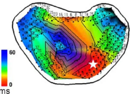
Figure 13. Example of repolarisation map during vagal stimulation. ... 36
Figure 14. Experimental protocol. ... 36
Figure 15. Experimental protocol timeline. ... 37
Figure 16. Designated atrial regions based on electrode plaque location. ... 39
Figure 17. Number of dogs presenting bradycardic response to LAGP stimulation at baseline and following sequential ablations. ... 42
Figure 18. Average change in cycle length with LAGP stimulation following each step of the ablative protocol. ... 42
Figure 19. Average cycle length change per ablation sequence in the antegrade group. ... 43
Figure 20. Average cycle length change in retrograde group, control versus LAGP stimulation ... 43
Figure 22. Surface area of activation along the right atrial free wall with progressive ablation in the antegrade group. ... 45 Figure 23. Cumulative incidence maps of REPOL∆ with antegrade group. ... 46 Figure 24. Isointegral changes in Bachmann’s bundle throughout ablation sequence.
... 47 Figure 25. Surface area of activation along the right atrial free wall with progressive ablation in the retrograde group. ... 48 Figure 26. Summary of LAGP pathways responsible for sinus bradycardia. ... 51
Abbreviations:
AF Atrial Fibrillation
ANS Autonomic nervous system Ao Aorta
AVN Atrioventricular node AVN atrio-‐ventricular BPM Beats per minute CL Cycle length
CNS Central nervous system
ICNS Intrinsic cardiac nervous system LAGP Left atrial ganglionated plexus RAFW Right atrial free wall
RAGP Right atrial ganglionated plexus
RAAS Renin-‐angiotensin-‐aldosterone system REPOL∆ Repolarisation changes
SAN Sinoatrial node SVC Superior Vena Cava
Acknowledgments:
To my wife, Adina, who supports me selflessly and unquestionably in all of my endeavours. Without you the sleepless nights and sometimes endless days would not be possible nor seem worthwhile.
To my daughters, Keira and Eden, having my two big girls to come home to always keeps me motivated.
To Dr Pagé, both in research and throughout my residency, your infinite patience and commitment to training the next generation of cardiac surgeons continually amazes me. You have become a mentor to me and an example of what it is to be a surgeon, a teacher, and a “mensch”.
To my collaborators: Dr Yalin Yin, without whom this work would not have been possible, your experience has been invaluable. Caroline Bouchard, who guided me through my foray into experimental surgery. This would not have been possible without your dedication to this project and the animals you care for.
Nervous System
1.1 What is atrial fibrillation?
Atrial fibrillation (AF) is characterized by disorganized atrial electrical activity leading to rapid, irregular activation of the atrium. This activation can be in the order of 400-600 pulses per minute in a given segment of atrial muscle and results in asynchronous and inefficient atrial contraction.1 It can be recognized on the surface electrocardiogram by the absence of the P wave (atrial wave), which is replaced by low amplitude irregular oscillatory waves, termed fibrillatory waves.2 AF is the most prevalent cardiac arrhythmia in humans, being responsible for 33% of hospitalizations for cardiac rhythm disturbances. The prevalence has best been studied in the American population and is estimated to be 0.4-1% of the general population, with a larger proportion of the elderly being affected.3 This corresponds to 2.2 million Americans and is likely to increase to 5.6 million by the year 2050 as a result of the aging population and increasing prevalence of cardiac disease.4
1.1.1 Classification
There have been several classification systems proposed for AF. One of the most commonly used in clinical practice is based on AF duration. If AF terminates spontaneously it is termed paroxysmal, while AF lasting 7 days or more is termed persistent. If it lasts more than 1 year it is designated permanent AF.5 Dr Jim Cox, a pioneer in the surgical treatment of AF has proposed a simplified classification, with Atrial fibrillation being either paroxysmal or chronic.6 He advocates this nomenclature because it will best separate patients according to the ideal therapeutic intervention. Another classification method is based on the underlying disease, for example, AF can be related to mitral stenosis, mitral regurgitation, or cardiomyopathy.7 AF that occurs in the absence of identifiable concomitant cardiac disease is termed “lone AF”, accounting for 10-30% of AF cases.7, 8 The autonomic
either parasympathetic or sympathetic hyperactivity.9 It is generally recognized that AF occurring in the postoperative period following cardiac surgery falls into a separate category. Postoperative AF occurs following 11% to 40% of coronary artery bypass graft (CABG) surgeries and as many as 50% of cardiac valvular surgeries.10 The onset of this subtype of AF is typically on the second or third postoperative day.11, 12 It is thought to be related to inflammation resulting from surgical manipulation and extracorporeal circulation fluid shifts and endogenous and exogenous catecholamine release.13-15 Although it is often a self limiting problem with 90% of patients returning to sinus rhythm at 6-8 weeks, it is associated a with significantly longer hospital stay as well as higher in-hospital and long term mortality.16-18
1.2 Clinical Implications
1.2.1 Morbidity and mortality
The clinical consequences of AF can be varied, ranging from slight discomfort due to palpitations to more severe complications such as stroke or death.19, 20 This morbidity is related to the three different problems created by AF. Firstly, the chaotic atrial activation leads to an irregular ventricular response resulting in the sensation of palpitations, which can lead to patient discomfort.21 These symptoms occur more frequently in patients with paroxysmal AF, that is, those having a rhythm that fluctuates between sinus rhythm and AF.22 Secondly, stasis of blood in the left atrium can lead to the formation of clot within the heart and predispose a patient to thromboembolic events, resulting in damage to the kidneys, intestine, extremities or brain.23, 24 For example, AF has been associated with a three to five time increase in the risk of stroke and as many as 30% of acute strokes occur in patients with AF.20, 25 This can have severe consequences leading to significant impairment of quality of life or even death.26 Finally, the loss of synchronous atrioventricular contraction can have important physiologic effects. Ventricular filling can be diminished thereby reducing exercise tolerance or leading to decompensated cardiac insufficiency by aggravating pre-existing compensated heart
failure.27 AF with an ill-controlled ventricular response can also result in “tachycardia-induced cardiomyopathy” brought on by prolonged rapid ventricular rates, a condition that is for the most part reversible.28 Overall, AF is associated with
a 1.5 and 1.9 fold increase risk of death in men and women, respectively.29
1.2.2 Cost
Although it is difficult to determine the true financial impact of AF on the health care system, it is clear that it is a costly public health issue. A study from the United Kingdom suggested the direct cost of AF on the health care system in the year 2000 to be over 700 million dollars, accounting for almost 1% of total National Health Service expenditures. This was a 50% increase from the cost five years earlier.30
Another European study estimated that AF costs the healthcare system between 2000$ and 3000$ per patient per year. With the high prevalence and increasing incidence of this disease, this amounts to an enormous burden on the healthcare system.31
1.3 Risk Factors
Although there are many factors associated with the development of AF, advanced age is the most significant independent risk factor. The exact cause for the increased prevalence in the elderly is incompletely understood but it is likely a combination of several phenomena associated with aging tissues, including atrophy of atrial muscle, atrial dilatation, and slowed or abnormal conduction.32 Aside from age, other important risk factors include male sex, hypertension, congestive heart failure, atrial dilatation, and valvular disease.33 Additional risk factors include
coronary artery disease, recent myocardial infarction, hypothyroidism, sleep apnea, diabetes, obesity, metabolic syndrome and the postoperative state.33-37 Many other factors have been identified that predispose a patient to atrial fibrillation following open-heart surgery, these include a history of atrial fibrillation, valvular surgery, and withdrawal from beta-blockers or ACE inhibitors18, 38
1.4 Pathophysiology
The pathophysiology and mechanisms of AF are complex and still incompletely understood. Despite this, beginning with a basic explanation of the fundamentals of the arrhythmia will permit a build-up to a comprehensive understanding of our knowledge to date. The onset of AF depends on the presence of two key elements: a trigger, i.e. an initiating event, and an anatomical substrate enabling AF maintenance1. The initiating event may result from a rapidly firing ectopic focus, multiple functional re-entrant circuits, or both.1 Atrial histological and electrical changes allowing for AF maintenance may be pre-existing or may be induced by the presence of continually firing triggers causing atrial remodelling.
Figure 1. Relationship between the various forms of AF.
The trigger will initiate the re-‐entry phenomenon. If the anatomical or electrophysiological substrate is insufficient to perpetuate re-‐entry, the sequence will terminate with an atrial extrasystole (APC, atrial premature complex). In paroxysmal AF, the arrhythmia will be initiated by a trigger,
however, the substrate is insufficient to maintain AF and a spontaneous cardioversion ensues. In persistent AF the atrial substrate is sufficient to maintain a fibrillatory state but the arrhythmia can still be terminated with a therapeutic intervention. Permanent AF is present when severity of atrial remodelling makes it no longer possible to return to sinus rhythm. CV= Cardioversion39
1.4.1 Mechanisms of atrial fibrillation
In the early twentieth century three competing theories regarding the mechanisms of AF were suggested. They postulated that AF is derived from 1) rapidly discharging atrial ectopic foci, 2) a single entry circuit, or 3) multiple re-entry circuits.40 These theories have been both disputed and modified over the years
and have helped shape our current understanding of this complex arrhythmia. Whatever the exact mechanism, it appears that AF begins with abnormal impulses arising from atrial tissue. While normal automaticity results from the spontaneously depolarizing sinus node cell reaching threshold potential, ectopy can result either from other cells depolarizing at an abnormally rapid rate, or from afterdepolarizations reaching threshold and causing premature action potentials. Conversely, re-entry can occur as a result of a premature impulse propagating between two zones of tissue with different refractory periods.1 In order for AF to be initiated, a premature activation must occur on the border between these two zones creating a unidirectional block when the impulse contacts one of the zones during its refractory period. Once the refractory period has terminated, the atrial tissue can then be re-excited and allow for propagation of the pathologic impulse. Once initiated, this circuit can continue indefinitely. With increased atrial tissue heterogeneity, the likelihood of unidirectional blocks occurring augments and the risk of chaotic activation leading to AF is amplified.41-43
These phenomena are the result of alterations in atrial ionic currents. Changes in the balance between the various potassium, sodium and calcium currents lead to varied action potential duration and a proarrhythmic state. As an example, in response to calcium overload, atrial cells will decrease the concentration of certain membrane bound calcium channels in order to preserve cell viability.44 However,
decreased membrane concentration will decrease the action potential duration, which in turn reduces the refractory period and promotes the maintenance of AF by multiple circuit re-entry.45, 46
There are two competing models regarding the mechanism of re-entry. Allessie et al proposed the “leading circle hypothesis” which relies on the theory that there is a small circuit established with the smallest diameter possible that will maintain continuous activity.41 This minimum circuit size is defined as the product of the refractory period and the conduction velocity. The core of the circle is continuously excited by impulses originating from the outer circle. Pertsov et al suggested the “spiral wave theory” in 1993.47 This model depends on a fully excitable core with the maintenance of spiral wave re-entry dependant on the curvature of the wavefronts at the tip of the spiral. Each of these theories come with inherent limitations, highlighting the fact that although we have come a long way in understanding this complex arrhythmia, there is still work to be done.
1.4.2 Electrical remodelling – AF begets AF
The idea of tachycardia induced electrical remodelling was first suggested by two independent animal studies performed in the mid 1990’s. In a dog model of chronic rapid atrial pacing, Morillo et al demonstrated a shortening of the atrial refractory period and more readily inducible sustained AF.48 Wijffels et al developed a chronic goat model of sustained AF by attaching implanted electrodes to a fibrillation pacemaker that automatically delivered bursts of stimuli whenever sinus rhythm occurred.49 By artificially maintaining AF, they showed even more marked refractory period shortening as well as increased inducibility and stability of AF. These observations led to the concept of “Atrial Fibrillation Begets Atrial Fibrillation”, that tachycardia induced remodelling creates a substrate for persistent AF.49 Although the results of these experiments opened a new avenue for researching and understanding AF, they failed to fully explain the phenomenon being witnessed. The changes of atrial refractory period demonstrated by Wijffels et al were near maximal after 24 hours, whereas the duration of AF continued to increase for 2 weeks.49 In the years since the publication of these seminal papers
additional factors explaining tachycardia induced remodelling, including conduction velocity, wavelength and regional heterogeneity, have been proposed.46 The basis of
these changes have since been linked to ionic mechanisms, some of which are described above, most important of which is a reduction in L-type calcium channels.50, 51
1.4.3 Histological changes
The alterations in atrial tissue most frequently seen as consequences of AF are atrial fibrosis and loss of atrial muscle mass. It is important to mention that before anatomical changes occur, electrical remodelling is brought on by the initial AF stimulus. If AF is allowed to continue, anatomical remodelling will occur subsequently. Atrial fibrosis favours the development of re-entry circuits by creating physical barriers within the atria impeding the propagation of electrical activity. This causes a heterogeneous slowing of atrial conduction, as is the case in heart failure.52 The renin-angiotensin-aldosterone system (RAAS) plays an important role in the development of atrial fibrosis. The RAAS is activated in response to atrial dilatation, with an increased expression of angiotensin converting enzyme within the atrial tissue. Angiotensin II and aldosterone facilitate the proliferation of fibroblasts and the deposition of proteins within the extracellular matrix, thus altering atrial structure.53 Angiotensin receptor blockers and angiotensin converting enzyme inhibitors, medications frequently used for the control of blood pressure and in the treatment of heart failure, have been shown to delay or reduce the development of fibrosis and AF.54, 55
1.4.4 Autonomic influences
The development of AF can be secondary to changes in cardiac nervous tone as well. Coumel was one of the first to suggest that AF could result from increase in signalling of either of the components of the autonomic nervous system (ANS), i.e. sympathetic or parasympathetic.56 He noted that when patients are asleep they can have AF episodes preceded by a sinus bradycardia, suggesting a vagal origin.57 Conversely, it has been shown that AF can be associated with increased sympathetic
tone in the presence of intense emotional or physical stressors.58 Coumel suggests that sympathetic AF occurs in patients with concomitant cardiac disease, while vagal AF in seen primarily in the absence of structural heart disease and generally yields paroxysmal AF.58 The role of the ANS in AF is evidenced by the RR interval variation, corresponding to the distance between the two R waves (positive deflection of the QRS complex of ventricular depolarisation) on the electrocardiogram. This parameter has been shown to reliably identify sympathetic and parasympathetic influences on the heart, and variations recorded immediately prior to the onset of AF have made it possible to link these episodes to autonomic nervous system activity.59, 60 The specific and relative contributions of each arm of the ANS remains to be determined, but the evidence is increasingly pointing to a synergistic effect between the sympathetic and parasympathetic systems.61, 62 Changes in nervous tone in localized areas of the atrium create various zones with heterogeneous refractory periods. The result is zones of functional block favouring re-entry and, consequently, AF.58 Hirose et al demonstrated that direct stimulation of the vagus nerves increases the heterogeneity of atrial refractory periods, thus facilitating induction of AF.63 In a series of canine preparations, Jayachandran et al demonstrated that rapid AF, induced by rapid atrial pacing, results in a heterogeneous increase in atrial sympathetic innervation, with greatest heterogeneity in the right atrium.64 Since the establishment of the role of the ANS in the pathophysiology of AF, further research has illuminated the importance of several anatomical sites as they relate to AF origin.
1.4.5 Anatomical origins of AF
The pulmonary veins
Various atrial regions have been reputed to play important roles in the initiation and maintenance of AF. The base of the pulmonary veins has emerged as a preferential site of micro and macro re-entry.65 Haissaguerre was the first to identify this area as an important source of AF. While performing electrophysiological studies on a series of patients with drug refractory AF, he noted multiple ectopic atrial beats originating at the base of the pulmonary veins.66 He demonstrated that
the origin of AF was found within a muscular band at the junction of the left atrium and the pulmonary veins.67 The relatively chaotic organization of the area, including
the orientation, sheathing, and ending of the muscular fibres, tends to favour the creation of local re-entrant circuits.67-69 The embryologic origins of the pulmonary veins partially explain the aforementioned “chaotic” arrangement of these fibres. The base of the pulmonary veins represent the junction between the trunk of the pulmonary venous system, derived from the embryonic anterior intestine, and the left atrium, derived the primitive cardiac tube. The ionic characteristics of myocytes in this area also favour re-entry as a result of shorter refractory periods.70, 71
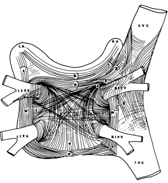
Figure 2. Schematic of superficial muscular fibres of the left atrium.
A main circular fascicle (a, a’, a”, and a”’) runs peripherally around the area of the openings of the pulmonary veins, while an interatrial fascicle (b) runs between the right (RA) and the left (LA) atrium. Some fibres (c) descend from the left atrium into the left part (a’) of the main circular fascicle. Circular fibres leaving the main fascicle turn around the openings of the pulmonary veins, forming sphincter-‐like structures; other fibres extend over the veins as myocardial sleeves. Loops of fibres coming from the atrium are seen over the right superior pulmonary vein (R.S.P.V.) and returning to the atrium. Oblique, vertical (e), and transverse (f, f’) fascicles of fibres are also seen on the
posterior atrial surface. L.A. = left atrium; R.A. = right atrium; S.V.C. = superior vena cava; I.V.C. = inferior vena cava; R.S.P.V. = right superior pulmonary vein; L.S.P.V. = left superior pulmonary vein; L.I.P.V. = left inferior pulmonary vein. (Reproduced from Nathan et al, Circulation 34(3):412-‐422, 1966).
Other sites of origin
There are several sites other than the pulmonary veins that have been identified as foci of AF initiation. In general, the junction between the atria and all major thoracic veins constitute possible sites of re-entry.72 Although the left atrium is generally considered to be the more common site of AF initiation, the right atrium is also a potential source. Examples of the right atrium’s arrhythmogenic potential have been seen following surgical or percutaneous interventions targeting only the pulmonary veins. Firstly, the onset of left atrial flutter can seen, possibly a result of incomplete ablation lines within the left atrium. Secondly, the elimination of left atrial AF mechanisms may allow for the emergence of right atrial sites that were otherwise overshadowed by their left atrial counterparts.73 Forleo et al demonstrated that complex fractionated atrial electrograms were present in the right atrium and coronary sinus during pulmonary vein isolation procedures.74 Even more interestingly, the authors were able to link these electrograms to an increased risk of AF recurrence. While there has previously been controversy regarding the right atrium’s ability to initiate AF in the absence of left atrial sources, there is an increasing amount of evidence supporting this theory. When performing electrophysiological studies in 172 patients with AF, Chen et al identified sites of focal AF within the right atrium of 8 patients.75 These sites were the crista terminalis (the right atrial free wall) and at the ostium of the coronary sinus. Similarly, Tsai et al demonstrated loci of ectopic beats at the SVC-right atrial junction that appeared to be sites of AF origin.76 In both of these studies the authors were able to eliminate AF
by radiofrequency ablation of these areas.
Interatrial conduction pathways comprise another group of atrial tissue with a tendency to initiate AF. Although any atrial tissue has the ability to propagate an electrical signal, conduction tends to occur via certain preferential tracts. The importance of Bachmann’s bundle, also known as the interatrial band, as it relates to interatrial conduction was first described in 1916 by George Bachmann.77 It is located at the upper extremity of the interatrial septum and the results of several studies have suggested its role in AF pathophysiology.78-80 Other atrial structures studied in this context include the coronary sinus and fossa ovalis.81 Finally, another
area of the atria that has been implicated in several studies as a site of AF origin is the ligament of Marshall. The ligament of Marshall is an atrial fold located adjacent to the coronary sinus in the inferior left atrium that is a remnant of the left horn of the sinus venosus in the foetus. It contains nerve fibres, muscle bundles, and the oblique auricular vein, the vein of Marshall.82 The Marshall bundle is considered the most important interatrial conduction pathway in the inferior portion of the atria and is likely the site of AF origin in certain patients.83, 84 Concordant with this assertion are the findings by Haissaguerre et al that the epicardial and endocardial regions bordering the coronary sinus are important in the maintenance of AF.85, 86 Considering the above, it is clear that the sites of origin of AF are closely related to the anatomy and development of cardiac conduction tissue; consequently, a brief discussion of the embryologic development of cardiac conduction tissue will follow.
1.4.6 Embryologic development of the cardiac conduction
system
Although the development of the cardiac conduction system remains controversial, one of the most accepted theories explaining the sequence of depolarisation of myocardial tissue is that of the integration of the neural crest during fetal development.87, 88 Neural crest cells are said to participate in the formation of the four cardiac chambers and in the development of the cardiac conduction system.88, 89 The ectomesenchymal neural crest cells migrate to the heart by routes along arterial and venous structures. The cells on the arterial side develop into the semilunar valves, while those on the venous side become integrated into the myocardium to form the atrio-ventricular node (AVN), the bundle of His, and the ventricular conduction branches.90, 91 In addition, other authors have demonstrated a high concentration of cells surrounding the pulmonary veins.92 In addition to its role in the morphogenesis of the cardiac chambers and the conduction system, the neural crest is implicated in the development of the intrinsic cardiac innervation.93 This link between the nervous system and the heart is of utmost importance both in normal and pathologic cardiac function. There are a multitude of nervous fibres and ganglionated plexi on the surface of the heart that are important for the beat-to-beat
functioning of the heart.94, 95 These autonomic nervous ganglia are preferentially distributed within epicardial adipose tissue at the base of the heart between the atria and ventricles, as well as around the great vessels. They have the ability to modify cardiac chronotropy, dromotropy, inotropy, lusitropy and bathmotropy.96 The details of the cardiac autonomic innervation are in constant evolution but a review of our understanding to date is necessary in order to appreciate the findings of this work.
1.5 The nervous system and the heart
1.5.1 Neurocardiology
The ANS is responsible for maintaining homeostasis in the body. Classically, cardiac nervous control is said to result from a delicate balance between the two branches of the ANS, sympathetic and parasympathetic. Sympathetic influence is said to stimulate the heart, resulting in increased heart rate and force of contraction. While the parasympathetic, or vagal, influence has the opposite effect.97
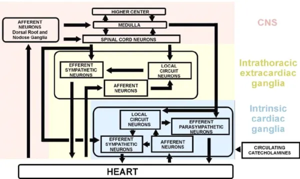
The “classic” view of cardiac nervous control entailed the assumption of the heart’s passive response to postganglionic sympathetic and parasympathetic signals, originating in the central nervous system (CNS).98 However, although this concept of neuroanatomic organization held for over a century, it is now known that this not the entire picture. There exists a complex hierarchy of nervous structures that interact to influence cardiovascular function. The nervous structures that are implicated in this process are located at different levels of the body, notably in the brain (CNS), thoracic cage (extrinsic cardiac nervous system), and on the heart (intrinsic cardiac nervous system). This hierarchy is composed of many levels of feedback control loops that includes parasympathetic and sympathetic efferent and afferent neurons, intraganglionic neurons, and interganglionic neurons (Figure 3).99 The notion of the heart’s capacity for local integration and processing of neuronal signals was first put forth in the early 1970’s by Armour and Ardell.100, 101 102-104 It allows for the local integration and control of the many cardiac parameters that can be affected by the autonomic nervous system.104-106
Figure 3 Proposed model for the cardiac neuronal hierarchy, emphasizing the
intrathoracic components.95
1.5.2 The extrinsic cardiac nervous system
The thoracic cage is rich with both efferent (toward the heart) and afferent (toward the brain) nervous structures that are active in coordinating the nervous influences of the heart. This is the substrate of the extrinsic cardiac nervous system, with the efferent structures acting on the heart consisting of those descending from the CNS as well as the intrathoracic extracardiac ganglia. The coordination and local processing of these neuronal signals is mediated by local circuit neurons located within the intrathoracic ganglia.105
Transmission of information occurs between the CNS and the end-organ via a connection of two neurons in series, termed preganglionic and postganglionic. The preganglionic neurons develop from the neural tube and postganglionic from the neural crest. Preganglionic sympathetic neurons synapse in the paravertebral ganglionic chains. The principal sympathetic efferent ganglia innervating the heart are the stellate and median cervical ganglia. Parasympathetic innervation of the heart stems primarily from the nucleus ambiguus of the brain stem and is relayed to the heart through the vagus nerve, also called the vagosympathetic complex, with the
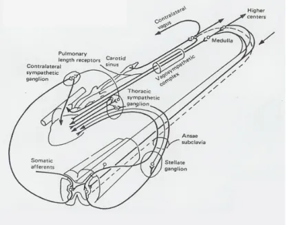
pre-post ganglionic synapse occurring at the level of the heart itself (Figure 4).107-109 The axons exiting the nucleus ambiguus are composed of large myelinated fibres allowing for rapid transmission of neural signals with short latency between activation and the end organ effect of vagal bradycardia.110 The principal parasympathetic afferent pathway, i.e. neuronal pathways leading away from the heart and toward the CNS, is the nodose ganglion, also known as the inferior ganglion of the vagus nerve. It has a cylindrical form and runs along the anterior aspect of the internal jugular vein. Cardiac sympathetic afferents reach the CNS via the dorsal root ganglia running along the spinal cord.111
Figure 4 Schematic representation of the extrinsic cardiac nervous system at
the level of the thoracic nerves.109.
The local circuit neurons mentioned previously are a sub-population of neurons contained within the autonomic ganglia of both the intrathoracic extracardiac and intrinsic cardiac systems. They have projections to adjacent neurons within their own ganglion, or in other regional ganglia, allowing for a “functional interconnectivity” within the intrathoracic nervous system.104, 112 The
result is a capacity to influence cardiac performance in a manner that would not be possible with simple neuronal connections in series.
1.5.3 The intrinsic cardiac nervous system
The intrinsic cardiac nervous system (ICNS) is a complex network of neuronal fibres where sympathetic and parasympathetic nerves do more than simply deposit their signal. It is an extra level of neuronal control located directly on the heart’s surface, otherwise known as the “little brain on the heart.”95, 96, 105 Neurons are located in the autonomic ganglionated plexi, which are clustered in discrete areas of the heart, primarily in the fat tissue on the epicardial surface of the heart. Four types of neurons can be found within these ganglia: efferent sympathetic and parasympathetic neurons, interneurons contained entirely within individual ganglion, local circuit neurons linking intracardiac ganglia, and sensory neurons mediating intracardiac reflexes.113 The anatomical details of the atrial ganglionated plexi will be discussed below.
1.5.4 Anatomy of the intrinsic cardiac nervous system
The most complete account of the anatomy of ICNS was first described for the canine heart by Yuan et al in 1994.114(Figure 5) Cardinal et al recently published
a reproduction of Yuan’s classification system.115(Figure 6) Armour et al. used this
work as a basis for their description of the human ICNS in 1997.96 They analysed the hearts of 18 subjects and elaborated on the gross and microscopic anatomical details. They were able to consistently identify ganglionated plexi in five atrial and five ventricular locations (Figure 7)(Figure 8). Each ganglion contained anywhere from one to over 200 neurons. It is important to note that the precise location and size of each ganglion varied somewhat between specimens. They also noted connections between ganglia within each region forming “regional neural networks.” The atrial ganglionated plexi as detailed by Yuan et al114 are described below:
1) The right atrial ganglionated plexus (RAGP) is located in the adipose tissue just cephalad to the right atrial-IVC junction and just ventral to the interatrial groove. It has been shown that autonomic nervous signals preferentially, although not exclusively, pass through this plexus en route to the sinus node.116
2) The left atrial ventral ganglionated plexus is located on the ventral surface of the left atrium, it is comprised of three components: cranial, intermediate, and caudal. These were originally described in detail by Armour et al in 1990.106
3) The dorsal atrial ganglionated plexus is situated on the dorsal surface of the heart between the two atria.
4) The inferior vena cava-inferior left atrium (IVC-ILA) ganglionated plexus is located on the inferior dorsal aspect of both atria, adjacent to the entrance of the IVC into the right atrium. This plexus has been linked to the autonomic control of the AVN.117
Figure 5. Distribution of ganglionated plexi on the canine heart, superior view. GP = ganglionated plexus, PA = pulmonary artery, IVC = inferior vena cava, SVC = superior vena cava. 114
Figure 6. Anatomical locations of intrinsic cardiac ganglionated plexi in the canine heart.
A: ventral, B: left lateral, C: dorsal views of the heart. RAGP = right atrial
ganglionated plexus (GP), RVGP = right ventricular GP, CMVGP = cranial medial ventricular GP, VSVGP= ventral septal ventricular GP, LAGP = left atrial GP, DAGP = dorsal atrial GP, IVC-‐ILA = inferior vena cava-‐inferior left atrial GP. VLCN=ventrolateral cardiac nerve, LPA=left pulmonary artery, Ao=aorta, IVC, SVC=inferior, superior vena cava. 115
Figure 7. Distribution of ganglionated plexi on the human heart, posterior view.96
Figure 8. Distribution of the ganglionated plexi on the human heart, superior view.96
These ganglionated plexi were initially thought to be comprised of purely parasympathetic postganglionic efferents 117-119, without intrinsic neuronal activity and without accompanying sympathetic neurons. Both of these assumptions have since been refuted by demonstrating spontaneous activity of sympathetic and parasympathetic neurons within each plexus.61
1.6 Management of Atrial Fibrillation
The treatment of AF generally involves any or all of 3 objectives – the control of ventricular rate, prevention of thromboembolism, and restoration of sinus rhythm. The goals of treatment are tailored to each patient based on symptoms and comorbid conditions. Subclassification according to therapeutic modality can aid in the explanation of AF treatment options.
1.6.1 Pharmacologic treatment
Pharmacologic therapy is generally accepted as the first line of treatment in AF5. A variety of medications exist that are used to convert AF to sinus rhythm, control ventricular response, or both.26, 120, 121 Accompanying their many benefits, these drugs can have significant side effects, including paradoxical proarrhythmic effects causing malignant arrhythmias or atrioventricular blockade as well as hypotension or exercise intolerance.122, 123 Additionally, depending on the time to successful conversion to sinus rhythm, the risk of thromboembolism persists while on these medications. Anticoagulation therapy, along with its own substantial complication risks, must be maintained.5, 124-126
1.6.2 Invasive treatment
The suboptimal pharmacologic treatment options listed above have led to much effort focused on developing invasive techniques to control AF over the past two decades.127-130 The objectives of these procedures are both to isolate arrhythmic foci and eliminate macro re-entry circuits. The maze procedure, described by Dr Jim Cox, is now considered the most effective treatment of AF.129 The original
procedure involved fragmentation of atrial tissue by the “cut and sew” technique. This entailed cutting apart the atrium at specific sites and suturing it back together in order to limit electrical continuity between certain areas. The resultant scar tissue barriers limit, in theory, macro re-entry circuits within the atria.
Figure 9. Ablation lines used in the invasive treatment of atrial fibrillation. A – Left sided Maze procedure. B – Pulmonary vein isolation.131
More recently, the less invasive and faster procedures using radiofrequency, cryotherapy, and microwave technology, attempt to reproduce the full thickness tissue scarring of the original Maze procedure.(Figure 9)132-135 In addition,
percutaneous techniques have attempted to reproduce the results seen with surgery either by mapping the atria and directly ablating complexes that may be precursors to AF, or by targeting and isolating the trial tissue giving rise to AF.136
1.6.3 Invasive treatment of AF – Are we there yet?
Although the number of AF procedures has been rising consistently, there are still questions to be answered about their effectiveness. Many reports boast excellent
success rates, however the follow-up in some has been called into question. For example, periodic electrocardiograms and telephone interviews regarding symptoms may not present an accurate picture of AF recurrence. This weakness is highlighted by a study from Hindricks et al that demonstrated patients who were very symptomatic from their AF before a percutaneous intervention continued to have asymptomatic AF episodes following the procedure.137 These episodes were found only with continuous electrocardiographic monitoring for 7 days using a portable monitoring device.
Regardless of how one might feel about the effectiveness of any given treatment, be it pharmacologic, percutaneous, or surgical, it is clear that the ideal treatment of atrial fibrillation is yet to be proven. The ideal treatment would be minimally invasive, very effective, and eliminate the negative consequences of AF, most notably symptoms and the need for anticoagulation. Pharmacologic treatment is incapable of returning many patients to sinus rhythm and when it does it is often at a price of significant complications, including malignant arrhythmias, severe side effects, and bleeding.138 Percutaneous and surgical treatment have improved
dramatically with few complications, however success rates are variable and reporting of results are inconsistent, as mentioned above. As a result of the abovementioned shortcomings, research in this area has become very active in recent years. One of the most active areas of study is the intrinsic cardiac nervous system and it’s associated autonomic ganglia.
1.6.4 The ICNS and control of cardiac functions
As discussed earlier, the ICNS is both the intermediary between the ANS and the heart, as well as an independent entity that contributes to beat-to-beat variations of cardiac activity.104 Intrathoracic nervous elements respond to intrinsic stimuli as well
as extrinsic electrical stimulation, thus raising the possibility of influencing the electrophysiological and dynamic properties of the heart.115 Furthermore, Chiou et al
demonstrated that selective denervation was possible when radiofrequency ablation was applied to epicardial ganglionated plexi. They were able to modify the atrial response to vagal stimulation by ablating three areas of cardiac ganglionated plexi
without affecting the ventricular response to vagal stimulation.139. Other studies using dog models have clearly demonstrated that electrical or chemical stimulation of various nervous elements can modify cardiac activity.95, 102, 105, 106, 115, 140, 141
Furthermore, it has been shown that there is considerable redundancy in the cardiac innervation, revealing the complexity of the cardiac nervous system.142 The idea of modifying cardiac arrhythmias by stimulation and ablation of atrial tissues has been exploited in percutaneous and surgical AF interventions.143-146 In summary, the ICNS is a potential therapeutic target for modulation of cardiac activity.
1.6.5 The ICNS as it relates to AF
As discussed earlier, the pathophysiology of AF involves structural, electrical, and neurophysiologic elements. The majority of pharmacologic and invasive therapies for AF target the former two elements. The importance of neurophysiology, particularly the ICNS, in AF pathogenesis and control is becoming more and more evident. In an animal model, Scherlag et al were able to induce AF by simulating ectopic firing from the pulmonary veins only if the adjacent ganglionated plexi were stimulated simultaneously.147 Huang et al induced
ventricular arrhythmias with stimulation of intrinsic cardiac neurons in a dog model.148 Several other experimental models have implicated ganglionated plexi and the ICNS in the initiation of both atrial and ventricular arrhythmias.149, 150 These findings have encouraged researchers and clinicians to target the ICNS in novel therapies to control or eliminate AF.
1.6.6 Neuromodulation by direct ablation to control atrial
arrhythmia.
There have been an overwhelming number of reports in recent years describing results following invasive treatment of AF. Many authors have suggested that much success is due to the phenomenon of neuromodulation.82, 136 Pappone et al reported on 297 patients who had underwent circumferential pulmonary vein ablation, with 34% of these patients having a bradycardic response to stimulation. This subgroup of patients seemed to have a better response to ablation with 99%
freedom from AF at 12 months compared to 85% in the group not presenting a bradycardic response to stimulation. 151 Mehall et al reported a minimally invasive
approach for atrial fibrillation treatment which included localization of epicardic ganglionated plexi by electrical stimulation followed by radiofrequency ablation.152 Like Pappone, the authors presumed that by localising and ablating ganglionated plexi they had eliminated additional arrhythmogenic foci. They propose this to be a form of vagal or autonomic denervation and suggest that these should become a routine addition to ablative treatment of AF.
Pappone’s observations led to increased interest in exploring AF treatment by direct neuromodulation through denervation. Scherlag et al described a canine model in which ganglionated plexus stimulation was thought to enable AF induction when performed concomitantly with pulmonary vein stimulation.153 Other authors have since reported similar experimental results, however, the causal link between ganglionated plexi and AF has yet to be established.154 Schauerte showed that direct stimulation of atrial ganglionated plexi led to a shortening of the atrial refractory period that facilitated AF.155 They also showed that ablation of those plexi reduced
the inducibility of AF through vagus nerve stimulation. Observations of this nature gave researchers the physiologic basis to modulate atrial arrhythmias through focal epicardial ablation. Other authors have reported conflicting results. Hirose, among others, found that partial denervation of the right atrium actually increased AF susceptibility.63, 156, 157 These observations potentially demonstrate not only the relationship between the autonomic nervous system and cardiac arrhythmias, but also the possibility to influence atrial arrhythmias through neuromodulation. The fact remains that, despite much research, the hypothesis of selective autonomic denervation to treat AF has not yet been proven. All studies relating AF to the ANS have been through indirect evaluation, relying mostly on heart rate variability to implicate ganglionated plexi in experimental or clinical result.
1.6.7 Direct neuromodulation: Many unanswered questions
An ill-defined target
Although there is some clinical evidence supporting the use of cardiac denervation in the treatment of AF, the technique has significant hurdles to overcome. Work by Tan et al suggests that the idea of selective neuromodulation employed during endovascular and surgical procedures is at best optimistic, since each ganglionic bundle contains both sympathetic and parasympathetic neurons.61 This theoretical problem can be reconciled by considering data from Patterson et al who demonstrated in experiments with isolated canine pulmonary veins that concomitant simulation of parasympathetic and sympathetic neurons may be more effective in induction of atrial ectopy. This suggests that it may in fact be favourable to target both types of autonomic influence together. In a human study, Cummings et al found that resection of the anterior aortic fat pad actually increased the incidence of postoperative AF.156 Finally, although it may be clear that autonomic nervous
elements are in some way implicated in AF, it is still unclear what these elements are, where they can be found, at what level of the ANS they must be targeted, intrathoracic versus the ICNS for example, and by what method they should be approached.
Durability of denervation?
The uncertainties of denervation procedures go beyond selecting the target. Even if the arrhythmogenic ganglia were clearly identified, there is significant evidence suggesting that effects of denervation on cardiac tissue are only transient.158, 159 In a canine model, Murphy et al found evidence of nerves crossing suture lines one year following cardiac transplantation.160 Evidence of sympathetic re-innervation has been described in the clinical transplant literature as well, with growth that occurs even up to 15 years following transplantation.161, 162 Even Pappone et al, in their seminal paper on pulmonary vein denervation, remarked that heart rate changes associated with vagal stimulation returned to baseline levels at 6 months, a sign of autonomic re-innervation.151 In an experimental model, Oh et al demonstrated that autonomic nervous changes induced by epicardial ganglionated
plexus ablation reverted to normal after 6 weeks.158 Therefore, it would seem that cardiac re-innervation might limit the durability of direct neuromodulation.
A third issue that may limit the applicability of direct neuromodulation in AF treatment is the paradoxical reaction that occurs following intervention termed “nerve sprouting”.156 Okuyama et al showed that radiofrequency ablation in a dog model produced a hyperinnervation of cardiac tissue rather than denervation.163 Chen et al showed that nerve sprouting following myocardial injury results in an electrophysiological heterogeneity that can increase the incidence of ventricular tachyarrhythmia.164
In summary, while direct neuromodulation may play a role in the treatment of AF, the ideal method has not yet been described. Furthermore, although some have reported clinical success using the bradycardic response as a road map for ganglionated plexus ablation, the neurophysiologic and anatomical basis for their success, or failure in some cases, is certainly not clear. Although advances in science, and medicine in particular, are often stumbled upon by chance, it is of utmost importance to explore the fundamentals of these advances in order to better understand why or how it has occurred and to help guide clinical practice and future development.
1.7 Hypothesis
As described above, the intrinsic cardiac nervous system is comprised of several ganglionated plexi located in areas of adipose tissue dispersed over the epicardial surface of the mammalian heart. According to Yuan et al’s classification, there exist 4 atrial ganglionated plexi on the canine heart.114 (Figure 6) It has been well established that atrial ganglionated plexi assert some control over various cardiac functions including, among other things, chronotropy. However, the anatomic pathways followed by these ganglionic nerve fibres have not been explored or characterized in any detail. Despite this, there are many authors who, during ablative interventions for AF perform electrical stimulation of these areas, and upon witnessing a bradycardic response, proceed to ablation of the area (See
sections 1.6.6 and 1.6.7). Although some publications report positive outcomes, the results are far from conclusive, leaving unresolved questions regarding the reproducibility of this technique and the overall arrhythmogenicity of the LAGP neurons. Understanding these pathways may play an important role in determining the ideal interventions, if any, targeting the intrinsic cardiac nervous system in the treatment of atrial fibrillation.
When considering the anatomic locations of each of the 4 atrial ganglionated plexi as they relate to the bradycardic response seen with epicardial stimulation, some obvious questions arise. The right atrial ganglionated plexus (RAGP) is in close proximity to the sinus node, making it easy to understand the short distance nervous structures must travel in order to arrive at the sinus node and effect changes in cardiac chronotropy. The left atrial ganglionated plexus (LAGP), however, is located lateral to the left pulmonary veins and anatomically distant from the sinus node. The physioanatomic pathways of the bradycardic response seen with stimulation of the LAGP are appreciably more difficult to comprehend.
There exist three possible neuroanatomic pathways that could explain the mechanism of the sinus bradycardia witnessed with stimulation of the LAGP. 1) A central reflex arc via the vagus nerve that travels from the LAGP, to the central nervous system and then back down to the right sided heart via the opposite vagus nerve. The bradycardic effect of the vagus nerve is beyond question and the neural and arrhythmogenic consequences of vagal stimulation have also been well established.141 2) A pathway through the pericardiac plexi, namely the periaortic or
superior vena caval plexi. Mediastinal neuronal projections have been found travelling along the superior vena cava and have been associated with arrhythmia induction.165 The peri-aortic plexi have been less thoroughly studied but may also play some role in arrhythmia formation96, 156, 165. 3) A direct intracardiac pathway that has not yet been described may play a role in LAGP-sinus node interactions. Finally, each of the above pathways may contribute in some capacity.
In addition, although we presume that the LAGP is responsible for the electrophysiological changes seen with direct epicardial ganglia stimulation, the
regional distribution of the response has not yet been characterized. It remains unknown whether neuronal activation occurs locally, across the entire atrium, or remotely from the stimulation site.
We hypothesise that the LAGP communicates with the sinus node via a central pathway along the vagus nerves and that repolarization changes occur in both the right and left atrium with LAGP stimulation.
1.8 Objectives
The objective of this work was to delineate the mechanism of the bradycardia induced by stimulation of the LAGP. To accomplish this, in addition to identifying the pathways responsible for the bradycardic response, we aimed to map the pattern of atrial repolarization changes following LAGP stimulation.
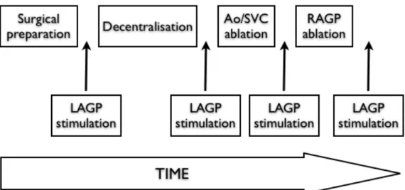
Twenty-two adult mongrel dogs of either sex, weighing 25-35kg, were studied during this experimental protocol. LAGP stimulation was performed at baseline and then following each ablation sequence.
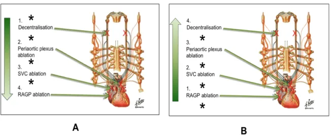
The pathways responsible for the bradycardic response were identified through sequential ablation of known autonomic pathways, including vagus nerve sectioning, radiofrequency ablation of periaortic and vena cava ganglionated plexi, and ablation of the RAGP, which is responsible for most autonomic input into the sinus node. Both antegrade and retrograde ablation sequences were performed (see below for details). Epicardial isointegral mapping was performed to accomplish the further goal of characterizing atrial repolarization changes following LAGP stimulation.
2.1 Ethics
Experiments were performed in accordance with guidelines set out by the Canadian Council of Animal Care166 and approval was obtained from the animal research ethics committee of the Sacré-Coeur Hospital Research Center.
2.2 Experimental protocol
2.2.1 Anaesthesia
Anaesthetic induction was performed using intravenous sodium thiopental (25 mg/kg) followed by hourly boluses of alpha-chloralose (25mg/kg) intravenously. Alpha chloralose was chosen because it is thought to have a limited effect on neuronal activity. Each bolus was administered over a period of 5 minutes to minimize hemodynamic disturbances. Depth of anaesthesia was evaluated continuously by monitoring of jaw tension and by variations in heart rate and blood pressure. Mechanical ventilation was maintained with a Harvard Apparatus (Millis, MA). Blood oxygen saturation was monitored continuously with lingual pulse-oxymetry (VetOx G2 Digital, Dolphin medical, Hawthorne, CA). Oxygen flow was
adjusted to maintain a partial oxygen saturation (SpO2) of 95% or greater. Body
temperature was maintained using a heated mat and infrared lamp. The left femoral artery and vein were cannulated to facilitate invasive blood pressure monitoring and fluid administration.
2.2.2 Surgery
Bilateral anterior thoracotomy was performed with the aid of electrocautery to ensure adequate hemostasis and both internal mammary arteries and veins were ligated with braided silk sutures. The animal is paralyzed (rocuronium 0.5mg/kg) during this time to facilitate the initial dissection. A pericardial cradle is created by a vertical midline pericardial incision and fixation to the lateral chest walls. This ensures exposure of the ascending aorta, superior vena cava, right and left atrial ganglionated plexi, and the entire heart. The vagus nerves are exposed in the cervical region and encircled with umbilical tape to aid in instrumentation. Epicardial electrodes were placed on the atrial and ventricular surfaces for monitoring and pacing, respectively. In order to facilitate data analysis, atrial electrograms were isolated by atrioventricular (AV) nodal blockade with formaldehyde injection (37%, 0.1-0.2ml) into the AV node. Right ventricular pacing (60bpm) was instituted to maintain adequate cardiac output between periods of nerve stimulation.
2.2.3 Neuronal stimulation
Stimulation of both vagus nerves, the RAGP, and the LAGP was performed at baseline and following each ablation sequence.
Vagus nerve stimulation
Two unipolar electrodes are installed on the vagus nerves bilaterally. These electrodes consist of two metallic wires (Medwire, Mount Vernon, NY), individually mounted on 25 gauge needles and inserted 1cm longitudinally into the vagus nerve. Electrical stimulus is transmitted via a programmable stimulator (Bloom Associates, Philadelphia, PA). A supramaximal stimulation (15 Hz, 1mA, 1ms) is transmitted to