The Long-Term Effects of Sports Concussion
par
Louis De Beaumont
Département de psychologie Faculté des Arts et des Sciences
Thèse présentée à la Faculté des Études Supérieures et postdoctorales en vue de l’obtention du grade de doctorat
en Psychologie – Recherche et Intervention option Neuropsychologie clinique
Octobre 2009
Université de Montréal
Faculté des études supérieures et postdoctorales
Cette thèse intitulée :
The Long-Term Effects of Sports Concussion
Présentée par : Louis De Beaumont
a été évaluée par un jury composé des personnes suivantes :
Michelle McKerral, président-rapporteur Maryse Lassonde, directeur de recherche
Miriam Beauchamp, membre du jury John Scott Delaney, examinateur externe Robert Forget, représentant du doyen de la FES
List of abbreviations
3D: three dimensionalAAN: America Academy of Neurology AD: Alzheimer’s disease AGA: Anterograde amnesia
AP: Anteroposterior
ApEn: Approximate entropy ApoE4: Apolipoprotein epsilon 4 BTVM: Brief Test of Visual Memory Ca+: Calcium ion
CIHR: Canadian Insitute of Health Research
CND: Canadian
COP: Centre-of-pressure
CS : Conditioning stimulus
CSF: Cerebrospinal fluid
CSP: Cortical silent period CT: Computerised tomography DTI: Diffusion Tensor Imaging
EEG: Electroencephalography
EMG: Electromyography
EOG: Electrooculogram
ERPs: Event-Related Potentials FDI: First dorsal interrosseus GABA: Gamma-Aminobutyric Acid
GCS : Glasgow Coma Scale
HEOG: Horizontal electrooculogram
Hz: Hertz
ICF: Intracortical facilitation ICI: Intracortical inhibition ISI : Interstimulus inhibition
K+: Potassium ion
LED: Light-emitting diode
LICI: Long-interval intracortical inhibition LOC: Loss of consciousness LTP: Long-term potentiation
M1: Primary motor cortex
MCI: Mild cognitive impairment MEP: Motor evoked potential
ML: Mediolateral
MMSE: Mini-Mental Status Examination MRI: Magnetic Resonance Imaging
Ms : milliseconds
MT: Motor threshold
mTBI: mild traumatic brain injury NCAA: National Collegiate Athletic Association
NFT: Neurofibrillary tangles
NSERC: Natural Sciences and Engineering Research Council of Canada PAS: Paired-associative stimulation
PCS: Post-Concussion Symptoms Scale PSU: Pennsylvania State University RAM Rapid Alternating Movement RCFT: Rey-Osterrieth Complex Figure Test
RGA: Retrograde amnesia
RMS : Root mean square
rMT: resting motor threshold S1: Primary somatosensory cortex SAI: Short-afferent inhibition SCAT: Sport Concussion Assessment Tool
SD: Standard deviation
SDMT: Symbol Digit Modalities Test
SE : Standard Error
TAI: Traumatic axonal injury TBI : Traumatic brain injury TMS: Transcranial Magnetic Stimulation
TS : Test stimulus
US: United States
Résumé
Questions : Cette thèse visait à répondre à deux questions fondamentales : 1) Est-ce que
les athlètes qui présentent un historique de commotions cérébrales du sport en conservent des effets délétères à long terme? ; et 2) Est-ce que les effets néfastes des commotions cérébrales récurrentes sur le fonctionnement tant cognitif que moteur sont cumulatifs?
Devis expérimental : À l’aide d’un plan d’investigation double-cohorte réalisé avec un
groupe d’athlètes évoluant au niveau universitaire et un autre formé d’anciens athlètes universitaires testés plus de trois décennies plus tard, les quatre études qui composent cette thèse ont employé des méthodes raffinées d’investigation des fonctions cognitives et motrices pour en déceler des atteintes persistantes. Méthodologie : Les potentiels évoqués cognitifs ainsi que les tests neuropsychologiques ont permis de sonder le fonctionnement cognitif de ces athlètes alors que la stimulation magnétique transcrânienne, une plateforme de force permettant de mesurer la stabilité posturale ainsi qu’un système d’enregistrement tridimensionnel des mouvements rapides alternatifs ont servi à l’évaluation de l’intégrité du système moteur. Résultats : Cette thèse a permis de déceler des altérations persistentes et cumulatives des fonctions cognitives et motrices. De plus, ces subtiles atteintes observées chez les jeunes athlètes, affectant essentiellement des marqueurs neurophysiologiques sous-cliniques du fonctionnement cognitif et moteur, s’étaient accentuées chez les anciens athlètes universitaires qui montraient un déclin quantifiable tant des fonctions cognitives que motrices. Discussion : Ces résultats suggèrent d’une part que les commotions cérébrales du sport entraînent des altérations cognitives et motrices chroniques qui s’accentuent en fonction du nombre de commotions cérébrales subies. D’autre part, les effets délétères des commotions cérébrales du sport sur le fonctionnement cognitif et moteur combinés à ceux associés au processus de vieillissement entraînent un déclin cognitif et moteur quantifiable en comparaison aux anciens athlètes n’ayant jamais subi de commotions cérébrales.
Mots-clés : Commotions cérébrales du sport, effets à long terme, neuropsychologie,
neurophysiologie, déclin cognitif, système moteur, excitabilité du cortex moteur, potentiels évoqués cognitifs, stimulation magnétique transcrânienne, stabilité posturale, mouvements rapides alternatifs.
Abstract
Question: This thesis aimed to address two fundamental issues: 1) Are there long-lasting
effects of sports-related concussion on cognitive and motor functions? and 2) Are the adverse effects of recurrent concussions cumulative? Experimental Design: The cross-sectional thesis design included a group of active university-level athletes as well as a group of former athletes recruited more than three decades after their university years who were tested on neurophysiological measures of both cognitive and motor system functions.
Methods: Event-Related potentials and neuropsychological tests were used to assess
cognitive functions while transcranial magnetic paradigms were used to assess motor cortex excitability, a force platform was used to assess postural stability and a 3-dimensional recording device was used to track hand position when performing a rapid alternating movement task. Results: This thesis disclosed persistent and cumulative alterations of both cognitive and motor functions after sports concussions. Furthermore, subclinical, neurophysiological alterations found in young concussed athletes were exacerbated in former athletes with concussions who displayed quantifiable cognitive and motor functions decline more than three decades post-concussion. Discussion: These results suggest that sports concussions induce cognitive and motor functions abnormalities that worsen as a function of the number of concussions sustained. Moreover, findings from the present thesis indicate that the deleterious effects of sports concussion on cognitive and motor system functions combined to those associated with the aging process lead to quantifiable decline on both cognition and motor functions.
Keywords : Sports concussions, long-term effects, neuropsychology, neurophysiology,
cognitive decline, motor system, motor cortex excitability, event-related potentials, transcranial magnetic stimulation, postural stability, rapid alternating movements.
Table des matières
1. INTRODUCTION ...1
1.1. WHAT A TRAUMATIC BRAIN INJURY IS… ...1
1.1.1. Definition and Prevalence...1
1.1.2. Severity of TBI...3
1.2. SPORTS CONCUSSION AS A SUBTYPE OF MILD TBI ...5
1.2.1. Prevalence and public awareness ...5
1.2.2. Definition...6
1.2.3. Classification of sports concussions ...7
1.3. HOWSPORTSCONCUSSIONSOCCUR ...8
1.3.1. Biomechanics of concussive injuries...8
1.3.2. The neuropathophysiology of concussion ...12
1.4. WHAT THE EFFECTS OF SPORTS CONCUSSIONS ARE…...14
1.4.1. In the first few minutes (on-field symptoms)...14
1.4.2. On the sidelines (from a few minutes up to 48 hours) ...16
1.4.3. From a few hours up to 14 days post-concussion ...20
1.4.4. From 14 days up to late adulthood ...26
1.5. THESISOBJECTIVES ...32 1.6. RESEARCH DESIGN ...33 1.6.1. Experiment 1 ...33 1.6.2. Experiment 2 ...34 1.6.3. Experiment 3 ...36 1.6.4. Experiment 4 ...39 2. EXPERIMENT 1...41 3. EXPERIMENT 2...74 4. EXPERIMENT 3...100 5. EXPERIMENT 4...118 6. DISCUSSION...156
6.1. MAIN THESIS FINDINGS...156
6.1.1. Experiment 1: Main findings...156
6.1.2. Experiment 2: Main Findings ...158
6.1.4. Experiment 4: Main findings...162
6.2. OVERALL THESIS DISCUSSION ...166
6.2.1. Alterations of cognitive functions after sports concussions...167
6.2.2. Alterations of motor system functions after sports concussions...173
6.2.3. Clinical implications of excessive intracortical inhibition in M1 ...175
6.2.4. Postural stability changes after sports concussions ...181
6.3. MAIN THESIS LIMITATIONS...182
6.4. OVERALL THESIS CONCLUSION ...183
7. REFERENCES ...184 7.1. INTRODUCTION...184 7.2. EXPERIMENT 1 ...220 7.3. EXPERIMENT 2 ...231 7.4. EXPERIMENT 3 ...237 7.5. EXPERIMENT 4 ...242 7.6. DISCUSSION...254
Liste des tableaux
Experiment 1, Table 1... 66 Experiment 1, Table 2... 67 Experiment 1, Table 3... 68 Experiment 1, Table 4... 69 Experiment 1, Table 5... 70 Experiment 2, Table 1... 95 Experiment 2, Table 2... 96 Experiment 2, Table 3... 97 Experiment 4, Table 1... 152 Experiment 4, Table 2... 152Liste des figures
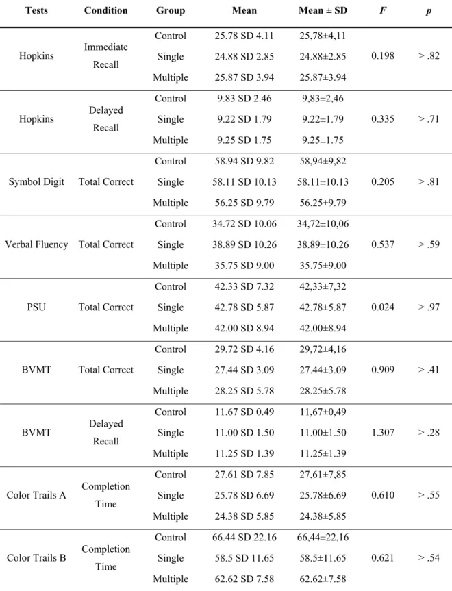
Figure 1: Glasgow Coma Scale ...4
Experiment 1, Figure 1 ... 71 Experiment 1, Figure 2 ... 72 Experiment 1, Figure 3 ... 73 Experiment 2, Figure 1 ... 98 Experiment 2, Figure 2 ... 99 Experiment 3, Figure 1 ... 116 Experiment 3, Figure 2 ... 117
This thesis is dedicated to those who suffer from this silent epidemic….
Remerciements
Devant l’impossibilité de remercier tous ceux qui ont influencé ma formation depuis le tout jeune âge jusqu’à son achèvement, je m’en tiendrai à souligner l’apport tout spécial de personnes qui se sont avérées essentielles à la réussite de mon parcours doctoral.
À ma directrice de recherche Maryse Lassonde. Premièrement, je vous remercie infiniment de la liberté et de la confiance que vous m’avez accordée. Vous avez été à travers ces années un modèle inspirant tant par vos accomplissements exceptionnels que par vos qualités humaines.
À Hugo Théoret. Je te remercie de tout le temps que tu m’as consacré depuis ces dernières années. Toujours à l’écoute, tu as su par la justesse et la profondeur de tes interventions m’inspirer à me dépasser.
À Martin Thériault. Ton amitié, humanisme et support inconditionnel m’ont été absolument nécessaires. Merci mon vieux !
À Sébastien Tremblay. Merci infiniment pour ton amitié, ton intégrité, ta loyauté, ta confiance, et ton éthique de travail. Travailler en étroite collaboration avec toi depuis les quelques dernières années a été pour moi un immense privilège. Tu me manqueras.
À mes parents. Merci pour votre amour. La rigueur et l’éthique de travail que vous m’avez transmises m’ont donné l’essentiel.
1. Introduction
1.1. WHAT A TRAUMATIC BRAIN INJURY IS…
1.1.1. Definition and Prevalence
A traumatic brain injury (TBI) is caused by a blow or jolt to the head or a penetrating head injury that disrupts the function of the brain. Not all blows or jolts to the head result in a TBI. The severity of a TBI may range from “mild,” i.e., a brief change in mental status or consciousness to “severe,” i.e., an extended period of unconsciousness or amnesia after the injury (Langlois, Rutland-Brown, & Wald, 2006).
TBI subtypes
i. Open (penetrating) head injury: An open head injury occurs when a high-velocity
object (projectile) fractures the skull and lesions brain tissues or the surrounding membranes. The open wound created by such insertion of a projectile into brain structures renders open head injury victims at risk from infection and contamination. Accidents involving firearms account for nearly 40% of all TBI-related deaths in the United States (Control, 2003).
ii. Closed head injury: A closed head injury occurs when kinetic energy of varying
magnitude is applied to the head without fracturing the skull. The ensuing consequences of a closed head injury are directly related to the force of the impact as the brain is damaged when colliding against the skull wall. Closed head injuries account for the vast majority of TBI cases (Control, 2003).
Recent data from the Centers for Disease Control and Prevention (2003) show that, on average, approximately 1.4 million people sustain a TBI each year in the United States. TBI represents the most prevalent cause of long-term or lifelong disability among adults and annual costs, whether direct medical costs or indirect costs such as lost productivity, amounted to 60 billion dollars in the United States in 2000 (Langlois et al., 2006). Males are about twice as likely as females to sustain a TBI (Langlois et al., 2006). About 75% of those accidents are concussions or other forms of mild TBI (Control, 2003) and leading causes of TBI are falls (28%), motor vehicle accidents (20%), collisions (20%), and assaults (11%). While similar Canadian statistics on TBI and its consequences are still lacking, recent evidence collected in the province of Ontario found roughly equivalent incidence rates of mild TBI admitted to Emergency Departments to those reported in the United States (Guerrero, Thurman, & Sniezek, 2000; Ryu, Feinstein, Colantonio, Streiner, & Dawson, 2009).
The conceptual definition of a mild traumatic brain injury proposed by the Centers for Disease Control and Prevention (2003) is an injury to the head as a result of blunt trauma or acceleration or deceleration forces that result in one or more of the following conditions:
● Any period of observed or self-reported:
- Transient confusion, disorientation, or impaired consciousness;
-Dysfunction of memory around the time of injury;
● Observed signs of neurological or neuropsychological dysfunction, such as: - Seizures acutely following injury to the head;
- Among infants and very young children: irritability, lethargy, or vomiting following head injury;
- Symptoms among older children and adults such as headache, dizziness, irritability, fatigue or poor concentration, when identified soon after injury, can be used to support the diagnosis of mild TBI, but cannot be used to make the diagnosis in the absence of loss of consciousness or altered consciousness.
1.1.2. Severity of TBI
The severity of a TBI may range from “mild,” i.e., a brief change in mental status or consciousness to “severe,” i.e., an extended period of unconsciousness or amnesia after the injury (Langlois et al., 2006). The Glasgow Coma Scale (GCS) is the TBI severity scale now used in most hospitals and emergency departments throughout the world (1988). Introduced in 1974 as a means of assessing depth and duration of impaired consciousness and coma, the GCS was also created for use in gauging deterioration or improvement at the emergent and acute stages of brain damage or lesions, as well as in predicting the ultimate outcome (G. Teasdale & Jennett, 1974). By observing a patients' eye opening, verbal performance, and motor response, the GCS allowed to more accurately define gradations in the comatose state than by only measuring duration of unconsciousness. Patient’ condition is assessed on three distinct aspects of consciousness (eye opening on a scale of 4 to 1, motor response on a scale of 6 to 1, and verbal response on a scale of 5 to 1) with the sum
total of scores of the three categories resulting in the total GCS score. When using the GCS as a classification measure of TBI severity, patients achieving total scores of 8 or less are classified as "in coma" or having a severe TBI, scores of 9 to 12 are classified as moderate TBI, and 13 to 15 as mild TBI or no head injury.
Figure 1: Glasgow Coma Scale
EYE OPENING Spontanous 4 To speech 3 To pain 2 No opening 1 VERBAL RESPONSE Conversation 5 Confused 4 Non Sense 3 To sounds 2 No Response 1 MOTOR RESPONSE To command 6 Localizing 5 Arm flexion 4 Arm extension 3 Generalizing 2 No response 1
1.2. SPORTS CONCUSSION AS A SUBTYPE OF MILD TBI
1.2.1. Prevalence and public awareness
Among causes of mild TBI which represent nearly 80% of all cases of TBI (Ruff, 2005), sports concussions are by far the most prevalent as recent epidemiological data estimated that nearly 50,000 to 300,000 contact sports athletes in the United States sustain a concussion every year (Control, 2003).
Epidemiological data gathered from 100 US high schools and 180 colleges were analyzed to calculate rates, describe patterns, and evaluate potential risk factors for sport-related concussion across different sports. This study showed that nearly 41% of all concussion cases resulted from participation in football (Gessel, Fields, Collins, Dick, & Comstock, 2007). However, a recent study showed that ice hockey, soccer and football had comparable yearly rates of concussions per 10 000 players who were admitted to the Emergency Departments in the United States (Delaney, 2004). It therefore seems that these three contact sports involve equivalent risk of concussions. The aforementioned epidemiological study also showed that the greatest incidence of concussion was found at the high school (5.6%) and collegiate division III (5.5%) levels, suggesting that there is an association between level of play and the proportion of players injured (Gessel et al., 2007). Moreover, another epidemiological study found that players who sustained one concussion in a season were three times more likely to sustain a second concussion in the same season compared with uninjured players (Guskiewicz, Weaver, Padua, & Garrett, 2000).
Most alarming, the number of concussions occurring is most likely underestimated. According to a study conducted by McCrea and colleagues (2004), 49% of athletes in high school who sustain a concussion did not report it. The main reason evoked is that the seemingly benign post-concussion symptoms are not severe enough to be reported. Likewise, a study conducted by Delaney and collaborators (Delaney, Lacroix, Leclerc, & Johnston, 2000) revealed that 44.8% of athletes in the Canadian Football League reported having experienced post-concussion symptoms in the years prior to the study. Among them, only 18.8% recognized having sustained a concussion.
In spite of these disconcerting findings, the reported incidence of sports concussions has grown at an accelerated pace over the last 15 years and is now considered a major public health concern (Kelly, 1999). New discoveries on the pervasive effects of sports concussions together with recurrent injuries occurring to high-profile professional athletes have contributed to raising unprecedented public awareness that the effects of concussions were not as benign as what was once thought.
1.2.2. Definition
According to the Prague Summary and Agreement Statement on concussion in sports (P. R. McCrory et al., 2005), sports concussion is defined as a complex pathophysiological process affecting the brain,induced by traumatic biomechanical forces. This definition was further broken down to include a range of clinical, pathological and biomechanical features intrinsic to sports concussions:
1. Concussion may be caused by a direct blow to the head, face, neck, or elsewhere on the body with an “impulsive” force transmitted to the head;
2. Concussion typically results in the rapid onset of a short lived impairment of neurological function that resolves itself spontaneously;
3. Concussion may result in neuropathological changes but the acute clinical symptoms largely reflect a functional disturbance rather than structural injury;
4. Concussion results in a graded set of clinical syndromes that may or may not involve loss of consciousness. Resolution of the clinical and cognitive symptoms typically follows a sequential course;
5. Concussions are typically associated with grossly normal structural anatomy as revealed by magnetic resonance imaging (MRI) and computerised tomography (CT) scan .
1.2.3. Classification of sports concussions
Based on recommendations from the Summary and Agreement Statement of the 2nd International Conference on Concussion in Sport (P. R. McCrory et al., 2005), concussion severity can be determined retrospectively and classified as either simple or complex concussion. Simple concussion, which accounts for most sports concussion cases, is defined as a concussion for which associated symptoms resolved within 7-10 days without complications. Complex concussions include cases where athletes suffer persistent
symptoms, specific sequelae (eg, concussive convulsions, prolonged loss of consciousness (>1 minute) or prolonged cognitive impairment following the injury. This group may also include athletes who suffer multiple concussions over time or where repeated concussions occur with progressively less impact force (Iverson, 2007; P. R. McCrory et al., 2005).
Very little is known about interindividual factors intervening to render an athlete more susceptible to fall into the category of complex concussion relative to those who sustain simple concussion. Future studies that systematically look at various concussion characteristics—clinical manifestations (confusion, memory problems, loss of consciousness), anatomical localization (such as cerebral vs. brainstem), biomechanical impact (rotational vs. linear force), genetic phenotype apolipoprotein epsilon 4 (ApoE4) positive vs. ApoE4 negative) and neuropathological changes (structural injury vs. no structural injury) (P. R. McCrory et al., 2005)— are likely to help shed some light on action mechanisms more common to each concussion subtype.
The following sections will discuss in further details the biomechanics and neuropathophysiology of concussive injuries.
1.3. HOW SPORTS CONCUSSIONS OCCUR
1.3.1. Biomechanics of concussive injuries
As most people agree that no two concussions are alike, they all share at least one feature in common. They all involve the near instant transfer of kinetic energy. The brain
floats within a protective shield filled with cerebrospinal fluid called subarachnoid space (Kandel, Schwartz, & Jessell, 2000). Its gelatinous and elastic properties guard the brain from colliding against the walls of the skull when the head is moved. However, when kinetic energy applied to the head exceeds the subarachnoid space cushioning properties, the brain will come into contact with the bones of the skull causing distortion, compression and deformation of neural tissue. The brain absorbs energy as a result of acceleration forces, while deceleration forces cause it to release kinetic energy when colliding with the skull (Shaw, 2002). To date, at least four distinct biomechanical processes have been identified in the induction of a concussive state: 1) Coup and contre-coup impact between the surface of the brain and the walls of the skull; 2) Traction of the brainstem neurons due to forceful movement of the hemispheres; 3) Compression of the skull bone responsible for compression of neural tissue and elevated intracranial pressure levels; and 4) Acceleration of the head about the axis of the brain (Shaw, 2002).
Coup and contre-coup were shown to occur as a result of such acceleration/deceleration forces applied to the brain (Ommaya, Goldsmith, & Thibault, 2002). Sequelae associated with coup injuries are those observed directly beneath the point of impact on the skull while those in contre-coup injuries occur elsewhere on the surface of the cortex, most predominantly opposite to the site of impact (Shaw, 2002). With sudden acceleration injury, movement of the brain lags behind that of the skull whereas deceleration injuries occur as the brain continues to move after the skull has abruptly halted. Football players will often escape being concussed from a direct impact to the head if they tense their neck muscles prior to collision so as to restrict the magnitude of head
motion thereby allowing kinetic energy to be dispersed throughout the whole body instead of being fully absorbed by the brain (R. C. Cantu, 1992). An accelerative/decelerative injury can be sustained either by impact or impulse. Impact involves a direct contact to the head, while impulse refers to an accelerative force that sets the head in motion without directly striking it (Shaw, 2002). Furthermore, acceleration/deceleration forces can either be transferred to the brain in a straight line passing through the head’s centre of gravity or in a tangential line and arc around its centre of gravity (Greenwald, Gwin, Chu, & Crisco, 2008). In football, concussions occurring as a result of head-on, helmet-to-helmet collisions are often due to an imbalance between the inertial linear forces carried by each colliding opponent (Broglio et al., 2009; Greenwald et al., 2008). In contrast, rotation of the brain within the skull induced by inertial force transferred to it in a tangential line occurs typically when a player rushing up the field gets hit by an opponent coming from an angle (Broglio et al., 2009).
Abrupt movement of the head due to inertial forces is implicated in more than coup contre-coup types of injuries. Although scarcely documented in the sports concussions literature, cortical, subcortical and brainstem white matter tracts are susceptible to stresses and strains as the cerebrum rotates about its junction with the fixed brainstem (Gentry, 1994; Gentry, Godersky, & Thompson, 1988; Hashimoto, Nakamura, Richard, & Frowein, 1993; Parizel et al., 1998; Paterakis, Karantanas, Komnos, & Volikas, 2000; Shibata, Matsumura, Meguro, & Narushima, 2000). Shearing and stretching of interconnected pathways are common manifestations of inertial forces applied to the brain (Besenski, 2002; Besenski, Broz, Jadro-Santel, Pavic, & Mikulic, 1996; Ommaya et al., 2002;
Ommaya, Rockoff, & Baldwin, 1964). Consequently, the maintenance of alertness and responsiveness, which are activities principally mediated by structures located in the midbrain and upper brainstem, are vulnerable to mechanical disruption by these stresses and strains.
Another type of mechanical brain injury that might possibly be involved in concussive injury is compression of the skull. In this instance, a concussive injury is incurred when the skull temporarily indents or bends at the site of impact without fracturing (Bayly, Black, Pedersen, Leister, & Genin, 2006; Bayly et al., 2005). The newly occupied space triggers pressure waves and pulses as a result of brain tissue compression. These pressure waves and pulses are thought to be diffusely transmitted to the brainstem and to other cranio-cervical junctions (McIntosh et al., 1996). Increases in intracranial pressure are associated with tissue shift or deformation and shearing stresses.
At this time, there is no existing animal or other experimental model that precisely reflects a sporting concussive injury. Experimental models of more severe concussive injuries have proposed that a complex cascade of biochemical, metabolic and gene expression changes occur (Hovda et al., 1995). Whether similar metabolic changes occur in sports concussion remains largely unknown at this time (McIntosh et al., 1996). However, the close resemblance between sports concussion and more severe forms of TBI in terms of neurologic impairments observed immediately post-injury and of residual post-concussion symptoms suggest that the latter share similar underlying mechanisms. Although probably to a lesser extent, it is likely that the biomechanical forces applied to the brain in sports
concussion could be sufficient to induce a sudden, uncontrolled chain reaction similar to the neurometabolic cascade triggered by more severe TBI.
1.3.2. The neuropathophysiology of concussion
Research on the pathophysiology of concussive injuries was initially instigated in order to elucidate the neurometabolic cascade that preceded diffuse axonal injury, which was held responsible for the symptoms of the post-concussion syndrome. Early work conducted by Courville (1953) suggested that the nature of these post-concussion symptoms was intimately related to the susceptibility of certain brain areas to traumatic blows to the head. As Courville (1953) demonstrated decades ago, neuronal tissues located within close vicinity of the frontotemporal bones are most vulnerable to mechanical strains and stresses induced by concussive injury which, in turn, prevent affected neuronal assemblies to play their usual role on brain function. For instance, memory deficits that often occur following concussive injuries could readily be explained by temporarily disturbed connections within the medial temporal lobe, while deficits in learning, emotional decoding, inhibition, difficulty related to decision-making in unstructured situations, and an inability to regulate behaviour according to internal goals and constraints could just as well be explained by momentarily compromised communication within the ventromedial portion of the prefrontal cortex (Pandya & Yeterian, 1996).
Although diffuse axonal injury is common subsequent to moderate or severe TBI (Povlishock, Becker, Cheng, & Vaughan, 1983; Povlishock & Katz, 2005), compelling evidence from animal work showed that the pathophysiology of mild TBI leaves neurons
and neuronal systems vulnerable but not destroyed (Iverson, 2005). Recent high-spatial resolution neuroimaging studies confirmed previous animal work suggesting preserved neuronal integrity following mild TBI (Aubry et al., 2002) despite the enduring belief that post-TBI symptoms are due to diffuse axonal damage. What is rather observed in mild TBI patients is a gradual process in which a very small proportion of badly damaged axons swell and eventually separate, while the vast majority of surrounding axons that are initially affected recover over time (Iverson, 2005). Incidentally, the term diffuse axonal injury has progressively been replaced by traumatic axonal injury as it more accurately describes the few damaged axons embedded among mostly intact axons seen in mild TBI (Mac Donald et al., 2007; Pettus, Christman, Giebel, & Povlishock, 1994). In light of accumulating evidence suggesting that neuronal systems are mostly preserved in mild TBI, the latter brain pathology appears to be a deficit of function rather than one of structure.
Accordingly, investigators have turned to the study of the neurometabolic cascade and its mediating effects on brain function in their effort to uncover potential mechanisms of action underlying post-concussion symptoms. The impending description of the multi-faceted metabolic cascade that immediately follows mild TBI has strongly been inspired by the work of Giza and Hovda (2001). Research has shown that immediately after a concussive injury, neurotransmitters are indiscriminately released and uncontrolled ionic fluxes ensue. The binding of excitatory neurotransmitters, such as glutamate, to the N-methyl-D-aspartate (NMDA) receptor leads to further neuronal depolarization with efflux of potassium and influx of calcium (Giza & Hovda, 2001). Due to severe disruptions in ionic gradients, ion-specific pumps are activated by the cells so as to restore resting
membrane potential. Because these ion-specific pumps require much energy to operate, a dramatic increase in glucose consumption ensues. This hypermetabolism occurs in the context of diminished cerebral blood flow, and the disparity between glucose supply and demand triggers a cellular energy crisis. The rapid exhaustion of glucose supply causes the concussed brain to undergo a period of depressed metabolism. Persistent increases in calcium were also shown to impair mitochondrial oxidative metabolism, thereby exacerbating the energy crisis (Awasthi, Church, Torbati, Carey, & Pryor, 1997; Deng, Thompson, Gao, & Hall, 2007). This period of depressed neurometabolism that immediately follows hyperexcitation in mild concussion is thought to gradually return to baseline levels within 6 to 7 days post-injury (Giza & Hovda, 2001). Interestingly, the duration of transient post-concussion symptoms concords with the time taken for the concussed brain to recover baseline metabolism levels (McCrea et al., 2003). This highly similar time course has been interpreted as evidence that the neurometabolic cascade of concussive injury underlies the post-concussion symptoms reported by athletes in the acute post-concussion phase (Giza & Hovda, 2001). More research that systematically controls for the progression of the neurometabolic cascade while assessing its concomitant clinical manifestations is required to validate this widespread belief.
1.4. WHAT THE EFFECTS OF SPORTS CONCUSSIONS ARE…
1.4.1. In the first few minutes (on-field symptoms)
In the first few minutes that immediately follow a concussion, the athlete may experience a wide range of symptoms that vary both in terms of intensity and duration.
Among them, disorientation, loss of consciousness and post-traumatic amnesia have been identified to index concussion severity. While several other scales are available, the American Academy of Neurology (1997) has perhaps been the most cited concussion severity grading scales in the scientific literature. Concussion severity assessment has traditionally been performed on-field during the first few minutes that immediately follow the accident. Sports therapists using the AAN grading scale have typically assessed concussion severity according to the following system:
• Grade 1: Trauma-induced alteration in mental status causing transient confusion for less than 15 minutes, without loss of consciousness;
• Grade 2: Trauma-induced alteration in mental status that lasted for more than 15 minutes but without loss of consciousness.
• Grade 3: Trauma-induced alteration in mental status accompanied with either brief or prolonged loss of consciousness.
Important limitations have been raised concerning the poor predictive value of concussion severity grades on post-concussion recovery. The traditional approach to severe traumatic brain injury utilizing loss of consciousness (LOC) as the primary measure of injury severity has met limitations in assessing the severity of sporting concussive injury. Findings in this field showed LOC association with specific early deficits but failed to predict post-concussion outcome (Lovell, Iverson, Collins, McKeag, & Maroon, 1999; McCrea, Kelly, Randolph, Cisler, & Berger, 2002). In parallel, recent evidence showed
that the presence of amnesia, not loss of consciousness, was predictive of symptom and neurocognitive deficits following concussion (Collins et al., 2003). However, published evidence suggests that the nature, burden and duration of post-concussion symptoms appear more important than the presence or duration of amnesia alone (Lovell et al., 1999; P. R. McCrory, Ariens, & Berkovic, 2000). Converging evidence on the lack of association between the presence and duration of on-field concussion severity markers and the course of recovery has rendered the use of traditional concussion severity grading scales obsolete. The newest Summary and Agreement Statement on concussions in sports (P.R. McCrory et al., 2009) supported the notion that such concussion severity grading scales should be abandoned in favor of combined measures of recovery. In fact, the expert panel maintained its prior position presented at the Vienna conference on concussion in sport (Aubry et al., 2002) suggesting that concussion severity could only be determined in retrospect after all concussion symptoms have cleared and cognitive function has returned to baseline. From a clinical perspective, this significant theoretical shift away from the immediate, on-field symptoms to assess concussion severity forced clinicians to thoroughly monitor recovery of acute symptoms occurring from the sideline up to their resolution most often taking place within 14 days from the injury (P. R. McCrory et al., 2005).
1.4.2. On the sidelines (from a few minutes up to 48 hours)
Concussion diagnosis is based on the sideline assessment of neurologic symptoms. A few minutes after the accident concussed athletes may be experiencing clinical symptoms, physical signs, cognitive impairment, and/or loss of consciousness (P. R. McCrory et al., 2005). These symptoms include:
Cognitive features
• Unaware of period, opposition,score of game • Confusion
• Amnesia
• Loss of consciousness
Typical self-reported post-concussion symptoms
• Headache or pressurein the head • Balance problems or dizziness • Nausea
• Feeling"dinged","foggy", stunned, or "dazed"
• Visual problems—forexample,seeing stars or flashinglights, double vision • Hearingproblems—forexample, ringing in the ears
• Irritabilityor emotional changes
Physical signs
• Loss of consciousness/impairedconscious state • Poor coordination or balance
• Concussiveconvulsion/impactseizure • Gait unsteadiness/loss of balance
• Slow to answerquestions or follow directions • Easily distracted,poor concentration
• Displaying inappropriate emotions—forexample, laughing,crying • Vomiting
• Vacant stare/glassy eyed • Slurred speech
• Personality changes
• Inappropriate playingbehaviour—forexample, running inthe wrong direction • Significantly decreasedplaying ability
The Sport Concussion Assessment Test (SCAT) is a 5-minute concussion diagnosis test battery specifically designed to detect anomalies in any of these spheres. In short, the SCAT (refer to Table 2) seeks for signs of concussions (LOC, unresponsiveness, balance problems, convulsive activity); symptoms commonly associated to sports concussions (Post-Concussion Symptom Scale is a 19-item Likert scale from 0 (none) to 6 (severe)); memory impairments (orientation questions and retrograde memory); cognitive impairments (episodic memory impairments on both an immediate and a 3-minute delayed recall of five words, attentional regulation, information processing speed); and neurologic impairments (speech, eye motion and pupil dilation, pronator drift and gait assessment).
Sideline evaluation of cognitive function is an essential componentin the assessment of this injury. Brief neuropsychological testbatteries such as the Maddocks questions (Maddocks, Dicker, & Saling, 1995) and the Standardised Assessment of Concussion (McCrea et al., 1998) that assess attention and memory function have been shown to be practical and effective. Previous evidence has shown that standard orientation questions (i.e.; time of day, location, person) are unreliablein the sporting situation when compared with memory
assessment (Maddocks et al., 1995; McCrea, Kelly, Kluge, Ackley, & Randolph, 1997). If any one of the previous symptoms or problems is detected with the SCAT,a head injury should be suspected (P. R. McCrory et al., 2005) and the athlete should be taken out of competition. This recommendation from the expert panel of the recent Summary and Agreement Statement on concussion in sport is supported by compelling evidence documenting catastrophic consequences of recurrent concussions in athletes who returned to competition during the same game (R. C. Cantu, 1998; P. McCrory, 2001a; P. R. McCrory & Berkovic, 1998). While the term second impact syndrome was used to discuss the highly controversial cases of high school and college football players who died after having sustained recurrent concussions before symptoms from the first head injury had resolved (R. C. Cantu, 1998), others have documented diffuse cerebral swelling following repetitive concussions that was associated with severe complications (P. R. McCrory & Berkovic, 1998). Along those lines, recent animal work demonstrated the existence of a temporal window of metabolic brain vulnerability to second mTBI that had profound consequences on mitochondrial-related metabolism (Vagnozzi et al., 2007).
Once these transient, gross neurologic impairments observed on the sideline in the first few minutes/hours after the accident have subsided, the athlete enters a slow, progressive recovery period. Recent estimates suggested that nearly 70% of concussed athletes no longer report experiencing post-concussion symptoms and have regained baseline performance levels on neuropsychological testing within 10-14 days post-injury (Collins, Grindel et al., 1999; R. J. Echemendia, Putukian, Mackin, Julian, & Shoss, 2001; Hinton-Bayre & Geffen, 2002; P. R. McCrory et al., 2005).
1.4.3. From a few hours up to 14 days post-concussion
Getting some rest during time out of competition is the usual treatment prescribed to the slow, progressive recovery period in uncomplicated cases of sports concussion (Ruben J. Echemendia, 2006). In addition to post-concussion symptoms that might be present during sideline assessment (refer to SCAT), symptoms specific to the post-concussion recovery phase might emerge. These symptoms include:
• Sadness • Nervousness
• Trouble falling asleep • Sleeping more than usual • Sensitivity to light • Sensitivity to noise
Although symptomatology varies widely during the acute post-concussion phase, headache is the most commonly reported and perhaps the most debilitating symptom occurring during this period (Guskiewicz et al., 2003; P. R. McCrory et al., 2000). Knowing that headache is associated to vasoconstriction consecutive to oxygen deprivation (Wilson, Foresman, Gamber, & Wright, 1998), the energy crisis consecutive to the neurometabolic cascade following concussive injury was proposed to at least partially mediate post-concussion headache (P. McCrory, 2000, 2001b). This is supported by recent evidence showing that the chronological course of the neurometabolic cascade and post-concussion symptoms recovery was very much alike. In fact, a nationwide National Collegiate Athletic Association (NCAA) concussion study has prospectively measured
immediate effects and natural recovery course relating to symptoms, cognitive functioning, and postural stability following sport-related concussion (McCrea et al., 2003). Out of the 1631 collegial football players who had undergone baseline testing on the aforementioned measures, 94 of them sustained a concussion throughout the course of a 3-year period. They were compared to 56 uninjured athletes who also underwent assessment of symptoms, cognitive functioning,and postural stability immediately, 3 hours, and 1, 2, 3, 5, 7, and 90 days after injury. The latter study revealed that on average, symptoms resolved byday 7, cognitivefunctioning improved to baseline levels within 5 to 7 days, and balance deficits dissipated within 3 to 5 days after injury (McCrea et al., 2003). Similarly to the gradual recovery of neurometabolic balance (Giza & Hovda, 2001), post-concussion symptoms recovery also follows a progressive course. In a prospective study that included 2905 football players from 25 US Colleges tested at preseason, 194 athletes sustained a concussion over three successive seasons. Headache was the most commonly reported symptom at the time of injury, followed by dizziness/balance difficulties and feeling cognitively "slowed down". Among the 167 playersexperiencing a headache at the time of their concussion, 149 (89.2%) still reported having a headache 3 hours after injury, 110 (65.9%) 24 hours after injury, 41 (24.5%) at postinjuryday 5, and 23 (13.8%) at postinjury day 7. On average, overallsymptom duration was about 3.5 days, and 87.8% achieved full symptom resolution within one week after injury (Guskiewicz et al., 2003).
Considerable emphasis is given to post-concussion symptoms in the management of sports concussions as the number and duration of post-concussion symptoms were shown to be somewhat reliable markers of injury severity and a valuable guide for return to play
(P. R. McCrory et al., 2000). In addition to assisting in sideline concussion diagnosis, the SCAT also proposes an empirically validated, stepwise return-to-play protocol. This stepwise process involves six incremental stages (see listed below) during which athletes should remain symptom free at all times before returning to competition. The SCAT is the return-to-play protocol endorsed by the last Summary and Agreement Statement on concussion in sport (P. R. McCrory, 1999).
SCAT return-to-play protocol
1. No activity, complete rest. Once asymptomatic, proceed to level2.
2. Light aerobic exercise such as walking or stationary cycling,no resistance training. 3. Sport specific exercise—forexample, skating in hockey,running in soccer;
progressive additionof resistance trainingat steps 3 or 4. 4. Non-contact trainingdrills.
5. Full contact training after medical clearance. 6. Gameplay.
According to the SCAT return-to-play protocol (P. R. McCrory et al., 2005), the athlete should only proceed to the next level if asymptomatic at the currentlevel for 24 hours. If any post-concussion symptoms occur, the patient shoulddrop back to the previous asymptomatic level and try to progressagain after being symptom free for at least 24 hours.
In parallel, recent data suggest that cognitive recovery may precede or follow clinical symptom resolution suggesting that the neuropsychological assessment of cognitive
function should be an important component in any return to play protocol (Bleiberg et al., 2004). Once considered as the gold standard to return-to-play decision making, current knowledge underlining neuropsychological test’s substantial vulnerability to serial testing together with known practice effects (Lovell et al., 2007; Moser et al., 2007) emphasize that it should not be the sole basis of a return to play decision but rather be seen as a complementary tool to clinical decision making (P. R. McCrory et al., 2005). In particular, having to rely on normative data when baseline testing is absent introduces numerous potential biases due to the singular nature of concussive injuries.
Although receiving general acceptance from clinicians in the sports concussion community, recommendations for return-to-play protocols proposed by consensus statements of expert panels do not benefit from solid empirical support. While not undermining the value of clinical judgment earned through years of experience working with concussed athletes, there is a clear lack of knowledge about the validity, reliability and clinical significance of return-to-play criteria proposed by the SCAT or any other concussion assessment battery. The management of concussion occurring in athleteswas recently characterized as an "anxietydisorder" typically occurring among sports medicine physiciansand athletic trainers (Mayers, 2008). Diagnosis, although defined in consensus statements by expert committees, cannot be precisely confirmed by currently available imaging or laboratory procedures, and no specific therapy has been proven effective (Mayers, 2008). Lacking reliable and specific measures of brain damage and/or dysfunction, physicians and trainers have relied on the resolution of symptoms (at rest and with exertion)and, where available, neuropsychological tests to provide estimates of the
appropriate time for athletes to resume practice and play. In addition to its lack of empirical support, the reliability of the two core components used to estimate post-concussion recovery can be questioned. In light of evidence showing that concussed athletes are under-reporters (Delaney, Lacroix, Leclerc, & Johnston, 2002; McCrea et al., 2004) who tend not to report experiencing post-concussion symptoms due to their seemingly benign nature (McCrea et al., 2004) coupled with their willingness to return to play as quickly as possible (P. R. McCrory, 1999), athletes who minimize and/or deny symptoms to promote their quicker return to play further complicate the utility of these recommendations. In addition, although alternate versions of neuropsychological tests are used by clinicians at post-concussion retests, these do not control for potential biases due to habituation with the testing procedure (Lezak, 1995). In fact, empirical data warn clinicians about the validity of such alternate versions of neuropsychological tests administered within a 2-year period (Lezak, 1995).
Despite a relative shift toward a more holistic approach to the management of concussion cases and the general consensus over limitations restricting the repetitive utilization of neuropsychological tests to assess recovery (P. R. McCrory et al., 2005), the overwhelming domination of the neuropsychology of sports concussion over the last two decades has had a profound impact on the sports concussion literature. Perhaps most importantly, a handful of landmark neuropsychological studies of sports concussions published in distinctly reputable scientific journals (Collins, Grindel et al., 1999; Collins, Lovell, & McKeag, 1999; R. J. Echemendia et al., 2001; Guskiewicz, 2002; McCrea et al., 2003) have contributed to instill the debatable practice of no longer assessing cognitive
functions in asymptomatic athletes whose neuropsychological tests performance has returned to baseline levels. Surprisingly enough, this invigorated consensus was reiterated in the last Summary and Agreement Statement on Sports concussions despite accumulating evidence of persistent effects of sports concussions using different methods to assess cerebral functions for at least one month in most concussed athletes tested (Catena, van Donkelaar, & Chou, 2007a, 2007b; Cavanaugh et al., 2006; Dupuis, Johnston, Lavoie, Lepore, & Lassonde, 2000; Gaetz & Weinberg, 2000; Gosselin, Theriault, Leclerc, Montplaisir, & Lassonde, 2006; Lavoie, Dupuis, Johnston, Leclerc, & Lassonde, 2004; Parker, Osternig, Van Donkelaar, & Chou, 2006).
Because the techniques that reveal persistent cerebral dysfunctions are not easily amenable to routine study of many patients, as they require complex equipment, skilled procedure performance, and sophisticated interpretation of results in the light of their clinical significance, it is not suggested that they should systematically be included in current return-to-play protocols. Since return-to-play criteria are still based on expert opinion rather than on empirical evidence, we can no longer ignore persistent cognitive and motor alterations consecutive to sports concussions. To this end, lengthening the time out of competition to allow better recovery should be envisaged considering the increased likelihood of recurrent concussions found in the first few weeks after the accident (Guskiewicz et al., 2003).
In short, a considerable amount of work remains to be undertaken to move from return-to-play decision making based on level 4 evidence (supported solely on expert
opinion) to evidence-based medicine (level 1) that integrates individual clinical expertise with the best available external clinical evidence from systematic research (Sackett, Rosenberg, Gray, Haynes, & Richardson, 1996).
1.4.4. From 14 days up to late adulthood
With the vast majority of the literature on sports concussion dedicated to improving the diagnosis, treatment, and recovery of sports concussions in the acute post-concussion phase (Kelly, 1999), the potential long-term sequelae of sports concussion have mostly been overlooked. The relatively transient nature of post-concussive symptoms reported by athletes coupled with the rapid recovery of gross cognitive and motor functions might explain the scarcity of studies detailing the long-term repercussions of sports concussions. However, four converging bodies of evidence have compellingly demonstrated that the effects of sports concussions on brain functions might not be as benign as we have long thought. Firstly, vast epidemiological studies have shown that athletes presenting with a prior history of sports concussions are more at risk of sustaining subsequent concussions (Guskiewicz et al., 2003; Zemper, 2003). Second of all, athletes with a prior history of concussions were found to suffer from more severe and longer-lasting post-concussion symptoms than those who suffered from their first concussion (Collins et al., 2002; Guskiewicz et al., 2003; Iverson, Gaetz, Lovell, & Collins, 2004). Thirdly, having sustained concussions in early adulthood has been associated with an increased prevalence of mild cognitive impairments (MCI) in retired professional contact sports athletes (Guskiewicz et al., 2005). Finally, the use of more sophisticated brain investigation techniques has revealed cognitive (Dupuis et al., 2000; Gaetz, Goodman, & Weinberg,
2000; Gaetz & Weinberg, 2000; Gosselin et al., 2006; Lavoie et al., 2004), balance control (Cavanaugh et al., 2006) and gait stability (Catena et al., 2007b; Parker et al., 2006) anomalies that outlast currently accepted return-to-play criteria (i.e.; the athlete no longer report experiencing post-concussion symptoms and perform normally on neuropsychological tests) (P. R. McCrory et al., 2005).
i. Concussed athletes show an increased vulnerability to recurrent concussions Perhaps the most compelling study on the increased susceptibility to subsequent concussions included nearly 3000 football players from the NCAA (Guskiewicz et al., 2003). This study revealed that college athletes with a history of three concussions or more and to a lesser extent one or two concussions, are significantly more at risk to sustain another concussion (Guskiewicz et al., 2003). In the same vein, a recent two-year prospective study reported that the risk of sustaining a concussion in football was 5.8 times greater if the athlete had already sustained a concussion (Zemper, 2003).
ii. Multiply concussed athletes suffer more severe post-concussion symptoms Athletes with a prior history of concussions were found to suffer from more severe and longer-lasting post-concussion symptoms than those who suffered from their first concussion. Along those lines, a study conducted with secondary school varsity football players found that athletes who had previously sustained a concussion that resulted in loss of consciousness were four times more likely to sustain another concussion involving LOC (Gerberich, Priest, Boen, Straub, & Maxwell, 1983). In a more recent study, Collins and collaborators (2002) found that athletes with a history of multiple concussions were
significantly more likely to experience an initial LOC combined with anterograde amnesia and confusion after a new concussive episode. They reported that only 5% of athletes with no prior history of concussion experience LOC, whereas 26% (5.2 odds ratio) of the repeated-concussion athletes experienced LOC after a subsequent injury. In parallel, the same study found that only 9.4% of players with no prior history of traumatic brain injury demonstrated prolonged post-injury mental status alterations, as opposed to 31.6% (3.36 odds ratio) of players with multiple concussions. When the four primary on-field severity markers were considered simultaneously (positive LOC, anterograde amnesia, retrograde amnesia, and confusion), only 3.7% of athletes with no history of concussion showed evidence of three to four markers, whereas 26.3% of the multiple concussion group suffered from three to four severity markers (9.3 odds ratio). In addition, recent data suggest that athletes with a history of more than three concussions recover significantly more slowly from the adverse effects of concussions than athletes who had sustained only one concussion at the time of testing (Guskiewicz et al., 2003). Furthermore, a recent neuropsychological study has looked at the additive deleterious impact of multiple concussions on cognitive measures immediately after a new concussion. This study showed that when tested two days post-injury, athletes with a history of multiple concussions scored significantly lower on memory tests than those who had sustained only one concussion (Iverson et al., 2004). In addition, multiply concussed athletes were 7.7 times more likely to show a drop in memory performance in the acute phase immediately following the injury than other concussed athletes with no prior history of concussion (Iverson et al., 2004).
iii. Retired concussed athletes at a higher risk of mild cognitive impairments A recent epidemiological study by Guskiewicz and colleagues (2005) not only supported the notion that the seemingly benign impact of sports concussion on long term brain function has traditionally been undervalued, but it established a relationship between a history of recurrent sports concussions and late-life cognitive impairments in retired professional athletes. More specifically, this study showed that athletes who had sustained three or more concussions throughout their career were five times more likely to develop mild cognitive impairment (MCI), a condition characterized by early memory impairments that convert at a rate of about 10-20% annually into dementia (Guskiewicz et al., 2005). Moreover, they observed an earlier onset of Alzheimer's disease (AD) in the concussed retirees than in the general American male population (Guskiewicz et al., 2005). Although specification of whether and how TBI may trigger a long-term process of neurodegeneration remains frequently debated (Levin, 1995), results from the mild TBI literature have provided plentiful support for the association between concussive injuries and the later development of Alzheimer’s disease. In fact, TBI was found to be among the most robust environmental AD risk factor in the general population (Guo et al., 2000; Heyman et al., 1984; Mortimer, French, Hutton, & Schuman, 1985; Plassman et al., 2000).
iv. Persistent brain function alterations beyond the acute post-concussion phase Finally, the use of more sophisticated brain investigation techniques has revealed cognitive (Gaetz & Weinberg, 2000; Gosselin et al., 2006), balance control (Cavanaugh et al., 2006) and gait stability (Catena et al., 2007a; Parker et al., 2006) alterations that persisted beyond the acute post-concussion phase.
a. Persistent cognitive functions alterations
Turning to more fine-tuned brain investigation methods helped uncover residual cognitive function alterations that went unnoticed on classic neuropsychological tests. A review on the clinical usefulness of electrophysiological procedures for the assessment of MTBI suggested that event-related potentials (ERPs) provide perhaps the most promising alternative to detect subtle cognitive function changes following the injury (Gaetz & Bernstein, 2001). To date, most of the literature on ERPs and MTBI has looked at the modulation of the classic P3 response as a result of concussion. Classic oddball paradigms typically yielded P300 amplitude reductions and latency delays after MTBI (Dupuis et al., 2000; Gosselin et al., 2006; Lavoie et al., 2004; Potter, Bassett, Jory, & Barrett, 2001; Reinvang, Nordby, & Nielsen, 2000; Solbakk, Reinvang, Nielsen, & Sundet, 1999; Werner & Vanderzant, 1991). Recent studies specifically conducted with asymptomatic concussed athletes showed persistent P300 latency delays (Gaetz & Weinberg, 2000) and amplitude attenuation (Gosselin et al., 2006). Reductions in the amplitude of the P300 component are thought to index memory updating (Donchin & Coles, 1988; Picton, 1992), subjective significance (Duncan-Johnson & Donchin, 1977) and stimulus probability (Donchin & Coles, 1988; Johnson & Donchin, 1978), whereas P300 latency delays are associated with reduced performance on neuropsychological tests that assess how rapidly attentional resources can be allocated for memory processing (Emmerson, Dustman, Shearer, & Turner, 1989; Polich, Howard, & Starr, 1983; Reinvang, 1999).
b. Pervasive balance control anomalies
Besides investigating cognitive functions impairments after sports concussions, assessment of postural stability (balance control) was introduced to assist clinicians in determining when concussed athletes who experienced balance problems immediately after an injury could safely return to play (P. R. McCrory et al., 2005). Although sophisticated measures have been developed to assess balance control in varying environmental contexts (Cavanaugh, Guskiewicz, Giuliani et al., 2005; Cavanaugh, Guskiewicz, & Stergiou, 2005; Guskiewicz, Perrin, & Gansneder, 1996; Guskiewicz, Ross, & Marshall, 2001), typical testing procedure requires participants to stand as steadily as possible in an upright position on a force platform with their eyes open and their feet side-by-side, parallel at pelvis width. Postural stability represents movement amplitude (in both mediolateral (ML) and anteroposterior (AP) directions) computed from the centre-of-pressure (COP) displacement (Guskiewicz et al., 2001). Approximate entropy (ApEn) value changes, which reflect abnormal randomness of centre-of-pressure (COP) oscillations (Cavanaugh, Guskiewicz, & Stergiou, 2005) in both ML and AP directions, were reliably found immediately following a sports concussion (Cavanaugh et al., 2006; Cavanaugh, Guskiewicz, & Stergiou, 2005). Compared to preinjury levels, COP oscillations of concussed athletes were less random (Cavanaugh et al., 2006; Cavanaugh, Guskiewicz, & Stergiou, 2005), and concussed athletes who showed increased regularity tended to display lower equilibrium scores (i.e.; postural instability) (Cavanaugh et al., 2006; Cavanaugh, Guskiewicz, & Stergiou, 2005). Day-to-day assessment of postural stability recovery revealed significantly depressed ApEn values still present at 4 days post-injury even among athletes whose initial postural instability had resolved (Cavanaugh et al., 2006).
From altered balance control in an immobile, upright position emerged the investigation of balance control during gait. Relative to controls, concussed athletes still exhibited significantly reduced gait stability at 28 days post-injury (Parker et al., 2006). This study also showed that performing simple or complex cognitive tasks exacerbated gait stability differences between concussed athletes and controls while also significantly reducing concussed athletes’ walking velocity when tested 28 days post-injury (Parker et al., 2006).
1.5. THESIS OBJECTIVES
The main objectives of the present thesis were twofold:
i. To cross-sectionally investigate lifelong changes in cognitive and motor systems functions using methods sensitive to the effects of mild TBI that outlast the acute post-injury phase;
ii. To systematically study the cumulative detrimental effects of recurrent sports concussions on cognitive and motor systems functions.
1.6.RESEARCH DESIGN
Four distinct experiments have been conducted in an attempt to meet these thesis objectives. The following sections will provide the rationale and justification for selecting the specific study methods/measures in each experiment.
1.6.1. Experiment 1
Long-term electrophysiological changes in athletes with a history of multiple concussions (published in Brain Injury, 2007).
In light of previous electrophysiological evidence suggesting that asymptomatic concussed athletes who perform normally on neuropsychological tests exhibit persistent reductions in P300 amplitude in addition to latency delays (Gaetz & Weinberg, 2000; Gosselin et al., 2006), the present study sought to systematically address whether these pervasive P3 alterations are accentuated as a result of the cumulative effects of sports concussions. In addition to looking at long-term P3 alterations, the present ERP study explored whether sports concussions sustained years earlier would exert persistent alterations of the N2pc component. The use of ERPs to investigate visual-spatial attention in concussed athletes stemmed from three main observations: 1) The deployment of visual-spatial attention for object detection in space is particularly important in contact sports, such that elite athletes usually perform significantly better than non-athletes on spatial attention tasks (Lum, Enns, & Pratt, 2002; McAuliffe, 2004); 2) ERPs provide unmatched sensitivity to subtle cognitive dysfunctions in asymptomatic concussed athletes (Gaetz &
Weinberg, 2000; Gosselin et al., 2006); and finally 3) a relatively new ERP component, namely the N2pc, provides an electrophysiological index of the moment-to-moment deployment of visual-spatial attention (Eimer, 1996; Luck, Chelazzi, Hillyard, & Desimone, 1997; Luck & Hillyard, 1994a, 1994b). In short, persistent N2pc alterations after sports concussions potentially exacerbated with recurrent concussions could be of significant clinical value because relying on altered visual-spatial attention skills could reduce an athlete’s ability to locate and track the position of teammates and opponents.
1.6.2. Experiment 2
Long-term and cumulative effects of sports concussion on motor cortex inhibition (published in Neurosurgery, 2007).
Only recently has the investigation of motor system abnormalities come to the forefront of the sports concussion literature in spite of the fact that transient balance impairments represent a reliable on-field predictor of post-concussion syndrome (McCrea et al., 2003). This lack of interest from the part of sports concussion researchers partly stemmed from the long-standing bias in the very definition of sports concussion that primarily focuses on mental status and cognitive functions alterations immediately after the injury (Aubry et al., 2002; P. R. McCrory et al., 2005). However, alarming findings from studies conducted with retired professional boxers raised awareness about the long-term repercussions of repeated blows to the head on the integrity of the motor system. A recent epidemiological study showed that nearly 17% of retired professional boxers go on to develop chronic TBI (dementia pugilistica) for which the earliest clinical manifestation is
ataxia symptoms (Rabadi & Jordan, 2001). In sum, these findings show that together with inducing quickly subsiding unsteadiness, the accumulation of repeated concussive and subconcussive injuries can lead to catastrophic motor functions impairments later in life.
Based on its capacity to provide a direct measure of central inhibitory/excitatory mechanisms of the motor system and knowing that these mechanisms are central elements to the production of movements (Abbruzzese & Trompetto, 2002; Cantello, 2002; Cantello, Tarletti, & Civardi, 2002; Reynolds & Pearson, 1993), transcranial magnetic stimulation (TMS) represents a particularly pertinent research tool to investigate persistent, modulatory effects of single/multiple concussive events on the motor system. The application of TMS to sports concussion is supported by previous findings that showed altered excitability of the motor system in the acute phase following a minor head injury (Chistyakov et al., 2001).
Where current evidence about the long-term neuropathophysiology of sports concussions is scarce
–
negative MRI findings in most cases (Aubry et al., 2002), equivocal evidence of diffuse axonal injury (Iverson, 2005) and neurometabolic balance typically restored within 10 days (Giza & Hovda, 2001)–
changes in motor cortex excitability consecutive to sports concussions would suggest that its neurophysiological underpinnings contribute to the pervasiveness of post-concussion sequelae. Finally, in order to partially address the issue of cause and effect–
namely that abnormalities in motor cortex function could have been a premorbid characteristic that may have renderedconcussed athletes more at risk of sustaining sports concussions
-
that cannot be excluded in retrospective studies of this nature, we sought to prospectively investigate whether sustaining another concussion would result in worsened motor system abnormalities, thereby providing further support for the contention that the effects of concussions are cumulative.1.6.3. Experiment 3
Persistent Motor System Abnormalities in Active Concussed Athletes (accepted in Journal of Athletic Training, 2010)
Having shown in Experiment 2 that sports concussions induced significant deleterious effects on motor cortex inhibition mechanisms that were exacerbated with recurrent concussions, the present study sought to further investigate motor system integrity consecutive to single versus multiple concussions by simultaneously looking at dynamic motor functions in relation to M1 inhibitory mechanisms.
To date, three aspects of dynamic motor functions have been introduced to the field of sports concussions: i) Postural stability; ii) Gait stability; and iii) Motor execution speed. Among the three, postural stability has perhaps been the most extensively studied partly due to the frequent gross balance deficits observed immediately after a sports concussion. However, the investigation of balance control recovery has strictly been limited to four days post-injury at which point concussed athletes are still showing significantly reduced COP oscillations randomness relative to their own baseline standards