3 Acknowledgments
I would like to thank Andy Russell and Camille Bonneaud who accepted to supervise my work and gave the freedom to pursue my ideas and taught me how to think and how to do research. I have learnt a lot of lessons from you and would never forget your helps and encouragements during the hard days of my work.
I owe a massive debt of gratitude to Jean Clobert, the head of Moulis lab, for his trust, for all his helps and supports, and all of the holidays and vacation hours he spent on my work along the way.
There is no word in any languages to help me to express my gratitude to Michel Baguette and Virginie Stevens. I would always remember the lessons of humanity that you have gave me during the hard days.
Prof. Claude Maranges, and Prof. H. Remignon the dean and the deputy of SEVAB doctoral school, and Mme. Dominique Pantalacci, the kind secretary of SEVAB, have helped in many ways and without their support, I would never been able to finish my PhD. Thank you for all your helps.
I also want to thank, Prof. P. Joly, Prof. D. Pontier, Prof. C. Thebaud and Prof. Claude Miaud for accepting to review my thesis and be part of my defense jury.
Olivier Clavez, Delphine Legrand, Michèle HUET, Alexis Chein, Fabian Aubert, and Simon Blanchet have always supported me by providing logistics, ordering and buying things and, importantly, by helping me to find the answer of my questions and troubleshooting of my experimental and field works. I also want to acknowledge Kim Rochas (and her colleagues at secretary) for being good friends and helping me to overcome the most complicated administrative processes of the world.
My lovely friend, Audrey TROCHET, was beside me from my very first moment in Moulis. Without her support, I would never be able to start, to continue and finish my work. I just can‟t find proper words to show how much I am thankful!
Jérémie CORNUAU, thank you for all your helps in the field and in the lab, all the data, photos and other materials that you kindly shared with me.
I would also like to thank other PhD students of Moulis, especially, Sophie Dardenne, Elvire Bestion, Sylvain Moulherat, Elise Maze, Louis and Alexis for their supportive helps during my work at Moulis.
Prof. Marcel Delpox I am now sure that you‟re one of the few male angels on the earth and it is impossible to thank you for all you‟ve done for me and my family in the hardest days of our lives. We all love you and we wish you a long healthy and happy life.
4 Vahideh and Afra, without you and your sacrifices I would never be able to finish my work. I love you with all my cells and would never find proper words to thank you for your presence at tough days…
Finally, I owe the greatest gratitude to my French mum, Françoise FERRIE. For me, you are the abstract and visualization of humanity and love. I thank you for all you have done for me and hope to learn more from you.
5 “The salamanders,
like tiny birds, locked into formation, fly down into the endless mysteries of the transforming water,
and how could anyone believe that anything in this world is only what it appears to be— that anything is ever final—
that anything, in spite of its absence, ever dies
a perfect death?
Mary Oliver, House of Light
Representative figure of a salamander above the door of an old house in Le
Mas-D‟Azil, Ariège, France
6
To the women who changed my life:
7 Preamble
The objective of this thesis was to conduct research that increase our understanding of the costs, environmental drivers, the outcomes, and the risks associate with the emergence of chytridiomycosis for an endemic amphibian of western Europe, the European palmate newt. The thesis is based on four standalone but interconnected manuscripts (published or in preparation):
1- Cheatsazan, H., A. P. de Almedia, A. F. Russell and C. Bonneaud (2013). "Experimental evidence for a cost of resistance to the fungal pathogen, Batrachochytrium dendrobatidis, for the palmate newt, Lissotriton helveticus." BMC Ecology 13(1): 27.
2- Cheatsazan, H., A. F. Russell and C. Bonneaud. “Reproductive costs of infection with the chytrid fungus, Batrachochytrium dendrobatidis, in an asymptomatic amphibian”. In preparation 3- Cheatsazan, H., A. F. Russell and C. Bonneaud.”Investigation of the direct and indirect effects of altitude on the cost of Batrachochytrium dendrobatidis infection in a wild amphibian”. In preparation for Journal of Experimental Biology
4- Cheatsazan, H., A. F. Russell and C. Bonneaud . “Host response to Batrachochytrium
8 Table of Contents Acknowledgments…….………..……...……… 3 Preamble …………..………...…...…………...………. 7 Table of contents .………..………..……….………..8 Figures List...….……… 10
1. Chapter I General introduction... 12
1.1 Global amphibian declines: the enigmatic driver of the sixth mass extinction on earth ... 12
1.2 Emerging infectious diseases and their threat to wildlife ... 15
1.3 Host-pathogen interaction: implications in amphibian declines ... 17
1.3.1 Host-pathogen coevolution ... 17
1.3.2 Host resistance vs. pathogen‟s virulence: how EID‟s can extinct a host? ... 18
1.4 Chytridiomycosis: largest infection threat to biodiversity ... 21
1.4.1 Emergence of chytridiomycosis ... 21
1.4.2 Taxonomy of Batrachochytrium dendrobatidis ... 23
1.4.3 Life cycle, pathogenesis and pathophysiology ... 23
1.4.4 Impacts of Bd on different amphibians ... 26
1.5 The palmate newt ... 26
1.5.1 Distribution, taxonomy and identification ... 26
1.5.2 Life cycle and habitat requirements ... 27
1.5.3 Threats and trend ... 28
1.5.4 Chytridiomycosis and the palmate newt ... 28
1.5.5 Palmate newt as model organism ... 29
1.6 Thesis objectives and outline ... 30
2. Chapter II Sub-lethal costs of chytridiomycosis for an asymptomatic species ... 33
2.2 Synopsis ... 33
2.3 Experimental evidence for a cost of resistance to the fungal pathogen, Batrachochytrium dendrobatidis, for the palmate newt, Lissotriton helveticus ... 37
2.3.1 Introduction ... 37
2.3.2 Methods ... 40
2.3.3 Results ... 45
2.3.4 Discussion ... 51
3. Chapter III Impact of asymptomatic Batrachochytrium dendrobatidis infection on host reproduction ... 57
9 3.2 Reproductive costs of infection with the chytrid fungus, Batrachochytrium dendrobatidis, in
an asymptomatic amphibian ... 61
3.2.1 Introduction ... 61
3.2.2 Materials and Methods ... 64
3.2.3 Results ... 69
3.2.4 Discussion ... 76
4. Chapter IV Environmental drivers of host- Batrachochytrium dendrobatidis interaction ... 83
4.1 Synopsis ... 83
4.2 Investigation of the direct and indirect effects of altitude on the cost of Batrachochytrium dendrobatidis infection in a wild amphibian ... 87
4.2.1 Introduction ... 87
4.2.2 Materials and Methods ... 90
4.2.3 Results ... 93
4.2.4 Discussion ... 98
5. Chapter V Host response to enzootic Batrachochytrium dendrobatidis: do asymptomatic amphibians acquire immunity against Bd? ... 102
5.1 Synopsis ... 102
5.2 Host response to Batrachochytrium dendrobatidis in a population with recent pathogen exposure history ... 106
5.2.1 Introduction ... 106
5.2.2 Materials and Methods ... 110
5.3 Results... 115
5.3.1 Discussion ... 121
6. Chapter VI Conclusions and future directions ... 128
6.1 Conclusions ... 128
6.1.1 Costs of asymptomatic Bd infection in resistant species ... 129
6.1.2 Fitness-consequences of Bd-infection ... 130
6.1.3 Environmental influence on infection‟s outcome ... 134
6.1.4 Impact of Batrachochytrium dendrobatidis on hosts with an exposure history ... 137
6.2 Implication of results ... 140
6.3 Perspective and future direction ... 142
7. References ... 146
8. Appendix ... 169
10 Figures list
Figure 1-1- Results of Global Amphibian Assessment ... 14
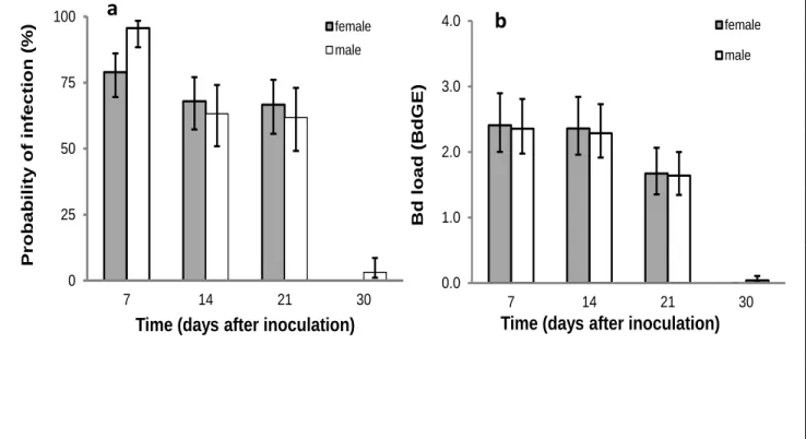
Figure 2-1- Changes in probability and intensity of the infection during the course of the experiment. ... 46
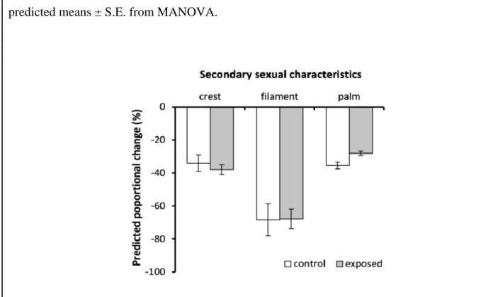
Figure 2-2- Effects of Bd exposure on male‟s secondary sexual traits. ... 48
Figure 2-3- effects of Bd exposure and load on body mass during the 30-day experiment. ... 50
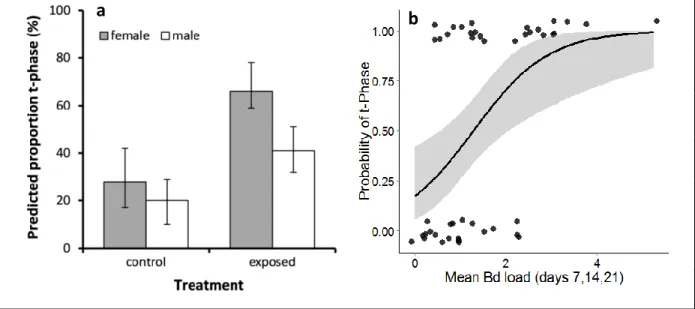
Figure 2-4- Consequences of Bd-exposure and Bd-infection for probability of termination of aquatic breeding season... 50
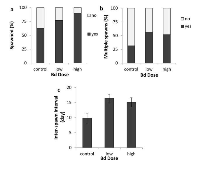
Figure 3-1- Impact of Bd infection on newts‟ spawning... 71
Figure 3-2- Impact of asymptomatic Bd infection on hatching success. ... 73
Figure 3-3- Consequences of Bd infection of parents for offspring... 75
Figure 4-1- Pathogen load over the course of the experiment. ... 95
Figure 4-2- Survival of newts over the course of the experiment. ... 95
Figure 4-3- Changes of mass in response to Bd. ... 97
Figure 5-1- Prevalence and intensity of Bd infection during aquatic phase. ... 117
Figure 5-2- transmission of Bd infection and infection intensity. ... 120
Figure 8-1- Histological section of severely infected digital skin of a wild Australian frog, Litoria caerulea (from Berger et al. 1998)... 169
Figure 8-2- Life cycle of Batrachochytrium dendrobatidis (from Rosenblum et al. 2008) ... 170
Figure 8-3- Schematic life cycle of the palmate newt... 171
Figure 8-4- Aquatic male palmate newt ... 172
Figure 8-5- Male secondary sexual traits ... 173
Figure 8-6- Female palmate newt in aquatic phase ... 174
Figure 8-7- Palmate newts from Puits d'Arius (chapter V) in Terrestrial phase ... 175
Figure 8-8- Experimental tanks were randomly distrusted in the housing facsility ... 176
Figure 8-9- Close contacts between uninfected newts and infected tadpoles (chapter V)... 177
Figure 8-10- Low altitude study site, the forest of Bouconne (chapter II and IV) ... 178
Figure 8-11- High altitude sites: above Puits d'Arius (chapter V); below Etang de Bouvet (chapter III and IV) ... 179
12
1. Chapter I
General introduction
1.1 Global amphibian declines: the enigmatic driver of the sixth mass extinction on earth
First concerns about widespread amphibian population declines are date back to 1970‟s in western United States (Drost and Fellers, 1996; Sherman and Morton, 1993), Puerto Rico (Burrowes et al., 2004), and other parts of the world. During 1980‟s a wave like simultaneous disappearance of mountain amphibians in several pristine habitats of Central America (Pounds and Crump, 1994; Pounds et al., 1997; Ron et al., 2003; Young et al., 2001) were again caught the attention of scientists to global amphibians decline (Stuart et al., 2004). These reports were initially received with some scepticism because amphibian populations often fluctuate widely (Pechmann and Wilbur, 1994). However, during 1990‟s long term experimental and quantitative studies showed that populations continued to decline, models indicated that declines are far more widespread and severe than would be expected under normal population dynamics (Blaustein et al., 1994; Drost and Fellers, 1996; Lips, 1998; Pounds et al., 1997; Tyler, 1991). By the end of 1990‟s retrospective studies indicated that declines may have been occurring since the late 1950s (Houlahan et al., 2000). The lack of sufficient data prompted the International Union for Conservation of Nature (IUCN) to sparkle a global amphibian assessment (GAA) for all known amphibians of the world, using IUCN Red List categories and criteria (IUCN, 2012). Results of GAA were astonishing in all respects (figure 1-1). While amphibians extinction rates since the year 1500 A. D. has been as low as 0.076 species per year (38 extinctions since then), currently, of the 6,260 amphibian
13 species assessed, 2030 species (32.4 %) are globally threatened or extinct and the situation of another 1,533 species is not known due to data insufficiency (IUCN, 2012) (figure 1-1a). The intensity and extension of global amphibian populations decline and the number of amphibian species at risk of extinction, which is more than those of any other taxon (Stuart et al., 2004), has made many scientists to argue that we are entering or in the midst of the sixth great mass extinction in the earth history (Gewin, 2008; Wake and Vredenburg, 2008).
Stuart et al. 2004 (Stuart et al., 2004) found that about 435 species that qualify for listing in IUCN categories of higher threat than they would have in 1980. They defined these species as “rapidly declining” species and divided them into three groups based on drivers of their decline: “overexploited”- declining because of heavy extraction (50 species); “reduced-habitat”- suffering significant habitat loss (183 species); and “enigmatic decline” - declining, even where suitable habitat remains, for reasons that are not fully understood (207 species). This was the first time in the history of conservational biology that enigmatic-declines have been recorded at such a considerable level. Interestingly, they also showed that the percentage of enigmatic-decline species increases with increasing extinction risk (figure 1-1-b), from 9.7% of the rapidly declining species in the IUCN Category Near Threatened, to 25.0% in Vulnerable, 47.3% in Endangered, 57.1% in Critically Endangered, and 92.4% in extinct in the wild. This observation suggests that the factors causing “enigmatic” declines are driving species toward extinction particularly rapidly. At that time, disease and climate change were the two plausible causes for enigmatic amphibians‟ decline (Berger et al., 1998; Burrowes et al., 2004; Daszak et al., 1999; Daszak and Cunningham, 2003; Lips, 1998). Nonetheless, in less than a decade, strong theoretical and empirical evidence were provided enough support to shed light on the wave-like amphibian extirpations in 1980‟s - 2000‟s.
14 Figure 1-1- Results of Global Amphibian Assessment (based on data from Stuart et al., 2004).
(a) GAA showed almost 32.4% of amphibians of the world are globally threatened or extinct and the situation of 40% of species is not known due to data deficiency. (b) in addition to the known causes of decline (e.g. habitat loss or anthropological activities) a considerable number of species are declining due to “enigmatic” reasons. The number of species with enigmatic reasons of decline is increases with the risk of the extinction.
Extinct 0% Extinct in the Wild 2% Critically Endangered 5% Endangered 11% Vulnerable 10% Near Threatened 9% Data Deficient 40% Least Concern 23%
a
0 100 200 300 400 500 600 Extinct in the Wild Critically EndangeredEndangered Vulnerable Near Threatened Num ber of Spec ies IUCN Category Enigmatic Decline
b
15 In effect, several independent studies in areas of amphibian declines revealed that rapid enigmatic declines are because of a new emerging infectious disease, the amphibian‟s chytridiomycosis (Berger et al., 1998; Cheng et al., 2011; Fisher et al., 2009b; Lips et al., 2008; Vredenburg et al., 2010; Wake and Vredenburg, 2008). This is the largest infectious disease threat to biodiversity in the history of the planet (Kilpatrick et al., 2010) and obviously, many facets underlying the devastating impact of chytridiomycosis on host populations are not thoroughly understood.
1.2 Emerging infectious diseases and their threat to wildlife
The term “Emerging Infectious Disease” (EID) was appeared in the infectious disease lexicon in 1990‟s and was referring to infections that have newly appeared in the population, or have existed but are rapidly increasing in incidence or geographic range (Morse, 1995; Morse and Schluederberg, 1990). Recent studies have shown that during less than a century (1940-2004), 335 infectious diseases have emerged in global human population (Jones et al., 2008) that have exerted, and are exerting a significant burden on global economies and public health (Binder et al., 1999; Morens et al., 2004). Among these, some EID‟s such as, acquired immunodeficiency syndrome, multidrug-resistant tuberculosis, Ebola, and tick-borne diseases are considered as threats to global human health and are still causing human mortality in some areas (Daszak et al., 2000). However, as for the humans, numerous EID‟s has been reported in a variety of wildlife populations which are decimating global biodiversity through population declines in multiple families of plants and animals. As such, amphibian‟s chytridiomycosis (Daszak et al., 1999; Fisher et al., 2009b), avian malaria (van Riper III et al., 1986), rinderpest in African ungulates (Plowright, 1982), sudden oak death in trees in western North America and Jarrah dieback or rootrot in trees in Australia (Anderson et al., 2004) are among the most
16 important traits to biodiversity and may account for at least some species extinction (Daszak et al., 2003; Fisher et al., 2009b). The increase of incident of wildlife EID‟s during the recent decades has primarily been attributed to anthropogenically induced changes in the ecology of the host and/or pathogen (Daszak and Cunningham, 2003) as well as human assisted disease introduction into naïve host populations (Daszak et al., 2001; Dobson and Foufopoulos, 2001).
EID‟s are found to be a major threat to global biodiversity (Daszak et al., 1999). First, they have the potential to cause dramatic decline in naïve host population. Second, after being established they may become enzootic and cause a chronic population depression (Daszak et al., 2000). In addition to the loss of biodiversity that is associated with some of these diseases, direct economic costs of EID‟s, such as EID induced collapse of fisheries‟ industry, livestock, and crop plant health costs (see (Daszak et al., 2000) for more details) are also reported from several countries. As opposed to human EID‟s very little is known about biotic and abiotic drivers of wildlife EID‟s and the interaction of hosts with these pathogens. During the past decades, wildlife diseases have only been considered when they were threating agriculture or human health (Daszak et al., 2000). However, the incidence of EID epidemics in endangered species, their veterinary impact and advances in host-pathogen population biology, nowadays, their threat is taken more seriously (Anderson and May, 1979; Daszak et al., 1999; Daszak et al., 2003; Fisher et al., 2009b; McCallum and Dobson, 1995; Thorne and Williams, 1988).
17 1.3 Host-pathogen interaction: implications in amphibian declines
1.3.1 Host-pathogen coevolution
At the heart of any parasitic relationship is a conflict of interest, but the battle for host resources is usually resolved in favour of the parasite (Price, 1980). Host resources may be invested in host energetic and cellular response to the infection (Bonneaud et al., 2003; Moret and Schmid-Hempel, 2000; Råberg et al., 2000), through the pathogens acquisition of host resources, or through inducing greater host susceptibility to other pathogens (Bonneaud et al., 2012; Casadevall and Pirofski, 2003; Hornef et al., 2002). As a consequence, even where the clinical symptoms of the infection and visible damage to the host are lacking, significant negative consequences arise through reduction in investment in traits associated with fitness (e.g., growth, survival and reproduction (Bonneaud et al., 2003; Burthe et al., 2008; Cheatsazan et al., 2013)). The ubiquity of parasites (Price, 1980), their ability to reduce host fitness (Sheldon and Verhulst, 1996) and deleterious effects on hosts populations (Anderson and May, 1979; Prado et al., 2009) have led to the evolution of host defence strategies, to minimize or neutralize negative fitness consequences of pathogens (Hedrick, 2004). On the other hand, in order to exist pathogens must successfully dodge the host‟s defences, at least for long enough to replicate and invade another host. Such interaction is generally associated with reciprocal, adaptive genetic changes between the host and the pathogen which are referred as antagonistic host-pathogen co-evolution (Woolhouse et al., 2002). The phenomenon is locked the host and the pathogen in an evolutionary “arms race” (van Valen, 1973) where the effects on the fitness of the host and pathogen act in opposite directions: what is good for the pathogen is bad for the host and vice versa (Woolhouse et al., 2002).
18 1.3.2 Host resistance vs. pathogen’s virulence: how EID’s can extinct a host?
As pathogens rely on host resources, evolution of resistance or avoidance in a host population exert a selective pressure on pathogen to choose between extinction and evolution of new traits to increase its ability to infect these resistant or avoidant hosts (Lion and Boots, 2010). In other words, pathogens need to trade-off between the rates of transmission from a host to another which would result in more rapid spread of the pathogen in the host population, and their virulence to sustain mobility and longevity of the host, at least for long enough for transmission to a new host (Anderson et al., 1992; Bull, 1994; Ewald, 1993; Read, 1994). Although increased transmission rate is likely to bring a short term fitness advantage for pathogen, but, it is usually induce clinical symptoms and threatens host life which has a negative impact on pathogen fitness (Combes, 1997; Ewald, 1995; Hurd, 2001). On the other hand, more rapid spread among host population may also be associated to rapid depletion of susceptible hosts form population (resistance/mortality) which will drive pathogen toward extinction (Grenfell and Harwood, 1997). In summary, the fitness of the pathogen would be maximized through a balanced transmission-virulence that would be achieved by maximizing the number of secondary hosts infected and optimization of the balance between replication and within host persistence.
Hosts have evolved distinct strategies to avoid pathogens, to reduce or remove their infection burden (resistance) or to decreases their susceptibility to damage and fitness costs caused by the pathogens or by the immune response against it (tolerance) (Råberg et al., 2009; Read et al., 2008). Costs may arise from collateral damage to host tissue during destruction and elimination of pathogens or alternations of normal tissue function (Majno and Joris, 2004; Medzhitov, 2010; Medzhitov et al., 2012). Collectively, negative effects of immune function on host fitness are referred as immunopathology (Graham et al., 2005). Insufficient immunity
19 results in a high rate of mortality from infections, thus, the optimal immune response is attained by a balance between efficient pathogen removal and an acceptable level of immunopathology (Casadevall and Pirofski, 1999, 2003). Given the vital benefits of the immune function for host survival, the acceptable level of immunopathology can be high causing the prevalent incidence of infectious disease symptoms (Graham et al., 2005; Medzhitov et al., 2012). Adding the costs associated to maintenance of surveillance and defensive immunity to the formers, possession of a complementary strategy to put forth a trade-off between costs of resistance and limiting tissue damage would increase the ability of the host to increase the magnitude and duration of the immune response than would have been otherwise possible (Hurd, 2001; Medzhitov, 2010; Medzhitov et al., 2012). This ability is refers as tolerance and consists the third major defensive strategy in animals and plants. Unlike resistance mechanisms, tolerance does not directly affect pathogen burden, thus, does not induce a direct selective pressure on pathogen. Rather, tolerance decreases the host susceptibility to tissue damage, or other fitness costs, caused by the pathogens or by the immune response against them (Råberg et al., 2009; Read et al., 2008).
Any negative impacts on the rates of host survival, reproductive output, immigration and emigration can lead to population decline and extinction. However, endemic pathogens alone rarely cause extinctions because of a co-evolutionary arms race between the host and pathogen that selects for more resistant host genotypes and less virulent pathogen genotypes, allowing them to coexist (Anderson and May, 1978). One of the most important features of wildlife‟s EID‟s is their ability to drive naïve populations toward extinction. The mechanism by which a pathogen can drive host population to extinction depends on the host-pathogen system (Anderson et al., 1986; De Castro and Bolker, 2005; Holt and Pickering, 1985;
20 McCallum and Dobson, 1995). In an obligatory single host-single pathogen system with density dependent transmission, pathogen cannot drive the host population to extinction. This is because a decline in host population size is equivalent to pathogen population decline which results in host population recovery cycle. Nonetheless, in such systems shrunk host populations are threaten by the risk of stochastic extinctions due to stochastic environmental or demographic events. Under an alternate circumstances, if the transmission of the pathogen is frequency dependent (e.g., sexually transmitted or vector-borne infections), pathogen infects multiple hosts or can survive outside the host body (e.g., commensal to other species or in an saprophytic state) decrease in (one) host population does not impose populations decline to the pathogen and pathogen can easily drive one or most hosts to extinction (Dobson, 2004; Mitchell et al., 2008). This latter is the case of many wildlife EID‟s which within a habitat, usually infect multiple hosts with different levels of susceptibility (De Castro and Bolker, 2005; McCallum et al., 2009).
Currently, emerging infectious diseases are one of the most important threats to human health and global biodiversity (see above). In order to understand pathogen abundance in natural host populations it is vital to integrate an understanding of how pathogens invade, replicate, and transmit along into an understanding of host ecology, including susceptibility, behaviour, and any infection-induced changes to these (Plowright et al., 2008; Tompkins et al., 2011). Moreover, understanding underlying mechanisms and consequences of host-pathogen interactions and co-evolution in these systems is a key first step in assessing the risk and the possibilities to mitigate infectious disease impacts on wildlife, livestock or agricultural systems (Woodhams et al., 2011) and importantly, to assess and predict the risk of emergence of new infectious diseases from endemic pathogens (Altizer et al., 2003).
21 1.4 Chytridiomycosis: largest infection threat to biodiversity
Chytridiomycosis was first identified in dead and dying frogs in late 1990‟s by two teams working independently on wild frogs from Central America and Australia (Berger et al., 1998) and captive frogs in united states (Longcore et al., 1999; Pessier et al., 1999). The fungal pathogen was then named Bathrachochytrium dendrobatidis (Bd) (Longcore et al., 1999) and soon was also known as the amphibian‟s chytrid fungus. Upon the discovery of the pathogen, Koch‟s postulates were then fulfilled by exposing healthy frogs to the new pathogen and re-isolation of the pathogen from diseased frogs (Berger et al., 1998; Longcore et al., 1999). Despite to the discovery of the pathogen and its association with chytridiomycosis, linking Bd to wild amphibian population declines was not easy. Evidences were attained through pattern identification of declines (Richards et al., 1993; Stuart et al., 2004), ecological modelling to explain patterns (Lips et al., 2003) and validation of the hypotheses under experimental circumstances (Berger et al., 2004; Longcore et al., 1999). As a consequent, so far, the fungus is found to infect more than 350 species on five continents from which more than 200 species were experiencing enigmatic declines and are among the highly threatened species of the world (Fisher et al., 2009b; Lips et al., 2008; Skerratt et al., 2007).
1.4.1 Emergence of chytridiomycosis
Two different theories are proposed on the origin of Bd (Fisher et al., 2009b). The debate is focused on whether Bd is a new, emerging pathogen. This is the novel pathogen hypothesis (NPH) or spreading pathogen hypothesis (Skerratt et al., 2007). The counter argument is that
22 environmentally driven changes has become has obtained more virulence. This is known as the endemic pathogen hypothesis (EPH). However, NPH receives support from observations that the distribution of Bd is patchy at many scales for decades prior to the onset of declines.
Current evidence suggests that the chytrid fungus is a novel pathogen that has spread globally outside of its native range (Rachowicz et al., 2005; Skerratt et al., 2007). The occurrence of sudden mass mortality events in amphibians in the absence of environmental pressures, is characteristic of introduction of infectious diseases into naïve host populations (Daszak et al., 1999).The wave-like pattern of amphibian declines in Australia (Laurance et al., 1996) and Central and Southern America (Catenazzi et al., 2010; Lips, 1999; Lips et al., 2004) are in agreement with movement of an infective agent through naïve landscapes (Rachowicz et al., 2005; Vredenburg et al., 2010).
Thus far the oldest record of Bd infection have been found in archived museum specimens of African clawed frog, Xenopus laevis, in 1938 (Weldon et al., 2004). This record precedes any recorded amphibian decline events and X. laevis shows no clinical effects of infection or population decline. It has therefore been suggested that the native range of the amphibian chytrid fungus is within Africa (Weldon et al., 2004). The NPH hypothesis explains that Bd was introduced to North America through international trading of the African clawed frog,
Xenopus laevis, from southern Africa began in the mid-1930s for use in pregnancy tests and
subsequently for laboratory experiments. Then it was introduced to other parts of the world by introduction of the North American bull frogs, Lithobates catesbeiana, that has also began in the 1930s for food and continued at least until late 1990s (Fisher and Garner, 2007; Garner et al., 2006; Weldon et al., 2004). Although, Bd genetics and genomics are pointed to a characterized globalized hypervirulent Bd lineage that suggests the emergence and spread of
23 chytridiomycosis largely owes to the globalization of this recently emerged recombinant lineage (Farrer et al., 2011), there are still some debates opposing NPH hypothesis. Finally, in addition to amphibians trade, transport of infected water and substrates such as soil and sand may also have contributed to the spread of the chytrid fungus (Johnson and Speare, 2003a, 2005; Longcore et al., 1999). However, despite to the above mentioned evidence, the origin of
Bd is still under debate and further detailed phylogeographic studies are necessary of
understanding the disease origin.
1.4.2 Taxonomy of Batrachochytrium dendrobatidis
The amphibian chytrid fungus is a member of the Chytridiales, one of five orders within the phylum Chytridiomycota, a group of primitive heterotrophic fungi that are characterised as having thalli with chitinous cell walls, an absorptive mode of nutrition and an unwalled flagellated zoospore stage that requires water for dispersal (James et al., 2000; Powell, 1993) (figure 8-2). They are important decomposers of keratin and chitin, and live saprophitically or as parasites of fungi, vascular plants and invertebrates (James et al., 2000; Powell, 1993). The amphibian chytrid fungus is distinct from other Chytridialean genera in thalli development and zoospore ultrastructure and so was placed into a new genus (Longcore et al., 1999). It is the only chytrid species described that infects vertebrates (Longcore et al., 1999).
1.4.3 Life cycle, pathogenesis and pathophysiology
The life cycle of Batrachochytrium dendrobatidis has 2 main stages: the motile, waterborne, short-lived zoospore for dispersal, and the stationary, monocentric thallus, which develops into a zoosporangium for asexual amplification (Berger et al., 2005a) (figure 8-2). Although occurrence of sexual reproduction leading to formation of a resistant or resting
24 zoospore is common in other chytridiomycota (Barr, 1978), sexual reproduction is not observed in Bd. However, loose evidence of a recombinant resting zoospore with thick cell wall has been found (Di Rosa et al., 2007; Morgan et al., 2007).
Zoospores are generally spherical with a diameter of 3-5 μm and a single posterior flagellum (Longcore et al., 1999). Motile zoospores disperse through liquid for up to 24 hours in search of a host, using positive chemotaxis to locate keratinised tissue (Moss et al., 2008; Piotrowski et al., 2004). Bd colonization occurs in the cells of the host epidermis (Berger et al., 2005b; Longcore et al., 1999). Then Bd enters into epidermal cells, encysts and fills them with up to three sporangia (Longcore et al., 1999). Hair-like rhizoids extending into host tissue to anchor the cell and absorb nutrients (Berger et al., 2005a). The zoospore then increases in size, becoming a zoosporangium. Zoosporangium grows to reach up to 40 μm in diameter before the cytoplasm cleaves into numerous uni-nucleate zoospores (Berger et al., 2005a). Upon maturity, zoospores are released into the environment through a discharge tube, completing the life cycle (Berger et al., 2005a). In culture, the amphibian chytrid fungus can complete its life cycle in less than 5 days, but this may differ in natural environments (Piotrowski et al., 2004).
Interestingly, the rate of Bd sporangia development is well matched to the maturing and keratinization rate of amphibian‟s epidermis cells. This adaptation causes the sporangia grow initially in living cells but complete their development in dead superficial keratinized cells that lack organelles when their discharge tubes can easily merge with and dissolve the epidermal cell membrane and open on to the surface of the cell, usually the surface distal from the body(Berger et al., 2005a). Berger et al. 2005 (Berger et al., 2005a) found these
25 specialized adaptations as an evidence that suggests the fungus has long evolved to live in amphibian skin.
The reproductive biology of Bd appears to be a critical factor in disease development and mortality (Voyles et al., 2011). In amphibian‟s skin local re-infection of an individual host seems to be the cause of an exponential increase in Bd load and development of severe infections and mortality (Briggs et al., 2010; Vredenburg et al., 2010). In infected frogs, clinical signs of disease and mortality occur in individuals with the highest zoospore loads (Voyles et al., 2007; Voyles et al., 2009; Vredenburg et al., 2010), suggesting that zoospore production and intensity of infection are critical for pathogenesis. Optimal growth of Bd is depending on temperature (17°–25°C), pH, and density (Longcore et al., 1999; Piotrowski et al., 2004). Although Bd can grow in either high or low temperatures, low temperature (4°C) induce a trade-off for lower population growth rates against increased zoospore activity and virulence (Voyles et al., 2012; Woodhams et al., 2008). This life history trade-off is proposed to be one the reasons of more devastating effects of Bd in high altitudes.
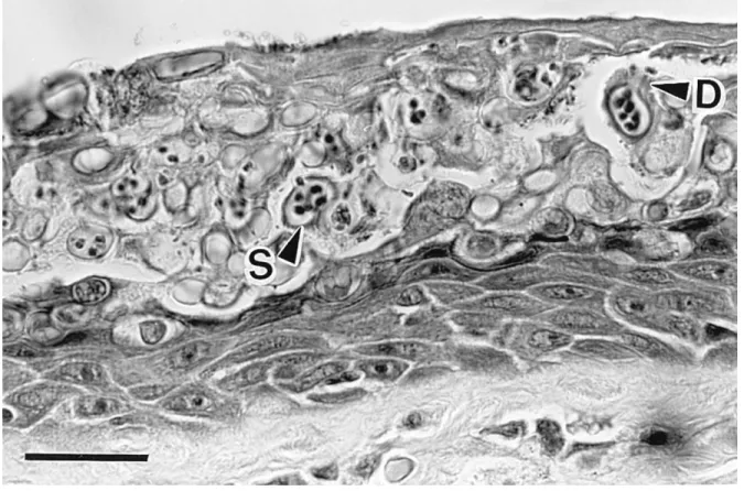
Primary pathological abnormalities include epidermal hyperplasia and hyperkeratosis (thickening of the stratum corneum; Figure 8-1) (Berger et al., 1998; Voyles et al., 2011). Other pathological changes in the epidermis include cytoplasmic degeneration and vacuolation in scattered cells in deeper layers (Longcore et al., 1999). Epidermal damage caused by Bd appears to disrupt critical cutaneous functioning such as electrolyte transport across skin which results in reductions in plasma electrolyte concentrations and deterioration of cardiac electrical functioning leading to a systolic cardiac arrest in clinically diseased animals (Voyles et al., 2007). These physiological malfunctions of the skin are associated with some clinical symptoms. As such, lethargy on skin, inappetence, cutaneous erythema,
26 irregular skin sloughing, abnormal posture (hind legs aBducted), and loss of righting reflex (Voyles et al., 2007; Voyles et al., 2009).
1.4.4 Impacts of Bd on different amphibians
Although Bd represents extreme host generality and is virtually capable to infect nearly all amphibians (Bonaccorso et al., 2003; Bosch et al., 2007; Bosch and Martínez-Solano, 2006; Green and Sherman, 2001; Kriger et al., 2006a; La Marca et al., 2005) amphibians‟ morbidity and mortality is highly host specific (Blaustein et al., 2005; Parris and Beaudoin, 2004; Voyles et al., 2011; Woodhams et al., 2007). Until now, several studies have shown some amphibians are able to carry the infection asymptomatically (Daszak et al., 2004; Davidson et al., 2003; Reeder et al., 2012; Retallick et al., 2004b; Rollins-Smith and Conlon, 2005) and serve as potential Bd reservoirs for other species. Histological examination of some of these carriers suggests that Bd infects their skin and they suffer from skin abnormalities similar to those observed with lethal Bd infection, but changes were usually limited to a patchy distribution (Reeder et al., 2012). However, the impacts of asymptomatic Bd infection, particularly for adults are not thoroughly investigated.
1.5 The palmate newt
1.5.1 Distribution, taxonomy and identification
The palmate newt, Lissotriton helveticus (Razoumowsky, 1789), formerly Triturus
helveticus, is a urodele from the family Salamandridae and Pleurodelinae subfamily. This
27 Luxembourg, and in the north of Portugal. Along this range, the species has an altitudinal range from sea level up to at least 2,400m asl in Pyrenees Mountains (IUCN, 2012).
Males‟ body size may reach up to 85 mm while females are usually longer and may reach to 95 mm length. Newts carry glandular ridges alongside the back give the male newts a square-backed appearance. Breeding males exhibit several secondary sexual traits: a low, smooth crest along the back that continues into a higher crest on the tail; a thin filament at the end of the tail; black webbing on the hind feet; and more swollen and darker cloaca than females (see Appendix) (Gasc, 1997; Griffiths and Mylotte, 1988; Nöllert and Nöllert, 2003).These characteristics become less distinct, and disappear during the terrestrial stage (Griffiths, 1996; Griffiths and Mylotte, 1988). The base colour of both sexes is olive-green or brown. A dark line runs from the nares, through the eyes, across the head. Both sexes have a yellow or pale orange belly that can sometimes show some spotting (See appendix). Males‟ secondary sexual characteristics have been shown to be associated with male reproductive success (Cornuau et al., 2012; Green, 1991a, b).
1.5.2 Life cycle and habitat requirements
Palmate newts can live till 8 to 11 years in the wild (Amat et al., 2010). Breeding and larval development take place in aquatic habitats with still or very slow-moving water during the breeding season, but otherwise have a terrestrial life style (Secondi et al., 2009) (appendix). It is found in a variety of habitats including marshes, heathlands, moorlands, forests (where it may be very common), pastures and agricultural land occupying moist habitats and refugia (Denoël and Ficetola, 2007; Nöllert and Nöllert, 2003). Reproduction occurs by transferring the male spermatophore that is placed on the ground and the female pick it up to her cloaca. Females can take several spermatophores several different males
28 during the breeding season (Gabor and Halliday, 1997). By termination of breeding season, males and females return to land feedings (Griffiths and Mylotte, 1988). Development of the eggs would be completed in 3-4 weeks and hatchlings will stay more than 8 weeks in the water before their transition to efts. Sexual maturity occur after the efts become newts after 3-4 years (Gasc, 1997; Nöllert and Nöllert, 2003).
1.5.3 Threats and trend
The species is very common in suitable habitats over much of its range. Nonetheless, it is endangered in the Netherlands, Belgium and Luxembourg and uncommon (with a fragmented range) in northern Portugal and declining in parts of Spain (Gasc, 1997). The species is locally threatened by general drainage, pollution and eutrophication of breeding pools, abandonment of traditional farming practices and the introduction of predatory fishes and crayfish (Procambarus clarkii). Desertification is a threat in southern parts of the range (IUCN, 2012). The palmate newts is listed as Least Concern in IUCN red list and also is listed on Appendix III of the Bern Convention and is protected by national legislation in most parts of its range (IUCN, 2012).
1.5.4 Chytridiomycosis and the palmate newt
Wood et al. (Wood et al., 2009) found that palmate newts in Bd infected sites of Luxembourg are lightly infected but their infection burden is quiet low, fairly above detection threshold of quantitative PCR technique, so, they proposed more comprehensive assessments. Nonetheless, based database of global Bd mapping project (www.Bd-maps.net, see (Olson et al., 2013) for details) in surveys of Iberia in 2006-2007, 3 out of 7 palmate newt samples were tested positive for Bd. Prevalence of Bd in these sites was 20% - 100% but due to small size
29 of their samples these numbers might not represent the infection status of this species. In French Pyrenees, Palmate newts were sampled in two sites; both samples were infected (95% and 100%). In none of these sites, Bd induced morbidity and mortality of the palmate newt was recorded, suggesting that the palmate newt is somewhat resistant to Bd. However, prior to our study, the consequences of exposure to Bd, which will most likely occur during the aquatic phase as a result of increased probability of exposure to waterborne Bd zoospores, have not been investigated in details.
1.5.5 Palmate newt as model organism
Palmate newt, possess several unique aspects that make it a proper model for this study: - Despite to presence in IUCN red list as Least Concern status susceptibility/resistance of this species to Bd is not studied
- Possess a distinctly separated reproductive phase (aquatic phase)
- A considerable amount of quantifiable morphological and behavioural sexually dimorphic traits
- Sexual morphology and reproductive behaviour is well studied in terms of ethology, ecology and physiology, provide a considerable amount of background data for a better understanding host specific drivers of the host response to Bd
- Long reproductive season that suits to our experimental design
- High reproductive output and short incubation period; this would let us to conduct experiments with offspring
- The species is dominant and consequently frequent in our study area, guaranteeing access to proper sample sizes for experimental studies
30 1.6 Thesis objectives and outline
A growing body of evidence suggest that asymptomatic Bd infection can sub-lethally affect larvae and tadpoles (Han et al., 2008; Venesky et al., 2009; Venesky et al., 2010b) leading to reduced survival or impaired development (Garner et al., 2009). Nonetheless, little is known about the sub-lethal costs of the infection for adult amphibians and, importantly, it is not clear whether Bd incurs any direct or indirect fitness cost to subclinically infected adults. The main aim of this thesis was to investigate the impact and the outcome of Bd infection for individuals of an asymptomatic species (Bd carrier), and to understand why some populations of some species persist with Bd while other populations of the same species decline to extinction. In order to disentangle the impact of Bd on asymptomatic host and factors which determine the final outcome of the infection for the host, we followed these primary goals:
1- Diagnosis of sub-lethal costs of Bd infection in the model amphibian
2- Understanding and quantifying the impact of sub-lethal costs on main fitness components (i.e. survival and reproduction)
3- The direct and indirect role of environment in determining the final outcome of the infection for the host
4- Development of host resistance against Bd
5- Costs of the infection for resistant (tolerant) individuals with a Bd invasion history In addition, wherever applicable, we also investigated probable sex-dependent differences in the impact of Bd, and evaluated the efficiency of the current methodological approaches in assessing the risk of the infection for asymptomatic amphibians.
The results of the thesis are organized in five standalone but interconnected chapters which are published or are in preparation to be published as a peer-reviewed scientific paper. In the second chapter we first establish the resistance of our model organism and then develop
31 a framework for quantification of costs associated with subclinical Bd infection. We then assess costs that might be related to reproduction and discuss the underlying drivers of the impact of Bd on naïve individuals in the light of our new findings.
In the third chapter, we investigate the reproductive costs of asymptomatic Bd infection for asymptomatic host, the intra-population variation in response to Bd, and test for consequences of the infection for the offspring. We discuss the significance of our results in the ecology and evolution of host-Bd interaction. Finally we outline the future directions for a more effective disease mitigation plans.
In the fourth chapter, we examine the role of the environmental context in determining the impact of the infection for the host. This is one of the most controversial issues in ecology of
Bd. Our results contribute to the former regional studies that emphasize on the indirect role of
environment on determining the final outcome of Bd infection for the host population. In addition to providing experimental evidence for among and within population variations response to Bd, we criticise utilization of infection prevalence for assessing the risk or severity of infection in amphibian populations.
In the fifth chapter, we address another highly controversial question: whether or not populations with a Bd invasion history acquire resistance against Bd. Using a unique experimental design with naturally infection individuals, we investigate infection patterns, prevalence and intensity of the infection in a palmate newt population with a recent outbreak of fatal chytridiomycosis. We also assess newts‟ defensive approach against Bd and show how resistance and tolerance to Bd contribute to minimize the detrimental cost of the infection in newts.
32 In the final chapter of the thesis, we discuss the connection among our results, and discuss their implications in understanding ecology of Bd and conservation of amphibians. Finally, we provide a short list of questions and directions for the future research.
33
2. Chapter II
Sub-lethal costs of chytridiomycosis for an asymptomatic species
2.2 Synopsis
It is well established that infection is costly for individuals that remain asymptomatic (i.e. subclinical infection). For example, not only does mounting an immune response require energy (Bonneaud et al., 2003; Råberg et al., 2000), but pathogens can use up host resources and induce tissue degradation, as well as increase host susceptibility to other pathogens (Bonneaud et al., 2012; Hornef et al., 2002). Furthermore, immune response to pathogen is usually associated with damage to the host tissue and impairing normal tissue functioning which are referred as immunopathology (Graham et al., 2005; Medzhitov, 2010; Medzhitov et al., 2012). The predicted consequences ensuing from sub-lethal costs of infections have been demonstrated empirically in a variety of systems, as decreased investment in several components of host fitness (e.g., growth and reproduction, (Bonneaud et al., 2003; Festa-Bianchet, 1989; Sheldon and Verhulst, 1996). Up to now, several studies have shown that subclinical Bd infection is costly for amphibians (e.g. Chatfield et al., 2013; Garner et al., 2009; Han et al., 2008; Retallick and Miera, 2007; Venesky et al., 2009). Nonetheless, it is not clear whether and to what extent these costs are likely to impair adults‟ reproduction or survival.
In this chapter, we report results of our study with a Bd-naïve population of the palmate newt that have been exposed to a very low dose of Bd (1000 Bd zoospores/ l) during their aquatic breeding season. Our study addresses the current gap in understanding the sub-lethal costs of Bd infection and its potential effect on reproduction. To our knowledge this study provides the first detailed observation of condition and reproductive costs of
34 asymptomatic Bd infection in an asymptomatic species. In addition, for the first time, we show how investment in clearing Bd infection may affect host reproduction and survival. Our results are recently published in BMC ECOLOGY journal (Cheatsazan et al., 2013). Given the importance and novelty of our results, the article became very successful and marked as “Highly Accessed” due to nearly 4500 access in less than five month after publication.
In this study, we inoculated a group of wild-caught naïve palmate newts with a single dose of Bd (18 controls and 50 exposed; 2000 zoospores per litre) to test: (1) whether palmate newts become infected by exposure to very low doses of Bd, (2) whether they are able to tolerate or clear the infection and prevent fatal chytridiomycosis; and (4) the consequences of
Bd infection and infection load for secondary sexual characters, condition and the probability
of an early entry into the terrestrial phase. Our results suggest that Bd has measureable, but sub-lethal effects, on adult palmate newts. Although we found no evidence of increased secondary sexual trait regression, through reducing individual body condition and potentially, breeding season duration, we predict that Bd exposure might have negative impacts on populations of palmate newts through reducing individual reproductive success and adult recruitment. This hypothesis receives experimental support from the results of the next chapter and will further be discussed there.
Hitherto, several explanations have been proposed to link asymptomatic chytridiomycosis with sub-lethal morphological and behavioural abnormalities of Bd infection. For example, shorter body size of subclinically infected individuals are proposed to be due to: (i) shorter individuals occupy habitats with higher suitability for Bd (Beard and O‟Neill, 2005); (ii) lower resistance of shorter individuals to Bd (Kriger et al., 2006b; Lamirande and Nichols,
35 2002); (iii) death of infected frogs before reaching maximal body size (Garner et al., 2009; Parris and Cornelius, 2004); (iv) Bd infection causes developmental constraints resulting in shorter body size (Burrowes et al., 2007; Kriger et al., 2006b; Parris and Cornelius, 2004); and (v) life history trade-offs imposed by costs of immune response, tissue degradation or immunopathology to growth and development (Garner et al., 2009). The latter is also suggested as the driver of the only known behavioural abnormality in subclinically infected adults (Chatfield et al., 2013). Despite to over a decade of research, the underlying driver(s) of sub-lethal costs of chytridiomycosis and whether they are likely to impair host fitness leading to the possibility of population decline in asymptomatic amphibians is not clear. Our findings contribute to this field by providing experimental evidence linking how and to what extent subclinical Bd infection is able to cause morphological and behavioural deficiencies. We showed that body size and condition are neither affecting probability of newts being infected nor their survival and mass investment to clear their infection load. Therefore, observed differences in morphology and behaviour of subclinically infected adults are not due to an age or size dependent increase in host ability to resist Bd or survive after the infection. Conversely, our study reveals sub-lethal costs of Bd infection are increased with infection intensity. In this way, our results support the life history trade-off hypothesis which predicts investment in host response to subclinical infection is associated with less investment in other physiological demands (reproduction, growth, body maintenance, etc.). Although for obvious ethical and conservation reasons, we were unable to measure the consequences of experimental infection for naïve individual in the wild, we suggest that Bd outbreak has a negative impact on naïve populations of palmate newts, and species with similar defensive strategy, through indirect impacts on survival, reproductive morphology and reproductive
36 behaviour (Burrowes et al., 2007; Garner et al., 2009; May et al., 2011; Richards-Zawacki, 2010).
37 2.3 Experimental evidence for a cost of resistance to the fungal pathogen,
Batrachochytrium dendrobatidis, for the palmate newt, Lissotriton helveticus
From: Cheatsazan, H., A. P. de Almedia, A. F. Russell and C. Bonneaud (2013). "Experimental evidence for a cost of resistance to the fungal pathogen, Batrachochytrium dendrobatidis, for the palmate newt, Lissotriton helveticus." BMC Ecology 13(1): 27.
2.3.1 Introduction
Batrachochytrium dendrobatidis (Bd), the causative agent of chytridiomycosis, has been
implicated in worldwide amphibian declines (Kilpatrick et al., 2010). Although Bd is able to infect a wide range of species and hence displays extreme host generality (Bonaccorso et al., 2003; Green and Sherman, 2001; Kriger et al., 2006a), amphibian morbidity and mortality in response to infection is highly variable and host-species specific (Gervasi et al., 2013; Voyles et al., 2011). For example, while some studies report devastating consequences of Bd infection (Retallick et al., 2004b; Schloegel et al., 2006; Skerratt et al., 2007), others have shown different levels of resistance to Bd infection (capacity to prevent or clear Bd infection) and have provided experimental evidence showing some species are able to clear Bd infection (Daszak et al., 2004; Davidson et al., 2003; Reeder et al., 2012; Retallick et al., 2004b; Rollins-Smith, 2009). In several species, infiltration of neutrophils, lymphocytes, macrophages and inflammation have been reported on the skin of susceptible (i.e., those which are not able to clear Bd infection or to prevent colonization of Bd) amphibians (Berger et al., 2005a; Davidson et al., 2003; Nichols et al., 2001; Pessier et al., 1999). Histological examinations of resistant hosts have revealed that Bd infections can cause skin injuries similar to those observed in susceptible species, albeit distributed more patchily on the skin (Daszak
38 et al., 2004; Davidson et al., 2003; Reeder et al., 2012). These results suggest that those amphibians that are apparently resistance to Bd infection might still experience sub-lethal costs but whether any such costs are likely to impair traits associated with reproductive success and survival is unclear.
It is well known that pathogens exert significant cost for the host (Lochmiller and Deerenberg, 2000; Sheldon and Verhulst, 1996). Costs may be manifest through the host‟s energetic and cellular response to the infection (Bonneaud et al., 2003; Moret and Schmid-Hempel, 2000; Råberg et al., 2000), through the pathogens acquisition of host resources, or through inducing greater host susceptibility to other pathogens (Bonneaud et al., 2012; Casadevall and Pirofski, 2003; Hornef et al., 2002). As such, hosts might show clinical symptoms and visible damage (Casadevall and Pirofski, 2003), but even where such effects are lacking, significant negative consequences can arise through reductions in investment in traits associated with fitness (e.g., growth, survival and reproduction, (Bonneaud et al., 2003; Sheldon and Verhulst, 1996). As a consequence, in species exhibiting apparent resistance to
Bd, we might also expect there to be sub-lethal costs of infection and for such costs to
influence populations. In support, a number of studies in frogs and toads have reported subclinical effects of Bd on tadpole growth (Davidson et al., 2007; Garner et al., 2009; Venesky et al., 2010a; Venesky et al., 2009), as well as adult body size (Burrowes et al., 2007) and body condition (Richards-Zawacki, 2010). Nevertheless, in such cases, tests of the consequences of Bd infection for traits important in reproduction (morphology, behaviour) of resistant amphibians are generally lacking.
The palmate newt, Lissotriton helveticus (Cuadata: Salamandridae; formerly Triturus
39 aquatic habitats with still or very slow-moving water during the breeding season, but otherwise have a terrestrial life style (Secondi et al., 2009), occupying moist habitats and refugia (Nöllert and Nöllert, 2003). Males develop large ornaments when they enter water for breeding, manifested as a low, straight edged crest which extends dorsally, a thread-like filament projecting from the end of the tail and conspicuous webbing between the toes of the hind feet (Griffiths and Mylotte, 1988). These secondary sexual characteristics have been shown to be associated with male reproductive success (Baker, 1992; Cornuau et al., 2012; Green, 1991b; Secondi et al., 2009). They then gradually start to regress as the newts leave their aquatic habitat, but may not necessarily have fully disappeared as individuals start their migration to their terrestrial feeding (Griffiths and Mylotte, 1988).
Interestingly, palmate newts have been reported in several Bd infected sites across Europe and have been found to test positively for Bd (WWW.Bd-maps.net (Imperial Clolledge, 2014). Despite this, no morbidity or mass mortality have been reported (Imperial Clolledge, 2014; Wood et al., 2009) and under laboratory conditions, excessive weekly exposures to more than 106 Bd zoospores per week for more than 4 weeks does not induce symptomatic chytridiomycosis even up to 40 days following a month-long exposure course (Cheatsazan, unpublished results 2011, n = 35 newts). These observations suggest that this species exhibits some level of resistance to Bd. However, the consequences of exposure to Bd, which will most likely occur during the aquatic phase as a result of increased probability of exposure to waterborne Bd zoospores, have not been investigated for this species. Here, we inoculated a group of wild-caught palmate newts experimentally with a single dose of Bd (18 controls and 50 exposed) and monitored Bd infection, survival and clinical symptoms, as well as changes in morphological and behavioural traits known to be associated with reproductive success in
40 this species. Our specific aim was to use this experiment to test: (1) whether palmate newts become infected by exposure to low doses of Bd, whether they show external symptoms and whether they are able to tolerate or clear the infection; (2) the consequences of Bd exposure and infection load for secondary sexual characters in males; (3) the consequences of Bd exposure and infection load for mass change in both males and females; and (4) whether exposure to Bd is associated with changes in the probability of an early entry into the terrestrial phase. Throughout, we consider sex differences, where possible.
2.3.2 Methods
In mid May 2011, 68 newts (34 males and 34 females) in aquatic phase were captured in narrow streams on the borders of Bouconne forest, Haute Garonne, France (43º 39‟ N and 1º 14‟ W, 190 m altitude). Our rationale for using wild-caught rather than laboratory-derived newts was that we wished to use individuals as close as possible to their natural state. The sites used are in a forest reserve with natural and man-made streams, and are located about 100 km outside the current extent of the known Bd infection zone. In addition, our inspections for Bd in this area since 2009 have failed to detect evidence of Bd infection of any amphibian species in the area, and all animals brought back to the laboratory tested negative for Bd. Male and female newts were randomly paired and transferred into plastic tanks (205× 205×140mm). Our reason for this is that wished to examine the effects of Bd exposure on male secondary sexual characteristics, which we considered to be likely influenced by the presence of a female. Each tank contained 1.0 litre of aged tap water and a hollow brick for shelter. We randomly assigned tanks to one of 2 treatment groups: controls (9 males, 9 females) and Bd-exposed (25 males and 25 females). All newts were maintained in the
41 laboratory for 7 days prior to experimentation and were provided with live midge larvae (blood worms), daphnia and/or tubifex every two days. Food was provided by „sucking‟ a known quantity of liquid (tube length x diameter = 55cmm x 0.5cm) containing roughly 75 larvae. Although the precise number of larvae was unknown, there is no reason to suppose this varied systematically between experimentally exposed and control tanks. Room temperature was maintained at 18.6±1.9°C throughout the experiment. Light exposure was adjusted weekly to equal the average day length of the first and of the last days of the focal week (approximately 14h light: 10h dark). Water was changed 3 days after inoculation and then weekly thereafter to prevent bacterial blooms in the tank. This also means that levels of
Bd loads detected were unlikely to be explained by zoospore survival in the water (Johnson
and Speare, 2003b).
Bd cultures were prepared from a Bd extract of an infected tadpole from an introduced
population of American bullfrog (Lithobates catesbeianus) in southern France (for extraction protocol see (Longcore, 2000). This extract was grown in 1.0% Trypton (SCHARLAU, cat. No 07-119) and 0.32 g × l-1 glucose (ROTH, cat. No X997) in ultrapure water. Cultures were incubated at 18.5˚C to optimize growth of the fungus (Voyles et al., 2011). Approximately one hour prior to inoculation, we counted approximately 160 active zoospores × ml-1 of the 5th passage of our culture using a haemocytometer. We then added 12 ml of this culture to each
Bd exposed tank (~2000 zoospores×l-1×tank-1). The experiment was stopped 30 days post-inoculation, when about approximately half of the newts had entered or were entering the terrestrial phase of the season (t-phase). When a newt consistently remained out of the water for five consecutive days, we considered the day the newt had left the water for the first time as the start of t-phase. Animal capture was with permission of the prefecture of Haute
42 Garonne (Permission No. 2009-12). Animal housing facility and experiments comply with the regulations of the housing organization (CNRS: National centre for scientific research) and the current rules of France.
Infection was detected by quantitative amplification of Bd-DNA content of weekly swab samples (from: forelimbs, hind limbs, aBdomen and cloaca) using fine tip dry swabs (Tubed sterile dryswabTM tip, Medical wire & Equipment, cat. No MW100). Swab samples were taken at upon arrival, day 7, 14, 21, and 30 after the first inoculation from all individuals. Quantitative PCR‟s were conducted as described in (Boyle et al., 2004), and after incorporating the changes suggested by Kriger et al. and Hyatt et al. (Bielby et al., 2009; Kriger et al., 2006b; Walker et al., 2007) except that the we reduced the final volume of reactions to 10 µl due to higher sensitivity of our q-PCR machine (Mastercycler ep realplex4, Eppendorf). Throughout this paper, results of all quantifications are presented in Bd Genome Equivalent (BdGE) per swab bout. In addition to standard duplicates and negative controls, we also included 6-12 samples of control newts in each test plates of exposed samples and utilized internal positive control reagent in all samples (TaqMan® Exogenous Internal Positive Control). To ensure the efficiency of inoculations, we also analysed swab samples of 66 individuals of the same population, after 2 weekly exposures to 106 zoospores of the same
Bd strain. The average Bd load of these newts, one week after the second inoculation, was
(mean ± S.E.) 442.2 ± 187.4 BdGE, showing the Bd strain and inoculation method was able to generate an infection in this population.
On days 0 and 30 post-inoculation, we measured body mass using a digital scale (±0.01 gram). Snout-to-vent length (SVL) and male secondary sexual traits were measured by ImageJ software on digital photographs of all individuals: area of tail crest, length of tail
43 filament, and average of webbed area of hind feet (left and right feet) one day before inoculation and day 30 after inoculation, according to (Cornuau et al., 2012). Photographs were taken on a single plastic board with a millimetre scaled area of 2×10 cm. All morphological measurements were taken twice by the same person (HC) and their average was used in statistical analyses. In all cases a strong correlation (r ≥ 0.90, p<0.001) was observed between replicates. Finally, we recorded survival, and the start of t-phase as well as symptoms of clinical chytridiomycosis (Voyles et al., 2011).
Infection patterns and symptoms-Changes in the probability of being infected during the
experiment were analysed by fitting whether (1) or not (0) a newt was infected on a given day as the response term in generalized linear mixed-effects model (GLMM) with a binomial error structure and logit link function. The binomial denominator and dispersion parameters were 1. Changes to infection loads during the course of the experiment, were analysed using a GLMM with Poisson error structure and log-link function in which load size (numbers of BdGE) was fitted as the response term. In both models, individual subjects‟ codes (ID‟s) nested within box number were fitted as random terms to account for repeated sampling of each. The SVL and body mass of individuals on day zero were fitted as covariates, while sex, days after the first inoculations (7, 14, 21, 30) and their interaction were fitted as fixed effects. Finally, throughout the experiment we examined the animals for signs to Bd-mediated symptoms.
Secondary sexual characteristics in males- Analysis of change in male secondary sexual
characters over the course of the experiment was conducting using multivariate analysis of variance, in order to account for multiple correlated terms of interest (crest area, tail filament length and rear foot webbing: Spearman‟s rank correlations between the traits, rs=0.4-0.86,