M´EMOIRE PR´ESENT´E
dans le cadre du programme de maˆıtrise en oc´eanographie en vue de l’obtention du grade de maˆıtre `es science
PAR
©JORDAN LATOUR
Jean-S´ebastien Deschˆenes, pr´esident du jury, Universit´e du Qu´ebec `a Rimouski (CAN)
Karine Lemarchand, directrice de recherche, Universit´e du Qu´ebec `a Rimouski (CAN)
R´ejean Tremblay, codirecteur de recherche, Universit´e du Qu´ebec `a Rimouski (CAN)
Bassem Allam, examinateur externe, Stony Brook University ( ´E.-U.)
Avertissement
La diffusion de ce m´emoire ou de cette th`ese se fait dans le respect des droits de son auteur, qui a sign´e le formulaire Autorisation de reproduire et de diffuser un rapport, un m´emoire
ou une th`ese . En signant ce formulaire, l’auteur conc`ede `a l’Universit´e du Qu´ebec `a
Ri-mouski une licence non exclusive d’utilisation et de publication de la totalit´e ou d’une par-tie importante de son travail de recherche pour des fins p´edagogiques et non commerciales. Plus pr´ecis´ement, l’auteur autorise l’Universit´e du Qu´ebec `a Rimouski `a reproduire, diffuser, prˆeter, distribuer ou vendre des copies de son travail de recherche `a des fins non commer-ciales sur quelque support que ce soit, y compris l’Internet. Cette licence et cette autorisation n’entraˆınent pas une renonciation de la part de l’auteur `a ses droits moraux ni `a ses droits de propri´et´e intellectuelle. Sauf entente contraire, l’auteur conserve la libert´e de diffuser et de commercialiser ou non ce travail dont il poss`ede un exemplaire.
Dans les ´ecloseries de bivalves, les pathog`enes opportunistes sont associ´es `a de grandes mortalit´es masse causant des pertes ´economiques importantes aux producteurs. Afin de contrˆoler ces ´ev`enements et stabiliser la production larvaire, des alternatives aux antibio-tiques sont propos´ees. Les probioantibio-tiques ou encore leurs mol´ecules bioactives sont associ´es `a des effets b´en´efiques pour les larves `a diff´erents niveaux, particuli`erement dans l’am´elioration de leur r´esistance aux stresseurs dont les pathog`enes bact´eriens. De nos jours, il est reconnu que la composition du microbiote influence l’´etat de sant´e de l’hˆote et qu’elle pourrait ˆetre une des cibles des probiotiques. Le but de cette ´etude est de mettre en ´evidence l’effet pro-tecteur d’une nouvelle mol´ecule bioactive, la marennine, sur les larves de Mytilus edulis durant des tests de provocations bact´eriennes avec le pathog`ene bact´erien opportuniste Vi-brio splendidus en relation `a une modification du microbiote larvaire des individus trait´es avec la marennine. L’hypoth`ese de ce m´emoire est que l’utilisation de la marennine du-rant l’´elevage larvaire pourrait modifier les conditions pr´evalant dans le milieu de culture et ainsi modifier la composition du microbiote larvaire menant `a une meilleure r´esistance aux infections bact´eriennes. La marennine est un pigment bleu produit par la diatom´ee Haslea ostrearia et a d´ej`a d´emontr´e un effet b´en´efique sur la survie larvaire lorsqu’utilis´ee `a une concentration de 500 µg L−1. Des larves-D et des post-larves ont ´et´e expos´ees `a V.
splendi-dus en pr´esence et en absence de marennine pendant 96 h. Nos r´esultats d´emontrent qu’`a cette concentration, la marennine n’a pas d’effet antibact´erien direct sur la croissance de V. splendidus. De plus, la pr´esence de marennine n’a pas modifi´e l’abondance de bact´eries dans le milieu de culture, indiquant que la marennine n’a pas eu d’effet antibact´erien sur la charge bact´erienne `a laquelle les larves ont ´et´e expos´ees durant les exp´eriences. Toutefois, la pr´esence de marennine a fait augmenter le taux de survie des larves-D, mais pas celui des post-larves. Les analyses mol´eculaires du microbiote larvaire ont permis de d´emontrer que l’augmentation du taux de survie larvaire ´etait accompagn´ee d’une modification de la ri-chesse sp´ecifique du microbiote. Ultimement, nos travaux permettront de mettre en ´evidence l’importance du microbiote larvaire dans la r´esistance aux pathog`enes durant l’´elevage des bivalves.
Mots cl´es : Microbiote, Vibrio splendidus, Pathog`ene opportuniste, Bivalve, Larve, ´Ecloserie
In bivalve hatcheries, opportunistic pathogens have been associated with important mass mortality events of larvae and important economic loss for producers. Alternatives to the use of antibiotics, such as probiotics, have been proposed to limit the occurrence of such events in bivalve hatcheries and thus to stabilize bivalve production. Probiotics and their natural molecules are associated with beneficial effects for larvae at different levels, especially to enhance their resistance to external stressors such as bacterial pathogens. It is now recognized that the composition of the host microbiota influences the host health status and could be a target of probiotics. The aim of this master’s thesis is to highlight the protective effect of a new bioactive molecule, marennine, on Mytilus edulis larvae during bacterial challenges in relation to a potential modification of the marennine-treated larvae microbiota. The main hypothesis is that the addition of marennine during larvae rearing could modify the conditions prevailing in the rearing medium and, as a consequence, the composition of the larvae microbiota leading to a better resistance to bacterial infections. Marennine is a blue pigment, originating from the diatom Haslea ostrearia, which has de-monstrated a positive effect on larvae survival at a concentration of 500 µg L−1. D-larvae
and post-larvae were exposed for 96 h to Vibrio splendidus with and without mareninne. Our results demonstrated that, at this concentration, marennine has no direct antimicrobial effect on V. splendidus growth kinetics. In addition, the presence of marennine did not mo-dify the abundance of bacteria in the rearing medium, suggesting no direct antimicrobial effect of marennine on the bacterial load to which larvae were exposed during the experi-ments. Nevertheless, the presence of marennine increased the survival of D-larvae exposed to the pathogen but have no effect on post-larvae survival. The molecular analysis of the larvae microbiota diversity allowed us to demonstrate that a modification in the larval mi-crobiota’s richness occurs while the survival rates increase. Ultimately, our work will enable us to shed light on the importance of the larval microbiota in pathogen-resistance during bivalves rearing process.
Keywords : Microbiota, Vibrio splendidus, Opportunistic pathogen, Bivalve, Lar-vae, Hatchery
R´ESUM´E . . . v
ABSTRACT . . . vi
TABLE DES MATI`ERES . . . vii
LISTE DES TABLEAUX . . . ix
LISTE DES FIGURES . . . x
INTRODUCTION G´EN´ERALE . . . 1
1.1 Les Vibrionaceae . . . 2
1.2 Microbiote et immunit´e . . . 4
1.3 Le microbiote des bivalves marins . . . 6
1.4 Contrˆole des ´ev`enements de mortalit´e de masse en ´ecloseries . . . 7
1.5 La marennine et son mode d’action potentiel . . . 9
1.6 Objectifs et hypoth`eses . . . 10
ARTICLE 1 IMPACT DE LA MARENNINE SUR LE MICROBIOTE DE LARVES DE MYTILUS EDULIS EXPOS´EES AU PATHOG`ENE VIBRIO SPLENDIDUS (IMPACT OF MARENNINE ON THE MICROBIOTA OF MYTILUS EDULIS LARVAE EXPOSED TO THE PATHOGEN VIBRIO SPLENDIDUS) . . . 11
2.1 R´esum´e . . . 11
2.2 Abstract . . . 12
2.3 Introduction . . . 13
2.4 Material and methods . . . 16
2.4.1 Marennine solution . . . 16
2.4.2 Bacterial culture condition . . . 17
2.4.3 V. splendidusgrowth kinetic . . . 17
2.4.4 Bacterial challenges . . . 18
2.5 Results . . . 23
2.5.1 V. splendidusgrowth kinetic . . . 23
2.5.3 Bacterial communities . . . 24 2.6 Discussion . . . 30 2.6.1 Antibacterial activity of marennine on V. splendidus 7SHRW . . . 31 2.6.2 Observed beneficial effects of marennine during bacterial challenges 32 2.6.3 Conclusion and perspectives . . . 37 CONCLUSION G´EN´ERALE . . . 39 R´EF´ERENCES . . . 42
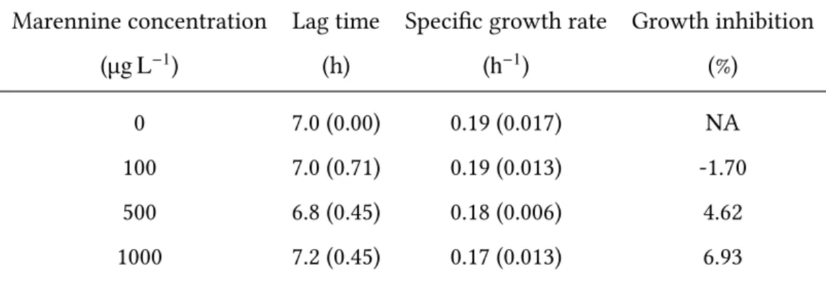
1 Effect of marennine on V. splendidus 7SHRW lag time, specific growth rate and growth inhibition (standard deviation is shown between parentheses). . 24 2 Two-way ANOVAs results for the cell abundance analyses for both larval
stages (D-larvae and post-larvae) with the treatment (C, M, V and MV) and the sampling time (1 h and 96 h) as factors and the interaction between both factors. . . 26
1 Larval survival rates (%) for the D-larvae and the post-larvae after 96 h of exposition to each treatment (C: control, M: marennine, V: vibrio and MV: marennine + vibrio; error bars: standard deviation; ***: significant difference (p<0.001)). . . 25 2 Bacterial abundance in the rearing medium after 1 h and 96 h of exposition
to each treatment (C: control, M: marennine, V: vibrio and MV: marennine + vibrio; standard deviation is shown with error bars; letters indicate groups formed by the Tukey’s HSD post-hoc analysis and one set of letters (a, b and c) were assigned to the analysis of D-larvae while another set (d, e and f) were assigned to the post-larvae analysis. . . 27 3 Dendrograms of the genetic fingerprints of the bacterial communities
sam-pled in the microbiota and the rearing medium of blue mussel D-larvae and post-larvae at the beginning of the experiment (T0) and exposed to four dif-ferent treatments (C: control, M: marennine, V: vibrio and MV: marennine + vibrio) during 96 h. The cluster analysis was based on the Jaccard coef-ficient similarity indicator and the dendrograms were constructed with the UPGMA algorithm using the vegan package (version 2.5-1) built for R (ver-sion 3.5). Numbers are the total number of OTUs recorded in each treatment. 28 4 Proportion of matches against mismatches in the comparison of the
com-munities of the D-larvae rearing medium and the larval microbiota for each treatment (C: control, M: marennine, V: vibrio and MV: marennine + vibrio) and the initial communities (T0). . . 29 5 Simultaneous comparison of the initial community (T0) and the control (C)
after 96 h of exposition with all other treatments (M, V and MV) in regard of a) the unique OTUs Gained in each treatment, b) the Conserved OTUs between T0 and the control (C), c) the Unique OTUs from the initial com-munity (T0) and d) the unique OTUs from the control (C). . . 31
L’aquaculture est un secteur d’activit´e en fort d´eveloppement `a l’´echelle mondiale en en raison de la hausse de la demande en produits aquatiques, mais aussi en raison et de la diminution des ressources naturelles marines disponibles (Sapkota et al., 2008). Au Canada, l’aquaculture s’est principalement d´evelopp´ee autour des salmonid´es et des bivalves, dont la moule bleue (Mytilus edulis) est le bivalve avec la production la plus importante repr´esentant un volume total de 25 800 tonnes produit entre 2011 et 2015 (Fisheries and Oceans Canada, 2017-03-15 2018). Au Qu´ebec, la tendance est similaire et la moule bleue est le bivalve le plus cultiv´e et cette culture repr´esentait une valeur `a la ferme `a 49,5 millions de dollars en 2013, soit une augmentation de 68% depuis 2009 (Fisheries and Oceans Canada, 2015-03-03 2015, 2017-03-15 2018). Pour r´epondre `a la demande grandissante de fruits de mer, les producteurs de bivalves marins doivent avoir acc`es `a un approvisionnement stable en juv´eniles (Helm et al., 2004). La technique de production de juv´eniles de moule utilise le cycle naturel couplant un stade larvaire p´elagique `a une phase de fixation sur un substrat pour capter les jeunes juv´eniles sur des collecteurs artificiels.
Comme le succ`es de l’approvisionnement en juv´eniles provenant du milieu naturel via le captage sur des collecteurs artificiels est tr`es variable, les juv´eniles produits en ´ecloseries sont susceptibles de devenir de plus en plus populaires aupr`es des conchyliculteurs (Helm et al., 2004). Or, cette m´ethode d’´elevage larvaire pratiqu´ee pour les bivalves comme la moule bleue concentre un grand nombre d’individus dans un volume d’eau restreint et peut causer un stress important chez les larves. De plus, ces conditions d’´elevage peuvent favoriser le d´eveloppement de bact´eries pathog`enes entraˆınant des ´ev`enements de mortalit´e de masse dans les ´ecloseries (Vadstein et al., 2004). Ces mortalit´es massives sont un enjeu repr´esentant un d´efi majeur pour la production aquacole et le contrˆole de leur occurrence est un en-jeu primordial afin d’assurer une production stable de juv´eniles. Ces ´ev`enements ont ´et´e reli´es `a la pr´esence de plusieurs pathog`enes opportunistes appartenant aux genres
Pseu-domonas, Roseovarius, Aeromonas et Vibrio (Tubiash et al., 1970; Beaz-Hidalgo et al., 2010). Les pathog`enes du genre Vibrio sont les agents infectieux les plus souvent identifi´es dans les ´elevages larvaires (Paillard et al., 2004). Afin de contrˆoler les pertes li´ees aux infections bact´eriennes, l’antibioth´erapie a longtemps ´et´e le moyen privil´egi´e dans l’industrie aqua-cole (Nicolas et al., 2007). Toutefois, le risque de d´eveloppement de souches bact´eriennes pouvant r´esister `a ces antibiotiques est consid´erable et doit absolument ˆetre pris en compte lors du d´eveloppement de nouveaux traitements (Defoirdt et al., 2011; Falaise et al., 2016). Par ailleurs, l’utilisation des antibiotiques en aquaculture est fortement r´eglement´ee autour du globe (Perez-Sanchez et al., 2018). Il est donc primordial de d´evelopper des techniques de contrˆole alternatives contre les infections bact´eriennes dans les ´elevages afin de limiter l’impact des ´ev`enements de mortalit´e de masse caus´es par des pathog`enes opportunistes et assurer le succ`es de d´eveloppement des ´ecloseries.
1.1 Les Vibrionaceae
Les Vibrionaceae sont des pathog`enes fr´equemment identifi´es lors d’´ev`enement de mortalit´e de masse chez les bivalves. Ces bacilles `a Gram-n´egatif, arborant un flagelle polaire et ayant besoin de sodium (Na+) pour leur croissance sont des microorganismes ubiquitaires
dans les milieux aquatiques ont ´et´e d´ecrits pour la premi`ere fois par V´eron en 1965 (V´eron, 1965). En 2013, 142 esp`eces de Vibrionaceae ´etaient r´epertori´ees (Sawabe et al., 2013). Cette famille comprend notamment l’esp`ece bien connue Vibrio cholerae, l’agent infectieux du chol´era qui repr´esente actuellement un probl`eme de sant´e publique majeur pour pr`es du tiers des pays du monde (Ali et al., 2015).
Le nombre important d’esp`eces class´ees dans la famille des Vibrionaceae en fait un groupe particuli`erement diversifi´e au sein duquel le pouvoir pathog`ene des esp`eces est tr`es variable. `A l’oppos´e de V. cholerae, on retrouve l’esp`ece bioluminescente V. fischeri qui s’as-socie avec la seiche Euprymna scolopes. Cette association symbiotique permet `a V. fischeri de coloniser un organe sp´ecialis´e de la seiche et d’y perdurer tout en permettant `a son hˆote
d’utiliser la lumi`ere que la bact´erie peut produire (Ruby, 1996). Les 2 esp`eces pr´ec´edemment mentionn´ees repr´esentent les deux extrˆemes du spectre des ph´enotypes (pathog`ene stricte et non-pathog`ene) que l’on peut retrouver pour les esp`eces de la famille des Vibrionaceae. Certaines esp`eces, quant `a elles, sont d´ecrites comme des pathog`enes opportunistes. Ces bact´eries infectent leur hˆote seulement lorsque ce dernier pr´esente une ou plusieurs condi-tions qui ne sont habituellement pas rencontr´ees chez les individus sains. Par exemple, une plaie ouverte peut servir de voie d’entr´ee pour ces pathog`enes ou encore un syst`eme im-munitaire compromis peut permettre `a une bact´erie normalement contrˆol´ee par le syst`eme immunitaire de d´evelopper son potentiel infectieux. `A titre d’exemple, l’esp`ece V. vulnificus est un pathog`ene opportuniste pouvant infecter les humains via l’ingestion de fruits de mer contamin´es ou encore via l’exposition `a de l’eau de mer contenant le pathog`ene. Il est re-port´e que les cas d’infections de V. vulnificus s’aggravent rapidement si les hˆotes ont d´ej`a des conditions m´edicales particuli`eres comme le diab`ete, l’alcoolisme ou encore la leuc´emie (Oliver, 2005). Les travaux de recherche sur cette infection se sont principalement concentr´es sur les humains (Strom and Paranjpye, 2000; Phillips and Satchell, 2017; Baker-Austin and Oliver, 2018) et sur la s´ecurit´e alimentaire concernant la production et la manutention des fruits de mer (Gopal et al., 2005). Or, la pr´esence de pathog`enes opportunistes dans les eaux naturelles (Mansergh and Zehr, 2014), les syst`emes aquacoles (Vandenberghe et al., 2003) et mˆeme dans les communaut´es microbiennes naturellement associ´ees aux tissus des animaux sains (Vandenberghe et al., 2003; Asmani et al., 2016; Vezzulli et al., 2018) soul`eve l’impor-tance de l’identification des facteurs biotiques et abiotiques d´eclenchant leur pathog´enie. Ce groupe de bact´eries est notamment responsable de maladies appel´ees couramment vibrioses qui affectent plusieurs ´elevages aquacoles entraˆınant des pertes ´economiques importantes (Beaz-Hidalgo et al., 2010). Dans les conditions particuli`eres associ´ees aux syst`emes aqua-coles (forte densit´e d’´elevage, stress de manipulation), les Vibrionaceae peuvent ´echapper `a la r´esistance du syst`eme immunitaire de nombreuses esp`eces aquacoles comme les poissons, les crustac´es et les bivalves et d´eclencher des infections pouvant conduire `a des ´ev`enements de mortalit´e de masse (Velji et al., 1992; Beaz-Hidalgo et al., 2010; Liu et al., 2013). Les larves
produites en ´ecloseries, qui est le stade ontog´enique le plus sensible (Rayssac et al., 2010), sont particuli`erement vuln´erables face aux pathog`enes du genre Vibrio (Elston and Leibo-vitz, 1980; Genard et al., 2013, 2014). Une meilleure compr´ehension des agents infectieux des vibrioses et de leurs interactions avec les larves et avec les autres bact´eries du milieu est donc n´ecessaire afin de d´evelopper de nouvelles strat´egies afin de contrer les effets n´efastes de ces infections bact´eriennes.
1.2 Microbiote et immunit´e
R´ecemment, beaucoup d’attention s’est port´ee sur la compr´ehension du rˆole du mi-crobiote (communaut´e microbienne naturelle associ´ee aux tissus d’un organisme) sur l’´etat physiologique de l’hˆote. Il est aujourd’hui reconnu que le microbiote contribue significati-vement au d´eveloppement et `a la condition physiologique de l’hˆote (Zilber-Rosenberg and Rosenberg, 2008; Rosenberg and Zilber-Rosenberg, 2016). Selon le concept de l’holog´enome, le super-organisme qui est compos´e de l’hˆote animal et du microbiote, la partie
micro-bienne de l’holobionte repr´esente l’´el´ement de l’holog´enome le plus variable. En effet, le microbiote peut r´eagir plus rapidement `a une variation dans les conditions environnemen-tales que l’hˆote. Consid´erant que le microbiote r´egule en partie les r´eponses physiologiques de l’hˆote face `a un stress externe, de nombreux travaux se sont pench´es sur le rˆole du mi-crobiote dans la colonisation des tissus par des agents infectieux (Stecher and Hardt, 2008; Ribet and Cossart, 2015). L’effet antagoniste des bact´eries intestinales contre la colonisation des pathog`enes bact´eriens dans les tissus animaux est bien connu et a d´ej`a ´et´e d´emontr´e par des exp´eriences utilisant des techniques de culture classiques (Freter, 1962; Bartlett et al., 1977). Des travaux plus r´ecents ont d´emontr´e l’implication du microbiote dans la virulence des pathog`enes, ce qui d´efi la vision classique du un pathog`ene, une maladie provenant
des postulats de Koch classiques. Par exemple, Duran-Pinedo et al. (2014) ont d´emontr´e que les bact´eries commensales du microbiote oral contribuaient significativement `a l’expression des facteurs de virulences des pathog`enes reconnus causant la p´eriodontite. Les auteurs ont
not´e qu’un d´ebalancement du microbiote, ou dysbiose, semblait ˆetre n´ecessaire pour l’ex-pression de la pathog´enie de ces bact´eries. Les m´ecanismes de la transition du microbiote d’un ´etat commensal `a un ´etat dysbiotique et l’implication de cette transition sur la suscepti-bilit´e aux pathog`enes demeurent difficiles `a ´etudier dˆu au grand nombre de facteurs pouvant influencer l’hom´eostasie `a l’interface hˆote-microbiote. Deux hypoth`eses non exclusives sont actuellement propos´ees : (1) l’hypoth`ese des nutriments (food hypothesis) et (2) l’hypoth`ese de la d´efense (killing hypothesis) (Stecher and Hardt, 2008). L’hypoth`ese des nutriments sup-pose que lorsque l’hˆote fait face `a un stress externe, son ´etat physiologique entraˆıne une mo-dification des nutriments disponibles pour les bact´eries de son microbiote, modifiant ainsi la capacit´e des tissus `a r´esister `a la colonisation par une bact´erie pathog`ene. L’augmenta-tion de la quantit´e de nutriments pourrait favoriser la croissance de bact´eries pathog`enes (reconnues pour croˆıtre rapidement dans un environnement enrichi en nutriments) et en-traˆıner en parall`ele un changement de diversit´e du microbiote (Ribet and Cossart, 2015). L’hypoth`ese de la d´efense stipule qu’une modification du microbiote peut ˆetre induite par la production importante de mol´ecules antimicrobiennes telles que la d´efensine, un peptide an-timicrobien, ou encore la phospholipase par l’hˆote (Stecher and Hardt, 2008). Ces mol´ecules sont peu sp´ecifiques et une production accrue de ces derni`eres dans l’environnement du microbiote peut accidentellement affecter grandement les bact´eries commensales du micro-biote (dommages collat´eraux). La r´eduction de la diversit´e sp´ecifique et de la densit´e totale de bact´eries du microbiote pourrait favoriser la colonisation de cet environnement par une bact´erie pathog`ene pr´esentant une r´esistance sup´erieure vis-`a-vis des mol´ecules antimicro-biennes. Afin de mieux comprendre cette dynamique complexe entre l’´etat physiologique de l’hˆote, son microbiote et un pathog`ene bact´erien opportuniste, une caract´erisation de la diversit´e sp´ecifique et fonctionnelle du microbiote des organismes hˆotes est n´ecessaire.
1.3 Le microbiote des bivalves marins
L’´etude du microbiote des bivalves marins se nourrissant en filtrant l’eau environnante est particuli`erement int´eressante parce que ces organismes maintiennent un lien ´etroit avec leur environnement, mais aussi parce qu’une partie de leur alimentation est constitu´ee de bact´eries planctoniques potentiellement pathog`enes. Les travaux de Beeson and Johnson (1967) sur la microflore normale du haricot de mer (Donax gouldii) en milieu naturel ont pu d´emontrer que ces bivalves maintenaient une communaut´e bact´erienne diff´erente de celle retrouv´ee dans leur environnement de croissance. Ces travaux font partie des premiers `a avoir sugg´er´e que les conditions physiologiques des organismes cr´eaient un environnement interne distinctif de celui de l’eau de mer environnante et que, contrairement `a la pens´ee populaire, la communaut´e bact´erienne dans l’eau environnante n’´etait pas le reflet de la communaut´e du microbiote. Parmi les bact´eries isol´ees des tissus des organismes hˆotes du-rant cette ´etude, plusieurs ´etaient de la famille des Vibrionaceae. R´ecemment, Chauhan et al. (2018) ont d´ecrit le microbiote d’huˆıtres am´ericaines (Crassostrea virginica) provenant du milieu naturel. Le microbiote des huˆıtres ´etait domin´e par quelques taxons comme Cyano-bacteria sp. et Pelagibacter sp., mais aussi par Photobacterium sp., un pathog`ene opportu-niste de la famille des Vibrionaceae qui peut ´egalement former une symbiose avec l’hˆote. La pr´esence de pathog`enes opportunistes dans le microbiote de bivalves sains n’est pas surpre-nante consid´erant que ces derniers sont retrouv´es naturellement dans les eaux naturelles (Mansergh and Zehr, 2014; Takemura et al., 2014). Toutefois, le rˆole des pathog`enes oppor-tunistes de la famille des Vibrionaceae dans le maintien de l’hom´eostasie des bivalves sains et les facteurs d´eclenchant leur pathog´enie demeurent `a ´elucider (Pruzzo et al., 2005).
Les travaux de caract´erisation de la diversit´e des Vibrionaceae du microbiote des valves se sont principalement int´eress´es aux stades adultes (Romalde et al., 2014). Or, les bi-valves ont la particularit´e d’avoir `a se m´etamorphoser pour passer du stade larvaire au stade juv´enile. Pendant cette m´etamorphose, le syst`eme de la prophenoloxidase (proPO) s’active (Smith and S¨oderh¨all, 1991). Ce syst`eme agit comme un syst`eme immunitaire inn´e
rudimen-taire qui permet aux bivalves de se d´efendre contre les pathog`enes par l’opsonisation des cellules ´etrang`eres. L’activation de ce syst`eme peut ˆetre enclench´ee par certaines mol´ecules comme le ß-1,3-glucane, le peptidoglycane et certains lipopolysaccharides (S¨oderh¨all and Cerenius, 1998). Ce syst`eme est partiellement d´ecrit chez les bivalves adultes et peu de donn´ees sont disponibles pour ce qui est des stades larvaires (Bassim et al., 2015b). Une meilleure compr´ehension des capacit´es immunitaires des larves de bivalves est n´ecessaire afin de mieux comprendre les facteurs biotiques et abiotiques r´egulant ces derni`eres et plus particuli`erement le rˆole possible du microbiote larvaire des bivalves dans leur immunit´e.
Consid´erant l’importance du microbiote dans la colonisation des tissus animaux par les pathog`enes et de son implication dans la physiologie de son hˆote, la caract´erisation de la diversit´e sp´ecifique du microbiote larvaire de bivalves sous diff´erentes conditions s’av`ere n´ecessaire. L’utilisation des esp`eces ´elev´ees en aquaculture comme mod`ele d’´etude est une avenue tr`es int´eressante pour ´etudier les relations entre les larves de bivalves, leur micro-biote et les pathog`enes bact´eriens opportunistes couramment retrouv´es dans les syst`emes d’´elevage (Vandenberghe et al., 2003). Plusieurs de ces pathog`enes bact´eriens opportunistes ont notamment ´et´e retrouv´es dans le microbiote d’individus sains, dont des membres de la famille des Vibrionaceae (Vandenberghe et al., 2003; Asmani et al., 2016; Vezzulli et al., 2018).
1.4 Contrˆole des ´ev`enements de mortalit´e de masse en ´ecloseries
L’occurrence d’´ev`enements de mortalit´e de masse dans les ´ecloseries repr´esente une des limitations majeures `a la production de juv´eniles en ´ecloserie. Les pathog`enes du genre Vibriosont les agents infectieux les plus souvent report´es dans le cas d’infections bact´eriennes dans les syst`emes d’´elevage de larves (Paillard et al., 2004). Les travaux d’Elston and Leibo-vitz (1980) sur la pathogen`ese de la vibriose chez les huˆıtres am´ericaines ont d´emontr´e que les infections progressaient diff´eremment selon l’isolat bact´erien utilis´e. Les observations faites durant cette ´etude ont permis de d´eterminer quels signes cliniques observer afin de diagnostiquer la vibriose lors des premi`eres ´etapes de l’infection des huˆıtres. Par exemple, il
est possible d’observer le d´etachement des cellules du manteau ou encore une atrophie des visc`eres (Elston and Leibovitz, 1980).
Depuis les travaux de Davis and Chanley (1956), l’utilisation d’antibiotiques a gagn´e en popularit´e aupr`es des aquaculteurs afin de limiter l’impact des infections dans les ´elevages. La streptomycine, la p´enicilline et le chloramph´enicol ont longtemps ´et´e les antibiotiques de choix pour r´eduire l’impact des infections bact´eriennes dans les ´elevages larvaires. Toute-fois, l’utilisation syst´ematique et intensive d’antibiotiques dans les milieux d’´elevage accroˆıt les risques li´es au d´eveloppement de r´esistance aux antibiotiques (Defoirdt et al., 2011; Fa-laise et al., 2016) et au transfert de cette capacit´e dans les r´eseaux trophiques (Sapkota et al., 2008). Les traitements `a l’aide d’antibiotiques sont devenus un enjeu majeur pour la pro-duction aquacole (Watts et al., 2017). En effet, lorsqu’une population de bact´eries devient r´esistante `a l’utilisation d’antibiotiques courants, cela peut mener `a la persistance des ma-ladies difficilement traitables dans les syst`emes d’´elevage.
L’utilisation de techniques de contrˆole biologique en aquaculture afin de limiter l’im-pact des pathog`enes bact´eriens est une voie int´eressante, car la diversit´e de leur mode d’ac-tion r´eduit significativement la probabilit´e de l’apparid’ac-tion de r´esistances sp´ecifiques (Ni-colas et al., 2007). Les probiotiques vivants, g´en´eralement compos´es de bact´eries vivantes ayant un effet b´en´efique sur la sant´e sur l’hˆote (Joint FAO/WHO Expert Consultation, 2001), sont une de ces alternatives. D`es 1907, les observations de Metchnikoff en lien avec la consommation de produits laitiers ferment´es lui ont permis de stipuler qu’il serait possible de moduler la flore (microbiote) humaine afin de remplacer les bact´eries n´efastes par des bact´eries b´en´efiques. Plus d’un si`ecle apr`es ces observations, les effets b´en´efiques de plu-sieurs bact´eries sur la sant´e de l’hˆote non seulement chez l’humain, mais ´egalement chez plusieurs esp`eces ´elev´ees en aquaculture furent d´emontr´es (Kesarcodi-Watson et al., 2016; Sohn et al., 2016a,b; Zorriehzahra et al., 2016; Sharifuzzaman and Austin, 2017). Les effets immunostimulants de certaines esp`eces bact´eriennes utilis´ees en aquaculture permettent de contrer l’implantation d’un pathog`ene sp´ecifique ou encore d’accroˆıtre la r´esistance aux
ma-ladies de mani`ere non sp´ecifique (Akhter et al., 2015). Par exemple, la bact´erie Pseudoaltero-monassp. X153, isol´ee de la surface d’une roche `a Saint-Anne-du-Portzic (Bretagne, France) a d´emontr´e un effet protecteur sur les palourdes (Ruditapes philippinarum) au stade larvaire (Longeon et al., 2004) . Les auteurs de cette ´etude ont d´emontr´e que non seulement Pseu-doalteromonas sp. X153 n’´etait pas toxique pour les larves de palourdes, mais ´egalement qu’elle avait un effet protecteur pour des larves de coquilles Saint-Jacques (Pecten maxi-mus). Une mol´ecule bioactive, P-153, se retrouvant `a la fois dans les cellules du probiotique et dans l’eau d’´elevage a ´et´e identifi´ee comme ´etant un des possibles acteurs principaux dans le mode d’action de Pseudoalteromonas sp. X153. Les probiotiques, ou encore leurs bio-mol´ecules actives sont donc une avenue int´eressante pour r´eduire l’utilisation syst´ematique des antibiotiques, mais ´egalement pour rendre les pratiques aquacoles plus durables et plus s´ecuritaires (Balcazar et al., 2006; Defoirdt et al., 2011; Beaz-Hidalgo et al., 2010; Gastineau et al., 2012).
1.5 La marennine et son mode d’action potentiel
La marennine, une biomol´ecule provenant de la diatom´ee Haslea ostrearia, a ´et´e pro-pos´ee comme traitement alternatif aux antibiotiques en aquaculture (Pouvreau et al., 2006, 2008). Ce pigment a d´emontr´e un effet inhibiteur de la croissance de diff´erentes bact´eries pathog`enes en culture pure dont plusieurs de la famille des Vibrionaceae comme V. tasma-niensis, V. aesturianus et V. splendidus (Defoirdt et al., 2011; Gastineau et al., 2012, 2014) `a des concentrations variant entre 19.14 mg L−1pour V. tasmaniensis (Falaise et al., 2016) et
2.89 mg L−1 pour V. splendidus (Gastineau et al., 2014). Toutefois, Turcotte et al. (2016) ont mis en lumi`ere un effet cytotoxique de la marennine sur les larves de bivalves `a ces concen-trations. C’est pourquoi la marennine a ´et´e utilis´ee `a une concentration de 500 µg L−1dans
les ´elevages larvaires durant leurs travaux. Leurs r´esultats ont pu d´emontrer le fort potentiel protecteur de cette biomol´ecule pour l’´elevage de bivalves marins. Les auteurs ont d´emontr´e que la marennine avait un effet b´en´efique sur le taux de survie des larves-D de M. edulis lors
d’expositions `a V. splendidus sans que l’abondance bact´erienne dans les milieux de culture des larves ne soit affect´ee. Le mode d’action de la marennine `a cette concentration ne semble donc pas venir d’un quelconque effet antimicrobien et demeure donc `a ´elucider.
1.6 Objectifs et hypoth`eses
Dans ce m´emoire nous validerons l’hypoth`ese qu’un des modes d’action possible de la marennine, lorsqu’utilis´ee `a des concentrations de l’ordre de celle utilis´ee par Turcotte et al. (2016), pourrait ˆetre l’interaction avec le microbiote larvaire de M. edulis. Le premier objectif sp´ecifique de ce projet est de d´emontrer l’absence d’un effet antimicrobien de la marennine contre V. splendidus `a une concentration de 500 µg L−1. Le deuxi`eme objectif sp´ecifique est
de mettre en ´evidence l’effet de la marennine `a une concentration de 500 µg L−1 sur les
communaut´es bact´eriennes indig`enes, dans l’eau d’´elevage et du microbiote larvaire, lors de tests de provocations bact´eriennes avec V. splendidus.
´Elucider les m´ecanismes d’action de la marennine lors de son utilisation en conchyli-culture permettrait non seulement d’optimiser son utilisation, mais ´egalement d’´etendre son potentiel `a plus grande ´echelle dans un contexte de d´eveloppement durable de l’exploitation des ressources marines.
IMPACT DE LA MARENNINE SUR LE MICROBIOTE DE LARVES DE
MYTILUS EDULISEXPOS ´EES AU PATHOG`ENE VIBRIO SPLENDIDUS (IMPACT OF MARENNINE ON THE MICROBIOTA OF MYTILUS EDULIS LARVAE EXPOSED
TO THE PATHOGEN VIBRIO SPLENDIDUS)
Jordan Latour1, Kim Doiron1, R´ejean Tremblay1and Karine Lemarchand1
1Institut des Sciences de la Mer de Rimouski, Universit´e du Qu´ebec `a Rimouski, 310, all´ee
des Ursulines, Rimouski (Qc, Canada)
2.1 R´esum´e
Dans les ´ecloseries de bivalves, les pathog`enes bact´eriens opportunistes ont ´et´e iden-tifi´es comme ´etant responsables des ´ev`enements de mortalit´e de masse causant des pertes ´economiques importantes pour les conchyliculteurs. De nouvelles techniques de contrˆole ont ´et´e propos´ees afin de remplacer l’utilisation d’antibiotiques dans les ´elevages. En effet, les probiotiques et les mol´ecules bioactives d’origine naturelle ont d´ej`a ´et´e sugg´er´es afin de limiter l’occurrence de ces ´ev`enements de mortalit´e de masse. Ces microorganismes et ces mol´ecules sont associ´es `a des effets b´en´efiques sur les hˆotes, plus particuli`erement sur leur r´esistance face `a des stress externes comme un pathog`ene bact´erien. De nos jours, il est bien reconnu que le microbiote joue un rˆole important dans le maintien de l’´etat de sant´e d’un organisme. Le but de cet article est de mettre en ´evidence l’effet protecteur d’une nouvelle mol´ecule bioactive naturelle extraite de la diatom´ee Haslea ostrearia en relation avec une potentielle modification du microbiote larvaire de Mytilus edulis. L’hypoth`ese principale est que cette modification du microbiote de larves trait´ees avec la marennine am´eliorera la r´esistance des larves contre les pathog`enes bact´eriens. L’effet protecteur de la marennine a
d´ej`a ´et´e d´emontr´e chez M. edulis `a une concentration de 500 µg L−1. Dans cette ´etude, des
larves-D (J9) et des post-larves (J29) ont ´et´e expos´ees au pathog`ene opportuniste Vibrio
splen-diduspendant 96 h en pr´esence et en absence de marennine. Nos r´esultats d´emontrent qu’`a une concentration de 500 µg L−1, la marennine n’a pas d’effet antibact´erien sur la croissance
du pathog`ene en culture pure. De plus, la pr´esence de marennine n’a pas d´emontr´e d’effet sur l’abondance bact´erienne dans le milieu de culture lors de l’exposition des larves indi-quant que la pr´esence de marennine n’influence pas la charge bact´erienne `a laquelle les larves sont expos´ees durant les exp´eriences. Toutefois, le taux de survie des larves-D a sig-nificativement augment´e en pr´esence de marennine lors des expositions aux pathog`enes. Les analyses mol´eculaires ont r´ev´el´e que l’augmentation du taux de survie larvaire ´etait accompagn´ee d’une modification significative de la richesse sp´ecifique du microbiote des larves. Ces travaux permettront de mieux comprendre le rˆole que le microbiote joue dans la r´esistance aux pathog`enes opportunistes en pr´esence de biomol´ecules en aquaculture.
2.2 Abstract
In bivalve hatcheries, opportunistic pathogens have been associated with important mass mortality events of larvae and important economic loss for producers. Alternatives to the use of antibiotics, such as probiotics, have been proposed to limit the occurrence of such events in bivalve hatcheries and thus to stabilize bivalve production. Probiotics and their natural molecules are associated with beneficial effects for larvae at different levels, especially to enhance their resistance to external stressors such as bacterial pathogens. It is now recognized that the composition of the host microbiota influences the host health status and could be a target of probiotics. The aim of this master’s thesis is to highlight the protec-tive effect of a new bioacprotec-tive molecule, marennine, on Mytilus edulis larvae during bacterial challenges in relation to a potential modification of the marennine-treated larvae micro-biota. The main hypothesis is that the addition of marennine during larvae rearing could modify the conditions prevailing in the rearing medium and, as a consequence, the
composi-tion of the larvae microbiota leading to a better resistance to bacterial infeccomposi-tions. Marennine is a blue pigment, originating from the diatom Haslea ostrearia, which has demonstrated a positive effect on larvae survival at a concentration of 500 µg L−1. D-larvae and post-larvae
were exposed for 96 h to Vibrio splendidus with and without mareninne. Our results demon-strated that, at this concentration, marennine has no direct antimicrobial effect on V. splen-didusgrowth kinetics. In addition, the presence of marennine did not modify the abundance of bacteria in the rearing medium, suggesting no direct antimicrobial effect of marennine on the bacterial load to which larvae were exposed during the experiments. Nevertheless, the presence of marennine increased the survival of D-larvae exposed to the pathogen but have no effect on post-larvae survival. The molecular analysis of the larvae microbiota diversity allowed us to demonstrate that a modification in the larval microbiota’s richness occurs while the survival rates increase. Ultimately, our work will enable us to shed light on the importance of the larval microbiota in pathogen-resistance during bivalves rearing process.
2.3 Introduction
During the last few decades, fish and shellfish farming industries have experienced an important increase in their production rates in response to the growing seafood demand worldwide. From 1960 to 2013, the world marine products consumption per capita almost doubled increasing from an average of 9.9 kg to 19.7 kg per year (FAO, 2016). As a large proportion of natural seafood stocks being fully fished (58.1%) or overfished (31.4%), the share of aquaculture products in the global seafood consumption also increased from 7% to 39% between 1974 and 2004 (FAO, 2016). In order to meet the demand, producers need to have access to a large and stable supply of juveniles (Helm et al., 2004). Consequently, hatcheries are most likely to gain popularity, especially among bivalve producers who often rely on hatchery-reared juveniles due to the high spatiotemporal variability of the natural recruitment success (Helm et al., 2004).
larvae survival in the rearing system. Bacterial infections are known to be a major bot-tleneck to hatchery-reared juvenile bivalve production by causing massive mortality (mass mortality events). Many opportunistic bacterial pathogens from the genera Vibrio, Pseu-domonas, Aeromonas and Roseovarius were reported to be linked to mass mortality events in bivalve hatcheries (Tubiash et al., 1970; Paillard et al., 2004). These organisms exert their pathogenicity only in specific environmental or larvae physiological conditions that are still poorly understood. The seemingly random nature of mass mortality events is a major ob-stacle to achieve steady production rates. Understanding and therefore controlling disease outbreaks in hatcheries is crucial to assure the stability of the hatchery production.
Prior to the 1980s, antibiotics were regularly used by producers in bivalve hatcheries to reduce the impact of bacterial infection on their production (Asmani et al., 2016). However, the development of antibiotic resistance (Defoirdt et al., 2011; Falaise et al., 2016) and the risk of transmission of these resistant strains in the food web (Sapkota et al., 2008) led to strict regulations in several countries (Hernandez and Serrano, 2005; FDA, 2007). The use of antibiotics is therefore not a sustainable solution for controlling the occurrence of mass mortality events in bivalve hatcheries.
Probiotics and natural bioactive molecules have been proposed as alternatives to an-tibiotics in animal farming (Balcazar et al., 2006; Defoirdt et al., 2011; Beaz-Hidalgo et al., 2010; Gastineau et al., 2012; Falaise et al., 2016). A wide range of organisms, or their cel-lular components, was tested as probiotics for their positive effect in aquaculture (Irianto and Austin, 2002). For example, Chilean scallop larvae (Argopecten purpuratus) were able to complete the larval pelagic phase without any antibiotic treatment when exposed to inhibitor-producing bacteria strains (Vibrio sp. C33, Pseudomonas sp. 11 and Bacillus sp. B2) (Riquelme et al., 2001). The strains Pseudoalteromonas sp. X153 (Longeon et al., 2004) and Phaeobacter gallaeciensisX34 (Genard et al., 2014) also demonstrated a positive effect on cul-tured scallop larvae (Pecten maximus) survival. Azadirachtin, an extract from the neem tree (Azadirachta indica), also demonstrated a beneficial effect acting as an immunostimulant
on goldfish (Carassius auratus) and increased their survival rate when challenged against Aeromonas hydrophila(Kumar et al., 2013). Recently, marennine, a blue-green pigment pro-duced by the diatom Haslea ostrearia, was suggested as an interesting bioactive molecule for bivalve hatcheries (Turcotte et al., 2016). This natural pigment has shown promising re-sults by reducing the mortality rate of blue mussels larvae (Mytilus edulis) challenged with V. splendidus. Unfortunately, little is known about the mode of action underlying the bene-ficial effects of marennine in bivalves farming (Pouvreau et al., 2008; Gastineau et al., 2012), except the study of Tardy-Laporte et al. (2013) demonstrating the interaction of marennine with lipopolysaccharides related to higher rigidity of membrane cells in Gram negative bac-teria. Improvement of the use of the feed, immunostimulation, antibacterial activity, alter-ation of the microbial metabolism and competitive exclusion are some of the proposed, but poorly documented, hypothetical modes of action of probiotics in aquaculture (Irianto and Austin, 2002; Prado et al., 2010). Unravelling the role of marennine as a water-additive in bivalve hatcheries is crucial in order to safely expand its utilization.
In this paper, we test the hypothesis that the interaction of marennine with the larval microbiota could be the mode of action contributing to the observed beneficial effects on bivalve larvae survival. The artificial addition of marennine in the rearing medium might modify the bacterial community within the rearing medium itself and induce a change in the bacterial community recruited by the larvae to form their microbiota. In addition, maren-nine could interfere with the quorum sensing system within the larvae resulting in a mod-ification of the microbiota formation (Kalia, 2013).
Nowadays, it is well known that the taxonomic diversity, the abundance and the physi-ological structure of the microbiota affect its host health condition (Lopez et al., 2014; Laterza et al., 2016; Marchesi et al., 2016). In the case of bivalves, it has been suggested that a shift in the structure and the specific diversity of the microbiota in adults might prevent bacterial pathogen to settle within the host (Froelich and Oliver, 2013). These microbiota modifica-tions might lead to a better pathogen resistance in larvae and contribute to the beneficial
effect of marennine addition observed in bivalve hatcheries. Therefore, interaction between probiotics or bioactive molecules with the natural larvae microbiota, the conditions prevail-ing in the rearprevail-ing medium and the diversity of bacterial communities might contribute to the observed protective effect of marennine on bivalve larvae in hatcheries.
The aim of this study is to investigate the importance of the larval microbiota in hatchery-reared bivalve larvae when exposed to the opportunistic bacterial pathogen V. splen-didus in presence or absence of marennine. Blue mussel larvae, the most important shell-fish aquaculture production in Canada from 1995 to 2016 (Fisheries and Oceans Canada, 2015-03-03 2015), were exposed to the opportunistic bacterial pathogen V. splendidus during bacterial challenge experiments with and without marennine added to the rearing medium. The bacterial community in the rearing medium and the blue mussel larval microbiota were characterized under different bacterial contamination and treatment conditions in order to better understand the importance of the larval microbiota in the prevention of mass mortal-ity events with marennine as a natural water-additive. The bacterial communmortal-ity abundance in the rearing medium was characterized with flow cytometry and the diversity of commu-nities in the rearing medium and larval microbiota were investigated using the denaturing gradient gel electrophoresis (DGGE) technique. Taken together, these analyses have helped to shed light on the host-pathogen-microbiota interplay during the rearing process of ma-rine bivalve’s larvae in hatcheries.
2.4 Material and methods
2.4.1 Marennine solution
Marennine was obtained from H. ostrearia culture produced as described in Gastineau et al. (2014) and Turcotte et al. (2016) and the pigment was purified by the method of Pou-vreau et al. (2006) to obtain a solution in nanopure water (pH 7.2) that was autoclaved. The solution was filtered on 0.2 µm pore-sized cellulose acetate membrane before determining
the solution concentration by spectrophotometry at 656 nm with the specific extinction co-efficient of 12 L g−1cm−1according to Turcotte et al. (2016).
2.4.2 Bacterial culture condition
V. splendidus 7SHRW, a strain isolated from the Gulf of St. Lawrence (Qc, Canada) (Mateo et al., 2009), was grown overnight in 10 mL of salty LB medium (25 g L−1NaCl, pH
7.2, autoclaved) at room temperature prior to each experiment (bacterial growth kinetic and bacterial challenges). This strain is recognized for its ability to infect blue mussel larvae (Turcotte et al., 2016). After incubation, cells were centrifuged at 3000 g for 5 min and the cell pellet was washed twice in sterile physiological water (9 g L−1NaCl, pH 7.2, autoclaved).
Then, bacterial cells were suspended in sterile physiological water to obtain a stock solution at 109cell mL-1.
2.4.3 V. splendidus growth kinetic
The effect of marennine on V. splendidus growth was assessed by spectrophotometry. The three final concentrations of marennine used were 100 µg L−1, 500 µg L−1, 1000 µg L−1.
Negative controls without bacterial cells were used for each of the concentrations tested. A marennine-free positive control with bacteria was also included. Each condition was repli-cated 5 times. The experiment was performed in a 96-wells microplate in a final volume of 200µL. Each well contained 100 µL of salty LB 2X medium (50 g L−1 NaCl, pH 7.2, auto-claved), 50 µL of Vibrio cells at 106cell mL-1(or physiological water, (9 g L−1 NaCl, pH 7.2,
autoclaved) and 50 µL of marennine (or nanopure water (pH 7.2) as a control) at desired concentrations. Cell growth was estimated by measuring the suspension’s OD at 595 nm every 30 min during 48 h. The OD reads were performed automatically with a SpectraMax 190 (Molecular devices, CA, USA) plate reader running the SoftMax Pro software. Growth curves were obtained by plotting OD values against time. Specific growth rate was
es-timated from the slope of the ln-transformed curves as recommended by Beaulieu et al. (2015). Growth inhibition was calculated by comparing the OD values of the curves from each tested marennine concentration after 24 h to the OD value of the positive control.
2.4.4 Bacterial challenges
2.4.4.1 Blue mussel larvae rearing conditions
Spawning adults from a pure population of M. edulis (Tremblay et al., 2011) were ob-tained from St. Peters Bay (Prince Edward Island, Canada; 46.4281° N, 62.6422° W) in June 2017. The latter were used to produce gametes and ultimately larvae following the usual protocol used by R. Tremblay’s laboratory (Turcotte et al., 2016). Spawning was induced by thermal shocks from 5°C to 20°C and D-larvae (9 days-old) and metamorphosed post-larvae (29 days-old) were both used for bacterial challenges. All the rearing process was carried out at the Station aquicole de Pointe-au-P`ere of the UQAR/ISMER (Rimouski, Qc, Canada).
2.4.4.2 Treatments
Bacterial challenges were carried out at the UQAR/ISMER’s marine microbial ecology laboratory (Rimouski, Qc, Canada) to assess the larvae’s microbiota response to the expo-sure to the opportunistic bacterial pathogen V. splendidus and marennine. The experiments were performed in 3 Fernbach flasks (Thermo Fisher Scientific, USA) for each treatment ap-plied at room temperature. A concentration of 10 larvae mL−1was used during the bacterial
challenges in a volume of 2.5 L.
Both D-larvae and metamorphosed post-larvae were exposed to four different treat-ments (three replicates of each): a control without V. splendidus or marennine (C), a pathogen treatment with only V. splendidus at a final concentration of 106cell mL-1(V), a marennine
in which larvae were exposed to both V. splendidus and marennine at the same concen-trations used in V and M treatments (MV). All treatments were performed in UV-treated and filtered (0.2 µm pore-sized filter) sea water from the St. Lawrence estuary (Qc, Canada) having a salinity of 23.09 for the first experiment (D-larvae) and 23.11 for the second exper-iment (post-larvae). For the purpose of this article, treatments C and M will be referred as unchallenged larvae and treatments V and MV will be referred as challenged larvae.
2.4.4.3 Sampling
Before the challenge experiments, the initial bacterial communities in the seawater inhabiting the rearing medium in the culture tanks (T0) were characterized by sampling 1 L seawater used to fill the tanks in triplicates that were filtered on a 0.2 µm pore-sized Durapore filter (Ø 47 mm) and frozen at -80°C before further analyses. The larval microbiota prior to the challenge experiment was characterized to assess the initial communities (T0). Approximately 10 000 larvae (10 larvae mL−1) were collected on 50 µm mesh, gently washed
with sterile seawater (filtered on 0.2 µm and autoclaved) and immediately frozen at -80°C until DNA extraction. During the challenge experiments, two samples of 1 L of each flask were collected after 1 h and 96 h of exposition after gently homogeneous mixing and filtered on 50 µm mesh to collect the larvae. The collected larvae were treated as already described and frozen at -80°C. A sample of 4 mL of the 50 µm mesh filtered water was fixed in the dark for 15 min with 0.2% glutaraldehyde at pH 7 and then frozen at -80°C until further bacterial abundance analyses. Finally, the remaining water was filtered on a 0.2 µm pore-sized Durapore filter (Ø 47 mm) to collect the bacteria present in the rearing medium and frozen at -80°C for DNA extraction.
2.4.4.4 Survival rate
After 96 h of exposition, three samples of 10 mL of rearing medium containing at least 30 individuals were taken to assess the larvae survival rate. The larval survival rate (%) was obtained by calculating the ratio (number of live larvae/total number of larvae) between the control (C) and the treatments (M, V and MV). The control survival rate was set as 100%. In addition, the shell’s length of 30 live larvae was measured to monitor the potential effect of marennine on the larval growth during the experiments. Larvae were examined and photographed under 100X with a microscope Olympus BX41 coupled to an Evolution VF camera and use of Image Pro Plus software v5.1 (Media Cybernetics, Silver Spring, MD, USA). The mean of the three counts from each tank was used for statistical analyses.
2.4.4.5 Total bacteria abundance
The total free abundance in the rearing medium was assessed by flow cytometry using a CytoFlex Flow Cytometer (Beckman Coulter Inc., Mississauga, Canada). Frozen samples were thawed at room temperature and then stained in the dark with 0.3 µL of SYBR Green I (10 000X, Invitrogen, Thermo Fisher Scientific, USA). Fluorescent beads (Fluoresbrite YG microsphere 1 µm, Polysciences) were added to each sample prior to their analyses as an internal standard (Lebaron et al., 2002). The sample volume analyzed was determined by weighing the tubes before and after the analyses. Then, the abundance was determined with the latter volume and the number of counted events. Data analyses were performed with the FCSalyzer software (version 0.9.14, Free Software Foundation Inc., Boston, USA). Total bacteria were detected by plotting the green fluorescence recorded at 530 nm (FL1) versus the side angle light scatter (SSC).
2.4.4.6 Bacterial diversity
Bacterial 16S rDNA gene amplification and purification
Total DNA was extracted from larvae and the filters containing the bacteria from the rearing medium using the E.Z.N.A. Mollusc DNA kit (Omega Bio-Tek, Norcross, USA) ac-cording to the manufacturer’s instructions. Prior to each DNA extraction, larvae were gently washed with physiological water and crushed in a sterile 1.5 mL Eppendorf tube containing 350µL of ML1 buffer from the extraction kit and the filters were cut into pieces then trans-ferred in a sterile 1.5 mL Eppendorf tube. The extracted DNA from the larvae contained the DNA from the larvae itself, the bacteria within the larvae (microbiota) and the bacteria attached to the shell of the larvae.
The bacterial 16S rDNA gene was amplified by PCR using the universal primers 341F-GC (5’-C341F-GC-CCG-CCG-C341F-GC-CCC-341F-GCG-CCC-GTC-CCG-CCG-CCC-CCG-CCC-341F-GCC-TAC- (5’-CGC-CCG-CCG-CGC-CCC-GCG-CCC-GTC-CCG-CCG-CCC-CCG-CCC-GCC-TAC-GGG-AGG-GGA-GAG-3’) and 907R (5’-CCG-TCA-ATT-CMT-TTG-AGT-TT -3’) from Sch¨afer and Muyzer (2001). The mix was composed of 5 µL of 10X PCR buffer (QIAGEN, Hilden, DE), 200 µM of DNTPs (VWR, Radnor, USA), 50 pmol of each primers, 1 U of HotStart Taq polymerase (QUIAGEN, Hilden, DE), 200 ng of DNA and sterile water (q.s. water 50 µL). Amplifications were performed in triplicates then pooled to minimize the effect of PCR bi-ases (Perreault et al., 2007). Briefly, a 500 bp fragment from the V4 region coding for the 16S sub-unit of the bacterial ribosome was amplified with the following PCR conditions: an initial denaturation at 94°C for 1 min, followed by 20 cycles consisting of a denaturation step at 94 °C for 1 min, a annealing step at 65°C for 1 min (touchdown of -0.5°C per cycle) and an extension step at 72°C for 3 min. Following these steps, there was 15 cycles consisting of a denaturation step at 94°C for 1 min, an annealing step at 55 °C for 1 min and an extension step at 72°C for 3 min. Finally, a last extension step at 72°C for 3 min. Amplicons were then purified using the MiniElute columns (QIAGEN, Hilden, DE). Purified amplicons were then kept at -20°C until bacterial diversity analyses.
Denaturing gradient gel electrophoresis (DGGE)
The bacterial diversity was assessed using the PCR-DGGE technique described in Sch¨afer and Muyzer (2001). Analyses were performed with a DGGE-4001-Rev-B (C.B.S. Scientific Company, CA, USA) following Sch¨afer and Muyzer (2001) recommendations. A denaturing gradient from 30 to 70% was used to allow a good discrimination of operational taxonomic units (OTUs). The migration was performed at 100 V for 16 h and temperature of 60°C. After migration, gels were stained with SYBR Gold at a final concentration of 3X (10000X, Invitrogen, Thermo Fisher Scientific, USA) during 1 h in the dark. A photograph was taken of each gel using UV light (AlphaImager HP, Alpha Innotech, CA, USA).
2.4.4.7 Statistical analyses
Larval survival rates were compared using a one-way ANOVA with the treatment (C, M, V and MV) as a factor. The shell’s lengths were compared between treatments using a mixed model setting the 30 replications of the measure as the random effect to account for the pseudo-replications, replicates were the three tanks used per treatment and the factor the four treatments. The bacterial abundances in the rearing medium were compared by performing a two-way ANOVA with treatment and sampling time as factors. Where differ-ences were detected, Tukey’s HSD test was used as multiple comparison test to determine which means were significantly different. For each model, residuals were screened for nor-mality using the normal probability plot and then tested using the Shapiro-Wilk’s statistic. Homogeneity of variances was graphically assessed using residual.
DGGE profiles were analyzed using the Phoretix 1D Software (Non-linear Dynamics, Newcastle upon Tyne, UK) to obtain a presence-absence matrix from the detection of OTUs. Because the triplicates were similar, further analyses were performed with a single sample for each treatment for each larval stage. The latter matrix was then used to calculate dis-tances between sample’s profiles from the Jaccard dissimilarity index. Hierarchical analyses
were performed with the UPGMA algorithm to form clusters based on the previously calcu-lated dissimilarity index. The fingerprints of the communities from the rearing medium and the D-larvae microbiota after 96 h of exposition were compared to investigate the resem-blance between both communities. The initial communities (T0) were also compared with each treatment. The proportions of matches (%) in terms of the number of shared OTUs between samples were retained.
The variation of the number of conserved, gained and lost OTUs for the D-larvae in the treatments M, V and MV in regard to the control (C) and the initial community (T0) after 96 hof exposition were used to identify variations of OTUs between treatments in regards of the match mismatch analysis presented previously. The total number of OTUs for each treatment, the number of unique OTUs in each treatment and the number of common OTUs between treatments were assessed.
2.5 Results
2.5.1 V. splendidus growth kinetic
When exposed to the 4 different marennine concentrations, the lag time of V. splen-didusranged from 6.8 h to 7.2 h and the specific growth rate ranged from 0.17 h−1to 0.19 h−1 (table 1). No statistical differences were found for the specific growth rate of V. splendidus be-tween tested concentrations (F3,16=0.27, p=0.84) in regards of the lag time (F3,16=2.92, p=0.07).
The exposition to different concentrations of marennine did not result in inhibition of V. splendidus growth. All calculated growth inhibitions (%) were under 10%, and there-fore marennine concentrations under 1000 µg L−1were considered as non-effective on the
Table 1: Effect of marennine on V. splendidus 7SHRW lag time, specific growth rate and growth inhibition (standard deviation is shown between parentheses).
Marennine concentration (µg L−1)
Lag time (h)
Specific growth rate (h−1) Growth inhibition (%) 0 7.0 (0.00) 0.19 (0.017) NA 100 7.0 (0.71) 0.19 (0.013) -1.70 500 6.8 (0.45) 0.18 (0.006) 4.62 1000 7.2 (0.45) 0.17 (0.013) 6.93
2.5.2 Larval growth and larval survival rate
The mean shell length measured on the D-larvae after 96 h was 183.5 µm. There was no statistical difference of the shell’s length between treatments for the D-larvae (F3,8=0.78,
p=0.54).
In contrast, D-larvae we observed after 96 h of exposition significant survival rate variation between treatments (near 25%; F3,8=16.44, p<0.001; figure 1), with a significant
lower value for Vibrio challenged larvae (p<0.001). No statistical difference was detected among the survival rates of the post-larvae from the different treatments with maximal variation observed lesser than 20% (F3,8=1.93, p=0.20).
2.5.3 Bacterial communities
2.5.3.1 Abundance
Cell abundance analyses in the rearing medium showed significant differences be-tween the treatments (C, M, V and MV), the sampling time (1 h and 96 h ) and the interaction of these factors (treatment:time) for the both larval stages (table 2).
Figure 1: Larval survival rates (%) for the D-larvae and the post-larvae after 96 h of exposition to each treatment (C: control, M: marennine, V: vibrio and MV: marennine + vibrio; error bars: standard deviation; ***: significant difference (p<0.001)).
In the flasks of unchallenged larvae (C and M), the abundance of bacteria after 1 h of exposition was under 0.5 x 10⁵cell mL-1and significantly increased by 3 to 5 fold after 96 h for
both treatments (figure 2). In the flasks containing challenged larvae (V and MV), bacteria abundances after 1 h of exposition were over 7 × 10⁵ cell mL-1, translating the artificial
addition of pathogen cells to attain a final concentration of 106 cell mL-1, and significantly
decreased to less than 2.5 × 10⁵cell mL-1after 96 h (figure 2). The presence of marennine
did not significantly affect bacteria abundances measured in the flasks after 96 h (figure 2) of exposition as the values are more similar than the Vibrio treatment (figure 2).
Table 2: Two-way ANOVAs results for the cell abundance analyses for both larval stages (D-larvae and post-larvae) with the treatment (C, M, V and MV) and the sampling time (1 h and 96 h) as factors and the interaction between both factors.
Larval stage DF F p-value
D-larvae Treatment 3 50.7 <0.001 Time 1 53.9 <0.001 Treatment:Time 3 88.1 <0.001 Residuals 16 - -Post-larvae Treatment 3 188.4 <0.001 Time 1 356.4 <0.001 Treatment:Time 3 228.0 <0.001 Residuals 16 - -2.5.3.2 Diversity Microbial diversity
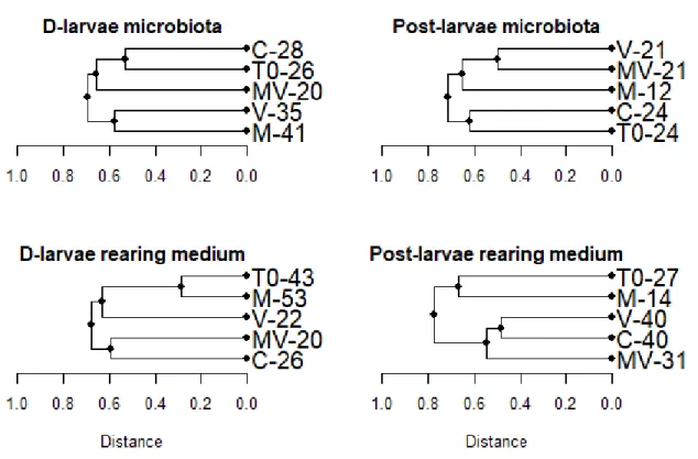
After 96 h of exposition, the D-larvae microbiota genetic fingerprints were separated in two clusters. A first cluster composed of the fingerprint from the initial larval microbiota (T0), the control (C) and the challenged marennine-treated D-larvae (MV) and a second one composed of the fingerprint from the challenged (V) and marennine-treated (M) D-larvae microbiota (figure 3). For the post-larvae microbiota, the fingerprints of the challenged larvae (V and MV) are clustered with the unchallenged marennine-treated post-larvae mi-crobiota (M) (figure 3).
The total number of OTUs decreased from 26 in T0 to 20 in the MV treatment. The total number of OTUs increased from 20 (T0) to 35 in V and 41 in M in the microbiota fingerprints from the second cluster (figure 3). In the rearing medium, the fingerprint of the challenged marennine-treated rearing medium of the D-larvae (MV) formed a cluster with
Figure 2: Bacterial abundance in the rearing medium after 1 h and 96 h of exposition to each treatment (C: control, M: marennine, V: vibrio and MV: marennine + vibrio; standard deviation is shown with error bars; letters indicate groups formed by the Tukey’s HSD post-hoc analysis and one set of letters (a, b and c) were assigned to the analysis of D-larvae while another set (d, e and f) were assigned to the post-larvae analysis.
the control (C), both with a decreased total number of OTUs representing respectively 20 and 26 compared to the initial community (T0) which had 43 OTUs in total (figure 3).
Notably, both communities from the rearing medium and the microbiota from the challenged D-larvae marennine-treated treatment (MV) has a total of 20 OTUs (figure 3) from which only 11 were common to the rearing medium and the microbiota. For the D-larvae and the post-D-larvae, the treatment M is less dissimilar from the initial communities (T0) in the rearing medium with 28.57% and 66.68% respectively (figure 3).
Figure 3: Dendrograms of the genetic fingerprints of the bacterial communities sampled in the microbiota and the rearing medium of blue mussel D-larvae and post-larvae at the beginning of the experiment (T0) and exposed to four different treatments (C: control, M: marennine, V: vibrio and MV: marennine + vibrio) during 96 h. The cluster analysis was based on the Jaccard coefficient similarity indicator and the dendrograms were constructed with the UPGMA algorithm using the vegan package (version 2.5-1) built for R (version 3.5). Numbers are the total number of OTUs recorded in each treatment.
Match-mismatch between the rearing medium and the larval microbiota di-versity
The proportion of matches between the community in the rearing medium and the community composing the microbiota of the D-larvae was 34.2% for T0 and 34.4% for the treatment C. This proportion was more important for the treatment M and V being respec-tively 67.5% and 41.9%. The proportion of matches in the MV treatment was lowest with 26.9% (figure 4).
Figure 4: Proportion of matches against mismatches in the comparison of the communities of the D-larvae rearing medium and the larval microbiota for each treatment (C: control, M: marennine, V: vibrio and MV: marennine + vibrio) and the initial communities (T0).
Comparison of microbial communities
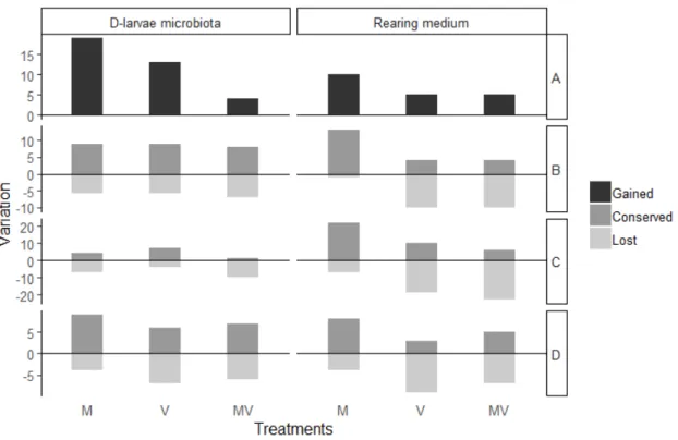
The simultaneous comparison of the initial community (T0), the control (C) and each treatment (M, V, and MV) allowed determining which OTUs were lost, conserved and gained among the different treatments. The treatment M, in regards of the larval microbiota and the rearing medium, was the one that lost the less OTUs that were either shared between T0 and C or unique to the treatment C (figure 5). The number of OTUs gained or conserved was dissimilar between the D-larvae microbiota and the rearing medium. For example, in the treatment M, 22 OTUs were conserved from the initial community in the rearing medium and only 4 in the larval microbiota. In terms of lost OTUs, the treatments V and MV were similar, except for the OTUs lost that were unique to the initial microbiota (T0). The challenged marennine-untreated D-larvae microbiota (V) conserved 7 OTUs from T0 and the challenged marennine-treated D-larvae (MV) conserved only 1 OTU from T0 (figure 6). This difference is less important in the rearing medium for the same treatments (figure 5). In the rearing medium, treatment M gained 10 unique OTUs while both V and MV gained 5 unique OTUs each (figure 5). The difference in the number of unique OTUs gained is even greater than in the microbiota for the treatment M. There were 19 unique OTUs found in the larval microbiota from the M treatment, 13 unique OTUs in the treatment V and 4 in the treatment MV (figure 5).
2.6 Discussion
The future of the development of shellfish aquaculture is often related to stable pro-duction juveniles potentially having controlled genetic characteristics. However, the larval rearing process can create an environment conductive to the development of opportunistic bacterial pathogens like V. splendidus. Controlling these pathogens in the rearing environ-ment is crucial to ensure a stable juvenile production. As new alternative methods to the harmful usage of antibiotics in aquaculture, marennine, a biomolecule extracted from a di-atom is becoming an interesting way of limiting the impact of pathogens (Gastineau et al.,
Figure 5: Simultaneous comparison of the initial community (T0) and the control (C) after 96 hof exposition with all other treatments (M, V and MV) in regard of a) the unique OTUs Gained in each treatment, b) the Conserved OTUs between T0 and the control (C), c) the Unique OTUs from the initial community (T0) and d) the unique OTUs from the control (C).
2014; Falaise et al., 2016; Turcotte et al., 2016). In our study, we confirm the beneficial ef-fects of marennine on blue mussels larvae challenged with V. splendidus and investigated the marennine effect in regards of the response of the bacterial communities in the system.
2.6.1 Antibacterial activity of marennine on V. splendidus 7SHRW
When investigating the beneficial effects of a new biomolecule against bacterial dis-eases, one of the first hypotheses to be tested would be that the molecule of interest exerts an antibacterial effect. Our results do not show a direct antibacterial effect of marennine at a concentration of 500 µg L−1and therefore an antimicrobial effect on the pathogen V.
splen-didus itself is unlikely to contribute to the observed beneficial effects documented by Tur-cotte et al. (2016). It has been previously reported that marennine demonstrates an inhibitor effect on the growth of several Vibrio species in previous experiments. Falaise et al. (2016) assessed the sensitivity of various species from the Vibrio genus (V. tasmaniensis, V. aestu-arianus, V. coralliitycus and V. tubiashii) to marennine at different concentrations ranging from 1 mg L−1to 100 mg L−1. Results showed concentration-dependant inhibitor effects on
the growth kinetics of all the tested species for marennine concentrations higher than the one used in our experiments. Hence, the main objective of the present study which is to investigate a potential larval microbiota modification leading to a better resistance against the pathogen V. splendidus. Turcotte et al. (2016) observed that marennine had a lethal effect on M. edulis D-larvae at a concentration as low as 1000 µg L−1. The marennine
concentra-tion used during their bacterial challenge experiments was therefore reduced to 500 µg L−1
to avoid the lethal effect of the pigment on the larvae during the rearing procedure.
2.6.2 Observed beneficial effects of marennine during bacterial challenges
The exposition of the larvae to marennine during bacterial challenges did not result in a measurable inhibition or an enhancement of the larval growth unlike the results obtained by Turcotte et al. (2016). The effect of marennine on larval growth might only be observable after a longer exposure to the bioactive molecule. Turcotte et al. (2016) exposed D-larvae to marennine for 20 days and observed significantly bigger shell when the larvae were exposed to marennine at 100 µg L−1and accumulation of energetic lipids.
The effect of marennine on the D-larvae survival after 96 h of exposition was simi-lar to the results published by Turcotte et al. (2016) with no significant difference between the survival rate of the control (C) and the corresponding treatments M (marennine) and MV (marennine and Vibrio). In the same way, the survival rate of the marennine untreated challenged D-larvae with Vibrio (V) was lower than the other treatments. Thus, our result confirms that marennine had a beneficial effect on the larval survival rate of the D-larvae