Science Arts & Métiers (SAM)
is an open access repository that collects the work of Arts et Métiers Institute of Technology researchers and makes it freely available over the web where possible.
This is an author-deposited version published in: https://sam.ensam.eu Handle ID: .http://hdl.handle.net/10985/19145
To cite this version :
Sylvain HAUPERT, Pascal LAUGIER, Sandra GUERARD, Françoise PEYRIN, David MITTON -Nonlinear ultrasound monitoring of fatigue microdamage accumulation in cortical bone - In: 2011 IEEE International Ultrasonics Symposium, Etats-Unis, 2011 - 2011 IEEE International
Ultrasonics Symposium - 2011
Any correspondence concerning this service should be sent to the repository Administrator : archiveouverte@ensam.eu
Nonlinear ultrasound monitoring of fatigue
microdamage accumulation in cortical bone
Sylvain Haupert, Pascal LaugierUPMC Univ Paris 06,
CNRS 7623, Laboratoire d'Imagerie Paramétrique, Paris, France,
sylvain.haupert@upmc.fr
Françoise Peyrin
CREATIS, Inserm U1044, CNRS 5220, INSA Lyon, Université Lyon I, Lyon, France,
ESRF, Grenoble, France peyrin@esrf.fr
Sandra Guérard Arts et Metiers ParisTech Laboratoire de Biomécanique
Paris, France sandra.guerard@ensam.eu
David Mitton
Université de Lyon, F-69622, Lyon, France ; IFSTTAR, LBMC, UMR_T9406, F-69675, Bron ;
Université Lyon 1, Villeurbanne david.mitton@ifsttar.fr
Abstract— Accumulation of bone micro-damage is suspected to
lead to severe impairment of mechanical properties with an increase in skeletal fragility and fracture risk. The objective of the study was to evaluate the potential of Nonlinear Resonant Ultrasound Spectroscopy (NRUS) for measuring micro-damage accumulation in cortical bone using four-point bending cycling fatigue. Sixteen human cortical bone specimens were machined as parallelepiped beams. Damage progression was controlled by measuring the linear elastic beam theory modulus (ELEBT), known
to reflect microdamage accumulation. Before and between each damage step, the nonlinear ultrasonic elastic coefficient was measured by NRUS. At the end of each cycling fatigue, a subset of bone samples was measured by µCT at the European Synchrotron Radiation Facility. Results showing a progressive increase of nonlinear ultrasonic elastic coefficient along fatigue cycling suggest that NRUS measurements are sensitive to micro-damage accumulation. The results mentioned above were validated using synchrotron radiation µCT. The variation of elastic nonlinearity was found to be significantly correlated to the variation of number density of small microcracks which almost doubled in damaged regions.
Keywords-component; nonlinear; NRUS; microcracks; bone; fatigue
I. INTRODUCTION
Bone microdamage is a natural phenomenon caused by daily loading, such as walking, jumping or carrying heavy load. Microdamage manifests as linear microcracks and diffuse damage. It is typically of little consequence under normal bone self-repair capability through targeted remodeling. Impaired repair capabilities following upon deficient turnover caused by disease or drug absorption result in an accumulation of microdamage which is then suspected to lead to severe impairment of mechanical properties such as decrease of bone toughness, stiffness and ultimate load. Such loss of biomechanical competence may lead ultimately to an increase in skeletal fragility and fracture risk [1].
Histomorphometry is the current gold standard to characterize damage accumulation in vitro. This technique is inherently invasive and destructive, is limited to the measurement of a small number of 2-D cross-sections and does not allow investigating the whole volume. These limitations have led to the emergence of alternative techniques. Micro-computed tomography (µCT) with contrast agent has been suggested for quantification and localization of microdamage in the whole specimen volume. However the resolution of about 10µm remains well below that allowed by histomorphometry and is not efficient to detect isolated microcracks and to assess their geometry. This limitation is overpassed by synchrotron radiation micro-computed tomography (SR-µCT) which is the only 3-D imaging technique capable to assess bone microcrack geometry at a micro-scale resolution [2].
At ultrasonic frequencies (approximately 100 kHz—2 MHz), linear characteristics of elastic wave transmission through bone, such as frequency-dependent attenuation and speed of sound, are widely used to assess skeletal status and predict osteoporotic fracture risk [3]. However, it was found that these parameters are insensitive to mechanically induced damage in human bone likely due to the fact that these parameters governing the wave equation fail to be sensitive to discontinuities at the meso or micro scale [4].
More recently, nonlinear acoustics techniques well known to be far more sensitive than linear acoustics to detect microcracks in diverse materials (e.g. concrete, composite), were applied to bone microdamage detection without quantification [5, 6]. A preliminary in vitro study by our group suggested that accumulation of damage induced by mechanical fatigue in compression of cortical bone was reflected by hysteretic nonlinear elastic properties measured by nonlinear resonant ultrasound spectroscopy (NRUS) [7] . The results mentioned above have to be validated against histology or high resolution µCT. In the present paper, we
1024
978-1-4577-1252-4/11/$26.00 ©2011 IEEE 2011 IEEE International Ultrasonics Symposium Proceedings 10.1109/ULTSYM.2011.0251
report the results of NRUS measurements in cortical bone specimens that were subjected to four-point bending fatigue. The results are compared to the level of damage measured by SR-µCT measurements.
II. MATERIAL AND METHODS A. Specimen preparation and measuring protocol
Sixteen human cortical bone samples were prepared from the femoral mid-diaphysis of four female donors (age = 88.5±9.8). Femurs were removed during multi-organ collection. Ethical approval for the collection of samples was granted by the Human Ethics Committee of the Centre du don des Corps at the University Paris Descartes (Paris, France). The tissue donors or their legal guardians provided informed written consent to give their tissue for investigation, in accord with legal clauses stated in the French Code of Public Health. The specimens were wet machined as parallelepiped beams (50*4*2mm), defatted and stored at -20°C until experiments. Initial NRUS measurements were performed for all samples to determine their initial nonlinear properties before any mechanical fatigue. The specimens were then taken through a fatigue protocol in four-point bending as described below, during which mechanical parameters were determined. NRUS measurements were repeated after each cycling session. Four damage steps were achieved. After each damage step, three specimens were removed for 3-D SR-µCT investigations of microdamage.
B. Nonlinear Resonant Ultrasound Spectroscopy (NRUS)
Microcrack accumulation in a material sample is responsible for a softening of the material for increasing excitation amplitudes, leading to a decrease of the resonance frequency when excitation amplitude increases. The NRUS technique exploits the resonant behavior of samples but with progressively increasing excitation levels to retrieve the nonlinear elastic behavior of the material. In resonance, it can be shown that the frequency shift Δf is proportional to the peak strain amplitude ∆ε via the nonlinear elastic (αf) parameter:
−
= =
where f is the resonant frequency at increased strain level, f0
its corresponding value at the lowest drive amplitude [8]. The parameter αf, so-called the nonlinear elastic hysteretic
parameter, characterizes the hysteretic nonlinearity that occurs for strain levels above approximately 10-5 [8] in damage materials and convey information about the amount of hysteretic nonlinearity (damage accumulation) in the material.
The principles of NRUS measurements have been extensively described elsewhere [9]. Briefly, a piezoceramic emitter glued on a backload was bonded at one end of the specimen to ensure free-fix boundary conditions for NRUS measurements. Each sample was probed by a swept-sine encompassing the first resonant modes of the cortical beam (assumed to be pure compression modes under symmetric loading conditions). The peak resonant frequency f is
measured as a function of strain applying increasing voltage drive level. The dynamic strain amplitude ε was calculated from the longitudinal particle displacement U at one end of the sample measured by a laser vibrometer (LSV, SIOS, Germany):
= = ∗ = ∗ 4
where k is the wave number and L is the specimen length. The nonlinear parameter αf can be calculated from the
strain-dependent resonance peak data, using Eq. 1. The usual NRUS measuring protocol was adapted to enhance the sensitivity to subtle variations of bone nonlinearity [10]. Toward this goal, the reference measurement f0 was repeated before each
excitation level and then used to compute αf. During the
NRUS measurements, specimens were kept at fixed temperature (37°C ±0.1°C) and relative humidity (15%±5%) into a climate chamber.
C. Biomechanical testing
The piezoceramic emitter attached to the specimen for NRUS measurements was removed before each mechanical testing. All specimens were progressively damaged by cyclic four-point bending at 2Hz in a saline solution at 37°C (±1°C) using a hydraulic testing machine (INSTRON, 8802, High Wycombe, England) with a 1kN loading cell (accuracy 0.5%) and the internal displacement transducer (accuracy 1%). In this configuration, damage is expected to occur specifically in the mid region of the sample [11], while the ends remaining intact may be used as control. Initial Young’s modulus was determined during pre-cycling after 20 cycles (Epre).
From the initial Young’s modulus, the load (Fmax) corresponding to 5000µε at the mid-span was computed for all specimens [11]. The four-point bending fatigue was then applied between -10N and –Fmax. During the cycling session, load and displacement curves were recorded to extract linear elastic beam theory (LEBT) modulus (ELEBT) as defined by
Landrigan [12]. ELEBT is a combination of elastic (secant
modulus) and plastic (residual strain) biomechanical parameters. After each damage step, the ELEBT modulus is
normalized by the initial value measured for the first loading cycle of the first damage step. ELEBT has been shown to
decrease as bone microdamage accumulates [11-13]. A progressive damage was performed in four steps (one step=one cycling session), each step corresponding to a reduction of 10% of ELEBT.
D. 3-D synchrotron radiation µCT (SR-µCT)
At the end of each cycling fatigue, a subset of 3 bone samples were measured by SR-µCT at the European Synchrotron Radiation Facility, Grenoble, France on beam-line ID19 [2]. The photon energy was 25 keV and the size of the region of interest (ROI) was 2.8x2.8x1.96mm3 with a voxel size of 1.4 µm. Two different ROIs were investigated: ROI1 located in the load-free region at one distal end of the sample assumed to be free of damage (except initial pre-fatigue damage) and ROI2 located in the central portion of the
This research was supported by the Agence Nationale pour la Recherche (ANR), France (Grant BONUS_07BLAN0197).
beam dam trans reco of m using Neur E. D Matl used (AN level fatig micr dam Wilc elast obta test. p<0. A. B T was the f for syste was varia 20 m where micr mage was ch sverse cross-nstructed bon microcracks ( g the softwa ronJ (Erik Me Figure 1. Tran Data analysis lab 7.8 with s d for statistic NOVA) was u ls of nonline gue protocol rodamage cha maged region coxon signed tic parameter ained using re The signific .05. Biomechanica The measurem assessed on final protocol. each sample. em stabilizati repeated 6 t ation was fou
00µm rodamage is a haracterized o sections (Fig ne volumes by (Cr.Dn [#.mm are ImageJ ( eijering, The N sverse 2-D high r microcracks ( statistics toolb al analyses. used to test fo ear elasticity l. The effe aracteristics i was investig rank test. Th r αf with mic egression anal cance level III. R al testing ment precision five dedicate . The initial Y Then they w on) of four-p times with rep
und to be 2.4 assumed to ac on 12 regul g. 1) extracte y measuring th m²]) and leng (NIH, USA)) Netherlands). resolution µCT cr (white arrows) box 7.6 (Math One-way ana for differences achieved at ect of fatig in the contro gated with a he relationship crodamage ch lyses and Spe
is measured RESULTS n of biomech ed specimens Young’s modu went through point bending positioning. T 4% for ELEBT ccumulate. M arly spaced ed from the he number de gth (Cr.Le [µ with the pl ross-section with hworks, USA) alysis of vari s among diff each steps of gue loading ol region and a non param p of the nonl haracteristics earman correl using a p-v hanical param not included ulus was estim h 20 cycles (
test. The pro The coefficien T. The numbe Micro-2-D 3-D ensity µm]), lugin h ) was iance ferent f the on d the metric inear was ation value meters d into mated (after ocess nt of er of cycl dam 1182 hom dens B. U The coef inter 8.5% first repo [10] step dam that decr spec sess relat fatig clari ANO step Figu C. D T imag stati Cr.D Cr.D in a Inde leng crac (Cr. p a ra m e te r v a ri a ti o n (Δα f /αf ) les required to mage step was
2±966 cycle mogeneous ini sity=1792±15 Ultrasonic (N measuremen fficient of rmediate deb % for αf. The t compression orted for und ]. Δαf/αf repres p of αf with res mage step in F the number o reased as a f cimens were sion. In parti tive increase gue steps are ity and were r OVA of Δαf/α
ps (F=7, p=0.0
ure 2. Nonlinea
Damage chara Taking into ges, the mea istically diffe Dn=1.92±1.19 Dn =2.20 ±1.2 a significant in eed, by taking gth falling in ck density alm Dn= 0.37±0 -20% 0% 20% 40% 60% 80% 100% 120% 140% pa ra m e te r va ri a ti o n (Δα f /αf )
No
o achieve the found to var s for the f itial biomecha 5g.mm-3 and E NRUS) measure nt precision variation of onding and r initial nonlin mode were co damaged cortisenting the rel spect to its ini ig. 2 using th of specimens i function of th removed for icular, three of αf higher t not represen removed for t αf showed a s 01) ar elastic coefficie st acteristics account all d asured microc erent between 9#/mm²) and 25#/mm²). In c ncrease of the g into account n the first qu most doubled b .26#/mm²) an 1 2 Dam
onlinear el
desired ELEBT ry between th first cycling anical proper ELEBT=15.1±3 rements of NRUS, f three me repositioning, near values αf onsistent with ical bovine b lative change itial value is r he Box and W included in ea he damage st SR-µCT acq outliers sho than 150% af nted in Fig. the ANOVA significant eff ent variation (Δαf/ tep detected mic cracks charac n ROI1 (Cr d ROI2 (Cr contrast, fatigu e density of s t only small m quartile (Cr.L between the co nd the dama 2 3 mage step #lastic coef
T reduction at he specimens session) de rties (apparen .0GPa). assessed by easurements was found t f= -5.6±2.7 fo h values previo bone (αf=-5.0±after each dam represented ag Whiskers Plot. ach cycling se tep, because uisition after owing a dram fter the 2nd an
2 for the sak analysis. One fect of the dam
f/αf) after each dam
crocracks on cteristics were .Le=71.8±13. .Le=65.3±10. ue cycling res small microcr microcracks w e<31.93±8.05 ontrol region R age region R 4
fficient
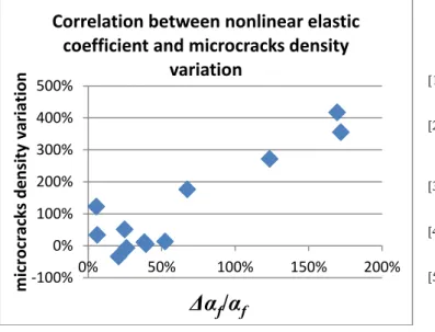
each (e.g., espite nt dry y the with to be or the ously ±2.5) mage gainst Note ession three each matic nd 3rd ke of e-way mage mage µCT e not 7µm; 3µm; sulted racks. with a 5µm), ROI1 ROI2(Cr.Dn= 0.63±0.45#/mm²) (p=0.01). Moreover, the relative variation of density of small microcracks between ROI2 and ROI1 was found to be significantly correlated to the relative variation ∆αf/αf (r²=0.6, p<0.05) (Fig.3).
Figure 3. Correlation between nonlinear elastic coefficient variation (Δαf/αf)
and tiny microcracks density variation IV. DISCUSSION AND CONCLUSION
This is the first study reporting the modulus ELEBT, the
nonlinear elastic hysteretic parameter αf and microdamage
characteristics derived from SR-µCT that were concurrently assessed in human cortical bone specimens during a four point-bending fatigue cycling protocol. The progressive 10% decrease of ELEBT after each fatigue cycling session is strongly
suggestive of microdamage accumulation [11, 13-15]. Our results showed a progressive decrease of the normalized hysteretic parameter αf, suggesting that NRUS measurements
are indeed sensitive to damage accumulation.
High resolution µCT investigations evidenced a significant increase in small microcracks number which almost doubled in damaged regions compared to their number in load-free (control) regions. These small microcracks, with length falling in the lowest quartile, are suspected to be newly formed microcracks as a result of fatigue cycling. The relative variation of nonlinear elasticity was significantly related to the relative variation of the number density of these small cracks. Altogether, our results evidence, for the first time, a relationship between the nonlinear elastic parameter αf
measured by NRUS and bone microdamage characteristics reflected by the density of supposedly newly formed microcracks during fatigue cycling. To conclude, our experimental results are indicative of the potential of NRUS measurements for monitoring non-invasively microdamage accumulation in cortical bone.
ACKNOWLEDGMENT
The authors want to thank G Renaud and J Rivière for their helpful comments and suggestions during the preparation of the experiments, as well as PA Johnson, Los Alamos National Laboratory, NM, USA for fruitful discussions and E Boller, M Zuluaga and M Langer for help during the ESRF experiment performed in the framework of LTP MD431.
REFERENCES
[1] P. Zioupos, "Accumulation of in-vivo fatigue microdamage and its relation to biomechanical properties in ageing human cortical bone," Journal of
Microscopy-Oxford, vol. 201, pp. 270-278, Feb 2001.
[2] A. Larrue, A. Rattner, Z. A. Peter, C. Olivier, N. Laroche, L. Vico, and F. Peyrin, "Synchrotron Radiation Micro-CT at the Micrometer Scale for the Analysis of the Three-Dimensional Morphology of Microcracks in Human Trabecular Bone," PLoS ONE, vol. 6, p. e21297, 2011.
[3] P. Laugier, "Instrumentation for in vivo ultrasonic characterization of bone strength," Ultrasonics, Ferroelectrics and Frequency Control, IEEE
Transactions on, vol. 55, pp. 1179-1196, 2008.
[4] P. H. F. Nicholson and M. L. Bouxsein, "Quantitative ultrasound does not reflect mechanically induced damage in human cancellous bone," Journal
of Bone and Mineral Research, vol. 15, pp. 2467-2472, Dec 2000.
[5] H. Moreschi, S. Callé, S. Guerard, D. Mitton, G. Renaud, and M. Defontaine, "Monitoring trabecular bone microdamage using a dynamic acousto-elastic testing method," Proceedings of the Institution of
Mechanical Engineers, Part H: Journal of Engineering in Medicine, vol.
225, pp. 282-95, 2011 .
[6] G. Renaud, S. Calle, J. P. Remenieras, and M. Defontaine, "Exploration of trabecular bone nonlinear elasticity using time-of-flight modulation," Ieee
Transactions on Ultrasonics Ferroelectrics and Frequency Control, vol.
55, pp. 1497-1507, Jul 2008.
[7] M. Muller, D. Mitton, M. Talmant, P. Johnson, and P. Laugier, "Nonlinear ultrasound can detect accumulated damage in human bone," Journal of
Biomechanics, vol. 41, pp. 1062-1068, 2008.
[8] P. Johnson and A. Sutin, "Slow dynamics and anomalous nonlinear fast dynamics in diverse solids," Journal of the Acoustical Society of America, vol. 117, pp. 124-130, Jan 2005.
[9] K. E. A. Van den Abeele, J. Carmeliet, J. A. Ten Cate, and P. A. Johnson, "Nonlinear elastic wave spectroscopy (NEWS) techniques to discern material damage, Part II: Single-mode nonlinear resonance acoustic spectroscopy," Research in Nondestructive Evaluation, vol. 12, pp. 31-42, 2000.
[10] S. Haupert, G. Renaud, J. Rivière, M. Talmant, P. A. Johnson, and P. Laugier, "High-accuracy acoustic detection of nonclassical component of material nonlinearity," Journal of the Acoustical Society of America, vol. 130, 2011.
[11] T. Diab and D. Vashishth, "Effects of damage morphology on cortical bone fragility," Bone, vol. 37, pp. 96-102, Jul 2005.
[12] M. Landrigan and R. Roeder, "Systematic error in mechanical measures of damage during four-point bending fatigue of cortical bone," Journal of
biomechanics, vol. 42, p. 1212, 2009.
[13] T. M. Boyce, D. P. Fyhrie, M. C. Glotkowski, E. L. Radin, and M. B. Schaffler, "Damage type and strain mode associations in human compact bone bending fatigue," Journal of Orthopaedic Research, vol. 16, pp. 322-329, May 1998.
[14] T. Diab, K. W. Condon, D. B. Burr, and D. Vashishth, "Age-related change in the damage morphology of human cortical bone and its role in bone fragility," Bone, vol. 38, pp. 427-431, Mar 2006.
[15] O. S. Sobelman, J. C. Gibeling, S. M. Stover, S. J. Hazelwood, O. C. Yeh, D. R. Shelton, and R. B. Martin, "Do microcracks decrease or increase fatigue resistance in cortical bone?," Journal of Biomechanics, vol. 37, pp. 1295-1303, Sep 2004.
[16] F. J. O'Brien, D. Taylor, and T. C. Lee, "Microcrack accumulation at different intervals during fatigue testing of compact bone," Journal of
Biomechanics, vol. 36, pp. 973-980, Jul 2003. -100% 0% 100% 200% 300% 400% 500% 0% 50% 100% 150% 200% microcracks density variation
Δα
f/
α
fCorrelation between nonlinear elastic
coefficient and microcracks density
variation
1027 2011 IEEE International Ultrasonics Symposium Proceedings Powered by TCPDF (www.tcpdf.org) Powered by TCPDF (www.tcpdf.org) Powered by TCPDF (www.tcpdf.org) Powered by TCPDF (www.tcpdf.org) Powered by TCPDF (www.tcpdf.org) Powered by TCPDF (www.tcpdf.org) Powered by TCPDF (www.tcpdf.org)