D
ECREASED GLUTAMATE TRANSPORTER
(GLT-1)
EXPRESSION IN FRONTAL CORTEX OF RATS WITH
ACUTE LIVER FAILURE
K Knecht
a, A Michalak
a, C Rose
a, J.D Rothstein
b, R.F Butterworth
a,a Neuroscience Research Unit, Hôpital Saint-Luc (University of Montreal), 1058 St-Denis, Montreal, Quebec, H2X 3J4, Canada b Department of Neurology, Johns Hopkins University, School of Medicine, Baltimore, MD 21287, USA
A
BSTRACT
It has been suggested that reduced astrocytic uptake of neuronally released glutamate contributes to the pathogenesis of hepatic encephalopathy in acute liver failure. In order to further address this issue, the recently cloned and sequenced astrocytic glutamate transporter GLT-1 was studied in brain preparations from rats with ischemic liver failure induced by portacaval anastomosis followed 24 h later by hepatic artery ligation and from appropriate sham-operated controls. GLT-1 expression was studied using reverse transcriptase-polymerase chain reaction (RT-PCR). Expression of GLT-1 transcript was significantly decreased in frontal cortex at coma stages of acute liver failure. Western blotting using a polyclonal antibody to GLT-1 revealed a concomitant decrease in expression of transporter protein in the brains of rats with acute liver failure. Reduced capacity of astrocytes to reuptake neuronally released glutamate, resulting from a GLT-1 transporter deficit and the consequently compromised neuron-astrocytic trafficking of glutamate could contribute to the pathogenesis of hepatic encephalopathy and brain edema, two major complications of acute liver failure.
Keywords
GLT-1; hepatic encephalopathy; Astrocytes; Neuron-astrocyte trafficking; Gene expression; RT-PCR; Western blottingAcute liver failure resulting from viral infections or ingestion of toxic substances results in severe neurological impairment progressing through stupor and coma within hours or days. The pathophysiologic mechanisms responsible for hepatic encephalopathy (HE) in acute liver failure are unknown but deficits of neurotransmission rather than primary cerebral energy failure are currently considered to be implicated [1 and 3]. In this regard, there is increasing evidence to suggest that abnormalities of the glutamate neurotransmitter system are involved in the pathogenesis of HE in acute liver failure. Brain concentrations of glutamate are reduced in both human [13]and experimental [7 and 17] acute liver failure and recent studies using in vivo cerebral microdialysis in both rats 2 and 9and rabbits [5]with acute (ischemic) liver failure have consistently revealed increased extracellular brain concentrations of glutamate. It was suggested that the increased extracellular concentrations of glutamate in the brain of these animals was the consequence of decreased astrocytic uptake [9]. The major high affinity astrocytic transporter for glutamate, GLT-1, has recently been cloned and sequenced 12 and 14. In order to directly assess glutamate uptake capacity in the brain in acute liver failure, GLT-1 gene and protein expression were measured in frontal cortex of rats with acute (ischemic) liver failure.
Male, Sprague-Dawley rats (175-200 g) were anesthetized with halothane and an end-to-side portacaval shunt (SH) was performed according to the guidelines of Lee and Fisher [6]. Sham-operated control rats (SM) were anesthetized and underwent a laparotomy and clamping of the portal vein. 24 h later, the same rats were reanesthetized with halothane and subjected to hepatic artery ligation (HAL) or laparotomy (SM). In this way, four groups of surgically operated animals were created with four rats in each group, namely acute liver failure (SH+HAL) and the three obligate control groups SH+SM, SM+HAL and SM+SM. After surgery, arterial blood glucose levels were monitored
and supplemental glucose was administered intraperitoneally, as needed. Control animals received an equivalent volume of 0.9% saline. Body temperature was maintained at 37°C.
Approximately 10-12 h after surgery, animals lost their righting reflex followed 2-3 h later by loss of corneal reflex (coma). At coma stages of ischemic liver failure, rats in acute liver failure together with their appropriate controls were killed by decapitation and brains were removed and stored at −80°C until use.
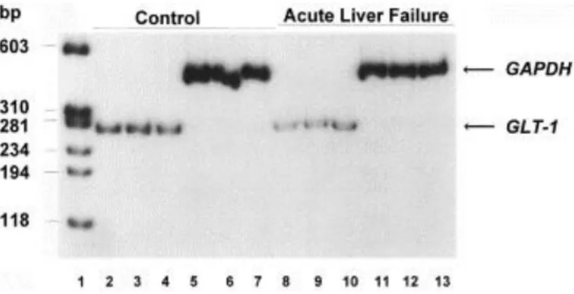
GLT-1 expression was quantitated by RT-PCR using purified total RNA from frontal cortex of animals from the four treatment groups (Fig. 1). Total RNA was extracted using the Trizol reagent (Life Technologies Inc., Gaithersburg, MD, USA) according to the manufacturer's protocol. DNA contamination of the RNA samples was removed by incubation with RNase-free DNase. Quantification of expression by RT-PCR requires a linear ratio between cycle number and resulting DNA amount. Thus, conditions for expression studies for each gene were performed.
Glyceraldehyde-3-phosphate dehydrogenase (GAPDH) was used as internal standard to evaluate RNA samples and to correct for loading variations. Optimal conditions were obtained after 22 amplification cycles for GLT-1 and 17 for GAPDH using 0.5 μg of RNA per amplification. The monospecificity of the oligonucleotide primers was verified by computer analyses using the PRIMER program (Genetics Computer Group, Madison, WI). Reverse transcription of mRNA and PCR amplification of resulting cDNA molecules were performed in a one-step reaction. The reaction mixture in a final volume of 50 μl contained 0.5 μg of the total RNA, which was incubated in 10 mM Tris–HCl (pH 8.3), 50 mM KCl, 0.1% (w/v) BSA, 1 mM MgCl, 200 μM dNTPs, 10 μCi/ml [α-32P]dCTP (3000 μCi/mmol), 320 U/ml of RNAsin, 100 U/ml of AMV reverse transcriptase, 20 U/ml of Taq DNA polymerase and the primers either for GLT-1 (1 μM of each forward and reverse, respectively) or GAPDH (0.5 μM of each). The mRNA was reverse transcribed at 50°C for 10 min followed by PCR including a denaturing step at 95°C for 30 s, primer annealing at 62°C for 1 min and DNA-elongation at 72°C for 1.5 min. Using these primer sets the amplification of GLT-1-mRNA resulted in a 285 bp fragment, whereas the GAPDH-product was 470 bp in length. The PCR products were separated on an 8%
polyacrylamide gel which was then apposed to film for visualization. The amplified GLT-1 and GAPDH-cDNA-fragments were subsequently excised from the gel and Cerenkov radiation was quantitated using a β-counter.
Fig. 1. Expression of the astrocytic glutamate transporter GLT-1 and GAPDH in frontal cortex of rats with acute liver failure versus control (sham operation). Total RNA was extracted from the brain of 3 animals with acute liver failure at coma stages of encephalopathy or at comparable times following sham operations. Lane 1, molecular weight standards; lanes 2-7, GLT-1 and GAPDH expression in sham-operated control rats; lanes 8-13, GLT-1 and GAPDH expression in rats with acute liver failure.
For the study of GLT-1 protein, brain tissue was homogenized in 50 mM Tris–HCl (pH 7.4), 300 mM NaCl, 0.1% Tween (Fig. 2). After centrifugation (10 000×g, 15 min) the supernatant was separated by a denaturing SDS-PAGE (7.5%) as previously described [14]. Blotting was performed with the semi-dry transfer cell from Bio-Rad at 15 V (1 A) for 45–60 min using ECL-nitrocellulose and Whatmann paper (3MM) each presoaked in transfer buffer (48 mM Tris (pH 8.3), 39 mM glycine, 0.037% SDS, 20% methanol) for at least 15 min. After the transfer the membrane was
briefly rinsed in 1×PBS/0.1% Tween-20 and air dried. For immunological protein detection the membrane was incubated in blocking buffer (10% horse serum, 5% nonfat dried milk, 1% BSA, 1× PBS/0.1% Tween-20) for 1 h followed by incubations with the antisera against GLT-1 and secondary antibodies (Amersham: 1:4000 diluted anti-rabbit IgG horseradish peroxidase-coupled) each in blocking buffer for 1 h. Subsequent to each incubation with the primary and secondary antibody, blots were washed three times (1×20 min and 2×5 min) with 1× PBS/0.1% Tween-20. For the detection of specific antibody binding the membranes were treated in accordance with the Amersham ECL-Kit instructions and apposed to photosensitive ECL-hybond film for 5–60 s. Signal intensities were measured with a densitometer.
Fig. 2. GLT-1 protein expression in frontal cortex of rats at coma stage of acute liver failure (ALF, lanes 5-7) and sham-operated controls (lanes 2-4). Lane 1, molecular weight markers.
Results of these studies reveal significant reductions in GLT-1 protein (by 31%, P<0.05) and in GLT-1 mRNA (by 60%, P<0.01) in frontal cortex of rats with ischemic liver failure at coma stages of encephalopathy compared to SM+SM, SM+HAL or SH+SM control groups of animals ( Fig. 1 and Fig. 2). These findings add to a growing body of evidence suggesting that acute liver failure results in altered glutamatergic synaptic regulation in brain. Previous studies using in vivo cerebral microdialysis demonstrated increased extracellular glutamate concentrations in frontal cortex [9]as a function of the deterioration of neurological status and of arterial ammonia concentrations in the same animal model of acute liver failure as that used in the present study. Such increases could result directly from diminished capacity for astrocytic uptake of glutamate as a consequence of decreased expression of GLT-1. Reduced expression of GLT-1 and the consequently increased extracellular brain glutamate concentrations also offers a possible explanation for the observed decrease in densities (down regulation) of AMPA/kainate binding sites also described in this model of acute liver failure [10]. Diminished uptake of neuronally released glutamate by the astrocyte as a consequence of decreased expression of GLT-1 would therefore constitute further evidence of impaired neuron-astrocytic trafficking of glutamate, a phenomenon which has previously been proposed to contribute to the pathogenesis of hepatic encephalopathy based on neurochemical studies [4]and by 13C-NMR spectroscopy [16]in portacaval-shunted rats.
Impaired removal of glutamate from brain extracellular space due to decreased GLT-1 expression could also be implicated in the pathogenesis of cytotoxic brain edema, a major complication of acute liver failure in both humans and in the experimental animal model used in the present study [17]. Exposure of cultured astrocytes to glutamate leads to significant cell swelling [15]. Finally, it is conceivable that, under certain circumstances, reduction in the capacity for glutamate uptake by astrocytes as a result of decreased GLT-1 expression could impair ammonia
removal by brain. Glutamine synthetase, the enzyme primarily responsible for ammonia detoxification by brain has a predominantly astrocytic localization [11]and a recent study provides evidence to suggest that the metabolic fate of glutamate in astrocytes (i.e. conversion to glutamine versus oxidation in the tricarboxylic acid cycle) is regulated by extracellular glutamate concentrations [8].
A
CKNOWLEDGEMENTS
The authors are grateful to Paul Desjardins for helpful discussions and to Dominique Roy for typing the manuscript. This study was funded by a grant (PG 11118) from The Medical Research Council of Canada. A.M. is recipient of a Research Fellowship from The Canadian Liver Foundation.
R
EFERENCES
1. T.E. Bates, S.R. Williams, R.A. Kauppinen, D.G. Gadian Observation of cerebral metabolites in an animal model of acute liver failure in vivo: a 1H and 31P nuclear magnetic resonance study J. Neurochem., 53 (1989), pp. 102–110 2. D.K. Bosman, N.E.P. Deutz, M.A.W. Maas, H.M.H. van Eijk, J.J.H. Smit, J.G. de Haan, R.A.F.M. Chamuleau Amino acid release from cerebral cortex in experimental acute liver failure, studied by in vivo cerebral cortex microdialysis J. Neurochem., 59 (1992), pp. 591–599
3. Butterworth, R.F., Hepatic encephalopathy. In: I.M. Arias, J.L. Boyer, N. Fausto, W.B. Jacoby, D. Schachter and D.A. Shafritz (Eds.), The Liver: Biology and Pathobiology, 3rd edn., Raven Press, New York, 1994, pp. 1193-1208.
4. R.F. Butterworth Portal-systemic encephalopathy: a disorder of neuron-astrocytic metabolic trafficking Dev. Brain Res., 15 (1993), pp. 313–319
5. R.J. de Knegt, S.W. Schalm, C.C.D. van der Rijt, D. Fekkes, E. Dalm, I. Hekking-Weyma Extracellular brain glutamate during acute liver failure and during acute hyperammonemia simulating acute liver failure: an experimental study based on in vivo brain dialysis J. Hepatol., 20 (1994), pp. 19–26
6. S.H. Lee, B. Fisher Portacaval shunt in rat Surgery, 50 (1961), pp. 668–672
7. A.M. Mans, M.R. DeJoseph, R.A. Hawkins Metabolic abnormalities and grade of encephalopathy in acute hepatic failure J. Neurochem., 63 (1994), pp. 1829–1838
8. M.C. McKenna, U. Sonnewald, X. Huang, J. Stevenson, H.R. Zielke Exogenous glutamate concentration regulates the metabolic fate of glutamate in astrocytes J. Neurochem., 66 (1996), pp. 386–393
9. A. Michalak, C. Rose, J. Butterworth, R.F. Butterworth Neuroactive amino acids and glutamate (NMDA) receptors in frontal cortex of rats with experimental acute liver failure Hepatology, 24 (1996), pp. 908–913
10. A. Michalak, R.F. Butterworth Selective loss of binding sites for the glutamate receptor ligands [3H]kainate and (S)-[3H]5-fluorowillardiine in the brains of rats with acute liver failure Hepatology, 25 (1997), pp. 631–635 11. M.D. Norenberg, A. Martinez-Hernandez Fine structural localization of glutamine synthetase in astrocytes of rat brain Brain Res., 161 (1979), pp. 303–310
12. G. Pines, N.C. Danbolt, M. Bjoras, Y. Zhang, A. Bendahan, L. Eide, H. Koepsell, J. Storm-Mathisen, E. Seeberg, B.I. Kanner Cloning and expression of a rat brain L-glutamate transporter Nature, 360 (1992), pp. 464–467
13. C.O. Record, B. Buxton, R.A. Chase, G. Curzon, I.M. Murray-Lyon, R. Williams Plasma and brain amino acids in fulminant hepatic failure and their relationship to hepatic encephalopathy Eur. J. Clin. Invest., 6 (1976), pp. 387–394 14. J.D. Rothstein, L. Martin, A.I. Levey, M. Dykes-Horberg, L. Jin, D. Wu, N. Nash, R.W. Kuncl Localization of neuronal and glial glutamate transporters Neuron, 13 (1994), pp. 713–725
15. G.D. Schneider, A. Baethmann, O. Kempski Mechanism of glial swelling induced by glutamate Can. J. Physiol. Pharmacol., 70 (1992), pp. S334–S343
16. U. Sonnewald, G. Therrien, R.F. Butterworth Portal-systemic encephalopathy, disorder of neuron-astrocytic metabolic trafficking: evidence from 13C-NMR studies J. Neurochem., 67 (1996), pp. 1711–1717
17. M. Swain, R.F. Butterworth, A.T. Blei Ammonia and related amino acids in the pathogenesis of brain edema in acute ischemic liver failure in rats Hepatology, 15 (1992), pp. 449–453