HAL Id: dumas-01315000
https://dumas.ccsd.cnrs.fr/dumas-01315000
Submitted on 12 May 2016HAL is a multi-disciplinary open access archive for the deposit and dissemination of sci-entific research documents, whether they are pub-lished or not. The documents may come from teaching and research institutions in France or abroad, or from public or private research centers.
L’archive ouverte pluridisciplinaire HAL, est destinée au dépôt et à la diffusion de documents scientifiques de niveau recherche, publiés ou non, émanant des établissements d’enseignement et de recherche français ou étrangers, des laboratoires publics ou privés.
Outcomes of covered balloon-expandable stent
placement in the treatment of atheroscerotic occusive
disease at the aortic bufircation
Amandine Elie
To cite this version:
Amandine Elie. Outcomes of covered balloon-expandable stent placement in the treatment of atheroscerotic occusive disease at the aortic bufircation. Human health and pathology. 2016. �dumas-01315000�
AVERTISSEMENT
Ce document est le fruit d'un long travail approuvé par le
jury de soutenance et mis à disposition de l'ensemble de la
communauté universitaire élargie.
Il n’a pas été réévalué depuis la date de soutenance.
Il est soumis à la propriété intellectuelle de l'auteur. Ceci
implique une obligation de citation et de référencement
lors de l’utilisation de ce document.
D’autre part, toute contrefaçon, plagiat, reproduction illicite
encourt une poursuite pénale.
Contact au SID de Grenoble :
thesebum@ujf-grenoble.fr
LIENS
Code de la Propriété Intellectuelle. articles L 122. 4
Code de la Propriété Intellectuelle. articles L 335.2- L 335.10
http://www.cfcopies.com/juridique/droit-auteur!""#$% '()* +!,-./0 10 2010,340 10 560478.0 #$%&% '(%&%)#%% '*+( ,-*.#%)#/*) 0+ 0*1#*(2# %) 3%0%1/)% 0/',43% 0-%#2#
0.30 !2!41340
#$%&% &*+#%)+% '+.,/A+%3%)# 2 ,2 B21+,#% 0% 3%0%1/)% 0% C(%)*.,%D ,6 E 3FG HI"J
06KFL: 76 MN8O PQ;9QR5 S6T
3QLRG6N8 76 9:;<$==$>: 2!540 ?$@"A.>BT '85RGS6L: SN MN8O 6: 0G86P:6N8 S6 :UVR6 3QLRG6N8 76 9:;<$==$>: 863,C74 9D$::$AEF$=
3QLRG6N8 76 9:;<$==$>: !.63, 9D$::$
3QLRG6N8 76 1;BG$>: H034/-6306 ,I:D=G;JI$ 3QLRG6N8 76 1;BG$>: ,7,C0/ 0KK@">$L
!"# %#&'()* +, -*+,&./, +, 01,/23(, /4,/),/+ +2//,1 #'&'/, #55123#).2/ /. .65123#).2/ #'7 25./.2/8 *6.8,8 +#/8 (,8 )9:8,8 ; &,8 25./.2/8 82/) &2/8.+*1*,8 &266, 51251,8 < (,'18 #'),'18=
!"#$%&'( %* $%+','- ./00%%12'34/1-/.0' (#'1#
40/$'&'1# 51 #6' #,'/#&'1# %* /#6',%($0',%#5$
%$$0"(5+' -5('/(' /# #6' /%,#5$ .5*",$/#5%1
Remerciements
À mon Maître, président du jury et directeur de thèse:Au Professeur Jean Luc MAGNE:
Je vous remercie de me faire l’honneur d’accepter la présidence de cette thèse. Merci de m’avoir tant aidé dans ce travail, pour votre disponibilité et les critiques, toujours justes, que vous avez pu émettre. Merci de m’avoir donné le goût de cette magnifique spécialité qu’est la chirurgie vasculaire et que vous exercez avec excellence, précision, calme, générosité et humilité. Merci de m’avoir accueillie dans votre service merci pour votre soutien, votre dévouement, votre bienveillance qui m’ont permis d’avoir un petit chez moi loin des miens... Votre expérience et votre sagesse resteront un exemple dans ma pratique future. Je vous adresse ce témoignage de ma reconnaissance et de mon plus profond respect. Merci enfin de prendre soin de nous comme si nous faisions partie de votre famille, famille dont je suis très honorée et fière d’être l’une de vos derniers rejetons…
À mes Maîtres et juges:
Au Professeur Pierre-‐Yves Brichon:
Je vous remercie de m’avoir fait l’honneur d’accepter de prendre part à ce jury. Merci de m’avoir fait découvrir votre merveilleuse spécialité qui n’en reste pas moins la plus difficile à maitriser à mes yeux. Merci pour la démonstration quotidienne de votre rigueur, de votre efficacité, de votre précision et justesse du geste chirurgical. Merci de nous montrer chaque jour ce qu’est d’atteindre l’excellence. Je vous remercie pour tous ces bons moments dans votre bloc ou lors des staffs, où vous savez allier sérieux, calme, gourmandise et humour. Merci enfin de me démontrer qu’il est possible d’être un père en chirurgie et un excellent père de famille…
Au Professeur Pierre Alric:
Je suis très honorée de votre présence dans mon jury. Je vous remercie de l’investissement et du temps que vous accordez à mon travail. Merci de m’avoir accueillie et de m’avoir permis de faire partie de votre école chirurgicale pendant un semestre et bientôt plus. Je vous suis très reconnaissante pour votre confiance. Vos connaissances, votre précision dans le travail et votre pédagogie vous honorent. Je retiens votre gentillesse et votre bienveillance à mon égard. Soyez assuré de ma sincère admiration et de mon plus grand respect.
Au Docteur Christophe Seinturier:
Vous m’avez fait l’honneur de juger cette thèse. Pour votre disponibilité, vos qualités professionnelles et votre gentillesse, je vous prie d’agréer mes remerciements les plus sincères et toute ma gratitude.
Au Docteur Emmanuel Cochet:
Merci de me faire l’honneur de prendre part à ce jury. Ce travail n’aurait pas vu le jour sans le tien, ta présence m’était donc évidente. Les quelques mois travaillés à tes côtés furent un réel plaisir. Merci pour ta rigueur, ta patience et ta maitrise chirurgicale qui te définissent au quotidien. Trouve ici le témoignage de ma plus grande estime.
Table of contents
Titre et résumé (français).………..……….…….8
Abstract……….9
Introduction………...……….…11
Materials and Methods………...………..15
Experimental design……………….15
Endovascular Procedure……….……….15
Follow up……….……….….17
Objectives and definitions………………17
Statistical analysis……….……18
Results………..………..19
Patient’s characteristics and demographics…..……….…….…….19
Intra and Peri-‐operative management………...………..20
Outcomes…………..……….23 Discussion……….31 Conclusion………39 Bibliography………42 Annexes………..………...46 Remerciements (français)...……….48
Résumé
Titre. Evaluation de la perméabilité des stents en acier couverts dans le traitement des
lésions occlusives aorto-‐iliaques.
Objectif. L’objectif était d’évaluer, à moyen et long terme, la perméabilité primaire des
stents couverts utilisés en première intention dans le traitement des lésions occlusives aorto-‐iliaques et de rechercher les facteurs prédictifs de resténose et de thrombose (FPRT).
Matériels et Méthodes. De janvier 2004 à décembre 2014, nous avons colligé
l’ensemble des patients ayant bénéficié d’une angioplastie ou recanalisation avec pose de stent couvert de type Advanta® V12 (Maquet) au niveau des artères iliaques
primitives. Il s’agit d’une série continue, mono centrique et rétrospective. Le suivi était effectué par une consultation chirurgicale avec un doppler à un mois, puis par un doppler annuel effectué par un médecin vasculaire. Le critère de jugement principal était l’absence de ré-‐intervention chirurgicale ou endovasculaire pour resténose ou thrombose du stent au niveau de l’artère cible. Les caractéristiques de la population, de la lésion et celles liés à l’intervention ont été analysées en multivarié afin d’identifier les FPRT.
Résultats. Un total de 151 artères iliaques ont été traitées chez 110 patients (86%
d’hommes, 41 kissing stents). L’âge moyen était de 61 ans (39-‐86). Le taux de succès technique était de 100%. Il n’y a eu aucun décès peropératoire et deux décès postopératoires : un à J13 de choc septique d’origine urinaire et l’autre à J5 d’infarctus du myocarde. La morbidité locale était faible. La durée de suivi moyenne était de 51,3 mois (0-‐120). Les taux de perméabilité primaire à 1, 2, 5 et 6 ans étaient respectivement de: 94,2%, 92,1%, 90,4% et 87,3%. Huit thromboses (7,3%) étaient observées dans un délai moyen de 31 mois (0 ; 118) et dix resténoses (9,1%) dans un délai de 47 mois [3 ; 95]. Parmi les 11 patients (10%) réopérés, étaient réalisés: 4 angioplasties itératives et 7 revascularisations par pontage. Seul le sexe féminin a été identifié comme facteur prédictif de thrombose et/ou de resténose de manière significative.
Conclusion. Dans notre expérience le traitement endovasculaire par stent couvert des
lésions occlusives aorto-‐iliaques est une technique fiable, efficace et durable. La morbi-‐ mortalité est faible. Dans la littérature, la perméabilité primaire à long terme des stents non couverts ne dépasse pas 80% à un an et 60% à 5 ans.
Absence de conflit d’intérêt.
Abstract
Title: Outcomes of covered balloon-‐expandable stent placement in the treatment of
atherosclerotic occlusive disease at the aortic bifurcation.
Objective: The objective was to evaluate at medium and long term, the primary patency
of covered stents used in first line treatment of aorto-‐iliac occlusive lesions and identify predictive factors of restenosis and thrombosis (FPRT).
Methods: Patients from a single institution with atherosclerotic occlusive disease at the
aortic bifurcation treated with covered balloon expandable stent type Advanta® V12
(Maquet) in iliac arteries were enrolled between January 2004 and December 2014. It was a retrospective study but patients were followed prospectively. A surgical consultation at one month with a control Doppler ultrasound and an annual follow up by the attending vascular physician were realized. The primary objective was primary patency. The characteristics of the population, of the lesion and those related to the intervention were analyzed by multivariate to identify FPRT.
Results: A total of 110 patients (151 limbs) were enrolled (86% of men, 41 kissing
stent). The mean age was 61 years (39-‐86). The technical success was 100%. There were no intraoperative deaths and two postoperative deaths: one to the 13th day a
severe sepsis because of urinary track infection and the otheron the 5th day of acute coronary syndrome. The morbidity was low.The mean follow-‐up time was 51,3 months (0 ; 120). The primary patency was at 1, 2, 5 and 6 years: 94,2%, 92,1%, 90,4% and 87,3% respectively. Eight patients presented a thrombosis in a mean time of 31 months
and ten stenosis in 47 months. Eleven patients (10%) required further surgery within an average of 37,8 months (0,8 ; 117,7): four iterative angioplasties and seven open surgeries (bypass). Only the female gender was identified as a predictive factor of restenosis or thrombosis significantly.
Conclusion: In our experience, endovascular treatment with covered stent for aorto
iliac occlusive lesions is a reliable technique, efficient and sustainable. The morbi-‐ mortality was low. In the literature, the primary patency at long-‐term of uncovered stents does not exceed 80 % at one year and 60 % at 5 years.
INTRODUCTION
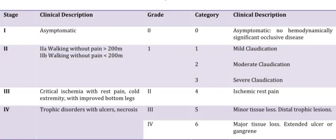
Peripheral Arterial Occlusive Disease (PAOD)is a complex disease of the arteries of the lower limbs. PAOD is defined as reduced arterial blood flow to the lower extremities. This reduction is mainly due to accumulation of plates limestone and cholesterol deposited on the arterial walls. This process defines atherosclerosis, which cause a reduction of arterial lumen (stenosis) or can lead to arterial occlusion (thrombosis). These injury lead to a reduction check oxygen and may lead to intermittent claudication (IC), critical limb ischemia (CLI) or ischemic skin lesions. Only one out of every four to five patients with PAOD will be symptomatic (1). Classification Leriche and Fontaine definedthe various symptoms of the disease (Table 1). PAOD is defined by measuring an index, ankle-‐brachial index (ABI): this index can be measured by taking systolic blood pressure at the wrists or arms and ankles, it is the ratio of systolic blood pressure of the right ankle systolic blood pressure on the right arm and the same ratio for the left side. When this ratio is less than 0.9 to the right and / or left, it is estimated that there PAOD.
Table 1. Fontaine and Rutherford Classifications for PAOD.
Fontaine Rutherford
Stage Clinical Description Grade Category Clinical Description
I Asymptomatic 0 0 Asymptomatic: no hemodynamically significant occlusive disease
II IIa Walking without pain > 200m
IIb Walking without pain < 200m 1 1 Mild Claudication 2 Moderate Claudication 3 Severe Claudication
III Critical ischemia with rest pain, cold
extremity, with improved bottom legs II 4 Ischemic rest pain
IV Trophic disorders with ulcers, necrosis III 5 Minor tissue loss. Distal trophic lesions. IV 6 Major tissue loss. Extended ulcer or
gangrene
Epidemiology
In the general population the prevalence of PAOD (asymptomatic and symptomatic) is around 15% in people over 65 years and over 20 % for more than 75 years (2). Framingham Study showed an incidence of intermittent claudication of 26/ 10,000 in men and 12/10,000 in women (3).The main risk factors of the disease are age, tobacco use, diabetes, arterial hypertension and dyslipidaemia. Anatomically, approximately 30% of the arterial lesions in PAOD are located in the iliac arteries (4).
Treatment
Historically, atherosclerotic occlusive disease involving the aortic bifurcation and proximal common iliac artery has been treated with open surgical by performing bypass. In 1964, Charles Dotter introduced the endovascular approach for treating aorto-‐iliac lesions (5,6). Then, Julio Palmaz in 1985 created the first intraluminal stent
which started a breakthrough in endovascular surgery (7). The open surgical treatment showed interesting results and good long-‐term patency but the perioperative morbidity and mortality was important(8–12). Furthermore theendovascular treatment with the “kissing stent” technique has shown early clinical results comparable to the surgical option (13–16). Technological innovation, improvement of materials and techniques have allowed the endovascular surgery to replace open surgery in the treatment of this pathology and have reduced morbidity and mortality.
In 2007, the Trans-‐Atlantic Inter-‐Society Consensus on the Management of Peripheral Arterial Disease (TASC II) published a classification of the Aorto-‐iliac Occlusive Disease (AIOD) (1).Lesions are divided into four subgroups (Annexe 1). This guideline recommends endovascular therapy for the treatment of choice for type A
lesions and surgery for the treatment of type D lesions. Endovascular treatment is preferred for type B lesions and surgery for good-‐risk patients with type C lesions. The patient's co-‐morbidities, fully informed patient preference and the local operator's long-‐ term success rates must be considered when making treatment recommendations for type B and type C lesions (1,6).
Endovascular treatment at the common iliac arteries consist of several techniques: the percutaneous transluminal angioplasty (PTA) (balloon dilatation), the primary stenting (to stent all lesions), or the selective stenting which consist balloon dilatation and place a stent. This technique can be used only in cases where there is a dissection or a residual stenosis. PTA is a simple and effective technique in the treatment of aorto-‐iliac occlusive lesions but doesn’t appear to protect against restenosis and it is not recommended for severe lesions.In 1998, in a randomized study by Tetteroo et al. showed no significant difference between direct stenting and selective stenting, in terms of patency (17).Bosch, meanwhile, published in 1997 a meta-‐analysis comparing the two methods and showed higher technical success and patency rates after direct stenting (18). The following year, in a randomized controlled trial, no significant difference was found between the two techniques but the selective stenting appears cheaper (17). Several authors propose the use of stent systematically especially in the presence of extensive lesions (TASC C or D).
There is no general consensus on which type of stent is best for which artery. However, we know that the balloon-‐expandable stents are more suited for lesions on rigid arteries such as the common iliac arteries. Indeed, balloon-‐expandable stents are characterized by much greater radial strength compared to self-‐expanding stents. Moreover, the arrival on the market of PTFE covered stent offers an attractive solution.
This type of stent realizes a real endopontage and appears to limit the effects of myo-‐ intimal hyperplasia intra stent and the risk of dissection and embolism during the procedure.
Problem statement
Some studies have published excellent results as regards the primary patency of PTFE-‐covered stents (Table 2). These studies are mostly retrospective, only three are comparative. They represent only a relatively small sample of patients with a short follow-‐up, except Chang in 2008, who compares retrospectively 71 stenting with covered stents versus 121 uncovered stents for 5 years but he study only self-‐ expanding stents (19).
Our study aims, by relating our experience to evaluate the effectiveness of balloon expandable covered stents Advanta®V12 in the treatment ofAIOD.
Table 2. Review ofPTFE-‐covered stents evaluation for PAOD in the iliac artery.
Author,
year Type of study Number patients Type of stent Primary patencyCovered Uncovered Lammer,
2000 (20) Prospective 61 Self-‐expanding 6 month: 98%
1 year: 91%
Wiesinger,
2005 (21) Prospective 60 Self-‐expanding 6 month: 94%
1 year: 91%
Bosier,
2007 (22) Prospective 91 Balloon-‐expandable 1 year: 91%
Chang,
2008 (19) Retrospective,comparative 71covered 122 uncovered
Mostly self-‐
expanding 5 year: 87% 5 year: 53%
Sabri,
2010 (23) Retrospective,comparative 26 covered 28 uncovered Balloon-‐ expandable 1 year: 92% 2 year: 92% 1 year: 78% 2 year: 62% Mwipatayi,
2011 (24) Randomized controlledtrial 83 covered 84 uncovered
Both 18 month: 92% 18 month: 75%
Bekken,
2012 (6) Randomized controlledtrial 87 covered 87 uncovered
Balloon-‐ expandable Self-‐expaning
In progress In progress
MATERIALS AND METHODS
Experimental design
A retrospective review of consecutive patients from a single institution with atherosclerotic occlusive disease at the aortic bifurcation treated with covered balloon expandable stent type Advanta®V12 (Maquet) in iliac arteries was undertaken by using
our database. Patients were enrolled between January 2004 and December 2014.
Endovascular therapy was considered as the first line treatment. Electronic medical records were reviewed to collect demographic data and patient follow up. Inclusion and exclusion criteria are summarized in Table 3. All study subjects had clinical and angiographic evidence of atherosclerotic occlusive disease involving the aortic bifurcation with unilateral or bilateral CIA stenosis or occlusion.
Table 3.Inclusion and exclusion criteria.
Inclusion criteria Exclusion criteria
Symptomatic patients according Leriche and Fontaine stages
2, 3 and 4 or sub acute ischemia or acute ischemia No atheromatic disease One or more significant aorto-‐iliac lesions (>60% stenosis or
thrombosis) listed according to TASC classification Asymptomatic lesion
Patient belongs to the French health care system Stenosis at an aorto-‐bi-‐femoral bypass segment or at the distal anastomosis of the bypass
Stenosis at an iliac limb extension of aortic stent graft
Pregnancy
Endovascular Procedure
All procedures were performed in the operating room by a certified vascular surgeon of the service. The endovascular indication was agreed upon during our weekly
vascular surgeon meeting. Informed consent was obtained before surgery. The procedure was performed under local anaesthesia with sedation in cases of isolated endovascular procedure and general anaesthesia for hybrid surgical procedures or when this one was recommended (agitated patients).
In most cases, femoral access was obtained by a percutaneous puncture guided by ultrasound, followed by insertion of 6-‐7 FR introducer sheaths in ipsilteral common femoral artery. For Kissing procedure, both common femoral arteries were punctured.
In very few cases, thecross-‐over technique(across the aorto-‐iliaccross-‐over) from the controlateral insertion point, under fluoroscopic guidance was realized. An intravenous bolus of 50 UI kg -‐1of heparin was administered. Under fluoroscopy, a hydrophilic 0,035-‐inch guide wire was advanced through the iliac lesions via the common femoral artery access sites, with the possible assistance of a straight diagnostic catheter 4F. A Digital Subtraction Angiography retrograde was performed by the introducer, in an anterior/posterior projection. Oblique X-‐ray of the pelvis arterial anatomy were also obtained. For proximal lesions, DSA was obtained by a pigtail placed in distal aorta.The stent dimensions were chosen by visual estimation to fit vessel diameter at best with a length exceeding the lesion length by 5mm proximally and 10mm distally. The diameters of the selected stent were usually oversized 10%-‐20% relative to the treated iliac arteries. Lesions were treated with as few stents as possible and adjacent stents over-‐lapped by 1 cm.Pre-‐dilation was performed when it was necessary to facilitate the deployment the stent. Advanta®V12 stents were deployed by inflation of the balloon to
the desired pressure according to manufacturer recommendations. Simultaneous stent deployment was performed when the kissing balloon/stent technique was necessary (Figure 1 and 2, Annexes). Post dilatation was realized with the same balloon,in case
of residual stenosis. The technical result of the procedure was evaluated by DSA. An additional endovascular or surgical procedure was performed if necessary. Groin closure was accomplished via manual compression during 15-‐20 minutes.
A prophylactic dose of low-‐molecular-‐weight heparin was given during hospitalisation to prevent venous thrombo-‐embolic events. Postoperatively, patients were prescribed Aspirin®(75 or 160 mg day_1) or Clopidogrel®(75 mg day_1). When
patients were under oral anticoagulant treatment, Aspirin® was the only anti-‐platelet
agent added.
Follow up
The patients were followed prospectively and different clinical and radiological data were stored in our database. Our monitoring protocol consisted of a surgical consultation after a month from the surgery with a control Doppler ultrasound and an annual follow up by the attending vascular physician. Finally each patient, physician, and vascular medicines were contacted by telephone at follow-‐up for evaluation of symptoms.
Objectives and definitions
The primary objective was primary patency. Secondary objectives were primary assisted patency, secondary patency, primary clinical success, technical success, morbidity and mortality in the immediate postoperative (<30 days) and remotely (>30 days), in-‐stent restenosis (ISR), in-‐stent thrombosis. Finally, we sought to identify predictive factors of restenosis or thrombosis.
Detailed definitions of outcome are:
• Primary patency was defined as patency without any percutaneous or surgical intervention in the treated segment or in the adjacent areas (25).
• Primary assisted patency was defined as a patent artery that underwent further endovascular intervention to improve patency.
• Secondary patency was defined as occlusion of the iliac segment requiring open or endovascular recanalization or bypass surgery (19).
• The rate of primary clinical success, defined as the disappearance or improving of the symptoms initially presented by the patient in the absence of any act of secondary revascularization or major amputation.
• Technical success was defined as achievement of a final residual diameter stenosis of <30% on the procedural completion angiogram.
• Morbidity was divided into minor and major complications.Minor complications are those following procedure (within the first month) and not requiring further treatment and not extending hospital stay. Major complications required re-‐ intervention or delay (more than 24 h) in patient discharge.
• ISR were assessed by duplex ultrasound and was defined by restenosis of more than 50%.
• The diagnosis of stent thrombosis was considered when occlusion was seen on duplex scan without any previous sign of restenosis.
Statistical analysis
Results were expressed as numbers and percentages for categorical parameters, and as the mean and standard deviation for continuous quantitative variables with normal distribution or median and percentiles if necessary. Univariate Cox regressions
were used to evaluate the role of the risks factors to detect restenosis or thrombosis of stent. A multivariate risk model was constructed using a backward stepwise Cox model strategy (P≤0.05 for model entry and P≤0.10 for model retention). To form this risk model for restenosis or thrombosis of stent, gender, BMI, HTA, dyslipidemia, diabetes, serum creatinine values, smoking, ASA score (American Society of Anesthesiologists), history of coronary disease, history of aortic surgery, or carotid artery surgery, or history of aortic bifurcation endovascular procedure, treatment with Plavix® or
Aspirine®, TASC classification, history of cancer, localization of lesion and degree of
stenosis were considered in the model selection process. The proportional-‐hazards assumption of the Cox model was checked separately for each covariate by the stcox PH-‐assumption test (stata procedure) after performing the cox analyses. The results were presented using the hazards ratio (HR) with a 95% confidence interval (CI). The level of significance was set at p<0.05. Statistics were conducted with STATA-‐13.1 software (StataCorp, College Station, TX, USA).
RESULTS
Patient’s characteristics and demographics
A total of 110 patients (151 limbs) were enrolled. The study population was composed of 95 men (86,4%) and 15 women (13,6%), and their median age at presentation was 61 years [55 ; 68]. Demographic characteristics are presented in Table 4. Indications for intervention included 89 patients (80,9%) for claudication, 4 patients (3,6%) for CLI, 12 (10,9%) for ischemic skin lesions and 5 (4,5%) for acute or sub-‐acute ischemia.
Table 4. Baseline patients’ characteristics and medical history. Variables
Age, years (mean ± SD) 61 ± 10
Male sex, n (%) 95 (86,4) Smokers, n (%) 105 (95, 5) Active 59 (53, 6) Previous 46 (41,8) Hypertension, n (%) 65 (59,1) Hyperlipidemia, n (%) 67 (60,9) Diabetes, n (%) 31 (28,2) Obesity (BMI > 25), n (%) 55 (50)
Serum creatinine values (mean ± SD) 93 ± 58
Coronary artery disease, n (%) 39 (35,5)
Myocardial infarction 13 (11,8)
Coronary Angioplasty/ stenting 14 (12,7)
Coronary artery bypass surgery 12 (10,9)
American Society of Anesthesiologists, n (%)
Grade I 8 (7,3)
Grade II 55 (50)
Grade III 43 (39,1)
Grade IV 4 (3,6)
Leriche and Fontaine stage of PAOD, n (%)
2 89 (80,9)
3 4 (3, 6)
4 12 (10,9)
Sub acute ischemia 3 (2,7)
Acute ischemia 2 (1,8)
BMI, Body Mass Index ; SD, Standard Deviation ; PAOD, Peripheral Arterial Occlusive Disease
Intra and Peri-‐operative management
Table 5 presents the outcomes of 110 endovascular procedures. Local anaesthesia with sedation was performed in 90 cases (81,8%), 11 general anaesthesia (10%) was performed for hybrid surgical procedures and 9 (8,2%) for patient comfort. An anterograde approach was realized for 150 limbs (99,3%), a cross over approach for one limb (0,7%) and 41 Kissing procedures (37,3%) were performed. Femoral access was obtained by a percutaneous puncture in 142 cases (94%). For 9 limbs (6%), femoral access was obtained by surgical approach. Seven lesions (4,7%) were restenosis.
According to the TASC II classification, 55 patients (50%) were classified as TASC A, 32 (29,1%) as TASC B, 5 (4,5%) as TASC C and 18 (16,4) as TASC D. A total of 120 lesions presented stenosis more than 70%. There were 19 thrombosis (12,7%). In 35,8% of the cases afemoro-‐popliteal lesion was associated. Mean stented segment was 44,4mm ± 7,9 and the mean number of the stent implanted per patient was 1,3 ± 0,3. Concomitant procedure was performed for 18 patients (16,4%): 6 (5,5%) by endovascular repair, 11 (10%) by open repair and 1 (0,9%) by mixt procedure. No conversion to open aorto-‐iliac surgery during the perioperative period were reported. The technical success was 100%. During the perioperative period two patients died: one patient aged 78, with coronary artery disease presented severe sepsis caused by urinary track infection to the 13th day post operatively, the second patient aged 85
years, suffering from severe coronary artery disease, died on the 5th day of acute coronary syndrome. Eight major complications (7,3%) led to an increase in the duration of hospitalization and/or re-‐intervention: two puncture site hematomas evolved to pseudo aneurysm, one deep vein thrombosis, one distal embolism, one occlusive syndrome, three early thrombosis. One minor hematoma appeared after a week and is absorbed without intervention. The average length of hospital stay was 3,1[1 ; 30].
Table 5. Intra-‐operative and perioperative data.
Variables
Characteristics of the lesions
TASC II classification of the lesions, n (%)
A 55 (50) B 32 (29,1) C 5 (4,5) D 18 (16,4) Approach, n (%) Crossover 1 (0,7) Anterograde 150 (99,3) Kissing, n (%) 41 (37,3) Degree of stenosis, n (%) 60-‐70% 12 (7,9) ≥70% 47 (31,1) ≥80% 48 (31,8) ≥90% 25 (16,6) Thrombosis 19 (12,6)
Location of the lesion, n (%)
Ostial 112 (74,2)
Proximal 37 (24,5)
Full axis 2 (1,3)
Femoro-‐popliteal lesions associated, n (%) 54 (35,8)
Characteristics of the stents
Length of stent, n (%)
22 mm 2 (1,3)
38 mm 135 (89,4)
59 mm 26 (17,2)
Number of stent per patient, n (mean ± SD) 1,3 ± 0,3 Number of stent implanted, n (%)
1 139 (92,1)
2 20 (13,2)
3 4 (2,6)
Diameter of implanted stent, n (%)
6 2 (1,3) 7 16 (10,6) 8 109 (72,2) 9 29 (19,2) 10 7 (4,6) Pre dilatation, n (%) 42 (27,8) Post dilatation, n (%) 5 (3,3) Associated procedures, n (%) Endovascular repair 6 (5,5) Aorta 1 (0,7)
External iliac artery 4 (2,3)
Common iliac artery controlateral 1 (0,7)
Open repair 11 (10)
Femoral endarteriectomy 7 (6,4) Femoro-‐popliteal bypass 1 (0,7) Iliac and popliteal thrombectomy ± aponevrotomy 3 (3,3)
Mixte 1 (0,9)
Technical Success, (%) 100
Intraoperative complications, n (%) 3 (2,7) Postoperative Serum creatinine values (mean ± SD) 76 ± 56 Duration of hospital stay, d (mean ± SD) 3,1 ± 4,9 Perioperative complications, n (%)
Minor 1 (0,9)
Major 8 (7,3)
Death (<30th day), n (%) 2 (1,8%) TASC, Trans-‐Atlantic Inter-‐Society Consensus
Outcomes
The mean follow-‐up time was 51,3 months (0 ; 120). All patient have been followed up. Twenty-‐two patients died during the follow up (Table 6).
Table 6. Outcomes post-‐operative
Variables
Death (>30thday), n (%) 22 (20)
Pulmonary neoplasia 7 (35)
Head and neck cancer 2 (10)
Gastric cancer 1 (5)
Hematologic malignancies 4 (20)
Bladder cancer 1 (5)
Unknown 7 (35)
In-‐stent restenosis (ISR), n (%) 10 (9,1) In-‐stent thrombosis, n (%) 8 (7,3)
The primary clinical success at the end of follow up was 82,7% (91 patients) (Figure 4).
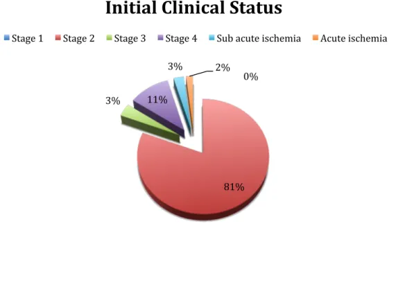
Among the 89 patients followed and treated for PAOD stage 2:
• A disappearance of intermittent claudication (n = 74) or an improvement of
claudication (n = 1) has been reported in 84,3% of cases, without re-‐intervention.
• Six patients did not have symptomatic improvement: they presented no stenosis or thrombosis and two of them had femoro-‐popliteal lesions.
• One patient treated for bilateral lesions TASC B presented a stenosis after 44,9 months. He still refuses further surgery.
• A disappearance of intermittent claudication has been reported in seven patients with re-‐intervention (Table 7): four patients presented a stenosis and three a thrombosis. Five of seven patients were active smokers.
asymptomatic at the end of follow up.
For the 12 patients followed and treated for PAOD stage 4, 11 patients (91,7%) were asymptomatic and one patient (8,3%) had healed but still presented with claudication. This patient was an active smoker, ached chronic renal failure on dialysis, in addition he had femoro-‐popliteal lesions, external and common iliac arteries. An external iliac artery angioplasty was realized while the procedure. The patient died of lung cancer after one year.
The two patients treated for acute ischemia 100% were asymptomatic. Finally, two of three patients (66,7%) treated for sub-‐acute ischemia were symptomatic, and one patient suffered to claudication.
Table 7. Characteristics of patients treated for claudication, improved with re-‐ intervention.
Lesion Occurrence delay (month)
Initial lesion TASC Length of stent Re intervention Delay (month) Thrombosis 117,7 Stenosis C 7 x 38 / 8 x 38 Kissing Aorto-‐bi-‐femoral bypass 118 Thrombosis 10,3 Thrombosis D 8 x (22+38+59) 7 x 38 Kissing Transfemoral amputation, Axillo-‐bi-‐femoral bypass Aorto-‐bi-‐femoral bypass 8 10 22 Thrombosis 15,1 Stenosis B 8 x 59 Thrombectomy +
Axillo-‐bi-‐femoral bypass 15,3 Stenosis 33,7 Pre thrombosis B 7 x 38 / 7 x 38 Kissing Angioplasty 36,9 Stenosis 8,6 Stenosis B 8 x 38 Angioplasty 8,7 Stenosis 11,3 Stenosis A 8 x 38 / 8 x 38
kissing
Angioplasty Left Aorto-‐femoral
bypass + femoro-‐ femoral cross over
bypass
11,3 88
Stenosis 70,3 Stenosis A 10 x 38 Transfemoral
amputation 72
Figure 4. Clinical outcomes at the end of the follow up. Patients classified by Leriche and Fontaine Classification.
Eighteen patients (16,4%) presented with stenosis or a thrombosis (Table 8), three were detected before the first month, three before the sixth month, two before the first year, and ten beyond twelve months. The initially treated iliac lesions were classified TASC A in 6 cases, TASC B in 7 cases, TASC C in 3 cases and TASC D in 2 cases.
0%
81%
3% 11%
3% 2%
Initial Clinical Status
Stage 1 Stage 2 Stage 3 Stage 4 Sub acute ischemia Acute ischemia
91% 9%
Clinical Status at the end of follow up
Table 8. Details of re-‐stenosis and thrombosis. Lesions Occurrence
delay (day) Initial PAODstage Kissing lesionsInitial TASC Length of sten Re intervention
Thrombosis 1 4 -‐ Thrombosis D 9 x 38 /
10 x 38
Iliac thrombectomy
Thrombosis 2 2 Kissing Thrombosis D 8 x (22+38+59)
7 x 38 Transfemoral amputation, Axillo-‐ bi-‐femoral bypass, Aorto-‐bi-‐femoral bypass
Thrombosis 14 4 -‐ Thrombosis B 8 x 38 Aorto common
femoral bypass
Thrombosis 67 2 -‐ Stenosis 70% B 7 x 38 -‐
Thrombosis 94 4 Kissing Stenosis 80% C 9 x 38 Bilateral iliac angioplasty Aorto-‐bi-‐femoral
bypass
Thrombosis 458 2 -‐ Stenosis 70% B 8 x 59 Thrombectomy,
Axillo-‐bi-‐femoral bypass
Thrombosis 2423 Sub acute ischemia
-‐ Stenosis 70% A 9 x 38 -‐
Thrombosis 3578 2 Kissing Stenosis 70% C 7 x 38 / 8 x 38 Aorto-‐bi-‐femoral bypass
Stenosis 90 2 Kissing Stenosis 90% A (9 x 38) x 2 -‐
Stenosis 103 2 Kissing Stenosis 70% A (8 x 38) x 2 -‐
Stenosis 263 2 -‐ Stenosis 80% B 8 x 38 Angioplasty
Stenosis 342 2 Kissing Stenosis 70% A (8 x 38) x2 Angioplasty, Left Aorto-‐femoral
bypass + femoro-‐ femoral cross over
bypass
Stenosis 497 Sub acute ischemia
-‐ Stenosis 80% A 8 x 38 Angioplasty
Stenosis 1024 2 Kissing Stenosis 90% B (7 x 38) x 2 Bilateral iliac angioplasty
Stenosis 1364 2 Kissing Thrombosis
(right) Stenosis 60%
B (8 x 38) x 2 -‐
Stenosis 2136 2 -‐ Stenosis 90% A 10 x 38 Transfemoral
amputation
Stenosis 2208 2 -‐ Stenosis 80% C 7 x 38 -‐
Stenosis 2425 2 Kissing Stenosis 80%
right, 60% left
B 8 x 38 / 8 x 59 -‐
Eleven patients (10%) required further surgery within an average of 37,8 months (0,8 ; 117,7): four iterative angioplasties and seven open surgeries (bypass).
The primary patency rate (Figure 5) was 98,1% (92,8 ; 99,5) at one month, 94,2% (87,5% ; 97,4%) at one year, 92,1% (84,7% ; 96%) at two years, 90,4% (82,2% ; 95%) at five years and 87,3% (75,9% ; 93,5%) at six years .
Figure 5. Primary patency.
The primary assisted patency rate was 98% (92,4 ; 99,5%) at one year, 97% (90,8% ; 99%) at two years, 95,3% (87,7% ; 98,3%) at five years and 95,3% (87,7% ; 98,3%) at six years (Figure 6).
Figure 6. Primary assisted patency.
The secondary patency rate was 98,2% (92,8% ; 99,5%) at one month, 96,2% (90,1% ; 98,5%) at one year, 95,1% (88,6% ; 97,9%) at two years, 95,1% (88,5% ; 97,9%) at five years and 91,9% (80,5% ; 96,8%) at six years (Figure 7).
Figure 7. Secondary patency
Significantly in univariate analysis, only female gender was identified as a predictive factor of restenosis or thrombosis Hazard ratio=4.20 [1.41 ; 12.48], p= 0,010
and arterial hypertension and dyslipidemia as protective factors Hazard ratio= 0.37 [0.14 ; 0.99], p= 0,049 and 0.39 [0.15 ; 0.99], p= 0,050 respectively. These results were still significant after adjustment with multivariate analysis (Table 9 and 10).
Table 9. Univariate Analysis – Risk factors of re stenosis or thrombosis. N=110 Absence of stenosis or thrombosis n=92 Presence of stenosis or thrombosis n=18 HR [IC à 95%] P value Female gender (2) 10.9% (10) 27.8% (5) 4.20 [1.41 ; 12.48] 0.010 ** BMI, mean ± SD 25.56 ± 3.50 25.32 ± 4.78 0.95 [0.83 ; 1.09] 0.460 HTA, % (n) 64.1% (59) 33.3% (6) 0.37 [0.14 ; 0.99] 0.049 * Dyslipidemia, % (n) 65.2% (60) 39.9% (7) 0.39 [0.15 ; 0.99] 0.050 * Diabetes, % (n) 32.6% (30) 5.6% (1) 0.16 [0.02 ; 1.22] 0.078 Créatinine value 79 [69 ; 100.5]1 75.5 [67 ; 87]1 0.98 [0.96 ; 1.01] 0.166 Smoker (Active), % (n) 51.1% (47) 66.7% (12) 1.65 [0.62 ; 4.41] 0.314 ASA, % (n) 1 2 3 4 7.6% (7) 48.9% (45) 40.2% (37) 3.3% (3) 5.6% (1) 55.6% (10) 33.3% (6) 5.6% (1) -‐ 2.24 [0.27 ; 18.7] 2.34 [0.25 ; 21.5] 5.62 [0.32 ; 97.6] -‐ 0.454 0.454 0.236 Coronary history, % (n) 39.1% (36) 16.7% (3) 0.40 [0.12 ; 1.40] 0.153 Carotid surgery history, % (n) 8.7% (8) 11.1% (2) 1.25 [0.29 ; 5.49] 0.764 Aortic surgery history, % (n) 1.1% (1) 5.6% (1) 2.56 [0.34 ; 19.48] 0.363 Aorto-‐iliac endovascular history,
% (n) 12% (11) 11.1% (2) 0.81 [0.18 ; 3.69] 0.781 Localization, % (n) 69.6% (64) 77.8% (14) 1.77 [0.58 ; 5.41] 0.319 Plavix®, % (n) 53.3% (49) 72.2% (13) 1.54 [0.54 ; 4.37] 0.421 Aspirin®, % (n) 59.8% (55) 44.4% (8) 0.89 [0.34 ; 2.33] 0.818 TASC, % (n) A B C D 53.3% (49) 27.2% (25) 2.2% (2) 17.4% (16) 33.3% (6) 38.9% (7) 16.7% (3) 11.1% (2) -‐ 2.13 [0.71 ; 6.40] 3.34 [0.77 ; 14.43] 1.02 [0.21 ; 5.09] -‐ 0.176 0.106 0.977 History of cancer, % (n) 17.4% (16) 22.2% (4) 1.40 [0.45 ; 4.30] 0.558 Degree of lesion, % (n) <70 ≥70 et <80 ≥80 et <100 100 6.5% (6) 31.5% (29) 48.9% (45) 13% (12) 5.6% (1) 33.3% (6) 44.4% (8) 16.7% (3) -‐ 0.97 [0.12 ; 8.13] 0.96 [0.12 ; 7.73] 1.37 [0.14 ; 13.66] -‐ 0.977 0.972 0.790
Univariate analysis: Cox Model; HR: Hazard Ratio [Confidence Interval à95%];1=median [25% ; 75%] ; BMI, Body Mass
Index ; SD, Standard Deviation; TASC, Trans Atlantic Inter-‐Society Consensus, ASA: American Society of Anesthesiologists;
Table 10. Univariate and multivariate analysis – Risk factors of re stenosis and thrombosis. N=110 Univariate analysis HR [IC 95%] P value Multivariate analysis HR [IC 95%] P value Female gender 4.20 [1.41 ; 12.48] 0.010 ** 5.74 [1.85 ; 17.81] 0.002 HTA 0.37 [0.14 ; 0.99] 0.049 * 0.32 [0.12 ; 0.89] 0.029 Dyslipidemia 0.39 [0.15 ; 0.99] 0.050 * 0.39 [0.15 ; 1.02] 0.056 Univariate and multivariate Analysis: Cox model, HR: Hazard Ratio [Confidence interval 95%]; HTA: arterial hypertension;
DISCUSSION
Our study shows excellent primary, primary assisted and secondary patency rate of covered stent in PAOD with low morbidity and mortality.
In 110 procedures we studied, the same technical problem occurred twice: the inability to pass the device through a calcified pre-‐thrombotic primary iliac stenosis, while the 0.035-‐inch hydrophilic guide had crossed the lesion without difficulty. This led to uncrimping of the stent that was released just below the lesion. The two patients in whom this occurred were being treated for bilateral pre-‐thrombotic lesions. A pre-‐ dilatation was performed and a new stent was implanted. During follow-‐up neither of these stents became stenosed or thrombotic.
For all treated lesions only 28% were pre-‐dilated, in contrast to the series reported by Bosiers et al. in which pre-‐dilation with a 5 mm diameter balloon was systematic and this type of problem was not encountered (22).
In our cohort there were two deaths (1.8%) within 30 days post-‐surgery but the causes were not directly related to the endovascular procedure. The rate of complications related to the procedure was 8.2%, including three early thrombosis.
This rate is comparable to that in the literature where the rate varies between 2.7 and 24% (26). The first case of early re-‐thrombosis occurred in an active smoker treated for bilateral claudication with very severe iliac lesions. He presented with a thrombosis of the entire right iliac axis and stepped tight stenosis along the whole left iliac axis requiring the implantation of several stents. This was probably a poor indication for stenting and a conventional surgical treatment should have been proposed according to the current TASC guidelines lesions as his lesions were classified as TASC D. Eight months later he underwent transfemoral amputation of the lower right limb, a year after this an axillo-‐femoral bridging graft and finally a left femoral artery bypass two years after the first procedure. The second case of early re-‐thrombosis also occurred in an active smoker treated for severe lesions (TASC class D). He presented thrombosis of the whole left iliac axis requiring the placement of two stents and a non-‐symptomatic contralateral primary iliac thrombosis. This patient would have received surgical treatment but was a fragile patient, ASA 4, and died less than a year after the procedure from acute leukemia. Finally, the last case of early re-‐thrombosis occurred in an active smoker, ASA 3, treated for TASC B lesions with left iliac thrombosis. No pre-‐dilatation before stenting had been done. The patient underwent aorto-‐left femoral coronary bypass surgery one month after the initial procedure. Thus, it was observed that early re-‐thrombosis occurred in patients with severe lesions for whom conventional surgery would have been proposed if they had not presented a contra-‐indication to aortic surgery.
Approximately 80% of patients in our study had TASC class A or B lesions; and those with TASC C and D lesions were for the most part fragile cases with an ASA score of 3 or 4, contra-‐indicative of aortic surgery. We had respected the current guidelines on the therapeutic management of AIOD in our operating procedures. However, some
authors report primary patency rates for the treatment of severe iliac lesions (TASC C and D) of 88% at 6 months, 82% at 12 months and 78% at 24 months with systematic use of self-‐expanding stents and of 87% at 6 months and 70% at 12 months when using covered stents (Wallgraft®or Viabahn®) (27,28). A review of the literature by Jongkind
et al. found primary and secondary patency rates of 60 to 80% and 80 to 98% respectively (29). These results must be considered alongside conventional surgery (aorto-‐femoral bypass) that achieves a primary patency rate at 5 years of 91% in claudication patients and 87% in patients with critical limb ischemia, with a perioperative mortality rate close to 2% (8,30).
In the literature, iliac stents are generally associated with good outcomes and for the most part intra-‐stent myo-‐intimal hyperplasia appears to be responsible for late failures of stenting (7). Although stents are used to correct an initial residual stenosis or localized dissection occurring after the fracture of the atherosclerotic plaque, they increase the formation of micro-‐thrombi, induce inflammatory reactions and myo-‐ intimal hyperplasia, all sources of re-‐stenosis. However, the use of steel stents covered with a PTFE membrane is considered to have several advantages. These stents can be used to treat an arterial rupture during the same procedure, a rare but extremely serious complication, which we were not confronted with in our series. The use of covered stents for the treatment of this complication has been described by Scheinert et al. with immediate success of 100% and a primary patency rate of 87% at 2 years (31).
The presence of the PTFE covering theoretically restricts fragmentation of the plaque during angioplasty and thus limits the risk of peripheral embolisms. However, in
our series, one patient, a 69 year-‐old male former smoker with dyslipidemia and treated for claudication, had this complication. He had severe lesions of the aortic bifurcation classified TASC D. Stenosis of the terminal aorta and iliac ostial stenosis was treated with one aortic stent, two iliac stents in a "kissing" configuration and one stent in right iliac artery. Sub-‐acute ischemia appeared immediate postoperatively in his left leg and a secondary femoral and popliteal embolectomy was performed. The risk of embolization appears greatest when the lesions are ulcerated or recanalization is attempted, with a rate of peripheral embolism of up to 24% in cases of simple angioplasty (32,33). This complication appears to be largely underestimated as it is most often asymptomatic, but it has been demonstrated in a histological study by Karnabatidis et al. to occur in up to 70% of cases (34). Moreover, it would appear that lesions of the aorto-‐iliac bifurcation are more at risk of embolism, such that some authors prefer using the "kissing" technique for this type of lesion (35). This is the case of our patient who should have been entitled to aortic surgery according to the TASC 2 guidelines.
Finally, the use of a covered stent seems to intuitively limit intra-‐stent myo-‐ intimal hyperplasia. It creates a mechanical barrier between the blood and the endothelium possibly restricting the stimulation and proliferation of smooth muscle cells and especially their migration through the mesh of the stent. According to some studies the non-‐porosity of the PTFE membrane appears to limit this hyperplasia and thus reduce the risk of restenosis (36,37). For Sabri, the difference in primary patency observed between the use of a covered stent and a bare stent is clearly due to the absence of intra-‐stent myo-‐intimal hyperplasia and better fluid dynamics (23). However, Sangiorni et al. in their anatomo-‐pathological study of pig peripheral arteries
did not find any difference when looking at micro-‐thrombi, local inflammatory phenomena or myo-‐intimal hyperplasia between the use of bare steel or stents covered with a PTFE membrane (38). All the same, it should be noted that the bare stents were deployed in the renal arteries while covered stents were placed in the iliac and renal arteries and the aortas. The comparison was thus probably biased. In addition, most of the results were not significant. Of the 10 cases of re-‐stenosis, 3 cases of restenosis were described as being at the end of the stent or downstream. While such lesions may be related to the progression of the atheromatic disease, they could also have been myo-‐ intimal hyperplasic lesions since they were at the end of the stent where the endothelium is traumatized by the stent, without the PTFE membrane performing its barrier role. Furthermore, two cases of intra-‐stent restenosis were detected by ultrasound but did not need iterative angioplasty. This type of restenosis can be explained by myo-‐intimal proliferation associated with excessive porosity or a tear in the PTFE membrane.
In our series the rates of primary, primary-‐assisted and secondary patencies at one month, one year and in the long term were slightly higher than those from the most recent series reported the literature that used iliac angioplasty with selective or systematic stenting (Table 10). The primary patency rate obtained by Bosiers et al, the only other study that evaluates the Advanta V12 covered stent, was 91.1% at one year compared with 94,2% in our study (22). This difference can be explained by the fact that Bosiers studied these stents predominantly in lesions of the aortic bifurcation, an area known to have a higher risk of restenosis or re-‐thrombosis. Chang et al. are the only authors to present long-‐term results with primary patency at 5 years of 87% against 90,4% in our series (19). However, in their series, Chang essentially assess self-‐
expanding stents in patients with severe lesions classified as TASC C or D, while in our series about 80% of patients had lesions classified as TASC A or B. A clear difference is also seen between our long-‐term results and those of bare stents. Indeed, the primary patency of bare stents observed by Chang was 53% at 5 years against 90,4% for the covered stents used in our series.
The various series described to date do not indicate whether the long-‐term decrease in patency is due to intra-‐stent restenosis, to stenosis at the ends of the stent or atheromatous lesions in the iliac axis. It is often said that intra-‐stent restenosis by myo-‐intimal hyperplasia is the main cause of late stent failure but its proportion is difficult to specify. This was the case in our study where the evaluation of restenosis was not standardized and the vascular doctors did not specify the location of all detected restenosis.
Table 10. Aorto-‐iliac Stenting. Literature review.
Stenting Primary or
Selective
Type of stent Number patients (limbs)
TASC, % Artery, % Primary patency
Kudo, 2005 (39) Selective -‐ Angioplasty (77%) -‐ Stents (23%) Balloon-‐ expandable 104 (151) A B C D 26 47 36 3 AIP AIE AIP+AIE 29 20 61 90% at 6 m. 76% at 1 y. 63% at 22 m. AbuRahma,
2007 (26) Primary Self-‐expanding 110 (149) A+BC+D 55,544,5 AIPAIE AIP+AIE 60 21 19 98% at 1 y. 94% at 2 y. AbuRahma,
2007 (26) Selective Self-‐expanding 41 (41) A+BC+D 6832 AIPAIE AIP+AIE 71 17 12 83% at 1 y. 78% at 2 y. Reekers,
2002 (40) Selective expandableBalloon-‐ 126 (143) -‐ AIPAIE AIP+AIE Aorta 49 31 17 3 94% at 6 m. 89% at 1 y. Becquemin,
1999 (41) Selective -‐ 235 (281) -‐ AIEAIP 43,556,5 86,6% at 1 y. 81,2% at 2 y. Timaran, 2001 (42) Both Both 189 (247) AB C D 19 51 25 5 AIP AIE AIP+AIE -‐ -‐ -‐ 88% at 1 y. 74% at 3 Y. Bosier,
2007 (22) Primary expandableBalloon-‐ (Advanta V12) 65 (91) -‐ AIP AIE AIP+AIE Aortic bifurcation 23,3 12,1 3,3 61,5 91,1% at 1 y. Lammer,
2000 (20) Primary Self-‐expandingcovered 127 (61) AB C 38* 56 7 AIP AIE 52,547,5 98,4% at 6 m. 91,2 at 1 y. Wiesinger,
2005 (21) Primary Self-‐expadingcovered 98 (60) -‐ AIP 94% at 6 m. 91% at 1 y.
Chang,
2008 (19) Primary Mostly self-‐expanding 171 (193)71 covered 122 uncovered C D 3961 AIP 87% at 5 y.Covered: Uncovered: 53% at 5 y. Sabri, 2010
(23) Primary Both 26 covered54 (54) 28 uncovered -‐ Aortic bifurcation Covered: 92% at 1 and 2 y. Uncovered: 78% at 1 y. 62% at 2 y. Mwipatayi,
2011 (24) Primary Both 125 (167)83 covered 84 uncovered B C D 62,1** 27,1 10,7 AIP Covered: 92% at 18 m. Uncovered: 75% at 18 m. Bekken,
2012(6) Primary Both 87 covered174 87 uncovered
-‐ AIP In progress
Our study,
2016 Both expandableBalloon-‐ (Advanta V12) 110 (150) A B C D 50 29,1 4,5 16,4 AIP 98,1% at 1 m. 94,2% at 1 y. 90,4% at 5 y. 87,3% at 6 y.
Among the 18 cases of restenosis or re-‐thrombosis 3 had lesions of the downstream external iliac arteries, of which 33.3% were TASC A lesions and 38.9% TASC B lesions. It is clear that lesions of the EIA (13,42) and severe pelvic disease (39) are associated with a decreased primary patency rate. Similarly, a degradation of the downstream arterial bed (13,43), female gender (42) and active smoking (39) are considered as factors that reduce primary patency. In our study only female gender was found to be predictive of restenosis and/or significant re-‐thrombosis. This may be due to an insufficient power of our study due to the small population size and the retrospective approach, a source of missing baseline data and making the data collection and follow-‐up difficult. Dyslipidemia and hypertension appear to be significantly protective of restenosis or thrombosis. This is probably due to the fact that
the corresponding patients were treated with Statins®and antihypertensive treatment,
known as protective treatments in this disease.
An analysis of systolic pressure indexes could not be performed because their measurements prior to the procedure and during follow-‐up were left to the discretion of the vascular physician. Finally, clinical assessment was not standardized and therefore could vary according to the medical examiner.
Our study evaluated the use of the only balloon-‐expandable covered stent commercially available at the time (2004), the Advanta® V12. The availability of new
models of balloon-‐expandable covered stents, such as LifeStream®(Bard), will probably
CONCLUSION
Despite the lack of consensus on the choice of type of stent for use in aorto-‐iliac occlusive disease (AIOD), the covered balloon-‐expandable stent used in our study appears to be a reliable, efficient and lasting solution. Morbidity and mortality are low. Primary, primary-‐assisted, and secondary long-‐term patency rates remain highly satisfactory and well above the long-‐term primary patency rate of bare stents, which according to the literature do not exceed 80% at one year and 60% at 5 years. Although, the mechanism of restenosis or thrombosis remains poorly understood, by achieving a true endopontage the covered stent remains promising and seems the most suitable type of stent for the treatment of AIOD. Further prospective studies with larger study populations and longer follow-‐up are needed to assess the benefits of this system for the prevention of in-‐stent restenosis compared to bare stents. The first results of the clinical trial by Bekken probably provide reliable and interesting information and will allow us to finally decide on the best choice of covered stent or bare stent in the treatment of this pathology (6).
THESE SOUTENUE PAR: Mlle ELIE Amandine TITLE:
Outcomes of covered balloon-‐expandable stent placement in the
treatment of atherosclerotic occlusive disease at the aortic
bifurcation.
CONCLUSION:
Objective: The objective was to evaluate at medium and long term, the primary patency
of covered stents used in first line treatment of aorto-‐iliac occlusive lesions and identify predictive factors of restenosis and thrombosis (FPRT).
Methods: Patients from a single institution with atherosclerotic occlusive disease at the
aortic bifurcation treated with covered balloon expandable stent type Advanta® V12
(Maquet) in iliac arteries were enrolled between January 2004 and December 2014. It was a retrospective study but patients were followed prospectively. A surgical consultation at one month with a control Doppler ultrasound and an annual follow up by the attending vascular physician were realized. The primary objective was primary patency. The characteristics of the population of the lesion and those related to the intervention were analyzed by multivariate to identify FPRT.
Results: A total of 110 patients (151 limbs) were enrolled (86% of men, 41 kissing
stent). The mean age was 61 years (39-‐86). The technical success was 100%. There were no intraoperative deaths and two postoperative deaths: one to the 13th day a
severe sepsis because of urinary track infection and the otheron the 5th day of acute coronary syndrome. The morbidity was low.The mean follow-‐up time was 51,3 months (0 ; 120). The primary patency was at 1, 2, 5 and 6 years: 94,2%, 92,1%, 90,4% and 87,3% respectively. Eight patients presented a thrombosis in a mean time of 31 months and ten stenosis in 47 months. Eleven patients (10%) required further surgery within an average of 37,8 months (0,8 ; 117,7): four iterative angioplasties and seven open
surgeries (bypass). Only the female gender was identified as a predictive factor of restenosis or thrombosis significantly.
Conclusion: In our experience endovascular treatment with covered stent for aorto iliac
occlusive lesions is a reliable technique, efficient and sustainable. The morbi-‐mortality was low. In the literature, the primary patency at long-‐term of uncovered stents does not exceed 80 % at one year and 60 % at 5 years.
BIBLIOGRAPHY
1. Norgren L, Hiatt WR, Dormandy JA, Nehler MR, Harris KA, Fowkes FGR. Inter-‐ Society Consensus for the Management of Peripheral Arterial Disease (TASC II). J Vasc Surg. 2007 Jan;45(1):S5–67.
2. Newman AB, Shemanski L, Manolio TA, Cushman M, Mittelmark M, Polak JF, et al. Ankle-‐arm index as a predictor of cardiovascular disease and mortality in the Cardiovascular Health Study. Arterioscler Thromb Vasc Biol. 1999;19(3):538–45. 3. Kannel WB, Skinner JJ, Schwartz MJ, Shurtleff D. Intermittent Claudication:
Incidence in the Framingham Study. Circulation. 1970 May 1;41(5):875–83.
4. Zeller T. Current state of endovascular treatment of femoro-‐popliteal artery disease. Vasc Med. 2007 Aug 1;12(3):223–34.
5. Dotter CT, Judkins MP. Transluminal Treatment of Arteriosclerotic Obstruction: Description of a New Technic and a Preliminary Report of Its Application. Circulation. 1964 Nov 1;30(5):654–70.
6. Bekken JA, Vos JA, Aarts RA, de Vries J-‐PP, Fioole B. DISCOVER: Dutch Iliac Stent trial: COVERed balloon-‐expandable versus uncovered balloon-‐expandable stents in the common iliac artery: study protocol for a randomized controlled trial. Trials. 2012;13(1):215.
7. Palmaz JC, Sibbitt RR, Reuter SR, Tio FO, Rice WJ. Expandable intraluminal graft: a preliminary study. Work in progress. Radiology. 1985 Jul;156(1):73–7.
8. de Vries SO, Hunink MG. Results of aortic bifurcation grafts for aortoiliac occlusive disease: a meta-‐analysis. J Vasc Surg. 1997;26(4):558–69.
9. Naylor AR, Ah-‐See AK, Engeset J. Aortoiliac endarterectomy: An 11-‐year review. Br J Surg. 1990 Feb;77(2):190–3.
10. Reed AB, Conte MS, Donaldson MC, Mannick JA, Whittemore AD, Belkin M. The impact of patient age and aortic size on the results of aortobifemoral bypass grafting1 1Competition of interest: none.Published online Mar 6, 2003. J Vasc Surg. 2003 Jun;37(6):1219–25.
11. Brothers TE, Greenfield LJ. Long-‐term results of aortoiliac reconstruction. J Vasc Interv Radiol JVIR. 1990 Nov;1(1):49–55.
12. Urayama H, Ohtake H, Yokoi K, Fujimori H, Kawaguchi M, Ishikawa T, et al. Long-‐ term results of endarterectomy, anatomic bypass and extraanatomic bypass for aortoiliac occlusive disease. Surg Today. 1998;28(2):151–5.
13. Galaria II, Davies MG. Percutaneous transluminal revascularization for iliac occlusive disease: long-‐term outcomes in TransAtlantic Inter-‐Society Consensus A and B lesions. Ann Vasc Surg. 2005 May;19(3):352–60.
14. Henry M, Amor M, Ethevenot G, Henry I, Mentre B, Tzvetanov K. Percutaneous endoluminal treatment of iliac occlusions: long-‐term follow-‐up in 105 patients. J Endovasc Ther. 1998;5(3):228–35.
15. Funovics MA, Lackner B, Cejna M, Peloschek P, Sailer J, Philipp MO, et al. Predictors of long-‐term results after treatment of iliac artery obliteration by transluminal angioplasty and stent deployment. Cardiovasc Intervent Radiol. 2002 Oct;25(5):397–402.
16. Gandini R, Fabiano S, Chiocchi M, Chiappa R, Simonetti G. Percutaneous treatment in iliac artery occlusion: long-‐term results. Cardiovasc Intervent Radiol. 2008 Dec;31(6):1069–76.
17. Tetteroo E, van der Graaf Y, Bosch JL, van Engelen AD, Hunink MG, Eikelboom BC, et al. Randomised comparison of primary stent placement versus primary angioplasty followed by selective stent placement in patients with iliac-‐artery occlusive disease. Dutch Iliac Stent Trial Study Group. Lancet Lond Engl. 1998 Apr 18;351(9110):1153–9.
18. Bosch JL, Hunink MG. Meta-‐analysis of the results of percutaneous transluminal angioplasty and stent placement for aortoiliac occlusive disease. Radiology. 1997 Jul;204(1):87–96.
19. Chang RW, Goodney PP, Baek JH, Nolan BW, Rzucidlo EM, Powell RJ. Long-‐term results of combined common femoral endarterectomy and iliac stenting/stent grafting for occlusive disease. J Vasc Surg. 2008 Aug;48(2):362–7.
20. Lammer J, Dake MD, Bleyn J, Katzen BT, Cejna M, Piquet P, et al. Peripheral Arterial Obstruction: Prospective Study of Treatment with a Transluminally Placed Self-‐ expanding Stent-‐Graft 1. Radiology. 2000;217(1):95–104.
21. Wiesinger B, Beregi J-‐P, Oliva VL, Dietrich T, Tepe G, Bosiers M, et al. PTFE-‐covered self-‐expanding nitinol stents for the treatment of severe iliac and femoral artery stenoses and occlusions: final results from a prospective study. J Endovasc Ther. 2005;12(2):240–6.
22. Bosiers M, Iyer V, Deloose K, Verbist J, Peeters P. Flemish experience using the Advanta V12 stent-‐graft for the treatment of iliac artery occlusive disease. J Cardiovasc Surg (Torino). 2007 Feb;48(1):7–12.
23. Sabri SS, Choudhri A, Orgera G, Arslan B, Turba UC, Harthun NL, et al. Outcomes of Covered Kissing Stent Placement Compared with Bare Metal Stent Placement in the Treatment of Atherosclerotic Occlusive Disease at the Aortic Bifurcation. J Vasc Interv Radiol. 2010 Jul;21(7):995–1003.
24. Mwipatayi BP, Thomas S, Wong J, Temple SEL, Vijayan V, Jackson M, et al. A comparison of covered vs bare expandable stents for the treatment of aortoiliac occlusive disease. J Vasc Surg. 2011 Dec;54(6):1561–70.e1.
25. Davaine J-‐M, Azéma L, Guyomarch B, Chaillou P, Costargent A, Patra P, et al. One-‐ year Clinical Outcome after Primary Stenting for Trans-‐Atlantic Inter-‐Society
Consensus (TASC) C and D Femoropopliteal Lesions (The STELLA “STEnting Long de L’Artère fémorale superficielle” Cohort). Eur J Vasc Endovasc Surg. 2012 Oct;44(4):432–41.
26. AbuRahma AF, Hayes JD, Flaherty SK, Peery W. Primary iliac stenting versus transluminal angioplasty with selective stenting. J Vasc Surg. 2007 Nov;46(5):965– 70.e2.
27. Leville CD, Kashyap VS, Clair DG, Bena JF, Lyden SP, Greenberg RK, et al. Endovascular management of iliac artery occlusions: extending treatment to TransAtlantic Inter-‐Society Consensus class C and D patients. J Vasc Surg. 2006 Jan;43(1):32–9.
28. Rzucidlo EM, Powell RJ, Zwolak RM, Fillinger MF, Walsh DB, Schermerhorn ML, et al. Early results of stent-‐grafting to treat diffuse aortoiliac occlusive disease. J Vasc Surg. 2003 Jun;37(6):1175–80.
29. Jongkind V, Akkersdijk GJM, Yeung KK, Wisselink W. A systematic review of endovascular treatment of extensive aortoiliac occlusive disease. J Vasc Surg. 2010 Nov;52(5):1376–83.
30. Cochet E. Chirurgie conventionnelle de l’aorte abdominale sous-‐rénale de 2001 à 2004 : analyse de la morbi-‐mortalité et comparaison des voies d’abord. [Grenoble]: UJF Grenoble; 2006.
31. Scheinert D, Ludwig J, Steinkamp HJ, Schroder M, Balzer JO, Biamino G. Treatment of Catheter-‐Induced Iliac Artery Injuries with Self-‐Expanding Endografts. J Endovasc Ther. 2000 Jun 1;7(3):213–20.
32. Leu A., Schneider E, Canova C., Hoffmann U. Long-‐term Results After Recanalisation of Chronic Iliac Artery Occlusions by Combined Catheter Therapy Without Stent Placement. Eur J Vasc Endovasc Surg. 1999 Dec;18(6):499–505.
33. Vorwerk D, Günther RW, Wendt G, Schürmann K. Ulcerated plaques and focal aneurysms of iliac arteries: treatment with noncovered, self-‐expanding stents. AJR Am J Roentgenol. 1994 Jun;162(6):1421–4.
34. Karnabatidis D, Katsanos K, Kagadis GC, Ravazoula P, Diamantopoulos A, Nikiforidis GC, et al. Distal Embolism During Percutaneous Revascularization of Infra-‐Aortic Arterial Occlusive Disease: An Underestimated Phenomenon. J Endovasc Ther. 2006 Jun;13(3):269–80.
35. Haulon S, Mounier-‐Vehier C, Gaxotte V, Koussa M, Lions C, Haouari BA, et al. Percutaneous Reconstruction of the Aortoiliac Bifurcation with the “Kissing Stents” Technique: Long-‐term Follow-‐up in 106 Patients. J Endovasc Ther. 2002 Jun 1;9(3):363–8.
36. Dolmatch B, Dong Y-‐H, Heeter Z. Evaluation of Three Polytetrafluoroethylene Stent-‐Grafts in a Model of Neointimal Hyperplasia. J Vasc Interv Radiol. 2007 Apr;18(4):527–34.
37. Marin ML, Veith FJ, Cynamon J, Parsons RE, Lyon RT, Suggs WD, et al. Effect of Polytetrafluoroethylene Covering of Palmaz Stents on the Development of Intimal Hyperplasia in Human Iliac Arteries. J Vasc Interv Radiol. 1996 Sep;7(5):651–6. 38. Sangiorgi G, Arbustini E, Lanzarini P, del Bello B, Maestri M, Gaspari A, et al.
Nonbiodegradable Expanded Polytetrafluoroethylene-‐Covered Stent Implantation in Porcine Peripheral Arteries: Histologic Evaluation of Vascular Wall Response Compared with Uncoated Stents. Cardiovasc Intervent Radiol. 2001 Jul;24(4):260– 70.
39. Kudo T, Chandra FA, Ahn SS. Long-‐term outcomes and predictors of iliac angioplasty with selective stenting. J Vasc Surg. 2005 Sep;42(3):466.e1–466.e13. 40. Reekers JA, Vorwerk D, Rousseau H, Sapoval MR, Gaines PA, Stockx L, et al. Results
of a European multicentre iliac stent trial with a flexible balloon expandable stent. Eur J Vasc Endovasc Surg. 2002 Dec;24(6):511–5.
41. Becquemin JP, Allaire E, Qvarfordt P, Desgranges P, Kobeiter H, Melliére D. Surgical transluminal iliac angioplasty with selective stenting: Long-‐term results assessed by means of duplex scanning. J Vasc Surg. 1999 Mar;29(3):422–9.
42. Timaran CH, Stevens SL, Freeman MB, Goldman MH. External iliac and common iliac artery angioplasty and stenting in men and women. J Vasc Surg. 2001 Sep;34(3):440–6.
43. Timaran CH, Ohki T, Gargiulo NJ, Veith FJ, Stevens SL, Freeman MB, et al. Iliac artery stenting in patients with poor distal runoff: influence of concomitant infrainguinal arterial reconstruction. J Vasc Surg. 2003 Sep;38(3):479–84.
ANNEXES
Figure 2. Right common iliac stenosis , before (A) and after deployment of the stent-‐ graft (B)
Figure 3. Bilateral iliac stenosis before (A) and after deployment of stents covered with Kissing procedure (B).