Université de Montréal
By
Ahmed Hachem
Department of Immunology and Microbiology Université de Montréal
Faculty of Medicine
Thesis presented to the faculty of graduate and postdoctoral studies in order to obtain the degree of Doctor in Immunology and Microbiology.
August 2012 © Ahmed Hachem
i
Université de Montréal
Presented By Ahmed Hachem
Evaluated By:
Dr. Jacque Thibodeau... Reporter President Dr. Yahye Merhi... Research Director Dr. Walid Mourad... Research Co-director Dr. Janos Filep... Jury Member
Dr. Éric Boilard... External Examiner Dr. Nathalie Arbour... Dean Representative
This entitled thesis:
ii
Résumé
Le CD40 est un membre de la famille des récepteurs du facteur de nécrose tumorale ("Tumour necrosis factor", TNF), initialement identifié sur des cellules de carcinome de la vessie. L'interaction du CD40 avec son ligand (CD40L) est d'une importance cruciale pour le développement des cellules B et de la commutation d'isotype au cours de la réponse immunitaire acquise. L'expression du complexe CD40/CD40L était initialement cru d'être limiter aux cellules du système immunitaire, mais aujourd'hui il est bien connu que ce complexe est également exprimé sur les cellules du système circulatoire et vasculaire, et est impliqué dans diverses réactions inflammatoires; de sorte que le CD40L est maintenant considéré comme une molécule thrombo-inflammatoire prédictive des événements cardiovasculaires. Les plaquettes expriment constitutivement le CD40, alors que le CD40L n'est exprimé que suite à leur l'activation. Il est ensuite clivé en sa forme soluble (sCD40L) qui représente la majorité du sCD40L en circulation. Il fut démontré que le sCD40L influence l'activation plaquettaire mais son effet exact sur la fonction plaquettaire, ainsi que les mécanismes cellulaires et moléculaires sous-jacents à son action demeurent inconnus. Ainsi, ce projet a été entrepris dans le but d’adresser les objectifs spécifiques suivants: 1) évaluer les effets in vitro du sCD40L sur l'activation et l'agrégation plaquettaire; 2) identifier les récepteurs plaquettaires impliqués dans l’action du sCD40L; 3) élucider les voies signalétiques intracellulaires induits par le sCD40L; 4) évaluer les effets du sCD40L sur la formation de thrombus in vivo.
Nous avons trouvé que le sCD40L augmente fortement l'activation et l'agrégation des plaquettes en réponse à de faibles concentrations d'agonistes. Les plaquettes humaines traitées avec une forme mutante du sCD40L qui n'interagit pas avec le CD40, et les plaquettes de souris déficientes en CD40 ne furent pas en mesure d'induire de telles réponses, indiquant que le récepteur principal du sCD40L au niveau des plaquettes est le CD40. En plus, nous avons identifié la présence de plusieurs membres de la famille du facteur associé du récepteur du TNF ("TNF receptor-associated factor", TRAF) dans les plaquettes et nous avons montré que seulement le TRAF2 s'associe avec le CD40 suite à la stimulation par le sCD40L. Nos résultats indiquent aussi que le sCD40L agisse sur les plaquettes au repos par l'entremise de deux voies signalétiques distinctes. La première voie implique l'activation de la petite GTPase Rac1 et de sa cible en aval, soit la protéine kinase p38 activée par le mitogène ("p38 mitogen-activated protein
iii kinase", p38 MAPK ), menant au changement de forme plaquettaire et à la polymérisation de l'actine; alors que la deuxième voie implique l'activation de la cascade signalétique du NF-kB. Par ailleurs, à la suite d'une lésion artérielle induite par le chlorure de fer, le sCD40L exacerbe la formation de thrombus et l'infiltration leucocytaire au sein du thrombus dans les souris du type sauvage, mais pas chez les souris déficientes en CD40.
En conclusion, ce projet a permis d'identifier pour la première fois deux voies signalétiques distinctes en aval du CD40 plaquettaire et a permis d'établir leur implication dans l'activation et l'agrégation plaquettaire en réponse au sCD40L. De manière plus importante, ce projet nous a permis d'établir un lien direct entre les niveaux élevés du sCD40L circulant et la formation de thrombus in vivo, tout en soulignant l'importance du CD40 dans ce processus. Par conséquent, l'axe CD40/CD40L joue un rôle important dans l'activation des plaquettes, les prédisposant à une thrombose accrue en réponse à une lésion vasculaire. Ces résultats peuvent expliquer en partie la corrélation entre les taux circulants élevés du sCD40L et l'incidence des maladies cardiovasculaires.
iv
Abstract
CD40 is a member of the tumour necrosis factor (TNF) receptor family, originally identified on human bladder carcinoma cells. Interaction of CD40 with its ligand (CD40L) is of crucial importance for B cell development and immunoglobulin isotype switching during the adaptive immune response. Expression of the CD40/CD40L dyad was initially thought to be restricted to cells of the immune system, but today it is known to be also expressed on cells of the circulatory and vascular systems, and have important implications in various inflammatory reactions, such that CD40L is now regarded as a thrombo-inflammatory molecule and a reliable predictor of cardiovascular events. Platelets constitutively express CD40, whereas CD40L is expressed upon activation and subsequently cleaved into its soluble form (sCD40L), accounting for the majority of circulating sCD40L. Soluble CD40L has been shown to influence platelet activation but its precise effect on platelet function, and the underlying cellular and molecular mechanisms remain undefined; hence the purpose of this project. The specific aims of this study are: 1) to evaluate the in vitro effects of sCD40L on platelet activation and aggregation; 2) to determine the receptor(s) on platelets involved in the action of sCD40L; 3) to elucidate the intracellular signalling pathways induced by sCD40L; and 4) to evaluate the in vivo effects of sCD40L on thrombus formation.
We have showed that sCD40L strongly enhances activation and aggregation of washed human platelets in response to sub-threshold concentrations of agonists. Human platelets treated with a mutated form of sCD40L that lacks CD40 binding, and platelets from CD40 deficient mice failed to elicit such responses, indicating that CD40 is the major platelet receptor for sCD40L. Moreover, we identified the presence of multiple members of the TNF receptor-associated factor (TRAF) in platelets and showed that only TRAF2 associates with CD40 after sCD40L stimulation. Interestingly, sCD40L primes resting platelets through two distinct signalling pathways. The first pathway involves activation of the small GTPase Rac1 and its downstream target p38 mitogen-activated protein kinase, leading to platelet shape change and actin polymerization; whereas the second pathway involves activation of the NF-κB signalling cascade. Furthermore, sCD40L exacerbates thrombus formation and leukocyte infiltration within the thrombus mass in wild-type mice but not in CD40 deficient mice following ferric chloride-induced arterial injury.
v In conclusion, we have identified for the first time two distinct signalling pathways downstream of platelet CD40, and established their implication in platelet activation and aggregation in response to sCD40L. Noticeably, we established a direct link between elevated levels of sCD40L and in vivo thrombus formation, while emphasizing the requirement of CD40 in this process. Therefore, the CD40/CD40L dyad plays an important role in platelet priming that predisposes platelets to enhanced thrombus formation in response to vascular injury. These results may partly explain the correlation between elevated circulating levels of sCD40L and the incidence of cardiovascular diseases.
vi Table of contents Jury members ... i Résumé ... ii Abstract ... iv Table of contents ... vi List of figures ... x
List of tables ... xiii
List of abbreviations ... xiv
Acknowledgments... xviii
Chapter 1: Platelets... 1-35 1.1 Introduction ... 2
1.2 Origin and structure ... 3
1.3 Platelet receptors and adhesion molecules ... 6
1.3.1 Protease activated receptors ... 7
1.3.2 Purinergic receptors ... 8 1.3.3 Thromboxane receptors ... 9 1.3.4 GPIb/IX/V complex... 10 1.3.5 GPVI ... 11 1.3.6 α2β1 ... 13 1.3.7 αIIbβ3... 14 1.3.8 P-selectin ... 17 1.4 Platelet function ... 18 1.4.1 Platelet adhesion ... 19 1.4.2 Platelet activation ... 21 1.4.3 Platelet secretion ... 23 1.4.4 Platelet aggregation ... 26
vii
1.5 Pathological role of platelets ... 28
1.5.1 Atherosclerosis ... 28
1.5.2 Thrombosis ... 30
1.5.3 Inflammation and immunity ... 33
Chapter 2: The CD40/CD40L Axis ... 36-79 2.1 The CD40/CD40L dyad ... 37
2.1.1 Structure of CD40... 37
2.1.2 Structure of CD40L ... 38
2.1.3 CD40/CD40L interactions ... 39
2.1.4 Alternative CD40L receptors ... 41
2.2 Cellular expression and function of the CD40/CD40L dyad ... 43
2.2.1 B lymphocytes ... 43 2.2.2 T lymphocytes ... 45 2.2.3 Dendritic cells ... 46 2.2.4 Monocytes/macrophages ... 47 2.2.5 Neutrophils ... 47 2.2.6 Platelets... 48 2.2.7 Endothelial cells ... 48
2.2.8 Smooth muscle cells ... 49
2.3 CD40 intracellular signalling ... 51
2.3.1 Structure and function of TRAFs ... 52
2.3.1.1 TRAF1 ... 53 2.3.1.2 TRAF2 ... 54 2.3.1.3 TRAF3 ... 55 2.3.1.4 TRAF4 ... 56 2.3.1.5 TRAF5 ... 56 2.3.1.6 TRAF6 ... 57 2.3.1.7 TRAF7 ... 57 2.3.1.8 JAK3 ... 58
viii
2.3.2 Structure and function of NF-κB ... 58
2.3.2.1 Canonical pathway ... 60
2.3.2.2 Non-canonical pathway ... 61
2.3.3 Other CD40 intracellular signalling molecules ... 62
2.4 Physiological role of the CD40/CD40L axis ... 62
2.4.1 Humoral immunity ... 62
2.4.2 Cell-mediated immunity ... 64
2.4.3 Apoptosis ... 65
2.5 Pathological role of the CD40/CD40L axis ... 65
2.5.1 Autoimmune diseases ... 66
2.5.1.1 Mechanism of action ... 66
2.5.1.2 Inflammatory bowel disease ... 67
2.5.1.3 Type I diabetes ... 68
2.5.1.4 Thyroiditis ... 69
2.5.1.5 Multiple sclerosis ... 70
2.5.1.6 Systemic lupus erythematosus ... 71
2.5.1.7 Rheumatoid arthritis ... 72 2.5.2 Cancer ... 73 2.5.3 Atherosclerosis ... 74 2.5.3.1 Plaque initiation ... 75 2.5.3.2 Plaque progression ... 75 2.5.3.3 Plaque instability ... 76
2.5.4 sCD40L as a marker of cardiovascular diseases ... 78
Chapter 3: The CD40/CD40L Axis in Platelets ... 80-83 3.1 Differential expression of the CD40/CD40L dyad in platelets ... 81
3.2 Platelet response following CD40 activation ... 82
3.3 Platelet CD40L in thrombus formation... 82
ix
Hypothesis and Objectives ... 84 Chapter 4: Scientific Contribution ... 84-140
Background for the first article ... 86 Authors contributions... 87 First article:
Enhanced Levels of Soluble CD40 Ligand Exacerbate Platelet Aggregation and Thrombus Formation through a CD40-Dependent Tumor Necrosis Factor Receptor-Associated Factor-2/Rac1/p38 Mitogen Activated Protein Kinase Signaling Pathway ... 88.121 Background for the second article ... 122 Authors contributions... 123 Second article:
Involvement of nuclear factor κB in platelet CD40 signaling ... 124-141
Discussion... 142-151 Conclusion and Future Directions ... 152-154 Bibliography ... 155-195 Publications ... 196-197 Annex I ... 198-201 Annex II ... 202
x
List of figures
Chapter 1:
Figure 1.1: Megakaryocytopoiesis ... 4
Figure 1.2: Structure of platelet cytoskeleton ... 5
Figure 1.3: Schematic representation of the GPVI receptor ... 12
Figure 1.4: Schematic representation of the α2β1 integrin ... 14
Figure 1.5: Schematic representation of the αIIbβ3 integrin ... 16
Figure 1.6: Schematic representation of the selectin family members ... 17
Figure 1.7: Platelet adhesion to the subendothelial matrix ... 20
Figure 1.8: The major signalling pathways implicated in platelet activation ... 22
Figure 1.9: Scanning electron microscopy image of platelet shape change ... 23
Figure 1.10: Schematic representation connecting agonist stimulation to αIIbβ3 activation in platelets ... 26
Figure 1.11: Model of the role of platelets in atherosclerotic plaque formation ... 30
Figure 1.12: Cartoon representation of thrombosis ... 33
Chapter 2: Figure 2.1: Human CD40 gene and protein structure ... 38
Figure 2.2: Human CD40L gene and protein structure... 39
Figure 2.3: Models of CD40/CD40L interaction ... 41
Figure 2.4: CD40L and its receptors ... 42
Figure 2.5: Effects of CD40 activation on different stages of B lymphocyte differentiation ... 45
Figure 2.6: The different CD40-mediated signalling pathways... 52
Figure 2.7: Structure of TRAF proteins ... 53
xi Figure 2.9: Schematic representation of the mammalian NF-κB, IκB and IKK family members
... .60
Figure 2.10: Role of CD40L in atherosclerotic plaque initiation, progression and stability ... 77
Article #1: Figure 1: sCD40L enhances platelet activation and aggregation through interaction with CD40 ... 111
Figure 2: sCD40L induces TRAF-2 association with CD40 ... 112
Figure 3: sCD40L induces platelet shape change and actin polymerization ... 113
Figure 4: The Rho-GTPase Rac1 is required for sCD40L signaling ... 114
Figure 5: The p38 MAPK is an important Rac1 downstream target in response to sCD40L.... 115
Figure 6: sCD40L exacerbates thrombus formation and leukocyte infiltration ... 116
Supplemental Figure I: Specific binding of CD40 ligand to CD40- or αIIbβ3-coated well plates ... 117
Supplemental Figure II: Effect of SB203580 (0.1 μM -10 μM) on sCD40L-induced p38 MAPK phosphorylation... 118
Supplemental Figure III: Effect of sCD40L on intracellular calcium flux ... 119
Supplemental Figure IV: Effect of sCD40L on platelet dense granule secretion ... 120
Supplemental Figure V: Effect of sCD40L on high dose collagen ... 121
Article #2: Figure 1: sCD40L induces TRAF2 association to CD40 and IκBα phosphorylation in platelets ... 138
Figure 2: IκBα phosphorylation downstream of CD40 is independent of p38 MAPK phosphorylation... 139
Figure 3: IκBα is required for sCD40L-induced platelet activation ... 140
xii
Annex I:
Figure 1: Thrombin and sCD40L induce TRAF2 association with CD40 and IκBα
phosphorylation in platelets ... 199 Figure 2: Effects of sCD40L and thrombin on TxA2 secretion ... 200 Figure 3: Schematic representation of the two divergent signalling pathways downstream of platelet CD40 ... 201
xiii
List of tables
Chapter 1:
Table 1.1: Platelet granule contents ... 24
Chapter 2: Table 2.1: Cell types expressing the CD40/CD40L dyad ... 43
Table 2.2: Cell type specific CD40L-induced CD40 signalling ... 50
Table 2.3: Genes induced by NF-κB signalling pathway activation ... 59
xiv
List of abbreviations
5-HT: 5- hydroxytryptamine (a.k.a. serotonin) ACS: Acute coronary syndrome
ADAM-10: A disintegrin and metalloproteinase domain-containing protein-10 AP1: Activator protein 1
APC: Antigen presenting cell ApoE: Apolipoprotein E
Bcl: B-cell leukemia
BCR: B cell antigen receptor
cAMP: Cyclic adenosine monophosphate Cbl-b: Casitas B-lineage lymphoma b c-Cbl: Casitas B-lineage lymphoma
CD40L: CD40 ligand
cFLIP: Cellular homolog of viral Fas-associated via death domain-like IL-1β converting-enzyme inhibitory protein
CHO: Chinese hamster ovary
cIAP: Cellular inhibitor of apoptosis COX-1: Cyclooxygenase-1 CRP: Collagen-related peptide CTL: Cytotoxic T lymphocyte
DAG: Diacylglycerol
DC: Dendritic cell
EAE: Experimental autoimmune encephalomyelitis EGF: Endothelial growth factor
Erk: Extracellular signal-regulated protein kinase FasL: Fas ligand
FcRγ : Fc receptor γ-chain FGF: Fibroblast growth factor
GM-CSF: Granulocyte/monocyte growth stimulating factor
xv GPCRs: G protein coupled receptors
HIGM: X-linked hyper-IgM syndrome ICAM: Intercellular adhesion molecule
Ig: Immunoglobulin
IKK: Inhibitor κB kinase
IKK: IκB kinase
IL: Interleukin
INF: Interferon
IP3: Inositol-1,4,5- triphosphate
ITAM: Immunoreceptor tyrosine-based activation motif
IκB: Inhibitor κB
JAK3: Janus kinase 3
JNK: c-jun N-terminal kinase LDL: Low-density lipoprotein
LFA: Lymphocyte function-associated antigen
LPS: Lipopoplysaccharide
LT-α: Lymphotoxin-α
Mac-1: Macrophage antigen-1
MAPK: Mitogen-activated protein kinase MCP-1: Macrophage chemotactic protein-1 MDC: Macrophage derived chemokine MEF: Mouse embryonic fibroblast
MEKK: Mitogen-activated protein kinase kinase kinase MIDAS: Metal ion-dependent adhesion site
MIP-1α: Macrophage inflammatory protein-1α MMP: Matrix metalloproteinase
NEMO: Nuclear factor-κB essential modulator NF-IL-6: Nuclear factor of interleukin-6
NF-κB: Nuclear factor-κB
NIK: Nuclear factor-κB-inducing kinase
xvi
NO: Nitric oxide
NOD: Non-obese diabetic
NSF: N-ethylmaleimide sensitive factor
NTPDase-1: Nucleoside triphosphate diphosphohydrolase-1 OCS: Open canalicular system
oxLDL: Oxidized low-density lipoprotein PAF: Platelet activating factor
PARs: Protease activated receptors PDGF: Platelet derived growth factor
PECAM: Platelet/endothelial cell adhesion molecule PF4: Platelet factor 4
PI3K: Phosphoinositide 3-kinase
PIP2: Phophatidylinositol 4,5-bisohosphate PKC: Protein kinase C
PLC: Phospholipase C
PLCβ: Phospholipase Cβ
PS: Phosphatidylserine
PSGL-1: P-selectin glycoprotein ligand-1 PSI: Plexin-semaphorin-integrin PSP: Platelet Sec1 protein
RA: Rheumatoid arthritis
RANK: Receptor activator of nuclear factor-κB
RANTES: Regulated upon activation normal T-cell expressed and released Rho GEFs: Rho guanine-exchange factors
RIP1: Receptor-interacting protein kinase 1 ROS: Reactive oxygen species
sCD40L: Soluble CD40 ligand SCR: Short consensus repeat siRNA: Small interfering RNA
SLE: Systemic lupus erythematosus SMC: Smooth muscle cells
xvii SNAP: Soluble N-ethylmaleimide sensitive factor-associated protein
SNARE: Soluble N-ethylmaleimide sensitive factor receptor STAT5: Signal transducer and activator of transcription 5 TAD: Transcription activation domain
TAP: Antigen peptide transporter TCR: T-cell receptor
TF: Tissue factor
TGF-β: Transforming growth factor β TLR: Toll-like receptor
TNF: Tumour necrosis factor
TRAF: Tumour necrosis factor receptor associated factor TxA2: Thromboxane A2
VASP: Vasodilator-stimulated phosphoprotein VCAM: Vascular cell adhesion molecule-1 VEGF: Vascular endothelial growth factor vWF: von Willebrand factor
xviii
Acknowledgments
I would like to give a special thanks to Dr. Yahye Merhi, my research director, for his unconditional support and supervision. Dr. Merhi, your passion for research, your scientific criticism and people skills make of you a great mentor.
I would also like to thank my work colleague, Dr. Daniel Yacoub, with whom I collaborated on the planning and execution of the experiments surrounding this project. Thank you Daniel, this project was a success, largely thanks to you. Your intellectual capacity and meticulous work created a challenging and stimulating work environment in which I was able to grow and become the researcher I am now. I wish you great success in your future projects.
I cannot forget to extend my thanks to Dr. Jean-François Théorêt for his relevant comments and humoristic personality. I also thank Ms. Lara Bou Khzam, Mr. Younes Zaïd and Mrs. Rahma Mrad for their friendly support and for creating a pleasurable work environment. I wish you all good luck in finishing your doctorate.
Finally, I greatly thank the staff of the Montreal Heart Institute: researchers, associates, assistants, nurses, research technicians and my student comrades for all the help they gave me in realizing this project.
Chapter 1
Platelets
2
1.1 Introduction
Platelets were considered as "red cell dust" mere fifty years ago, but now they are viewed as sentinels of the vascular system, where they react to damage to the vascular wall by forming a haemostatic plug. Beyond their role in haemostasis, platelets have emerged as active players in inflammation as well as modulators of both the innate and adaptive immunity. Their involvement in all facets of atherosclerosis, which is now considered as a chronic inflammatory disease, is probably the most relevant example in which platelets are viewed as true thrombo-inflammatory cells. The initiation, development, and progression of the atherosclerotic plaque depend largely on the interaction of platelets with the endothelium and subsequent recruitment of leukocytes to the lesion site, which is mediated by a plethora of molecules expressed and secreted by platelets. The thrombotic events resulting from atherosclerotic plaque rupture rely mainly on the activation and aggregation of platelets, and their subsequent involvement in mediating activation of the coagulation cascade.
CD40 ligand (CD40L) is among the important inflammatory molecules expressed by activated platelets. This inflammatory mediator was initially thought to be exclusive to activated T lymphocytes, but now it is known to be expressed on a wide array of cells, including endothelial cells, macrophages, smooth muscle cells, and platelets.1 Interaction of CD40L with its cognate receptor, CD40, on B lymphocytes is of major importance in the immune response. The pioneering work by Henn et al.2, 3 demonstrated the presence of the CD40 and CD40L dyad in platelets, as well as the role of platelet CD40L in inducing an inflammatory reaction in endothelial cells. Platelet CD40L has since been viewed as a key player in atherosclerosis development, and now a plethora of evidence point to multiple roles of platelet CD40L, including the stabilization of thrombus formation by sustaining platelet/platelet aggregates, activation of antigen presenting cells (APC), and differentiation and Ig isotype class switching in B cells.4-6
Platelets are estimated to account for more than 95% of circulating soluble CD40L (sCD40L).7 In addition, there is a close link between elevated levels of circulating sCD40L and clinical complications related to multiple vascular diseases, including acute coronary syndromes (ACS), peripheral arterial occlusive disease, hypercholesterolemia, and diabetes.8-12 Indeed, elevated levels of sCD40L are now considered as a risk factor for future cardiovascular events.13
3 Therefore, a better understanding of the interplay between CD40L and platelets could shed light on future diagnostic and preventive cardiovascular disease treatments.
1.2 Origin and structure
Platelets were first described by Osler in 1873 for their disk-like structure,14 then in 1881 Bizzozero identified platelets anatomically and associated them with a haemostatic and experimental thrombotic role.15, 16 Bizzozero was also the first to identify bone marrow megakaryocytes without recognizing them as the precursors of platelets, which was later discovered by Wright in 1906.17, 18 Actually, platelets are not only recognized for their physiological haemostatic role, but also for their contribution in multiple pathological conditions such as vascular thrombosis, atherosclerosis, inflammation, immunity, oncology, coronary and cerebral-vascular diseases, diabetes and psychiatric diseases.
Platelets are derived from highly specialized cells called megakaryocytes, which have the sole purpose of generating platelets. Megakaryocytes develop from pluripotent hematopoietic stem cells that are characterized by their surface expression of CD34 and CD41, and have become committed to the megakaryocyte lineage as indicated by expression of CD61 (integrin β3, GPIIIa) and CD41 (integrin αIIb, GPIIb) according to the model in Figure 1.1.19
Although it is well established that platelets originate from megakaryocytes, the mechanisms by which they are formed and released remains controversial. Three models of platelet formation have been proposed: 1) cytoplasmic fragmentation, 2) platelet budding, and 3) pro-platelet formation. The cytoplasmic fragmentation model describes the formation of mature platelets within the megakaryocytes. These platelets are then released following the defragmentation of the megakaryocytic membrane.20, 21 However, this model lost support because of several inconsistent observations. For instance, the platelet territories within the megakaryocytes do not exhibit structural characteristics of platelets. In the platelet budding model, platelets are formed by blebs pinching off from the megakaryocyte surface.22, 23 This model was also rejected because examination by electron microscopy revealed that these blebs lack platelet organelles. Accumulating evidence now support the current proposed pro-platelet formation model, in which megakaryocytes in the bone marrow form long cytoplasmic extensions through junctions in the lining of blood sinuses, thereby releasing barbell shaped pro-platelets into the circulation that undergo further fragmentation into individual pro-platelets.24, 25
4
Figure 1.1: Megakaryocytopoiesis. (CFU-GEMM: Colony-forming
unit-granulocyte-erythroid-macrophage-megakaryocyte; BFU-Meg: burst-forming unit-unit-granulocyte-erythroid-macrophage-megakaryocyte; CFU-Meg: colony-forming unit-megakaryocyte. Michelson A. D. (2007). Platelets. Second Edition. Burlington: Academic Press. p. 24.
Platelets circulate in the blood at a concentration of 150-450 x 106 per mL in adults and have a life span averaging 10 days. Senescent platelets are removed from the circulation in the spleen, liver and the mononuclear phagocyte system. Platelets are disc shaped with an average dimension of 3 μm x 0.5 μm. They are anucleated cells and their cytoplasmic organelles, such as mitochondria, granules, lysosomes and residual membranes of the endoplasmic reticulum originate from megakaryocytes.26 The platelet structure is divided into three essential components: the plasma membrane, the cytoskeleton and the secretory granules.
The platelet plasma membrane is a smooth surface containing invaginations that form the open canalicular system (OCS). This system forms an elaborate circuit of plasma membrane allowing entry of certain molecules inside the cell. In addition, during platelet activation, the granules secrete their contents to the cell surrounding through fusion with the membrane of the OCS. Moreover, the OCS provides additional plasma membrane reserve that is used for platelet shape change and spreading. Finally, as for other cells, the plasma membrane is an anchoring site for multiple receptors and adhesion molecules.27
5 The platelet cytoskeleton is formed by a monolayer of microtubule filaments underlining the plasma membrane, a spectrin membrane cytoskeleton and a cytoplasmic cytoskeleton formed by actin filaments (Figure 1.2).28-30 The microtubule filament confers the discoid shape to resting platelets while the spectrin membrane cytoskeleton, as well as the actin filaments, helps in maintaining the shape of resting platelets. In addition, the actin filaments constitute anchoring sites for different membrane glycoproteins (GP).31 As a whole, the platelet cytoskeleton harbours a site where multiple biochemical reactions, involved in platelet activation, occur.32 During platelet activation, there is reorganization of the cytoskeleton that allows platelets to change from their discoid structure into a spherical one containing multiple filopodia protrusions.33, 34
Figure 1.2: Structure of platelet cytoskeleton. A) A monolayer of microtubule filaments surrounding the
cytoplasmic cytoskeleton, which is formed by actin filaments. B) Structure of the membrane cytoskeleton formed by spectrin. C) Interaction between spectrin of the membrane cytoskeleton and actin filaments. D) Schematic of the membrane and cytoplamic cytoskeletons of platelets. Michelson A. D. (2007). Platelets. Second Edition. Burlington: Academic Press. p. 76.
6 The cytoplasm of platelets is occupied by a large number of secretory granules (see section 1.4.3). Platelets contain three types of granules; alpha granules, dense granules and lysosomes, which contain a plethora of molecules that play an important role in platelet activation, adhesion and aggregation.
1.3 Platelet receptors and adhesion molecules
Platelets play a pivotal role in haemostasis by maintaining the integrity of the blood vessels. In order to accomplish this task, platelets contain multiple receptors and adhesion molecules that allow them to respond to external stimuli and to interact with the endothelium, the sub-endothelial matrix and other platelets. Extensive research on the understanding of platelet receptors, adhesion molecules and the underlying signalling cascades shed light into the mechanisms by which platelets contribute not only to their physiological role in haemostasis but also to their pathological involvement in thrombosis.
Among the most important platelet receptors are the seven transmembrane domain receptors also known as G protein coupled receptors (GPCRs). Platelets express five types of GPCRs: the protease activated receptors (PARs), the purinergic receptors, the thromboxane receptors, the serotonin receptor (also known as the 5-hydroxytryptamine receptor; 5-HT receptor), and the adrenergic receptor. Human platelets contain PAR1, PAR4 and traces of PAR3, while mouse platelets contain PAR4 and PAR3.35-37 The purinergic receptors expressed on platelets are the ADP receptors P2Y2 and P2Y12, and the ATP receptor P2X1.38-40 The thromboxane receptors expressed by platelets are the two TP splice variants TPα and TPβ.41, 42 The 5-HT and adrenergic receptors expressed by platelets are the 5-HT2A and α2A, respectively.43, 44
In addition to the GPCRs, platelets also contain four types of adhesion molecules: the integrins, the immunoglobulins, the selectins and the sialomucins. The integrins include the integrin αIIbβ3 (GPIIb/IIIa), the integrin α2β1 (GPIa/IIa), the integrin αvβ3 (vitronectin receptor), the integrin α5β1 (fibronectin receptor) and the integrin α6β1 (laminin receptor). Among the immunoglobulins, platelets contain the GPVI, the Fc receptors (FcγRIIA and FcεRI), the intercellular adhesion molecule-2 (ICAM-2) and the platelet/endothelial adhesion molecule-1 (PECAM-1). Among the selectins, platelets contain only P-selectin (CD62P) and among the
7 sialomucins, platelets express only GPIb/IX/V. In this section, only the most relevant receptors and adhesion molecules will be discussed.
1.3.1 Protease activated receptors
Activation of platelets by thrombin was first discovered by Wright and Minot in 1917.45 Now it is well established that thrombin is the most potent platelet activator.46 Platelets contain two types of thrombin receptors: GPIb/IX/V (see section 1.3.4) and PARs (PAR1, PAR3, and PAR4). There are four known ubiquitously expressed members of the PAR family (PAR1, PAR2, PAR3, and PAR4).47 PAR1 was first discovered in human platelets following cloning of a mRNA encoding this receptor in platelets.48 The presence of PAR3 in murine platelets was put into evidence by the delayed thrombin response of platelets derived from PAR3 deficient mice. The PAR4-activating peptide (AYPGKF-NH2) was shown to induce activation and aggregation of human platelets as well as platelets derived from PAR3 deficient mice, indicating the presence of PAR4 as a second PAR receptor in platelets.37 This section will focus on PAR1 and PAR4, since PAR3 does not induce signalling but is rather a cofactor for PAR4 in murine platelets, and PAR2 is absent from platelets.49
PARs are seven transmembrane GPCRs that are irreversibly activated by serine proteases such as thrombin and trypsin, which cleave the receptors' amino terminal extracellular domain at a specific arginyl site, leading to unmasking of a previously cryptic tethered ligand domain that binds to a region in the second extracellular loop of the receptor, thereby inducing self activation of the receptor.48 Activation of PAR1 by thrombin rapidly transmits the signal across the plasma membrane to internally located G proteins. G12/13 activation by PAR1 allows them to bind Rho guanine-exchange factors (Rho GEFs) which are responsible for the change in platelet shape.50, 51 Activation of Gi by PAR1 induces a rapid rise in intracellular Ca2+ due to inhibition of cyclic adenosine monophosphate (cAMP), while activation of Gq stimulates phospholipase Cβ (PLCβ)-induced hydrolysis of membrane phosphoinositides (such as phophatidylinositol 4,5-bisohosphate [PIP2]), thereby releasing inositol-1,4,5-triphosphate (IP3) and diacylglycerol (DAG).52, 53 DAG in turn activates protein kinase C (PKC), which causes phosphorylation of proteins such as the vasodilator-stimulated phosphoprotein (VASP).54 IP3 increases cytosolic levels of Ca2+ by binding to the IP3 receptor on the endoplasmic reticulum and triggering the
8 release of intracellular Ca2+ stores, and inhibits adenylyl cyclase, leading to decreased cAMP levels.52, 55
PAR4, on the other hand, is also coupled to G12/13 and Gq, but signalling through this receptor is different from that of PAR1. Following cleavage by thrombin, PAR4 generates a slower response than PAR1, therefore, providing the majority of the intracellular calcium flux.46, 56
Moreover, unlike PAR1, PAR4 does not require additional signals from the P2Y12 receptor to sustain stable platelet-platelet aggregates.57, 58
It is now clear that thrombin acts on human platelets through both PAR1 and PAR4. It seems that thrombin cleavage of PAR1 initiates the signals necessary for platelet activation, while subsequent activation of PAR4 may be necessary to sustain the signalling events that optimize propagation of platelet activation. In addition, it has recently been shown in human platelets that PAR1 and PAR4 form a stable complex, which enables thrombin to act as a bivalent functional agonist.59
1.3.2 Purinergic receptors
ADP released form damaged blood vessels, red blood cells and secreted from platelet granules (in addition to ATP secreted from platelet granules) (see section 1.4.3) induces platelet activation through the purinergic receptors. These extracellular nucleotide receptors are subdivided into two groups: the P2X ligand-gated cation channels and the GPCR P2Y receptors.60 Up to date, seven members of the P2X receptors (P2X1 - P2X7) have been identified, and each member exhibits a distinct agonist profile.61 The P2Y group includes twelve members (P2Y1 - P2Y12).62, 63 It was thought that platelets express a single ADP receptor that was designated as the P2T receptor (thrombocyte P2), but now it is well known that platelets express three distinct purinergic P2 receptor subtypes (P2Y1, P2Y12 and P2X1).64, 65
The P2Y1 is expressed in a wide range of tissues, including the heart, blood vessels, smooth muscle cells (SMC), neural tissues and platelets.66 About 150 copies of this receptor are expressed per platelet, which probably explains why ADP is a weak platelet agonist. Nonetheless, the P2Y1 receptor is crucial for the initiation of platelet activation induced by ADP or collagen.67, 68 The P2Y1 receptor is a 373 amino acid protein that exhibits the classical seven transmembrane GPCR structure. Given that P2Y1 is coupled to Gq, it triggers mobilization of calcium from internal stores resulting in platelet shape change and a weak, transient aggregation
9 in response to ADP, which were put into evidence through pharmacological inhibition and in a murine P2Y1 genetic deficient model.69-71 In addition, P2Y1 deficient mice exhibit a prolonged bleeding time, which is due to a defect in in vivo thrombus formation.72
The identity of the P2Y12 receptor on platelets was elusive for a long time, and it was designated as the P2YADP, P2YAC, P2Tac, P2T and P2Ycyc. Molecular cloning confirmed this receptor to be the novel P2Y12 receptor, which is uniquely and abundantly expressed on platelets, and to a minor extent in brain tissue.73 The P2Y12 receptor is a GPCR coupled to Gi and its activation by ADP induces inhibition of adenylyl cyclase. The important role of the P2Y12 receptor is in the amplification of platelet aggregation induced by all known platelet agonist such as thrombin, collagen, thromboxane A2 (TxA2) and ADP.74 The P2Y12 is also involved in platelet dense granule secretion and TxA2 generation.75, 76
In platelets, it was initially thought that the P2X1 receptor was activated by ADP, but experiments with αβ-meATP (a specific P2X1 and P2X3 agonist) demonstrated an antagonistic effect of ADP in human platelets.77 As P2X1 is a ligand-gated cation channel, activation of this receptor by ATP induces a transient Ca2+ influx that is responsible for platelet shape change but not for aggregation.78
In summary, co-activation of the P2Y1 and P2Y12 receptors is necessary for normal ADP-induced platelet aggregation.71 Hence the targeting of the P2Y12 receptor by the thienopyridine compounds ticlopidine and clopidogrel, which are used as antithrombotic drugs.79 On the other hand, the P2X1 receptor participates in collagen-induced platelet aggregation under shear conditions, which are a requirement for this receptor to fully play its role in thrombus formation.77
1.3.3 Thromboxane receptors
Thromboxane A2 is generated in platelets through the sequential enzymatic conversion of arachidonic acid, from the phospholipid bilayer, by the cyclooxygenase (COX-1 in platelets) and thromboxane synthase enzymes following activation by agonists, such as thrombin, collagen or ADP.80 Thus, TxA2 is considered as a secondary mediator that is involved in the second wave of platelet activation as well as in amplifying the response to more potent platelet agonists.81 In contrast to TxA2, prostacyclin (PGI2), which is also generated through the sequential enzymatic conversion of arachidonic acid (mainly by endothelial cells in the vasculature), inhibits platelet
10 function through the prostagladin I2 (IP) receptor.82 There is much discrepancy regarding the TxA2 receptors on platelets. Although cDNAs for both receptors TPα and TPβ splice variants have been shown to be present in platelets, only the TPα variant was demonstrated to be expressed on platelets using a specific splice variant antibody, which is probably due to a low level of expression of the TPβ variant.41, 42
The TPα receptor cDNA was cloned from human placenta from which a 343 amino acid sequence revealed that it belongs to the GPCR superfamily.83 The TPβ receptor cDNA was recently cloned from human endothelial cells from which a 407 amino acid sequence revealed that it is a splice variant of the TPα receptor.84 Therefore, the TPα and TPβ receptors differ only in their C-terminal domains where the 15 amino acid sequence in the carboxyl end of TPα is replaced by a 79 amino acid sequence in TPβ. The G proteins that are coupled to either TPα or TPβ in platelets remain unclear. Reports suggest that both TP receptors are coupled to Gq, Gi, G12 or G13, and that signals from either TP receptor result in activating PLC, thereby releasing IP3 and DAG.52, 85, 86
1.3.4 GPIb/IX/V complex
The importance of the GPIb/IX/V complex in haemostasis was put into evidence with the discovery of a rare but often severe bleeding disorder, Bernard-Soulier syndrome, where the complex is either absent, expressed at low levels or dysfunctional.87 The GPIb/IX/V complex is the second most abundant receptor on platelets, as compared to the αIIbβ3 integrin, with approximately 25 000 copies per platelet.88 The complex consists of GPIbα (CD42b) disulfide linked to GPIbβ (CD42c), non-covalently complexed with GPIX (CD42a) and GPV (CD42d) at a ratio of 2:2:2:1.87 The GPIb/IX/V complex does not have a built-in tyrosine kinase activity. It is not directly coupled to G proteins, and it does not contain phosphorylatable tyrosine residues that recruit signalling molecules. Nevertheless, it utilizes all these avenues to transmit signals by associating with other signalling molecules. Indeed, the cytoplasmic tail of GPIbα associates with 14-3-3ζ, calmodulin, phosphoinositide 3-kinase (PI3K), the Src family tyrosine kinases and filamin A (actin-binding protein).89-93 Interaction of the GPIb/IX/V complex with other proteins is mostly due to the globular domain of GPIbα, where it binds von Willebrand factor (vWF), thrombin, integrin αMβ2 (CD11b/CD18, macrophage antigen-1 [Mac-1]) on leukocytes, P-selectin, coagulation factors XI and XII, and high molecular weight kininogen.
11 Platelet adhesion under high shear depends on the interaction of GPIb/IX/V with multimeric vWF deposited on the sub-endothelial matrix. vWF is a large glycoprotein synthesized by endothelial cells and megakaryocytes and stored in platelet α-granules and Weibel-Palade bodies of endothelial cells.94, 95 Upon a damage to the vessel wall, vWF is secreted into the circulation in a form incapable of interacting with GPIb/IX/V. vWF then forms multimers on exposed collagen of the sub-endothelial matrix, thereby allowing its binding with the GPIb/IX/V complex, which then favours platelet adhesion, activation, secretion and aggregation. As mentioned earlier, the GPIb/IX/V complex can also interact with thrombin. In fact, it has been demonstrated that this complex is actually the high affinity receptor for thrombin on platelets. However, it is still unclear if the GPIb/IX/V complex induces signals that activate platelets following its interaction with thrombin.96 On the other hand, interaction of GPIbα with thrombin allows it to act as a cofactor that localizes thrombin to the platelet surface to support thrombin cleavage of PAR1.97 Moreover, through interaction with Mac-1, the GPIbα subunit of the GPIb/IX/V complex favours recruitment of leukocytes to the site of injury where platelets are already adhered.98 Finally, the GPIb/IX/V complex seems to favour platelet rolling on the activated endothelium through its interaction with endothelial P-selectin.99
1.3.5 GPVI
GPVI is a valuable adhesion molecule that is uniquely expressed on platelets, and in addition to α2β1, is a major collagen receptor. Because of the low level of expression of GPVI on platelets as well as the difficulties in finding a suitable agonist (due to the second collagen receptor α2β1), it was difficult to determine the structure of this receptor. However, with the discovery of a specific GPVI agonist, the collagen-related peptide (CRP), it was possible to identify the structure of the receptor, which revealed that GPVI belongs to the immunoglobulin (Ig) superfamily with two Ig-like domains.100 GPVI was also shown to form a complex with the Fc receptor γ-chain (FcRγ) through a salt bridge at the Arg252 residue in the transmembrane domain of GPVI, which is responsible for stabilizing the complex in two Ig-like domains and one FcRγ configuration (Figure 1.3).101 Moreover, the two Ig-like domains of GPVI harbour the collagen binding site. Although the exact region that interacts with collagen is still unclear, it seems that there are more than one interaction site. The Ig-like domains also contain a high level of
12 glycosylation that allows GPVI to extend over the polysaccharide layer of the platelet surface, in a structure similar to GPIbα, thereby increasing its accessibility to collagen.102
Figure 1.3: Schematic representation of the GPVI receptor. Mori M. et al. Thrombosis Research. 2004;
114:221-233.
The FcRγ is essential for both expression and function of GPVI. Upon activation of GPVI by collagen, the immunoreceptor tyrosine-based activation motif (ITAM) within the FcRγ becomes phosphorylated by the Src kinases, Fyn and Lyn that are constitutively bound to the cytoplasmic tail of GPVI.103 In turn, the phosphorylated ITAM phosphorylates the tyrosine kinase Syk, which then induces the activating downstream signalling events, including activation of PKC and increase in intracellular Ca2+ by PLCγ2.102
It is now clear that GPVI is a major collagen receptor on platelets, but its physiological role is still unclear. There are two common haplotypes of GPVI known as GP6a and GP6b with observed frequencies of 0.85 and 0.13, respectively, that differ by 5 amino acid substitutions, 3 in the extracellular domain (Ser199Pro, Lys217Glu, Thr229Ala) and 2 in the cytoplasmic domain (Gln297Leu and His302Asn).104 Although there is no difference in the expression level between the low- and high-frequency alleles, homozygosity for GP6b is associated with decreased functional responses of the receptor, as well as a reduced risk for recurrent cardiovascular events and mortality.104, 105 Moreover, it has been shown that in GPVI-deficient
13 patients and GPVI-depleted mice, there is a moderate increase in bleeding time and an impairment of platelet aggregation in response to collagen.106, 107 In addition, GPVI-deficient platelets exhibit decreased adhesion to collagen under flow conditions, which is abolished upon α2β1 blockade.108 Therefore, it seems that GPVI and α2β1 make each their contribution to the platelet response to collagen.
1.3.6 α2β1
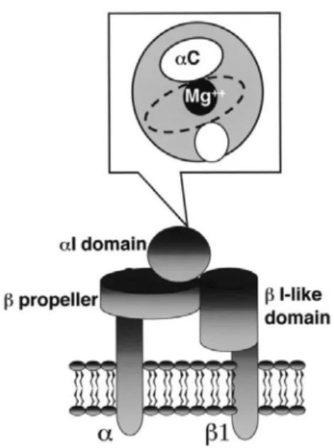
The α2β1 is the first collagen receptor to be identified on platelets and serves mainly as an adhesion molecule.109 The crystal structure of the α2β1 integrin allowed identification of the structures of the α2 and β1 subunits. The extracellular domain of the α2 subunit has a β propeller-like structure and a collagen-binding αI domain. The extracellular domain of the β1 subunit has an I domain-like fold, but it does not directly participate in collagen binding.110, 111 Interaction of collagen with the αI domain of the α2 subunit is dependent on the Mg2+ metal coordination site, named metal ion-dependent adhesion site (MIDAS), located in the bottom groove of the αI domain, while the αC helix in the αI domain seems to guide the collagen molecule into the groove in the right position, which is necessary for proper binding (Figure 1.4).112, 113 Signalling through the α2β1 integrin is similar to that induced by GPVI stimulation, which includes phosphorylation of the Src family kinases and the subsequent activation of PLC.114 Moreover, as for most integrins, the α2β1 integrin exists in two conformational configurations, an inactive conformation and an active one capable of bind collagen.111
14
Figure 1.4: Schematic representation of the α2β1 integrin. Heino J. Matrix Biology. 2000; 19:319-323.
It is now evident that α2β1 and GPVI are the two collagen receptors on platelets. However, their relative function in collagen-mediated platelet response is still unclear. The GPVI induced collagen response seems to be the most important. On the other hand, the α2β1 integrin is responsible for platelet adhesion to the exposed collagen surface in the sub-endothelium and in β1-deficient mice, the thrombi consist of loose platelet aggregates.115, 116 In addition, genetic or pharmacological depletion of GPVI highlight the capacity of α2β1 to mediate collagen signalling and haemostasis.108
1.3.7 αIIbβ3
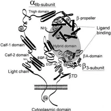
The αIIbβ3 integrin is the most important and most abundant integrin on platelets, with approximately 80 000 copies per resting platelet and with important αIIbβ3 reserves within platelet α-granules and the OCS, which increase the integrin's surface expression by 25-50% upon platelet activation.117-119 The crucial importance of αIIbβ3 in primary haemostasis is evidenced by the bleeding disorder of patients with Glanzmann's disease, where genetic disorders in the αIIb and β3 gene result in functional abnormalities and/or prevention of surface expression of the αIIbβ3 integrin.120 The bleeding disorder is due to absence or improper haemostatic plug formation because of lack of platelet aggregation, which is mediated by the cross-linking of αIIbβ3 on adjacent platelets by soluble fibrinogen at low shear rate or vWF at high shear rate.121, 122
15 The αIIb and β3 subunits of the αIIbβ3 integrin are both products of a single gene located on chromosome 17, giving rise to a 1008 and a 762 amino acid αIIb and β3 subunits, respectively.123 The αIIb is proteolytically processed into a heavy and a light chain. The light chain contains a 20 amino acid cytoplasmic tail, a transmembrane helix, and an extracellular segment that is disulfide linked to the heavy chain, which is entirely extracellular. Within the heavy chain, a large domain composed of a series of 60 amino acid repeats, which are arranged to form the seven blades of the propeller, extend outward from a central core. At the base of the β-propeller there are four divalent cation (Ca2+) binding motifs.124-126 The β3 subunit contains a 48 amino acid cytoplasmic tail and three major extracellular domains: the A domain, the PSI (plexin-semaphorin-integrin) domain, and four endothelial growth factor (EGF)-like domains (Figure 1.5).127 The PSI domain seems to be involved in integrin activation, because mutation of the cysteine linking it to the EGF-like domain, results in a constitutively active integrin.128 The A domain contains 2 or 3 divalent cations (Ca2+) sites, including a MIDAS motif that is highly involved in ligand binding. In addition, the A domain of the β3 subunit also contains two recognition sites, one for the γ-chain sequence and another for the RGD (arginine-glycine-aspartic acid) sequence, which is present in ligands of the αIIbβ3 integrin, including fibrinogen, fibrin, vWF, vitronectin, fibronectin and autotaxin.121, 129-133 Finally, the EGF-like domains are formed by four cysteine rich loops (EGF-1 to EGF-4), which seem to have a regulatory function because a cysteine mutation within this region causes activation of the αIIbβ3 integrin.134, 135
16
Figure 1.5: Schematic representation of the αIIbβ3 integrin. Quinn M. J. et al. Arteriosclerosis, Thrombosis, and Vascular Biology. 2003; 23:945-952.
As most integrins, the αIIbβ3 integrin exist in two conformations; an inactive low affinity ligand binding conformation, and an active high affinity ligand binding one. Activating signals mediated by platelet agonists such as collagen and thrombin induce "inside-out" signals, which shifts the αIIbβ3 integrin from its inactive to active conformation. In the active conformation, ligand binding to αIIbβ3 stimulates "outside-in" signalling that promote firm platelet adhesion and spreading on the extracellular matrix, fibrin clot retraction, and development of platelet pro-coagulant activity and microparticle generation.136-138 Moreover, there is a close association between αIIbβ3 and the platelet cytoskeleton, where the cytoskeleton is involved in regulating the structure and activation of the αIIbβ3 integrin. In resting platelets, the αIIbβ3 integrin is associated with the membrane cytoskeleton, which favours anchoring of the integrin to the plasma membrane, and upon platelet activation and ligation of the αIIbβ3 integrin with fibrinogen, the αIIbβ3 integrin associates with cytoplasmic actin, thereby stabilizing ligand/integrin interactions, which ultimately lead to stabilizing the platelet aggregates.139 The exact role of the αIIbβ3 integrin in platelet adhesion, aggregation and signalling will be discussed in more detail in section 1.4 of this chapter.
17
1.3.8 P-selectin
P-selectin belongs to the selectin family, which is composed of three members that are named according to their main expression site: L-selectin is expressed in leukocytes, E-selectin is expressed on endothelial cells, and P-selectin is mainly found in platelets but also in endothelial cells.140-142 The human selectin family is encoded by genes located on chromosome 1, and all the members share a conserved structure consisting of an N-terminal Ca2+-dependent lectin recognition motif followed by an EGF-like motif, a series of short consensus repeats (SCRs), a transmembrane domain, and a short cytoplasmic tail.143 The main structurally differentiating factor between all members of the selectin family is the variation in the number of SCRs, with L-selectin, E-L-selectin, and P-selectin having 2, 6 and 9 SCRs, respectively (Figure 1.6).
Figure 1.6: Schematic representation of the selectin family members. Image is from the laboratory of Dr. Merhi.
In platelets, P-selectin is stored in the α-granules, and upon activation it is translocated to the plasma membrane, where approximately 10 000 copies of the molecule are then expressed on the surface of an activated platelet. This translates to a density of 350 molecules/μm2, which exceeds that of activated endothelial cells by approximately 10 fold.144 One of the major roles of platelet P-selectin is mediating interactions between platelets and leukocytes through their constitutively expressed high affinity P-selectin ligand, the P-selectin glycoprotein ligand-1 (PSGL-1).145 Therefore, with such a high density of P-selectin molecules expressed on activated platelets, this interaction might have an even more important role in recruiting leukocytes to the
18 site of injury/inflammation than the interaction between endothelial P-selectin and leukocyte PSGL-1. In fact, it has been demonstrated that in P-selectin deficient mice, there is lack of leukocyte recruitment onto the platelet monolayer at the site of vessel injury.146 Moreover, the platelet/leukocyte interaction is of major importance not only in a physiological setting, but also in pathological ones such as in ischemia-reperfusion injury and in atherosclerosis.147, 148
P-selectin also has an important role in platelet/platelet interactions, as shown by its involvement in stabilizing platelet aggregates and thrombus formation; however, its ligands on platelets are still a matter of debate.149, 150 Although platelets express three potential ligands for platelet P-selectin; PSGL-1, GPIbα and sulfatides, sulfatides and GPIbα seem to be the most probable ligands. P-selectin and GPIbα but not PSGL-1 blockade, inhibits platelet rolling onto an activated endothelium and affects the stability of platelet aggregates, indicating a possible role for GPIbα as a P-selectin ligand on platelets.99, 149 By contrast, it has recently been shown that blocking antibodies against P-selectin and sulfatides, but not PSGL-1 or GPIbα inhibited platelet adhesion to P-selectin and platelet aggregation, indicating that sulfatides, and not PSGL-1 or GPIbα are the major P-selectin ligands on platelets.149, 151, 152 Such discrepancy in identifying the platelet P-selectin ligand could be attributed to the experimental settings. Furthermore, P-selectin expressed on activated platelets promotes fibrin and thrombus formation by recruiting tissue factor (TF) bearing monocytes and monocyte derived microparticles to the site of vessel injury.153, 154
1.4 Platelet function
As mentioned earlier in this chapter, platelets play a pivotal role in haemostasis by maintaining the integrity of blood vessels through the formation of a haemostatic plug that prevents blood loss. In order to accomplish such task, platelets respond to external stimuli mediated by the interaction of their receptors with the respective ligands. These interactions lead to a sequence of events consisting of adhesion, activation, secretion, and aggregation. In brief, GPIb/IX/V, GPVI, α2β1 integrin and αIIbβ3 integrin mediate platelet adhesion and activation, while αIIbβ3 is responsible for platelet aggregation, clot retraction and thrombus stability.
19
1.4.1 Platelet adhesion
Under normal circumstances, platelets circulate in the blood in an inactive state unable to adhere or become activated, a process mediated by an important function of the endothelium. The endothelial cells not only create a physical barrier preventing platelet and sub-endothelial matrix contact, but also actively participate in inhibiting platelet activation by secreting inhibitory molecules such as nitric oxide (NO), PGI2, and nucleoside triphosphate diphosphohydrolase-1 (NTPDase-1, CD39).82 On the other hand, damage to the vessel wall exposes the sub-endothelial matrix and activates endothelial cells, thereby initiating the haemostatic mechanism. Platelet adhesion at the site of vessel injury is initiated by platelet rolling on the activated endothelium, followed by firm adhesion of platelets to the components of the sub-endothelial matrix (collagen and vWF).155
Platelet rolling on the activated endothelium
Platelet rolling on the activated endothelium is the first step in initiating the haemostatic plug. Before adhering to the sub-endothelial matrix, circulating platelets undergo a deceleration process on activated, P-selectin (present in Weibel-Palade bodies) expressing endothelial cells neighbouring the injured vasculature.156 This process is mediated by the interaction of endothelial P-selectin with its yet unresolved ligand on platelets, albeit platelet PSGL-1 and the GPIb/IX/V complex could be the two possible candidates.99, 151
Platelet adhesion to the sub-endothelial matrix
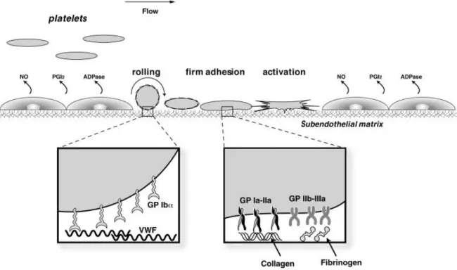
At the site of the injured vasculature, following the rolling process onto the activated endothelium, platelets come to an arrest upon contact with the sub-endothelial matrix. The arrest is made possible by the interaction of platelet GPIb/IX/V, GPVI, α2β1 and αIIbβ3 with the components of the sub-endothelial matrix. The importance of the relative initial interactions of these receptors with their respective ligands is dependent on the shear force present at the site of injured vasculature. For instance, at high shear levels such as in stenotic arteries, initial tethering of platelets onto the sub-endothelial matrix fully depends on the weak interaction between the GPIb/IX/V complex and vWF, then followed by the firm adhesion of platelets to the exposed collagen matrix, which is GPVI and α2β1-dependent.157-159 By contrast, at low shear levels such as in the venous circulation, platelet adhesion to the sub-endothelial matrix depends on the
20 interactions of GPVI and α2β1 with collagen fibers, even though the GPIb/IX/V and vWF interaction is still present, it is of less importance.160 The αIIbβ3 integrin also participates in platelet adhesion by binding fibrinogen and vWF molecules present in the sub-endothelial matrix. Figure 1.7 summarizes the major events in platelet adhesion to the sub-endothelial matrix.
It is of interest to mention that circulating vWF molecules are not able to bind the GPIb/IX/V complex, but once in contact with collagen in the sub-endothelial matrix and in the presence of shear forces, the vWF molecule undergoes conformational change, which allows it to bind the GPIb/IX/V complex.161, 162 Although the vWF/GPIb/XI/V interaction is of a weak nature, at high shear levels, it occupies a crucial role in slowing down circulating platelets, which then favours GPVI and α2β1-dependent platelet arrest at the sites of injury.
Figure 1.7: Platelet adhesion to the sub-endothelial matrix. Under normal conditions, platelet activation is prevented
by the secreted endothelial mediators (NO, PGI2 and NTPDase-1). At the site of vessel lesion, platelet rolling,
tethering, adhesion and activation is mediated by the interaction of platelet receptors/integrins with the components of the sub-endothelial matrix (see above text). Conde I. D. et al. Catheterization and Cardiovascular Interventions. 2003; 60:236-246.
21
1.4.2 Platelet activation
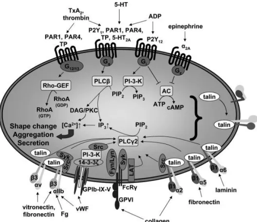
Interaction of GPIb/IX/V, GPVI, α2β1 and αIIbβ3 with components of the sub-endothelial matrix is not only responsible for platelet adhesion, but also mediates signals that induce platelet activation, secretion and aggregation. As mentioned in the previous section, ligation of GPIb/IX/V, GPVI and the α2β1 integrin with their respective ligands mainly induce activation of Src family kinases which ultimately activate PLC, specifically PLCγ2, leading to the generation of DAG and IP3, that activate PKCs and increase cytosolic Ca2+ levels, respectively. Furthermore, ligation of fibrinogen to the αIIbβ3 integrin also induces activation of the Src family kinases that activate PLCγ2, and the interaction of talin with the cytoplasmic tail of the β3 subunit, which is involved in clot retraction.163
Following the initial wave of platelet activation induced by ligation of GPIb/IX/V, GPVI, α2β1 and αIIbβ3 with the components of the sub-endothelial matrix, activated platelets secrete secondary mediators such as TxA2 and ADP (see section 1.4.3) that recruit and activate additional circulating platelets at the site of vascular lesion. In addition, TF expressed on recruited monocytes, endothelial cells and activated platelets leads to the generation of thrombin at the site of injured vasculature, which also participates in recruitment and activation of additional platelets.154, 164, 165 Activation of platelets by TxA2, ADP and thrombin is mediated by ligation of these secondary mediators with their respective receptors; TPα and TPβ, the purinergic receptors, and PARs, which are coupled to G proteins (see section 1.3). Briefly, Gq and G12/13 lead to the generation of DAG and IP3 by PLCβ, and to the activation of the Rho GEFs, respectively. DAG in turn activates PKCs, which lead to protein phosphorylation, including VASP phosphorylation. VASP functions as an anti-capping protein, which is involved in crucial cellular functions, including shape change by directly modulating the actin ultra-structure.166 IP3 binds to its receptor on the endoplasmic reticulum thereby inducing release of Ca2+ from intracellular stores. The Rho GEFs; RhoA, Rac1, and Cdc42 are regulators of signalling pathways that control actin organization by the formation of stress fibers, lamellipodia and filopodia.167 In summary, activation of platelets is mediated by two major signalling pathways, one including activation of PLC (PLCγ2 or PLCβ), and the second mediated by activation of G proteins through GPCRs. Figure 1.8 summarizes the major signalling pathways implicated in platelet activation.
22
Figure 1.8: The major signalling pathways implicated in platelet activation. Varga-Szabo D. et al. Arteriosclerosis,
Thrombosis, and Vascular Biology. 2008; 28:403-412.
One of the early hallmarks of platelet activation is the morphological shape change induced by the reorganization of the platelet actin cytoskeleton, which transforms platelets from their disk-like structure into a spherical one containing multiple filopodia protrusions (Figure 1.9).33, 34 The change in platelet shape as well as the formation of filopodia allows platelets to expand and cover a larger surface area at the site of vessel injury. Platelet activation also leads to the expression of P-selectin and the activation of the αIIbβ3 integrin (through an "inside-out" signalling mechanism), where they each make their contribution to platelet aggregation.
23
Figure 1.9: Scanning electron microscopy image of platelet shape change. Left: Resting platelet. Right: Activated
platelet. Images are from the laboratory of Dr. Merhi.
1.4.3 Platelet secretion
Platelet activation is accompanied with secretion, where platelets secret a plethora of molecules stored in their granules, as well as newly enzymatically synthesised compounds, such as TxA2. Platelet secretion is an indispensible process linked to their pathophysiological role, including recruitment and activation of additional circulating platelets, initiation of thrombus formation, mediating intracellular adhesion and triggering cell proliferation and migration. Platelets contain three types of secretory granules; α-granules, dense granules and lysosomes, which are formed, pre-packaged, and sorted into pro-platelets during platelet formation from megakaryocytes.168
Megakaryocytes synthesise most of the protein contents present in platelet granules, and during the platelets' life span, platelets incorporate proteins into their existing granules through endocytosis. For instance, incorporation of fibrinogen is mediated by its interaction with the αIIbβ3 integrin, and its subsequent internalization and sorting into α-granules.169 The α-granules are the largest and most abundant granules in platelets with approximately 80 α-granules per platelet, and they contain coagulation proteins (e.g. fibrinogen, factor V), soluble adhesion molecules (e.g. vWF), growth factors (e.g. platelet derived growth factor [PDGF]), protease inhibitors (e.g. plasminogen activator inhibitor-1), and membrane adhesion molecules (e.g. P-selectin and αIIbβ3).170 The dense granules are less abundant and less voluminous than α-granules, with about 3-8 dense granules per platelet; however, they contain substances indispensible for platelet activation, such as ADP, serotonin, Ca2+, and magnesium.171, 172 Although the contents of the α and dense granules differ from each other, recent studies have shown the presence of P-selectin, αIIbβ3, and GPIb in both granules.173, 174 Finally, lysosomes contain enzymes that have a role in degrading and digesting multiple proteins.175, 176 Table 1.1 summarizes the major contents of platelet granules.177
24
Table 1.1: Platelet granule contents.
α-granules Dense granules Lysosomes
P-selectin αIIbβ3 integrin GPIb/IX/V GPVI αvβ3 integrin PECAM-1 Stomatin PDGF EGF VEGF
Transforming growth factor β (TGF-β) Albumin Fibrinogen Fibronectin Vitronectin Osteonectin vWF
von Willebrand antigen II Thrombospondin Platelet factor-4 (PF4)
IgG, IgA, IgM C1 inhibitor Plasminogen
Plasminogen activator inhibitor-1 Platelet-derived collagenase inhibitor
High molecular weight kininogen Protein S
α2-antitrypsin
α2-macroglobulin
α2-antiplasmin
Multimerin Platelet basic protein
β-thromboglobulin Histidine-rich glycoprotein Connective tissue-activating protein III
Neutrophil-activating protein II Coagulation factor V Coagulation factor VIII
GPIb αIIbβ3 integrin P-selectin CD107a (LAMP-1) CD107b (LAMP-2) CD63 (LAMP-3) Serotonin Histamine ATP ADP GTP GDP Pyrophosphate Calcium Magnesium Cathepsin D Cathepsin E Carboxypeptidase A Carboxypeptidase B Proline carboxypeptidase CD107a (LAMP-1) CD107b (LAMP-2) CD63 (LAMP-3) Acid phosphatase Arylsulphatase β-D-glucuronidase β-D-galactosidase β-D-fucosidase β-D-glucosidase α-D-mannosidase α-D-galactosidase α-L-arabinofuranosidase α-L-fucosidase
Activation of platelets by physiological agonist such as thrombin, ADP, TxA2 and collagen leads to intracellular signalling events that mediate platelet secretion, where platelets discharge their granule contents into the extracellular environment by exocytosis.172 Although most of the platelet physiological agonists can mediate α and dense granule secretion, it seems that only thrombin, the most potent agonist, is capable of inducing lysosome secretion.178 As mentioned earlier in this chapter, ligation of platelet agonists with their cognate receptors induces activation of two major signalling pathways (PLC and G protein activation), of which PLC
25 activation seems to be of significant importance for platelet secretion. The increase in cytosolic Ca2+ levels by IP3, as well as the activation of DAG following PLC mediated degradation of PIP2, leads to the activation of PKCs, which are crucial signalling molecules involved in platelet secretion. Platelets contain multiple PKCs (α, β1, β11, δ, ζ, η, and θ) and it seems that each of them has a specific role in platelet secretion, where they induce phosphorylation of intracellular signalling molecules implicated in the exocytotic machinery.172, 179-183
The molecular machinery involved in platelet secretion, which mediates granule fusion with surface connected membranes of the OCS or the plasma membrane, remained obscure for a long time.184, 185 However, insights into similarities between neuronal and platelet exocytosis, revealed molecular components of the secretory mechanism involved in both cell types.186 The molecular machinery responsible for membrane fusion during exocytosis is composed of the core soluble N-ethylmaleimide sensitive factor (NSF) associated protein receptor (SNARE) complexes. The SNAREs associated with granules are termed vesicular SNAREs (vSNARE), while those associated with target membranes (e.g. OCS and plasma membrane) are termed tSNAREs. In platelets, the identified vSNAREs include the vesicle-associated membrane proteins (VAMP) -2, -3, -7, and -8, while the identified tSNAREs include the syntaxins 2, 4, 7, and 11, and the soluble NSF-associated proteins (SNAP) -23, -25, and -29.187-193 Platelets also contain the Sec1 (platelet Sec1 protein, [PSP]) and Rab proteins that regulate SNARE function.183, 194 Platelet exocytosis is mediated by orchestrated steps involved in granule and plasma membrane fusion. Briefly, in resting conditions, PSP binds to tSNAREs and prevents formation of the SNARE complex required for membrane fusion. Upon platelet activation, the increase in cytosolic Ca2+ levels, as well as the activation of the PKCs, induces phosphorylation of PSP, which in turn relieves its inhibitory effects on SNARE complex formation.183 Concurrently, NSF disassembles the cis-conformation of SNAREs on the same membrane thereby allowing formation of the trans-conformation of the SNAREs so that they are able to interact with their respective SNAREs on opposing membranes.195 Finally, the association of the vSNAREs and tSNAREs, in addition to the modulating role of Rab proteins, which facilitates docking of opposing membranes and modifies SNARE protein function, allow fusion of granule and plasma membranes thereby releasing the granule contents into the extracellular milieu.196-198
In addition to granule exocytosis, platelets also synthesize and secrete new molecules such as TxA2, upon activation. Platelet activation by physiological agonists such as thrombin,