Comparative study of commercially available anti-α-synuclein antibodies
E. Croisier*, D. Elfant MRes*, M. Deprez‡, K. Goldringt†, D. T. Dexter†, R. K. B. Pearce*, M. B. Graeber* and F. Roncaroli*
*Department of Neuropathology, Imperial College London, †UK Parkinson's Disease Society Tissue Bank, Department of Cellular and Molecular Neuroscience, Imperial College London, UK, and ‡Laboratory of Neuropathology, Department of Pathology, University of Liège, Belgium
Abstract
Immunohistochemistry for alpha-synuclein has become the histological technique of choice for the diagnosis for Parkinson's disease, Dementia with Lewy bodies and Multiple System Atrophy (www.ICDNS.org).
Nevertheless, no standardised protocol has been proposed. We have reviewed 242 of the 270 studies published until June 2005 that mentioned immunohistochemistry for anti-alpha synuclein on human tissue and we found that only 75 (31%) used commercial antibodies. We also noted that protocols, particularly dilution and antigen unmasking, varied between studies, even when the same antibody was employed. In order to establish a standardised protocol for alpha-synuclein immunohistochemistry, which can be applied in diagnostic neuropathology we tested seven commercial monoclonal antibodies in brains of subjects with Parkinson's disease, dementia with Lewy bodies, multiple system atrophy, multiple sclerosis with incidental Lewy bodies and aged-matched normal brain and determined for each antibody the best suited protocol for antigen unmasking. We evaluated the intensity of immunolabelling in Lewy bodies, neuropil threads, dendrites, pre-synaptic terminals, granular cytoplasmic positivity, peri-axonal positivity, glial inclusions and non-specific immunolabelling. Although our results showed that all the antibodies detected alpha-synuclein inclusions, differences were noted between antibodies, particularly with regard to the detection of glial inclusions. From our study, the best antibodies of the seven tested appeared to be those directed against amino acids 116-131 and 15-123 and we suggest them to be used in routine diagnostic practice for alpha-synucleinopathies.
Keywords : alpha-synuclein ; immunohistochemistry
Alpha-synuclein (αSN) is a highly conserved, natively unfolded 18 kDa protein [1] which represents the major component of Lewy bodies (LBs) [2], and the glial cytoplasmic inclusions (GCIs) associated with Multiple System Atrophy (MSA) [3]. The increased sensitivity of αSN immunohistochemistry over conventional histological stains has established the technique as a standard tool in the diagnosis of Parkinson's disease (PD), Dementia with Lewy Bodies (DLB) and MSA [4], and led to new insights regarding the extent and variation of
αSN-containing structures and their formation. αSN aggregations vary throughout the brain, with brainstem
pathology characterized by compact LBs and neuropil threads. Pigmented neurones of the substantia nigra may also contain diffuse fine or coarse granules, which likely represent the state of aggregation preceding LBs [5,6]. Cortical pathology consists of more diffuse LBs and punctate granular positivity that corresponds to pre-synaptic terminals [7].
Despite the growing frequency with which immunohistochemistry is used as a diagnostic tool for PD, DLB and MSA, a standardized protocol for αSN has not been established. Pretreatment with proteinase K or formic acid has been reported to increase the immunoreactivity of αSN; however, the only studies to investigate this systematically were conducted with non-commercial antibodies [8] or brain tissue unaffected by
neurodegenerative disease and devoid of any pathologically associated protein aggregations [9]. To assess the use of commercial vs. non-commercial anti-αSN antibodies and different antigen-unmasking pretreatments in research, we performed a search of the PubMed database updated to 30 June 2005 for 'alpha-synuclein'. We found 1567 citations pertaining to the protein, and after reviewing all of these abstracts, we selected studies that mentioned the use of αSN immunohistochemistry on human tissue (270 overall). We screened the methodology of 242 articles for type of antibody used (commercial or non-commercial), dilution and pretreatment for antigen unmasking. Only 75 studies (31%) used commercial anti-αSN antibodies. Thirty-seven studies (15%) reported using a pretreatment for antigen unmasking and 17 (7%) included no details at all regarding the antibodies used. Also, many studies performing immunohistochemistry with the same commercial anti-αSN antibodies used different dilutions and protocols.
We observed that different anti-αSN antibodies varied in how effectively they identified different types of inclusions. In order to standardize the protocol for a more rigorous and reproducible neuropathological diagnosis of PD, DLB and MSA, we have compared seven commercially available monoclonal anti-αSN antibodies (mAbs) representative of the clones and immunogens currently available. Details of the mAbs' manufacturer, clone, the dilution and antigen-unmasking technique used, and the epitope they recognize or immunogen against which they were raised are summarized in Table 1. According to manufacturers' data sheets, four mAbs are specific to human αSN (A, B, F, G), one recognizes human, mouse and rat isoforms (C), one was untested in species other than human (D), and the cross-reactivity of one was unspecified (E). Using a protein BLAST search, we compared epitopes or immunogens against which the mAbs were raised and found no sequence homology to any other known human proteins. The seven mAbs were tested on tissue from five post mortem brains selected on the basis of neuropathological diagnosis. One case each of idiopathic PD, DLB and MSA, and one age-matched control were provided by the Parkinson's Disease Society Tissue Bank (PDSTB). A case of multiple sclerosis with numerous incidental brainstem LBs was provided by the UK Multiple Sclerosis Tissue Bank. Brains were fixed in 10% buffered formalin for at least 2 weeks, and sample blocks taken during the fixed tissue dissection were processed routinely up to paraffin embedding. Ten micron sections from the superior frontal gyrus and the substantia nigra from each case were stained with haematoxylin-eosin and with each of the seven anti-αSN mAbs using the avidin biotin complex/peroxidase method (ABC, Vector, Burlingane, CA, USA). All primary mAbs were incubated overnight at 4°C. We first followed manufacturers' instruction for dilution and pretreatment for antigen unmasking and subsequently tested different dilutions and pretreatments to optimize the protocols. For antigen unmasking, we tested incubation in 100% formic acid for 5 min, and heating in a
microwave oven at 3 50 W for 20 min using 10 mM citrate buffer at pH 6 and 1 mM ethylenediamine tetra-acetic acid (EDTA) at pH 8. Immunoreactions with omission of the primary mAb were performed for every case as controls. All immunoreactions were evaluated independently by two authors (E.C. and F.R.). We rated LBs, cytoplasmic granular positivity neuropil threads, presynaptic terminals, periaxonal deposition and GCIs using a four-point system from - (no reactivity) to +++ (intense staining). A rating of +⁄- was also added to our
assessment on GCIs to indicate that, regardless of the intensity of staining, the mAb failed to identify nearly as many GCIs as were observed with other mAbs on adjacent sections, suggesting only a partial affinity for this form of αSN aggregate. We also assessed nonspecific background staining of the neuropil, rating it either none, low (light non-specific staining, generally confined to white matter tracts) or high (non-specific staining throughout the neuropil, dark enough to compete with specific labelling). The immunostaining profiles for each antibody are outlined in Table 2.
Table 1. Details of the seven monoclonal anti-αSN antibodies used in this study
Manufacturer [clone] Immunogen [epitope] Dilution Pretreatment
A. Abeam [Syn204] Human recombinant αSN [aa 87-110 (12)] 1:50 100% FA 5 min
B. Alexis [15G 7] Synthetic peptide aa 116-131 1:10 100% FA 5 min
C. Becton-Dickinson[42] Rat synuclein aa 15-123 1:300 100% FA 5 min
D. Chemicon [7B 2,12] Human recombinant αSN 1:400 100% FA 5 min
E. NovoCastra[KM51] Human recombinant αSN 1:40 H-T EDTA
F. SantaCruz[Syn211] Human recombinant αSN [aa 121-125 (12)] 1:100 100% FA 5 min
G. Zymed [LB509] Purified LBs [aa 115-122 (13)] 1:200 H-T citrate
LBs, Lewy bodies; FA, formic acid; H-T, high temperature (20 min in microwave at 350 W); αSN, alpha-synuclein; EDTA, ethylenediamine tetra-acetic acid.
Table 2. Immunostaining profiles for each monoclonal anti-αSN antibody (mAb)
mAb LBs Neuropil threads Dendrites Terminals Granular cytoplasmic
Peri-axonal GCIs Non-speciflc
A ++ ++ +++ ++ ++ - +⁄- High B +++ ++ ++ +++ +++ + +++ None C +++ +++ ++ + +++ + +++ None D + + - + ++ - +⁄- High E ++ ++ - +++ ++ ++ + None F +++ +++ + +++ +++ + +++ High G ++ ++ + + + ++ ++ None
Our results showed that immunostains using mAbs A, C, D and F were not significantly affected by pretreatments for antigen unmasking. In contrast, mAbs E and G showed far intenser immunostaining after microwaving in EDTA and citrate buffer, respectively. All mAbs demonstrated good results in detecting LBs (both brainstem and cortical), neuropil threads and cytoplasmic granular deposition as well as the diffuse, granular deposition characteristic of cortical grey matter (Figures 1 and 2). However, a broad range of staining intensity was observed across the mAbs. The intensity of immunolabelling of large neuropil threads paralleled that observed in LBs. Smaller threads likely corresponding to dendrites rather than axons were only clearly highlighted by three mAbs (A, B and C). Two mAbs (B and F) demonstrated intra-axonal, neurite-like αSN deposition in the cortical white matter of the DLB case. Four mAbs (B, E, F and G) showed finely granular periaxonal positivity in the white matter of the PD and DLB cases, which appeared denser and more evenly distributed than that observed in the cortical grey matter. Double immunolabelling with one of these anti-αSN mAbs (G) and anti-myelin basic protein (polyclonal, DAKO, Carpinteria, CA, USA, 1 : 200, no pretreatment), suggests that this type of αSN deposition may co-localize with the myelin sheath (data not shown).
The most distinct, and diagnostically relevant, variability between the mAbs evaluated was observed in their labelling of GCIs. Three mAbs (A, D and E) barely labelled these, while the other four labelled them clearly (Figure 3). Chemical differences have been reported between αSN aggregates in MSA and PD [10], which may explain the marked differences in reactivity we observed across our panel of mAbs. An earlier
immunohistochemical study of MSA tissue suggests that immunoreactivity for GCIs is maximized by antibodies directed to the carboxyl-terminal of αSN, but that immunoreactivity can be improved for other antibodies after formic acid pretreatment [11]. Three of the mAbs in the present study (A, F and G) are identical to
non-commercial clones used in earlier study [12,13], and, with respect to these three mAbs and mAb B, our findings support the observations of Duda et al. [11] in that our immunostaining appeared progressively intenser as epitopes moved progressively closer to the carboxyl terminus. However, the other clones raised against full-length recombinant αSN (D, E) showed only weak immunoreactivity, and mAb C, raised against amino acids 15-123 of rat synuclein, demonstrated excellent results. Because the specific epitopes recognized by these last mAbs have not been reported, we cannot generalize staining intensity as a factor of proximity to the carboxyl-terminus. As a result of this comparative study, we now preferentially use mAbs that recognize amino acids 116-131 or 15-123 (A and C) for diagnostic purposes based on their strong immunoreactivity for a variety of αSN-containing structures, with the dilutions and pretreatment recommended in Table 1. Depending on the experimental design, other mAbs may be preferable. For example, although it is not a primary concern in studies of post mortem human tissue, cross-reactivity with mouse synuclein would be a major concern for investigators studying human
αSN expression in transgenic mice.
The wide variety of staining patterns observed across the seven antibodies emphasize the need for
standardization prior to the interpretation of immunohistochemical results. Previous analyses of glial fibrillary acidic protein [14] and prion protein [15] have demonstrated that variability across antibodies could account for discrepancies between research studies and affect diagnostic accuracy. All mAbs in this study adequately labelled LBs and are suitable for diagnosis of idiopathic PD or use in research; however, as the
neuropathological nuances differentiating alpha-synucleinopathies become increasingly refined, the use of a panel of antibodies, or one which is known to label multiple structures of diagnostic relevance, may become necessary.
Figure 1. Substantia nigra of the case of idiopathic PD contains intensely stained Lewy bodies (a, ×400) and neuropil threads (b, ×200). Diffuse and finely granular immunolabelling corresponds to presynaptic terminals (mAb C). PD, Parkinson's disease; mAb, monoclonal anti-α-synuclein antibody
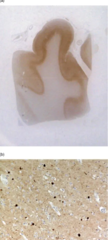
Figure 2. Diffuse cortical αSN immunolabelling is present in the superior frontal gyrus of the case of DLB (a, whole mount). Many neurones are seen to contain LBs (b, × 100) (mAb B). DLB, Dementia with Lewy bodies;
αSN, alpha-synuclein.
Figure 3. The monoclonal antibody raised against amino acids 15-123 (mAb C) strongly stains oligodendroglial inclusions in the case of MSA (a, ×100). In the same region, the antibody directed against amino acids 87-110 stains consistently fewer GCIs (mAb A) (b, ×100). mAb, monoclonal anti-αSN antibody; αSN, alpha-synuclein;
MSA, Multiple System Atrophy; GCI, glial cytoplasmic inclusion.
Acknowledgements
This work was supported by a Programme Grant from the UK Parkinson's Disease Society. We would like to thank Mrs Louisa McGuinness and Miss Helen C. Cairns for technical support.
References
1 Uversky VN, Fink AL. Amino acid determinants of alpha-synuclein aggregation: putting together pieces of the puzzle. FEBS Lett 2002; 522: 9-13
2 Spillantini MG, Crowther RA, Jakes R, Hasegawa M, Goedert M. Alpha-synuclein in filamentous inclusions of Lewy bodies from Parkinson's disease and dementia with Lewy bodies. Proc Natl Acad Sci USA 1998; 95: 6469-73
3 Wakabayashi K, Yoshimoto M, Tsuji S, Takahashi H. Alpha-synuclein immunoreactivity in glial cytoplasmic inclusions in multiple system atrophy. Neurosci Lett 1998; 249: 180-2
4 Dickson DW. Required techniques and useful molecular markers in the neuropathologic diagnosis of neurodegen-erative diseases. Acta Neuropathol (Berl) 2005; 109: 14-24
5 Saito Y, Kawashima A, Ruberu NN, Fujiwara H, Koyama S, Sawabe M, Arai T, Nagura H, Yamanouchi H, Hasegawa M, Iwatsubo T, Murayama S. Accumulation of phosphorylated alpha-synuclein in aging human brain, J Neuropathol Exp Neurol 2003; 62: 644-54 6 Kuusisto E, Salminen A, Alafuzoff I. Ubiquitin-binding protein p62 is present in neuronal and glial inclusions in human tauopathies and synucleinopathies. Neuroreport 2001; 12: 2085-90
7 Maroteaux L, Campanelli JT, Scheller RH. Synuclein: a neuron-specific protein localized to the nucleus and presynaptic nerve terminal, J Neurosci 1988; 8: 2804-15
8 Takeda A, Hashimoto M, Mallory M, Sundsumo M, Hansen L, Masliah E. C-terminal alpha-synuclein immunoreactivity in structures other than Lewy bodies in neurodegenerative disorders. Acta Neuropathol (Berl) 2000; 99: 296-304
9 Mori F, Tanji K, Yoshimoto M, Takahashi H, Wakabayashi K. Demonstration of alpha-synuclein immunoreactivity in neuronal and glial cytoplasm in normal human brain tissue using proteinase K and formic acid pretreatment. Exp Neurol 2002; 176: 98-104
10 Campbell BC, McLean CA, Culvenor JG, Gai WP, Blumbergs PC, Jakala P, Beyreuther K, Masters CL, Li OX. The solubility of alpha-synuclein in multiple system atrophy differs from that of dementia with Lewy bodies and Parkinson's disease, J Neurochem 2001; 76: 87-96
11 Duda JE, Giasson BI, Gur TL, Montine TJ, Robertson D, Biaggioni I, Hurting HI, Stern MB, Gollomp SM, Grossman M, Lee VM, Trojanowski JO. Immunohistochemical and biochemical studies demonstrate a distinct profile of alpha-synuclein permutations in multiple system atrophy. J Neuropathol Exp Neurol 2000; 59: 830-41
12 Giasson BI, Jakes R, Goedert M, Duda JE, Leight S, Trojanowski JO, Lee VM. A panel of epitope-specific antibodies detects protein domains distributed throughout human alpha-synuclein in Lewy bodies of Parkinson's disease. J Neurosci Res 2000; 59: 528-33
13 Jakes R, Crowther RA, Lee VM, Trojanowski JO, Iwatsubo T, Goedert M. Epitope mapping of LB509, a monoclonal antibody directed against human alpha-synuclein. Neurosci Lett 1999; 269: 13-16
14 Halliday GM, Cullen KM, Kril JJ, Harding AJ, Harasty J. Glial fibrillary acidic protein (GFAP) immunohistochemistry in human cortex: a quantitative study using different antisera. Neurosci Lett 1996; 209: 29-32
15 Kovacs GG, Head MW, Hegyi I, Bunn TJ, Flicker H, Hainfellner JA, McCardle L, Laszlo L, Jarius C, Ironside JW, Budka H.
Immunohistochemistry for the prion protein: comparison of different monoclonal antibodies in human prion disease subtypes. Brain Pathol 2002; 12: 1-11