AVIS
Ce document a été numérisé par la Division de la gestion des documents et des archives de l’Université de Montréal.
L’auteur a autorisé l’Université de Montréal à reproduire et diffuser, en totalité ou en partie, par quelque moyen que ce soit et sur quelque support que ce soit, et exclusivement à des fins non lucratives d’enseignement et de recherche, des copies de ce mémoire ou de cette thèse.
L’auteur et les coauteurs le cas échéant conservent la propriété du droit d’auteur et des droits moraux qui protègent ce document. Ni la thèse ou le mémoire, ni des extraits substantiels de ce document, ne doivent être imprimés ou autrement reproduits sans l’autorisation de l’auteur.
Afin de se conformer à la Loi canadienne sur la protection des renseignements personnels, quelques formulaires secondaires, coordonnées ou signatures intégrées au texte ont pu être enlevés de ce document. Bien que cela ait pu affecter la pagination, il n’y a aucun contenu manquant.
NOTICE
This document was digitized by the Records Management & Archives Division of Université de Montréal.
The author of this thesis or dissertation has granted a nonexclusive license allowing Université de Montréal to reproduce and publish the document, in part or in whole, and in any format, solely for noncommercial educational and research purposes.
The author and co-authors if applicable retain copyright ownership and moral rights in this document. Neither the whole thesis or dissertation, nor substantial extracts from it, may be printed or otherwise reproduced without the author’s permission.
In compliance with the Canadian Privacy Act some supporting forms, contact information or signatures may have been removed from the document. While this may affect the document page count, it does not represent any loss of content from the document.
Cardiovascular and sensory abnormalities in a rat model of
insulin resistance: beneficial effect of an antioxidant and an
angiotensin-l converting enzyme inhibitor
by
Mahmoud Ali Ismael
Department ofPhysiology
Faculty of Medicine
In partial fulfillment of the requirements
F or the Degree in Master of Science
(M.Sc.) in physiology
August 2007
Université de Montréal
F acuIté des études supérieures
Thesis title:
Cardiovascular and sensory abnormalities in a rat model of
insulin resistance: beneficial effect of an antioxidant and an
angiotensin-l converting enzyme inhibitor
Presented by:
Mahmoud Ali Ismael
The thesis has been evaluated by the following persons:
Madhu Anand-Srivastava
President
Réjean Couture
Research director
Pierre Beaulieu
Member
Abstract
Glucose-fed rat is a model of insulin resistance that displays hypertension and sensory polyneuropathy. This study aimed at comparing the beneficial effects of an antioxidant (N-acetyl-L-cysteine, NAC) and an angiotensin-l converting enzyme inhibitor (ramipril) in glucose-induced tactile and cold allodynia, hypertension, plasma levels of glucose, insulin, malondialdehyde (MA) and 4-hydroxynonenal (4-HNE), liverlaortic superoxide anion, changes of skeletal muscle insulin receptor substrate-l (IRS-l) protein expression
and of tissue kinin BI receptor mRNA. Methods: Male Wistar rats (50-75 g) were given
10% D-glucose in their drinking water during Il and 20 weeks. NAC (1-2 g/kg/day
orally) and ramipril (1 mg/kg/day in drinking water) were administered for the last 4-5 weeks. Results: Systolic blood pressure, plasma levels of insulin and glucose as well as insulin resistance (HOMA index) were significantly higher in rats treated with glucose for 20 weeks. This was associated with a higher production of superoxide anion and NADPH oxidase activity in aorta and liver and with a marked reduction of IRS-l protein expression in the gastrocnemius muscle. Tactile and cold allodynia occurred after six
weeks of glucose treatment and BI receptor mRNA was increased in the spinal cord and
renal cortex at Il weeks. NAC restored all these alterations in glucose-fed rats and
decreased plasma MA and 4-HNE levels. Although ramipril provided the same therapeutic effect as that ofNAC on blood pressure and allodynia, it was less effective in reducing insulin resistance and failed to reduce liverlaortic NADPH oxidase activity and plasma levels of MA and 4-HNE. Ramipril normalized superoxide anion only in the aorta.
Conclusion: The beneficial effects of NAC and ramipril on insulin resistance,
hypertension and allodynia were linked to the reduction of the oxidative stress and kinin
BI receptor expression. The antioxidant effect ofNAC involved the inhibition ofNADPH
oxidase and lipid peroxidation while that of ramipril was exerted most strongly in vascular tissue independently ofNADPH oxidase and lipid peroxidation.
Key words: allodynia, diabetes, hypertension, insulin resistance, kinin BI receptor, N-acetyl-L-cysteine, oxidative stress, polyneuropathy
Résumé
L 'hypertension et les polyneuropathies apparaissent chez le rat recevant du glucose, un modèle de résistance à l'insuline. Cette étude a pour but de comparer, chez le rat recevant du glucose, les effets bénéfiques d'un antioxydant (N-acétyl-L-cystéine, NAC) et d'un inhibiteur de l'enzyme de conversion de l' angiotensine-1 (ramipril) sur l' allodynie tactile et au froid, l'hypertension, la glycémie, l'insulinémie, les taux plasmatiques de malondialdéhyde (MA) et de 4-hydroxynonenal (4-Hl\JE), l'anion superoxyde dans l'aorte et le foie, les changements d'expression protéique de «insulin receptor substrate-1» (IRS 1) dans le muscle gastrocnemius ainsi que sur l'expression tissulaire (ARNm) du récepteur BI des kinines. Méthodes: Des rats mâles de 50-75 g ont reçu 10% de glucose
dans l'eau de boisson pendant Il et 20 semaines. NAC (1-2 g /kg/jour oralement) et
ramipril (1 mg/kg/jour dans l'eau de boisson) ont été administrés pendant les 4 et 5 dernières semaines. Résultats: La pression systolique, la glycémie, l'insulinémie ainsi que la résistance à l'insuline (indice HOMA) étaient significativement augmentées chez le rat recevant du glucose pendant 20 semaines. Ceci était associé avec une plus grande production de l'anion superoxyde et de l'activité de la NADPH oxydase dans l'aorte et le foie et avec une réduction marquée de l'expression de l'IRS-l dans le gastrocnemius. L' allodynie tactile et au froid apparaissaient après six semaines de traitement au glucose et l'ARNm du récepteur BI était augmenté dans la moelle épinière
et le cortex rénal à Il semaines. Le NAC a corrigé toutes ces anomalies chez le rat
recevant du glucose et a diminué les taux de MA et 4-HNE. Bien que le ramipril ait produit les mêmes effets thérapeutiques que le NAC sur la pression artérielle et
réduit l'activité de la NADPH oxidase dans le foie ou l'aorte ou encore les taux de MA et de 4-HNE. Le ramipril a toutefois normalisé la production d'anion superoxide dans l'aorte.
Conclusion: Les effets bénéfiques du NAC et du ramipril sur la résistance à l'insuline,
l'hypertension et l'allodynie sont liés à la réduction du stress oxydatif et à l'expression du
récepteur BI des kinines. L'effet antioxydant du NAC implique l'inhibition de la NADPH
oxydase et de la peroxydation des lipides, tandis que celui du ramipril est exercé principalement sur les vaisseaux indépendamment de la NADPH oxidase et de la peroxydation des lipides.
Mots clés: allodynie, diabète, hypertension, résistance à l'insuline, récepteur BI, stress
Table of Contents
Abstract ... 111
Résumé ... V Table of content ... VII List of figures ... XI List of abbreviations ... XII Dedication ... XV Acknowledgment ... XVI CHAPTERONE INRODUCTION 1.1 Diabetes ~ellitus ... ... 2
1.2 Type 1 Diabetes mellitus ... 2
1.3 Type 2 Diabetes mellitus ... 4
1.4 Global prevalence of diabetes ... 4
1.5 Complications associated with Diabetes ~ellitus ... 5
1.5.1 Microangiopathy ... 5
(a) Diabetic retinopathy ... 5
(b) Peripheral neuropathy... ... ... ... ... ... 6
(c) Erectile dysfunction ... 7
(d) Diabetic nephropathy ... 7
(e) Diabetic cardiomyopathy ... 7
1.5.2 Macrovascular complications ... 8
(a) Hypertension ... 8
(b) Peripheral vascular disease ... 9
1.6 Diabetes ~ellitus and oxidative stress ... ... ... ... 10
1.6.1 Source of oxidative stress in diabetes ... 10
1.7 Antioxidants and diabetes ... 14
1.7.1 Defense system against oxidative stress ... 14
(a) Primary antioxidants: active detoxification ... 14
1.8 N-acetyl-L-cysteine (NAC) ... 19
1.8.1 Mechanism of action ofNAC as antioxidant and anti-inflammatory agent... 19
1.8.2 Therapeutic uses of NAC ... 20
1.9 Angiotensin-converting enzyme (ACE) inhibitor ... 22
1.9.1 Ramipril ... 23
1.10 Diabetic neuropathic pain ... 23
1.11 Insulin signaling and action ... 27
1.11.1 Insulin receptor ... 27
1.11.2 Glucose transporters... ... 31
1.12 Hypothesis (article #1)... 32
1.13 Hypothesis (articl #2) ... 33
CHAPTERTWO ARTICLE # 1: Comparative effects ofN-acetyl-L-cysteine and ramipril on arterial hypertension, insulin resistance and oxidative stress in chronically glucose-fed rats 2.0 Abstract ... 36
2.1 Introduction ... 37
2.2 Materials and Methods ... 40
2.2.1 AnimaIs and protocols ... 40
2.2.2 Laboratory analysis ... 41
2.2.3 Aortic and hepatic superoxide anion measurment ... 41
2.2.4 Ske1etal muscle IRS-1 prote in level.. ... .41
2.2.5 Malondialdehyde and 4-hydroxynonenal analysis... ... ... ... ...42
2.3 Drugs ... 43
2.4 Statistical analysis of data ... 43
2.5 Results ... 44
2.5.1 Systolic blood pressure and body weight.. ... 44
2.5.2 Metabolic parameters ... 44
2.5.3 Oxidative stress parameters ... .45
2.7 Acknowledgment ... 48
2.8 References ... 49
CHAPTER THREE ARTICLE #2: Blockade of sensory abnonnalities and kinin BI receptor expression by N-acetyl-L-cysteine and ramipril in a rat model of insulin resistance 3.0 Abstract ... 64
3.1 Introduction ... 65
3.2 Materials and methods ... 68
3.2.1 Experimental animaIs and treatments ... 68
3.2.2 Measurement ofblood glucose and blood pressure ... 68
3.2.3 Behavioural testing ... 69
3.2.4 Tactile allodynia ... 69
3.2.5 Cold allodynia ... 69
3.2.6 SYBR green-based quantitative RT-PCR ... 70
3.3 Drugs ... 71
3.4 Statistical analysis of data ... 71
3.5 Results ... 72
3.5.1 Effect ofNAC and ramipril on body weight, blood glucose and blood pressure in glucose-fed rats ... 72
3.5.2 Effect ofNAC and ramipril on food and water intake in glucose-fed rats ... 72
3.5.3 Effects ofNAC and ramipril on tactile and cold allodynia in glucose-fed rats ... 73
3.5.4 Effects ofNAC and ramipril on kinin BI receptor expression in glucose-fed rats ... 73
3.6 Discussion ... 74
3. 7 Acknowledgements ... 77
CHAPTER FOUR
GENERAL DISCUSSION
4.0 Diabetes and oxidative stress ... 89
4.1 Advantages and disadvantages ofthe model of glucose-fed rats •••...••.•... 91
4.2 Observations made in the two articles ... " ••.• " ... " ... " ... 94
4.3 Perspectives ....•....•..•..•.••...•...•...•... 99
SUMMARY AND CONCLUSION ..•...•.•... 99
List of figures
Page
Figure 1
Geographie variation in annual incidence of type 1 diabetes ... 3
Figure 2
Relationship between oxidative stress and the development of type 2 diabetes ... 12
Figure 3
Pathogenic cascade of hyperglycaemia, oxidative stress, LDL-oxidation, arteriogenic plaque formation and myocardial infarction rate ... 13
Figure 4
Possible sites of action ofN-acetylcysteine ... 20
Figure 5
Multifactorial etiology of diabetic neuropathy ... 25
Figure 6
Structure of the insulin receptor ... 28
Figure 7
AGEs ACEI AngIl ANOVA BIR BzR BH4 CAP COQIO CNS Cu/Zn-SOD COz DNP DPNP Gab-l GLUT Grb-2 GPX GSH GSSH "HR0 2-H202
List of abbreviations
Advanced glycation end products
Angiotensin-converting enzyme inhibitor Angiotensin II Analysis of variance Kinin BI receptor Kinin B2 receptor Tetrahydrobiopterin Cbl-associated protein Coenzyme QIO
Central nervous system Cu/Zn superoxide dismutase Carbon dioxide
Diabetic neuropathic pain
Diabetic peripheral neuropathic pain
Growth factor receptor- binding protein 2-associated binder-l Glucose transporter
Growth factor receptor-binding prote in 2 Glutathione peroxidase
Reduced glutathione Oxidized glutathione
Hydroperoxyl Hydrogen peroxide
HOMA Homeostasis Model assessment
HOCI Hydrochlorous acid
iNOS Inductible Nitric Oxide Synthase.
IP3 Inositol 1; 4, 5-triphosphate
IRS-I Insulin receptor substrate-l
LA Lipoic acid
LDL Low-density lipoprotein
MAPKs Mitogen-activated protein kinase
MDA Malondialdehyde
NAD(P)H Nicotinamide adenine dinucleotide phosphate
NAC N-acetyl-L-cysteine
NO Nitric oxide
.02 Superoxide anion
OH Hydroxyl group
PARP Poly ADP-ribose polymerase
PBN Phenyl-N-tert-butylnitrone
PI3-kinase Phosphatidylinosito13-kinase
PKC Protein kinase C
ROS Reactive oxygen species
RNS Reactive nitrogen species
R02 Peroxyl group
SOD Superoxide dismutase
SH SH2 SHR SREBPs ZDF Sulfhydryl group Sre homology 2
Spontaneous Hypertensive Rats
Sterol Regulatory Element Binding Proteins Zueker Diabetie Fatty rats
Dedication
To my wife, 8alma
Acknowledgment
1 wish to express my smcere appreciation to my supervisor Professor Réjean Couture for his assistance in the preparation of this the sis and who read my numerous revisions and helped to make sorne sense out of the confusion. Thanks to the EI-Mergeb University, Elkhoms-Libya which provided me with the tinancial means to complete this project. Finally, thanks to my wife, children, and numerous friends who endured this long process with me, always offering support and love.
1. Introduction
1.1 Diabetes Mellitus
Definition: Diabetes mellitus is a chronic disease caused by inherited and/or acquired deficiency in production of insulin by islets of pancreas, or by the ineffectiveness of the insu lin produced. Deficiencies lead to increased concentrations of glucose in the blood, which in tum damage man y of the body's systems, in particular the blood vessels and nerves. It is probably the most important metabolic disease and is widely spread ail over the world. Diabetes mellitus is a chronic disease that requires long-term medical consideration both to Iimit the development of its harmful complications and to manage them when they do occur. Because of the huge premature morbidity and mortality associated with the disease, prevention of complications is the key issue.
1.2 Type 1 Diabetes Mellitus
It was formerly known as insulin-dependent diabetes, in which the pancreas fails to produce the insulin which is essential for survival. Most cases of type 1 diabetes are immune-mediated characterized by autoimmune destruction of insulin-producing B cells in the islets of langerhans of the pancreas by CD4+ and CD8+ T cells and macrophages infiltrating the islets (Foulis et al., 1991). This form develops most frequently in children and adolescents, but is being increasingly noted in adult people. The disease accounts for about 10% of ail cases of diabetes, occurs most commonly in people of European descent and affects 2 million people in Europe and North America.
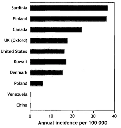
There is a marked geographic variation in incidence, with a child in Finland being about 400 times more likely than a child in Venezuela to acquire the disease (Gillespie, 2006). (Figure 1) Sardinia Finland Canada UK (Oxford) United States Kuwait Denmark Poland Venezuela China 0 10 20 30 40
Annual incidence per 100 000
Figure 1 Geographie variation in annual incidence oftype 1 diabetes.
The CUITent global increase in incidence of 3% per year is weIl reported. Furthermore, the incidence of type 1 diabetes will be 40% higher in 2010 than in 1998 (Onkamo et al., 1999). This rapid ri se strongly suggests that the action of the environment on susceptibility genes contributes to the evolving epidemiology of type 1 diabetes (Gillespie, 2006). People with type 1 diabetes depend on regular insulin injections with multiple self care tasks to achieve best blood glucose control.
1.3 Type 2 Diabetes Mellitus
It was formerly named non-insulin-dependent diabetes, which results from the
body's inability to respond properly to the action of insulin produced by the pancreas.
Type 2 diabetes is much more common than type 1 and approximately accounts for
90% of ail diabetes cases worldwide. It occurs most frequently in adults, but is being
noted increasingly in adolescents as weil (Likitmaskul et aL, 2003; Hotu et aL, 2004).
Most of type 2 diabetic patients are not diagnosed until the individual has had the
disease for many years, and the microvascular complications of diabetes (retinopathy,
nephropathy and neuropathy) are already present (Spijkerman et al., 2003). Lifestyle
modification is the cornerstone of both treatment and attempts to prevent type 2
diabetes (Mann et aL, 2004).
1.4 Global prevalence of diabetes
The prevalence of diabetes for aIJ age-groups worldwide was estimated to he
2.8% in 2000 and 4.4% in 2030. The number of people with diabetes is expected to
increase from 171 million in 2000 to 366 million in 2030. The proportion of diabetic
patients is increasing due to growth of population, aging, change in life style, and
increasing prevalence of obesity and physical inactivity (Woods et aL, 2004). The
morbidity and mortality associated with diabetes are related to the short- and
long-term complications. These complications include hypoglycemia and hyperglycemia,
1.5 Complications associated with Diabetes Mellitus 1.5.1 Microangiopathy
The microangiopathy is arising from small blood vessels disease which includes:
(a) Diabetic retinopathy
Diabetic retinopathy is one of the most common microvascular complications,
the most frequent cause of new cases of blindness in Europe and North America in
age group of 30 to 70-74 years (Villar et al., 1999; Stitt et al., 2002). Recent cl inical
studies have revealed that the presence of diabetic retinopathy is predictable of
diabetic nephropathy (Villar et al., 1999; EI-Asrar et al., 2001; Rossing et al., 2002).
Due to its high content of unsaturated lipids and high oxygen demand, the retina
represents a site which is partially prone to hyperglycemia induced free radical
generation and lipid oxidation. Furthermore, recent studies have shown that NF-kB
activation in retinal pericytes is responsible for hyperglycemia-induced loss of
pericytes observed in diabetic retinopathy. Loss of vision due to certain types of
glaucoma and cataract occurs primarily due to age, but is more common in diabetes.
Moreover, cataract is common in diabetic person where superoxide anion in the
mitochondria is elevated as a result of hyperglycemia (Vinson, 2006). Clinical trials
suggest that blood glucose control and control of hypertension can delay the onset and
progression of diabetic retinopathy and loss ofvision associated with diabetes (Grassi,
2003; Fong et al., 2004a; Fong et al., 2004b). Morover, loss of vision due to diabetic
retinopathy can be prevented by early detection and treatment of vision-threatening
retinopathy by regular eye examinations and timely intervention with laser treatment,
(b) Peripheral neuropathy
Neuropathy is an early clinical sign of diabetes affecting sensory and autonomic peripheral functions. Studies suggest that about 30-50% of people with type 2 diabetes are affected to sorne degree of neuropathy (Dickinson et al., 2002; Feldman, 2003). Moreover, diabetic neuropathies are the most common chronic disturbing complications of diabetes mellitus because of the pain, discomfort, and disability. Pain or numbness in the legs or feet may be the most common complaint from diabetic neuropathic patients (Park et al., 2004), who also display autonomic dysfunction (especially erectile dysfunction and altered cardiac vagal response). The pathogenesis of diabetic neuropathic pain is still unknown, yet several mechanisms were proposed. These include: (1) axonal degeneration/regeneration, (2) neuroma properties, which cause ectopic impulse generation and ephaptic transmission, (3) small-fiber diseases, which involve the AB and C-fibers, (4) dorsal root ganglion involvement, and (5) central sensitization and neural plasticity. The early detection and diagnosis of diabetic neuropathies are important to reverse and prevent their progression (Park et al., 2004). Diabetic patients who are inadequately treated have higher morbidity and complication rates related to neuropathy than patients whose blood glucose is closely controlled. Peripheral neuropathy represents the main etiologic factor involved in the development of diabetic foot ulceration and lower limb amputation.
c) Erectile dysfunction
Erectile dysfunction is commonly associated with diabetes and occurs at an
earlier age in such patients than in the general population. Hyperglycemia has
pathologic effect on peripheral tissue innervation and vascularization, both of which
are critical for erectile function. Oxidative stress to cavernous tissue may be an
important causative factor to erectile dysfunction in diabetic patients (Ryu et al.,
2005).
(d) Diabetic nephropathy
Nephropathy is an irreversible complication of diabetes representing 30% of
ail new cases of end-stage renal failure and the most distressing and
money-consuming complication in patients with diabetes throughout the world. The principal
lesion of diabetic nephropathy occurs in renal glomeruli and is called diabetic
glomerulosclerosis. Hyperglycemia is responsible for the development and
progression of diabetic nephropathy through different metabolic pathways, including
increased oxidative stress, renal polyol formation, activation of protein kinase C
(PKC)-mitogen-activated protein kinases (MAPKs), and accumulation of advanced
glycation end products, as weil as hemodynamic factors such as systemic
hypertension and increased intraglomerular pressure (Kikkawa, 2000).
(e) Diabetic cardiomyopathy
This prominent cardiovascular complication has been recognized as a
microvascular disease that may lead to heart failure. Pathogenesis of diabetic
cardiomyopathy involves vascular endothelial cell dysfunction, and cardiomyocyte
changes including increased non-enzymatic glycation, sorbitol-myoinositol-mediated
changes, redox potential alterations, and protein kinase C (PKC) activation, ail of
which have been implicated in diabetic cardiomyopathy (Farhangkhoee et al., 2006)
1.5.2 Macrovascular complications
The development of macrovascular complications, including cardiac,
cerebrovascular, and peripheral vascular complications, is an important concern
considering that a substantial proportion of premature deaths in patients with type 1
diabetes mellitus (Deckert et al., 1978), and most deaths in type 2 diabetes mellitus
are related to macrovascular disease (Morrish et al., 1990; Morrish et al., 1991).
(a) Hypertension
Patients with diabetes have a much higher rate of hypertension th an would be
expected in the general population. There is an increased prevalence of hypertension
among diabetic patients (Sowers et al., 2000). Population studies suggest that blood
pressure in excess of 140/90 mmHg is found in nearly 30% of adults havi ng type 2
diabetes. Moreover, both conditions are strongly age-dependent, and exhibit large
geographical variations. These two medical conditions tend to occur together in the
same patients; approximately 2/3 of diabetics will have hypertension while
hypertensive persons, have a substantially increased risk of diabetes. Elevated blood
pressure is known to contribute to diabetic microvascular and macrovascular
complications. Hypertension in patients with diabetes increases the risk of coronary
artery disease. In the general population, the prevalence of coronary artery disease lies
at around 1 % to 4 %, but this may increase by as much as fourfold in older adult
risk of heart failure has been shown to increase twofold for diabetic men and fivefold for diabetic women, relative to nondiabetic individuals (Krum, 2003). ln addition, more than one third of patients with myocardial infarction also suffer from clinically diagnosed type 2 diabetes (Norhammar et al., 2002). Hypertension increases the incidence of stroke in patients with diabetes. Survival rates and recovery from stroke are reduced in patients with diabetes compared with patients without diabetes; also hypertension increases the risk of peripheral vascular disease and subsequent foot ulcers and amputations in patients with diabetes. Hypertensive diabetic patients are also at increased risk for diabetes-specific complications including retinopathy and nephropathy. Heart disease accounts for approximately 50% of ail deaths among people with diabetes in industrialized countries. Diabetes mellitus induces abnormal changes in the structure of different components of the heart including the plasma membrane and other cytoplasmic organelles of cardiomyocyte. Pathological findings include cell hypertrophy, neuropathy, interstitial fibrosis, myocytolysis, apoptosis and Iipid deposits in the heart of diabetic patients (Adeghate, 2004).
(b) Peripheral vascular disease
The diabetic foot disease, due to changes in blood vessels and nerves, often leads to ulceration and subsequent limb amputation. It is one of the most costly complications of diabetes, especially in communities with inadequate footwear. lt results from both vascular and neurological disease processes. The impairment of microcirculation of diabetic patients leads to secondary complications in lower limbs, as foot infections and ulcerations. These microcirculatory changes, which are mainly functional rather than structural, are responsible for the impaired ability of the microvasculature to vasodilate in response to injury, and nerve reflex related
microvascular vasodilatation is also impaired in the diabetic population (Schramm et al., 2006). Diabetes is the most common cause of non-traumatic amputation of the
lower limb, which may be prevented by regular inspection and good care of the foot.
1.6 Diabetes Mellitus and oxidative stress 1.6.1 Source of oxidative stress in diabetes
Oxidative stress has been considered to be a common pathological factor of diabetes complications and appears a target for therapeutic treatments (Shih et al., 2002). Tissue exposure to hyperglycemia results in increased production of reactive oxygen species (ROS). Furthermore, ROS and reactive nitrogen species (RNS) are products of normal cellular metabolism, and recognized for playing a double role as both harmful and beneficial to living systems (Valko et al., 2006). Beneficiai effects of ROS occur at low/moderate concentrations and involve physiological roles in cellular responses to anoxia, in defense against infectious agents, in a number of cellular signaling systems, and induction of a mitogenic response. Oxidative stress is defined in general as excess formation and/or inadequate removal of highly reactive molecules such as ROS and RNS (Turko et al., 2001; Maritim et al., 2003). The sources for the overproduction of ROS in diabetes are widespread and include enzymatic pathways, auto-oxidation of glucose, and mitochondrial superoxide production. ROS include free radicals su ch as superoxide anion (02.), hydroxyl (OH), peroxyl (R02), hydroperoxyl (HR02-) and nonradical species such as hydrogen peroxide (H202) and hydrochlorous acid (HOCI) (Turko et al., 2001; (Evans et al.,
2002). RNS include free radicals like nitric oxide (NO) and nitrogen dioxide (N02-) as weil as nonradicals such as peroxynitrite (ONOO-), nitrous oxide (N20) and alkyl peroxynitrate (RONOO) (Turko et al., 2001; Evans et al., 2002). Reactive molecules,
superoxide anion, "NO and ONOO- are the main species which play important roles in
diabetic complications. In diabetes, the antioxidant defense is blunted whereas the
generating system is stimulated (Dickinson et al., 2002). Induction of ROS formation
can result from different additive mechanisms. These mechanisms include direct
intracellular effect of glucose in cells subjected to increase glucose uptake during
hyperglycemia (renal, retinal, and some nerves cells) and indirect via the extracellular
formation of advanced glycation end products (AGEs). Excess generation of oxidative
stress has pathological consequences including damage to proteins, Iipids and DNA.
'02- can activate several damaging pathways in diabetes including accelerated
formation of AGE, polyol pathway, hexosamine pathway and PKC, nicotinamide
adenine dinucleotide phosphate (NAD(P)H) (Kitada et al., 2003), mitochondrial
electron-transport chain (Brownlee, 2001), ail ofwhich were proven to be involved in
micro- and macrovascular complications. In addition, -02-and H202 stimu1ate
stress-related signalling mechanisms such as NF-KB, p38-MAPK and STAT-JAK resulting
in vascular smooth muscle cells migration and proliferation. In endothelial cells, H202
1 t Free Fatty Acids 1 1 1 Hyperglycemia 1 1
t
Mitochondrial ROS Macromolecule DamageJ
•
t
Oxidative Stress 1•
t
NF-KB 1t
p38 MAPK 1t
JNKJSAPK 1..
•
•
..
1 t Sorbitol 1 t AGE tOAG t Cytokines
J, J, t Prostanoids
-RAGE tPKC
~
Ilnsulin Resistance
1p
Cell Dysfunction
11
: Diabetic Complications:
1Figure 2 Relationship between oxidative stress and the development of type 2 diabetes (Evans et al., 2002).
Figure 2, shows the proposed causative link between hyperglycemia, elevated free fatty acid (FFA), mitochondrial ROS generation, oxidative stress, activation of stress-sensitive pathways (NF-kB, p38 MAPK, JNK/SAPK, and others), insulin resistance, B-cell dysfunction, and diabetic complications O\Jishikawa et al., 2000). The activation of NAD(P)H oxidase by protein kinase C produces the predominant source of reactive oxygen species in vasculature that directly lead to diabetic complications and cardiovascular disease ( Kitada et al.,2003; Feldman, 2003). ROS can stimulate oxidation of low-density lipoprotein (LDL), and oxidized-(ox)-LDL, which is not recognized by the LDL receptors. The ox-LDL which formed can be taken up by scavenger receptors in macrophages leading to foam cel! formation and atherosclerotic plaques as shown in Figure 3 (Taniyama et al., 2001).
LDL
ROS". +Hyperglycemia
OX-LDL
ROS" • +Free radicals
OX-LDL overloaded macrophages
Foam cell in arterial walls
Atherogenic plaques
Figure 3 Pathogenic cascade of hyperglycemia, oxidative stress, LDL oxidation, arteriogenic plaque formation and myocardial infarction rate.
Under normal conditions, .02- is immediately eliminated by natural defense
mechanisms, but in excess .02-reacts with "NO immediately and generates cytotoxic
ONOO- , which is a strong oxidant. This reaction has several consequences. First, ONOO- alters functions of biomolecules by protein nitration and Iipid peroxidation. Increase levels ofnitrotyrosine are associated with apoptosis ofmyocytes, endothelial cell and fibroblasts in diabetes (Turko et al., 2001). Second, ONOO- causes single-strand DNA breakage which in tum activates nuclear enzyme poly (ADP-ribose) polymerase (PARP) (Soriano et al., 2001). Third, it decreases "NO bioavailability causing impaired relaxation and inhibition of the antiproliferative effects of ·NO (Maritim et al., 2003). Furthermore, ONOU oxidizes tetrahydrobiopterin (BH4), an
2-instead of 'NO (Maritim et al., 2003). AlI these pathological modifications contribute to the pathogenesis ofvascular complications of diabetes.
1.7 Antioxidants and diabetes
1.7.1 Defense system against oxidative stress
Exposure to free radicals from different sources has led organism to develop a series of defense mechanisms (Cadenas, 1997). The defense mechanisms against free radical-induced oxidative stress involve: (1) preventive mechanisms, (2) repair mechanisms, (3) physical defenses, and (4) antioxidant defenses (Valko et al., 2006). Under normal physiological conditions, our body constantly produces ROS and RNS, which are eliminated by antioxidant enzymes as primary antioxidants. But when the production of ROS and RNS is significantly increased, the enzyme systems are rapidly overloaded. The oxidation may be slowed down by secondary antioxidants provided in the diet. Under normal conditions, there is a balance between the activities and the intracelIular levels of these antioxidants. This balance is essential for the survival of organisms and their health.
(a) Primary antioxidants: active detoxification
The celI has antioxidant enzymes, which are very effective defense systems because enzymes have the property to eliminate free radicals in a constant manner. This line of defense is composed of three major antioxidant enzymes, superoxide dismutase (SOD), glutathione peroxidase (GPX), and catalase, which differ from each other in structure, tissue distribution, and cofactor requirement. Their substrates are reactive species; they change superoxide anions and hydrogen peroxide into non-harmful products.
SOD: O2-
+
O2-+
2H+ -+ H202+
O2GPX: ROOH + 2GSH -+ ROH + H20
+
glutathioneSOD is the first line of defense against ROS by preventing changes in NF-kB,
polyol pathway, AGE formation and PKC activity. There are 3 different forms of
SOD: Mn-SOD, a tetramer molecule present in mitochondria, Cu/Zn-SOD, a dimer
molecule present in the plasma and a tetramer form in the cytosol. SOD works
immediately to convert the superoxide radical O2- to hydrogen peroxide (H20 2),
which is toxic for the cell as it is involved in the formation of hydroxyl radical. H202
is then detoxified to water either by catalase in the lysosomes or by glutathione
peroxidase in the mitochondria.
Catalase is a tetramer containing NAPH molecule which stabilizes the active
site and heme molecule necessary for enzymatic activity. Catalase reacts very rapidly,
without requiring any energy. Contrary to catalase, peroxidases need energy from the
cell and cofactors as ascorbate for ascorbate peroxidase and both glutathione and
selenium for selenium-dependent glutathione peroxidase (GPX). GPX is a tetramer
present in plasma and cytosol; it mediates the transformation of GSH to GSSH
(Bharath et al., 2002). GSH plays a critical role as a cellular antioxidant, reacting with
free radicals nonenzymatically, and also in the reduction of peroxides, catalyzed by
GPX.
Prolonged exposure to hyperglycemia increases the generation of free radicals
and reduces capacities ofthe antioxidant defense system. The pathogenesis of diabetic
complications is strongly related to cellular injury caused by intracellular alterations
in the metabolism of natural defense system against oxidative stress. Simple
lead to DNA cleavage (Kaneto et al., 1994). The most common antioxidant
deficiencies reported in diabetes are lower levels of ascorbate, glutathione and
superoxide dismutase, also the concentration of reduced glutathione has been seen in
diabetic neutrophils and monocytes (Venugopal et al., 2002). Aiso the activity and
expression of SOD and glutathione peroxidase are decreased in diabetic models
(Maritime et al., 2003).
b) Secondary antioxidants: passive detoxification
The passive detoxification is a second corn plementary line of defense
including compounds able to significantly slow down the etfects of free radicals that
have not been eliminated by the enzymatic defense systems. Non enzymatic systems
include vitamins A, C and E; glutathione; a-Iipoic acid; carotenoids; trace elements
Iike copper, zinc and selenium; coenzyme QIO (COQIO); and cofactors Iike folic acid,
uric acid, albumin, and vitamins B1, B2, B6 and B12 • These systems are located in cell
membrane, cystol, and plasma where they play specific functions. Numerous studies
demonstrated that antioxidant vitamins and supplements can help lower the markers
indicative of oxidative stress and lipid peroxidation in diabetic patients and animal
models (Mayne, 2003). A number of studies have reported vitamins C and E and
beta-carotene deficiency in diabetic patients and experimental diabetic animais (Penckofer
et al., 2002; Naziroglu and Butterworth, 2005). Vitamin E (tocopherols and
tocotrienols) is a lipophilic vitamin that prevents lipid peroxidation. It exists in 8
ditferent forms, of which alpha-tocopherol is the most active form in humans.
Alpha-tocopherol mainly eliminates Iipid peroxyl radicals while gama-Alpha-tocopherol is able to
scavenge peroxinitrites. Hydroxyl radical reacts with tocopherol forming a stabilized
dependent reductase enzymes (Hensley et al., 2000; Hensley et al., 2004). Vitamin C
is a hydrophilic molecule and is the strongest physiological antioxidant acting in the
organism's aqueous environment. It has been shown to be an important antioxidant, to
regenerate vitamin E through redox cycling, and to raise intracellular glutathione
levels (Zaidi and Banu, 2004). Thus vitamin C plays an important role in protein thiol
group protection against oxidation (Rahimi et al., 2005). In contrast to vitamin A, the
combination of vitamins C and E can also be safely used in high doses to prevent
diabetes and cardiovascular disease (Hatzigeorgiou et al., 2006).
COQIO is a lipid soluble antioxidant, endogenously synthesized compound that
acts as an electron carrier in the Complex 11 of the mitochondrial electron transport
chain. At higher concentrations, it scavenges '02' and improves endothelial
dysfunction in diabetes. Furthermore, it inhibits lipid peroxidation by either
scavenging free radicals directly or reducing tocopheroxyl radical to
alpha-tocopherol (Kagan et al., 1990; Emster and Dallner, 1995; Forsmark-Andree et al.,
1995; Lass and Sohal, 1998; Abusheikha et al., 1998). The concentration of CoQ
homologues in plasma, tissue homogenates and mitochondria can be increased by
dietary COQIO supplementation. CoQ supplementation can modulate the plasma
aminothiol redox status towards antioxidants and lower protein oxidative damage in
skeletal muscle mitochondria (Vahle et al., 2002). Furthermore, CoQ intake enhances
the antioxidative potential of tissues by elevating the endogenous amounts of alpha
-tocopherol (Kamzalov et al., 2003).
Glutathione, a water-soluble tripeptide (y-L-Glu-L-Cys-Gly) is the most
abundant intracellular nonprotein thiol compound in mammalian cells (Sies, 1999). It
pathways essential for the whole body homeostasis (Deneke and Fanburg, 1989; Kretzschmar, 1996). It occurs in reduced thiol (OSH) and oxidized disulfide forms (OSSO). OSH is linked to many physiologic processes including detoxification of xenobiotics, modulation of signal transduction, prostaglandin metabolism, regulation of immune response, and enzyme activities. Synthesis of glutathione depends on the intake of cysteine. Cysteine availability is known as the rate-Iimiting factor in OSH synthesis. Glutathione deficiency leads to increase oxidative stress and may therefore play a key role in the pathogenesis of many diseases. Low levels of glutathione are found in persons with arthritis, diabetes, and cardiac injuries (Julius et al., 1994) or in neurodegenerative pathologies including dementia (Jenner, 1994).
Alpha-Lipoic acid (LA), a dithiol compound derived from octanoic acid, is used as a pote nt antioxidant, and has special criteria making it a powerful antioxidant. These criteria include radical quenching, metal chelation (Packer et al., 1995), amphiphilic character, bioavailability and safety, interaction with other antioxidants, and metabolic regeneration (Packer et al., 1995). LA scavenges hydroxyl radicals, hypochlorous acid, peroxynitrite, and singlet oxygen. Dihydrolipoic acid also scavenges superoxide and peroxyl radicals and can regenerate thioredoxin, vitamin C, and glutathione, which in turn can recycle vitamin E. In addition to its antioxidant properties, LA increases glucose uptake through recruitment of the glucose transporter-4 to plasma membranes, a mechanism that is shared with insu lin-stimulated glucose uptake by activating elements of the insulin-signaling pathway. Furthermore, recent trials have demonstrated that treatment of insulin-resistant fatty Zucker rats with LA increased both oxidative and nonoxidative glucose metabolism and enhanced insu lin sensitivity (Jacob et al., 1996). In experimental and clinical studies, LA markedly reduced the symptoms of diabetic pathologies, including
cataract formation (Maitra et al., 1995), vascular damage (Hofmann et al., 1999), and improved neural blood flow, endoneural glucose uptake and metabolism and nerve conduction (Ruhnau et al., 1999; Smith et al., 2004). Treatment with a-lipoic acid has also been shown to prevent diabetic nephropathy (Siu et al., 2006).
1.8 N~acetyl-L-cysteine (NAC)
N-acetyl-L-cysteine (NAC) developed in the 1960s, is a sulfhydryl-containing compound, which is a stable derivative of the amino acid cysteine, has antioxidant properties and makes up part of the tripeptide glutathione. NAC is rapidly absorbed into various tissues following an oral dose, is deacetylated and metabolized in the intestine and liver, and incorporated into disulfide protein peptides. A peak plasma level of NAC occurs approximately one hour after an oral dose and at 12 hours post-dose it is undetectable in plasma (De et al., 1989).The biological activity of NAC is attributed to its sulfhydryl group while its acetyl substituted amino group affords its protection against oxidative and metabolic processes. NAC can be administered orally, intravenously and via respiratory nebulizer.
1.8.1 Mechanism of action of N AC as antioxidant and anti-inflammatory agent
NAC is rapidly metabolized to cysteine, which is a direct precursor in the synthesis of intracellular GSH. In this way, it acts as an antioxidant by restoring the pool of intracellular reduced GSH (Santangelo, 2003). NAC can also have reducing and antioxidant properties by acting as a direct scavenger of free radicals such as OH· and H202 and 02-· (Aruoma et al., 1989; Benrahmoune et al., 2000). Moreover, as a direct consequence of its antioxidant and SH-donating properties, NAC restores cellular redox-status and can in this way modulate the activity of redox-sensitive
cell-signaling and transcription pathways su ch as NF-KB which regulates a variety of
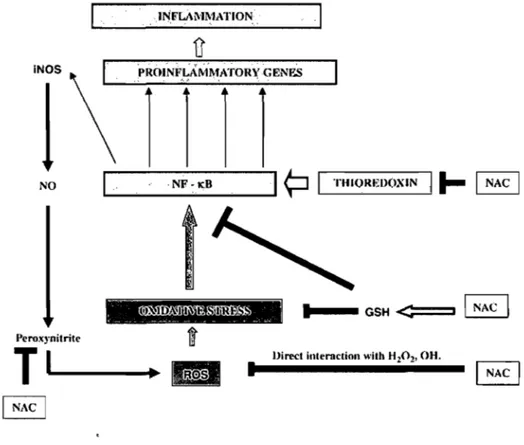
proinflammatory genes (Desaki et al., 2000), and the p38, ERK 1 /2, SAPKlJNK, c-Jun and c-Fos pathways (Zafarullah et al., 2003; Wuyts et al., 2003). The possible sites of action of NAC as antioxidant and anti-inflammatory agent on chronic obstructive pulmonary disease are shown in Figure 4 (Sadowska et al., 2007).
INFLAMMATION
.1
, ~Th~~ 1-1 _ _ GSH <:<:::=::::::11 1 NAC 1
..
-~- -~-~- -- -- _. ~ -~Peroxynitrite
T
1
_ _ _
-._
îlisi
1
lJirect interaction ",lib H202• OH.
Figure 4 Possible sites of action of N-acetyl-L-cysteine (NAC). NAC inhibits the oxidative stress by acting as direct ROS scavenger and by changing the cellular redox status. This, in tum, may influence NF-KB activation and modulate the inflammatory response (Sadowska et al., 2007).
1.8.2 Therapeutic uses of NAC
NAC has been in clinical practice since 1960s (Ziment, 1986; Flanagan, 1987; de and De, 1993; van, ] 995). Initially, NAC was introduced as a mucolytic agent for
the treatment of respiratory diseases such as chronic bronchitis and cystic fibrosis (Webb, 1962; Richardson and Phipps, 1978). It acts as an expectorant by stimulating both ciliary action and the gastro-pulmonary vagal retlex, thereby clearing the mucus from the airway (Sheffner, 1966). For this reason, NAC is used clinically in bronchopulmonary diseases to reduce both the viscosity and the tenacity of mucus, as weil as to facilitate its removal. In the late 1970s, NAC was recognized as an antidote for the therapy of acute acetaminophen intoxication (Prescott et al., 1977; Prescott et al., 1979). Recent studies have shown an effect of NAC in the prevention of atheromatous plaque formation, NAC inhibits the oxidation of LDL which accumulates in the vascular wall and promotes a local intlammatory process contributing to the progression of atheromatous plaque (Van et al., 2005). More recently NAC was found to prevent fructose-induced insulin resistance and hypertension in rats (Song et al., 2005). NAC also increased fat degradation and decreased body weight gain in conditions of excess sucrose intake (Diniz et al., 2006). Chronic treatment with NAC in Spontaneously Hypertensive Rats (SHR) decreased blood pressure by improving sympathetic functions and p-adrenergic pathway (Girouard et al., 2003). Furthermore, NAC exerts protective effect against glucose toxicity on pancreatic p-ceUs in various models of diabetes, reduces blood glucose and increases glucose-induced insulin secretion (Ho et al., 1999; Kaneto et al., 1999; Tanaka et al., 1999). NAC has been shown to be a strong antioxidant, to exert antigenotoxic and anticarcinogenic properties, and to detoxify free radicals that cause DNA changes in diseases (e.g., cancer). These effects of NAC have been attributed to its ability to act as an analogue of cysteine and precursor of reduced glutathione (GSH), to improve the activities of glutathione S-transferases, glutathione peroxidase, glutathione reductase, NADH- and NAD(P)H-quinone reductase, and probably, to
promote DNA repair by protecting ADP-ribosyltransferase activity (Albano et al., 1984; De et al., 1984; De et al., 1984; De et al., 1985; De et al., 1985; De et al., 1986; Perchellet et al., 1987; Dorsch et al., 1987; De and Ramel, 1988; Cesarone et al.,
1988).
1.9 Angiotensin-converting enzyme (ACE) inhibitor
Angiotensin Tl (Ang Il), is a potent vasoconstrictor that is involved in the regulation of vascular functions such as cell growth, apoptosis, migration, inflammation, and fibrosis (Touyz and Schiffrin, 2000; Wolf and Wenzel, 2004). Ang Il is an important growth modulator of blood vessels and renal organogenesis during development. It plays a critical role in regulating blood pressure and fluid homeostasis in physiological conditions. Ang 11 contributes to altered vascular tone, endothelial function, structural remodeling and to vascular inflammation, characteristic features of vascular damage in hypertension, atherosclerosis, vasculitis, and diabetes (Griendling and Ushio-Fukai, 2000; Touyz and Schiffrin, 2000). Angiotensin-l converting enzyme (ACE) inhibitors are widely accepted as vascular protective agents. They are frequently used as the first line of treatment of hypertension in type 2 diabetic patients, and have been shown to improve insulin sensitivity and to reduce cardiovascular complications in diabetes (Henriksen et al., 1996; McFarlane et al., 2003). The mechanism by which ACE inhibition improves insulin sensitivity and reduces the development of type 2 diabetes in patients with essential hypertension is not yet completely elucidated and goes beyond the blockade of the renin-angiotensin system (Couture and Girolami, 2004). Treatment with ACEI attenuated ROS formation and prevented phenotypic changes in the heart (cardiomyocyte hypertrophy, perivascular fibrosis) and in the aorta of diabetic rats (Fiordaliso et al.,
2006). ACE is a major link between the renin-angiotensin system and the kinin system, because it not only con verts Ang 1 to Ang II but also degrades kinins.
1.9.1 Ramipril
Ramipril is an ACE inhibitor. As inactive prodrug, ramipril is converted to ramiprilat in the liver by esterase enzymes, and is used to treat hypertension and heart failure, to reduce proteinuria and renal disease in patients with nephropathies, and to prevent stroke, myocardial infarction, and cardiac death in high-risk patients. Ramiprilat is mostly excreted by the kidneys. The half-Iife of ramiprilat is variable (3 - 16 hours), and is prolonged by heart and liver fai 1 ure, as weil as kidney failure. Ramiprilat, the active metabolite, competes with angiotensin 1 for binding at the angiotensin-converting enzyme, blocking the conversion of angiotensin 1 to angiotensin II. As angiotensin Il is a vasoconstrictor and a negative-feedback mediator for renin activity, lower concentrations result in a decrease in blood pressure and an increase in plasma renin. Ramiprilat may also act on kininase II, an enzyme identical to ACE that degrades the vasodilator bradykinin. Recent studies have shown that ramipril, reduced the accumulation of advanced glycation end products in experimental diabetic nephropathy (Forbes et al., 2002).
1.10 Diabetic neuropathic pain
Functional and structural impairments of peripheral nervous system in diabetic individual are generally defined as diabetic neuropathy, which is a common complication of both type 1 and type 2 diabetes (Boulton et al., 2005). Periphera1 neuropathy affects about 30% of people with diabetes mellitus. Between 16% and 26% of diabetes patients experience chronic pain. This may be referred to as diabetic
neuropathic pain (ONP) or diabetic peripheral neuropathic pain (DPNP) (Jensen et al., 2006). Most commonly, it manifests as sensory loss, which predisposes to foot abnormalities and high risk of ulceration. It has been reported that between 4 and 33% of patient with diabetes mellitus suffer from the painful type of neuropathy, which is recognized as one of the most difficult type of pain to treat (Ziegler et al., 1992; Oaousi et al., 2004). Oiabetic neuropathic pain can occur either spontaneously or as a result of exposure to mild painful stimuli (hyperalgesia) or to abnormal non painful stimuli (allodynia) (Brown and Asbury, 1984; Clark, Jr. and Lee, 1995). Diabetic neuropathy, during its natural course progresses from initial functional changes to late poorly reversible structural changes. Various interconnected pathogenetic concepts of diabetic neuropathy have been proposed based on metabolic and vascular factors, mostly derived from long-term hyperglycemia. The etiology of diabetic neuropathy is multifactorial, long term hyperglycemia, increased oxidative stress and altered protein kinase C activity and poly AOP-ribose polymerase (PARP) activation, all these lead to development of neuropathy (Yagihashi, 1995; Sima and Sugimoto, 1999; Yagihashi, 2006; Yagihashi et al., 2007) (Fig 5).
1
Hyperglycemia
lPolyol Pathway
AGE
(AR-Sorbitol) ,H Glycation NOl & Glutothione , ,H Protein change
Free rodîcals t
...
/I~
Oxidative
r-C-yt-o-ki-n-es~~
stress
Inflammationf NO, Growth factor 1 PARPf •• -..
..
....
.•...
..-.
• ,. •
....
• •••• ;e
.
..
.
... . ..
..
. -.
,
..
-
....
~.......
.-..
..
-..-
.,
,.
....
~. . "
l'.
._ • .
~~Qj
...
::.,~:
... : ..
~Obility,r.l ~~~;,r 1Peripheral Neuropathy
Figure 5 Multifactorial etiology of diabetic neuropathy. Hyperglycemia increases polyol pathway, AGE formation, oxidative stress and cytokine release. These factors interact or operate independently in diabetic neuropathy. They can affect nerve tissues either directly or indirectly through nutrient vascular tissues (Yagihashi et al., 2007).
These pathogenic mechanisms have been targeted in several experimental and
clinical trials (Yagihashi et al., 2007). The experimental rat model of chronic glucose
feeding presents aIl the hall marks of type 2 diabetes such as the increases of plasma
levels of glucose and insulin, arterial hypertension, insulin resistance and increases in
the production of superoxide anion (marker of the oxidative stress) in the heart and
aorta (Midaoui and de Champlain, 2002; Midaoui and de Champlain, 2005). Further
studies have shown sensory abnormalities, namely tactile and cold allodynia, after 4
weeks of treatment with glucose. However, glucose-fed rats did not exhibit thermal
hyperalgesia up to 20 weeks of treatment. Thermal hyperalgesia is mediated by
myelinated Ab and unmyelinated primary afferent neurons, which include both
peptidergic and non peptidergic C-fibers. Foot withdrawal responses evoked by 10w
Kobal, 1993; Yeomans and Proudfit, 1996). Whereas C-fi bers are thought to be
involved in the transmission of warm sensations, Ao-fibers are stimulated by cold
stimulus (Pierau and Wurster, 1981). On the contrary, tactile aBodynia is a central
phenomenon mediated by large myelinated A~-tibers (Woolf et al., 1992; Shortland et
al., 1997; Pitcher and Henry, 2000). Thus, sensory neuropathy measured in
glucose-fed rats may affect primarily sensory A~ and Ao tibers and less Iikely polymodal
C-tibers.
A recent study showed an impairment of cutaneous endothelium-related
vasodilation and C-tiber-mediated vasoconstriction in peripheral diabetic neuropathy.
Peripheral nerve perfusion is reduced by diabetes and this makes an important
contribution to neuropathy in patient and animal models (Tuck et al., 1984; Cameron
et al., 2001; Quattrini et al., 2007). Treatment of diabetic neuropathic pain is based on
four cornerstones: (a) causal treatment aimed at (near)-normoglycemia, (b) treatment
based on pathogenetic mechanisms, (c) symptomatic treatment, and (d) avoidance of
risk factors and complications (Ziegler, 2006). A c1inical study in diabetic
polyneuropathic patients has shown that the oral treatment with alpha-lipolic acid for
tive weeks improves the neuropathic symptoms and deticits (Ziegler et al., 2006).
Inhibition of xanthine oxidase (an important source of ROS that contributes to
neurovascular dysfunction in experimental diabetes) could be a potential therapeutic
approach to diabetic neuropathy and vasculopathy (lnkster et al., 2007). Systemic
injection of the ROS scavenger phenyl-N-tert-butylnitrone (PBN) relieved mechanical
allodynia in a model of neuropathic pain (Kim et al., 2004). Intraperitoneally
administration ofNAC resulted in signiticant reduction of hyperalgesia after chronic
1.11 Insulin signaling and action
Insu 1 in is the most potent ana bol ic hormone and is essential for normal tissue
development, growth, and maintenance of body glucose homeostasis. It is secreted by
the pancreatic B cells of islets of Langerhans in response to increased circulating
levels of glucose and amino acids after a meal. Insulin regulates glucose homeostasis
at different organ sites, reducing hepatic glucose output (via decreased
gluconeogenesis and glucogenolysis) and increasing the rate of glucose uptake,
primarily into striated muscle and adipose tissue (Goal stone and Draznin, 1997).
Gluconeogenesis represents the generation of glucose [rom non-sugar carbon
substrates and occurs during periods of fasting, starvation or intense exercise.
Glycogenolysis refers to enzymatic breakdown or catabolism of the polysaccharide
glycogen into molecules of glucose and molecules of glucose I-phosphate.
Insulin signaling at the target tissue results in a large array of biological
functions. It is essential for normal growth and development and for normal
metabolism of glucose, fat and protein. The insu lin pathway is critical for the
regulation of intracellular and blood glucose levels and the avoidance of diabetes.
Moreover, studying the signaling pathways involved in insulin action increases our
understanding of the pathophysiology of insulin resistance related to type 2 diabetes
and can help to identify the key molecules for the development of more effective
therapeutic.
1.11.1 Insulin receptor
The insu lin receptor (IR) belongs to a family of transmembrane receptors with
intrinsic tyrosine kinase activity (Schlessinger, 1993). 1t is an heterotetrameric protein
COOH
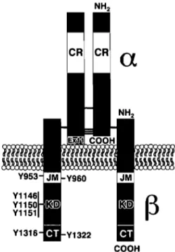
Figure 6 Structure of the insu lin receptor. CR, Cysteine-rich domain; JM, juxtamembrane domain; KD, kinase domain; CT, carboxyl-terminal domain. The positions of the tyrosine autophosphorylation sites are indicated. The left side depicts IR-B, which includes l2-amino acids alternatively spliced exon Il (ExIl) at the carboxyl terminus of the
a
subunit. The right side depicts IRA. The extracellular -and intracellular B-subunits are indicated. The horizontal black bars represent disulfide linkages (De Meyts and Whittaker, 2002).Binding of insulin to a subunits induces a conformational change resulting in the autophosphorylation of a number of tyrosine residues present in ~ subunits (Van, Baron et al., 2001). These residues are recognized by phosphotyrosine-binding (PTB) domains of adaptor proteins such as members of the insulin receptor substrate family (lRS) (Lizcano and Alessi, 2002). Receptor activation leads to the phosphorylation of key tyrosine residues on IRS proteins, sorne of which are recognized by the Src homology 2 (SH2) domain of the p85 regulatory subunit of PI 3-kinase (a lipid
kinase). The binding of insu lin to the a subunit of IR not only concentrates insulin at its site of action, but also induces conforrnational changes in the receptor, which in turn stimulates the tyrosine kinase activity intrinsic to the p subunit of the IR and triggers the signaling cascades (Fig 7). Insulin receptors trans phosphorylate several immediate substrates (on Tyr residues) including IRS 1 - 4, Shc, and Gab 1, Cbl, APS, and P60dok. Each of these provides specifie docking sites for other signaling
proteins containing Src homology 2 (SH2) domains (White and Yenush, 1998). These events lead to the activation of downstream signaling molecules including PI-3 kinase.
The four IRS proteins are highly homologous with overlapping and differential tissue distribution. Studies with genetic deletion in mouse models and cell lines indicate that the IRS proteins serve complimentary functions in different tissues as immediate substrates for insulin and IGF-I receptors. Combined heterozygous deletions of insulin receptor, IRS-l, and IRS-2 in different tissues develop severe insulin resistance in skeletal muscle and liver and marked p-cell hyperplasia. A recent study suggested tissue-specifie differences in the roles of IRS proteins to mediate insulin action, with IRS-I playing a prominent role in skeletal muscle and IRS-2 in liver (Kido et al., 2000). Aiso rRS-2 promotes p cell replication, function, and survival, especially during metabolic stress (Park et al., 2006). Furtherrnore, recent studies showed that IRS-3 and IRS-4 impair IGF -l-mediated IRS-I and IRS-2 signal ing in cells (Tsuruzoe et al., 2001).
Activation of the insu lin receptor evokes increased transcription of SREBP and the phosphorylation of members of the IRS family, SHC and Cbl. Upon tyrosine phosphorylation, these proteins interact with signaling molecules through their SH2
domains, which results in the activation of a variety of signaling pathways, including Pl 3-kinase signaling, MAPK activation and the activation of the Cbl/CAP complex. These pathways act in a coordinated manner to regulate glucose, lipid and protein metabolism.
•
•
•
[PTPtu
SOSIRas/
•
MfK MAP kinase Gene expression growth regulat/on • InsuUn Receptor IRS1/213/41 ----.
PI-3 K+·tpriN]
1
_S,~/!'2 }DK,\- -
--.
aPKC Akt•
/
l
l
-...
- -
--.
GSK3 p70S6k PP1 Signai Transduction 11
1
•
t
i
Glycogen SY1thase<€~co~
Glucose Transporter (GLUT-4)~
Oxldatlve Glucose Metabollsm Glucose Utlllzatfon Glycogen/llpldlproteln syntheslsFigure 7. Activation of the insulin receptor evokes increased transcription of SREBP and the phosphorylation of members of the IRS family, SHC and Cbl, Gab l, SIRPS, and adaptor protein containing PH and SH2 domains APS. This results in the activation of a variety of signaling pathways, including PI 3-kinase signaling, MAPK activation and the activation of the Cbl/CAP. Taken from
1.11.2 Glucose transporters
Glucose is cleared from the bloodstream by transporters (GLUTs) which
include a family of highly related 12 transmembrane domain-containing proteins
(Joost and Thorens, 2001). The GLUT family contains 13 known members and can be
classified into three classes based upon their structural characteristics (Joost et aL,
2002). Class 1 includes GLUTs 1-4 which are, by far, the best characterized
transporters of the family. Class II includes GLUT5 (a fructose-specific transporter),
and GLUTs 7, 9, and Il (Joost and Thorens, 2001). Class III includes GLUTs 8, 10,
12, and the proton-myoinositol symporter H+-myo-inositol cotransporter (HMITI)
(Joost and Thorens, 2001). GLUT4 is expressed primarily in striated muscle and
adipose tissue and, unlike most other GLUT isoforrns, is sequestered in specialized
intracellular membrane compartments under basal conditions (Bryant et al., 2002).
GLUT4 is the only known insulin-responsive GLUT which is highly and specifically
expressed in muscle and adipose tissue, the major sites of postprandial glucose
disposaI. Interestingly, overexpression of the human GLUT4 gene in muscle and fat
tissue of the diabetic db/db mou se, which lacks the leptin receptor, protects these