Response to Copper Stress in Streptomyces lividans Extends
beyond Genes under Direct Control of a Copper-sensitive
Operon Repressor Protein (CsoR)
*
□SReceived for publication, February 13, 2012, and in revised form, March 17, 2012 Published, JBC Papers in Press, March 26, 2012, DOI 10.1074/jbc.M112.352740 Srivatsa Dwarakanath‡, Amanda K. Chaplin‡, Michael A. Hough‡, Se´bastien Rigali§1, Erik Vijgenboom¶, and Jonathan A. R. Worrall‡2
From the‡School of Biological Sciences, University of Essex, Wivenhoe Park, Colchester, CO4 3SQ, United Kingdom, the§Centre for Protein Engineering, Institut de Chimie B6a, University of Lie`ge, B-4000 Lie`ge, Belgium, and¶Molecular Biotechnology, Leiden Institute of Chemistry, Gorlaeus Laboratories, Leiden University, P. O. Box 9502, 2300RA Leiden, The Netherlands
Background:Balancing copper homeostasis and bioavailability is critical for morphological development in Streptomyces lividans.
Results:In Streptomyces lividans, a copper-sensitive operon repressor (CsoR) protein regulates a three-locus copper homeo-stasis regulon.
Conclusion:Genes regulating copper homeostasis are more extensive than the regulon under direct control of CsoR. Significance:CsoR is part of a regulatory network controlling copper homeostasis that extends beyond the CsoR regulon.
A copper-sensitive operon repressor protein (CsoR) has been identified in Streptomyces lividans (CsoRSl) and found to regu-late copper homeostasis with attomolar affinity for Cu(I). Solu-tion studies reveal apo- and CuI-CsoRSlto be a tetramer
assem-bly, and a 1.7-A˚ resolution crystal structure of apo-CsoRSl reveals that a significant conformational change is necessary to enable Cu(I) binding. In silico prediction of the CsoR regulon was confirmed in vitro (EMSA) and in vivo (RNA-seq), which highlighted that next to the csoR gene itself, the regulon consists of two Cu(I) efflux systems involving a CopZ-like copper metal-lochaperone protein and a CopA P1-type ATPase. Although deletion of csoR has only minor effects on S. lividans develop-ment when grown under high copper concentrations, mutations of the Cu(I) ligands decrease tolerance to copper as a result of the Cu(I)-CsoR mutants failing to disengage from the DNA tar-gets, thus inhibiting the derepression of the regulon. RNA-seq experiments carried out on samples incubated with exogenous copper and a⌬csoR strain showed that the set of genes respond-ing to copper stress is much wider than anticipated and largely extends beyond genes targeted by CsoR. This suggests more control levels are operating and directing other regulons in cop-per homeostasis beside the CsoR regulon.
For many bacterial organisms, maintaining cellular metal ion homeostasis is an essential requirement for viability.
Sophisti-cated cellular machinery has evolved and consists of an exten-sive network of specialized proteins and transporters that respond to either metal ion deprivation or overload (1–3). Cop-per is an essential transition metal ion in biology and is required by many proteins and enzymes that function in electron-trans-fer chemistry and in the catalytic oxidation of substrates. The ability of copper to donate or accept single electrons is also detrimental as “free” cellular copper ions are complicit in a Fenton-like reaction, catalyzing the production of damaging hydroxyl radicals (4). This deleterious behavior leads to bacte-ria avoiding copper toxicity through buffering copper in the cytosol very tightly (2, 3, 5). Once the buffering capacity for cytosolic copper is exceeded, this is sensed by transcriptional regulatory proteins termed copper sensors that trigger the expression of genes encoding for efflux systems such as copper-exporting P1-type ATPases and copper metallochaperones that act in tandem to return the cytosol to a buffered copper state (3, 6, 7).
The filamentous Gram-positive soil-dwelling bacterium
Streptomycesis important to both ecological and human wel-fare and is also one of the model systems for bacterial morpho-logical and physiomorpho-logical development. The morphomorpho-logical differentiation of Streptomyces has three characteristic devel-opment stages as follows: 1) formation of branched vegetative mycelium, 2) formation of aerial hyphae, and 3) the production of spores. Concomitant or just before the morphological switch from vegetative to aerial mycelium, the metabolic development is initiated by the production of secondary metabolites. These compounds comprise a large percentage of the clinically useful antibiotics, anti-fungals, and even some of the anti-tumor agents used to date. The bioavailability of copper ions has been shown to be of crucial importance for morphological develop-ment in certain streptomycetes (8, 9). Copper dependence is restricted to the reproductive growth phase (aerial mycelium and spores), whereas vegetative growth proceeds under strongly copper -limiting conditions. Evidence from genetic *This work was supported by the University of Essex (to S. D., A. K. C., M. A. H.,
and J. A. R. W.), The Biochemical Society (to A. K. C.), and Leiden University and NWO (to E. V.).
□S
This article contains“Materials and Methods,” supplemental Figs. S1–S9, and Tables S1–S6.
The atomic coordinates and structure factors (code4adz) have been deposited in the Protein Data Bank, Research Collaboratory for Structural Bioinformatics, Rutgers University, New Brunswick, NJ (http://www.rcsb.org/).
1Research associate of the Fonds de la Recherche Scientifique-Fonds National de la Recherche Scientifique.
2To whom correspondence should be addressed. Tel.: 44-1206-872095; E-mail: jworrall@essex.ac.uk.
THE JOURNAL OF BIOLOGICAL CHEMISTRY VOL. 287, NO. 21, pp. 17833–17847, May 18, 2012 © 2012 by The American Society for Biochemistry and Molecular Biology, Inc. Published in the U.S.A.
at UNIV OF ESSEX, on May 21, 2012
www.jbc.org
Downloaded from
knock-out studies has suggested that secreted cuproproteins or cuproenzymes are needed for the development switch from vegetative to aerial mycelium to occur, but their identification and role in the development switch have not so far been eluci-dated (10, 11).
The copper proteome of Streptomyces coelicolor has been determined using bioinformatics approaches, revealing a rich assortment of putative extracellular cuproenzymes, redox pro-teins, and copper metallochaperone-like proteins (10, 12). An exciting yet unexpected finding was the identification of a cuproenzyme along with a dedicated copper metallochaperone in the cytosol (10, 13). A metabolic requirement for copper in the bacterial cytosol has not yet been documented; however, in
vitrobiochemical evidence indicates that this cytosolic tyrosi-nase-like enzyme (MelD2) is active in the oxidation of mono-and di-phenols (13). The cytosol of Streptomyces coelicolor also contains proteins putatively involved in copper detoxification systems (10). Two operons have been identified that contain genes encoding for a CopZ-like copper metallochaperone and a CopA-like P1-type ATPase transporter. In certain bacteria, the copAand copZ resistance genes are transcriptionally regulated by a copper sensor protein belonging to the copper-sensitive operon regulator (CsoR)3family (7). In S. coelicolor, the gene SCO4136has been proposed to encode for a CsoR orthologue (10). However, the genomic environment of SCO4136 does not contain any known copper resistance genes and instead is located among genes encoding for proteins involved in phos-phate transport (10). Therefore, if the gene product of SCO4136 functions as the cytosolic copper sensor in S. coelicolor, it is not genetically linked to copper resistance genes such as the two putative copZA-like operons.
The founding member of the CsoR family was discovered in
Mycobacterium tuberculosis (CsoRMtb) (7). Under elevated
copper levels, the expression of CsoRMtb was found to be
strongly induced, with the binding of Cu(I) triggering the dere-pression of the rv0967–rv0970 operon in which rv0969 encodes for a P1-type ATPase involved in copper transport and rv0967
for CsoRMtb(7, 14). It has subsequently been found that
mem-bers of the CsoR family are widespread in bacterial genomes. Examples of CsoR proteins that transcriptionally regulate
copZA copper resistance operons have been identified and
characterized to varying degrees in Bacillus subtilis (15),
Liste-ria monocytogenes, and Staphylococcus aureus (16, 17). Fur-thermore, a recent report has identified a CsoR member that is not thought to be involved in copper homeostasis but instead appears to function in response to sulfur stress (17).
To understand the role copper plays in morphological differ-entiation and development, it is necessary to understand how cellular copper is handled under normal and elevated condi-tions. S. coelicolor and Streptomyces lividans both display a dis-tinct dependence on copper for development with that of S.
lividansbeing more pronounced (18). They also share a high level of sequence identity and genome organization (19, 20), and the gene numbering of the S. coelicolor genome database is used for S. lividans in this study. Sequence alignment of the S.
lividans 4136 gene product with known CsoR orthologues reveals conservation of the amino acids that act as Cu(I)-bind-ing ligands and two amino acids considered important for the Cu(I)-dependent allosteric regulation of DNA binding (Fig. 1). In this study, we have biochemically and structurally character-ized the S. lividans 4136 gene product and find it to be a tetra-meric CuI-CsoR (CsoRSl). In vivo studies with a genetic
knock-out (⌬csoR) and CsoRSlmutants indicate CsoRSlto be essential
for the response of S. lividans to elevated copper levels. Further-more, we have coupled bioinformatics and RNA-seq experi-ments to characterize the regulon under the direct control of CsoRSl. RNA-seq data suggest that the regulation of copper
homeostasis is much more extensive than only the genes repressed by CsoRSl.
MATERIALS AND METHODS
Streptomyces Strains, Media, and Growth Conditions—The
Streptomyces strains used are as follows: S. lividans 1326 (S.
lividans66, stock number 1326 John Innes Collection), S.
livi-dans ⌬csoR (this study), and S. coelicolor A3(2) strain M512
(21). The agar media soy flower mannitol, complex medium (R5), minimal agar medium (MM), the liquid complex medium tryptic soy broth with 10% sucrose, and the liquid defined medium NMMP were prepared according to Ref. 22. If required, glucose and/or mannitol were added to 0.5%. Agar plates were incubated at 30 °C, and liquid cultures were grown in 250-ml baffled flasks with 0.2-m vent caps (Corning Glass) with shaking at 160 rpm. Liquid cultures were inoculated with spores to a final concentration of 2⫻ 107spores/ml. Spore stocks were obtained from cultures grown on soy flower man-nitol plates and stored in 20% glycerol at⫺20 °C. Growth was 3The abbreviations used are: CsoR, copper-sensitive operon repressor;
AAS, atomic absorption spectroscopy; MM, minimal agar medium; BCS, bathocuproine disulfonate; BCA, bicinchoninic acid; AUC, analytical ultracentrifugation.
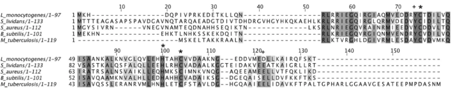
FIGURE 1. ClustalW2 multiple amino acid sequence alignment of copper-sensing CsoR proteins for which biochemical or structural data have been reported. Completely and partially conserved residues are boxed in dark and light gray, respectively. The conserved Cu(I)-binding ligands are indicated (*) along with residues reported to play a role in the Cu(I)-binding allosteric regulation of DNA (⫹). The UniprotKB accession numbers are Q8Y646 (L. monocyto-genes), A6QIT1 (S. aureus), O32222 (B. subtilis), and P71543 (M. tuberculosis).
at UNIV OF ESSEX, on May 21, 2012
www.jbc.org
recorded by determining the dry biomass from 1.5-ml samples collected in pre-dried Eppendorf tubes, and mycelium was pel-leted by centrifugation at 13,000 rpm for 10 min. After 12–16 h at 105 °C, the dry weight was determined using an analytical balance.
Generation of a CsoR Deletion Mutant (⌬csoR) of S. lividans 1326—The 4136 gene encoding for CsoRSlwas deleted in S. lividans1326 in a two-step process using the CRE-lox system (23). First, the gene (nt⫹4 to 399) was replaced by homologous recombination with an apramycin resistance cassette flanked by loxP sites. For this purpose, the upstream flanking region of
SL4136 (⫺1370 to ⫹3) and the downstream flanking region (⫹399 to 1680) were amplified from genomic DNA by PCR, including EcoRI, XbaI, XbaI, and HindIII sites, respectively, for cloning purposes. These two fragments and the apramycin resistance cassette flanked by loxP sites were cloned in the delivery vector pWHM3 that is unstable in Streptomyces (24). Following protoplast transformation, recombinants that were apramycin-resistant but had lost the vector (thiostrepton resistance) were isolated. Second, an unstable plasmid encod-ing the Cre recombinase was introduced (23) and allowed for the excision of the apramycin resistance cassette on the lox sites. The resulting strain,⌬csoR, has no coding sequence for CsoRSland has only a 61-nt “scar,” including two XbaI sites left
in the genome. The⌬csoR strain was analyzed by PCR to con-firm the loss of 4136, the apramycin resistance cassette, and vector sequences.
RNA Isolation and Transcriptome Analysis by RNA-seq —To-tal RNA was isolated with Kirby mix according to standard procedures from mycelium in early log phase grown on NMMP supplemented with 0.5% glucose and mannitol (22). Cultures were induced with 400MCu(II) for 2 h followed by total RNA
isolation. RNA integrity was confirmed by agarose gel electro-phoresis, and the absence of genomic DNA was checked by PCR. For the removal of ribosomal RNA, an Ambion kit was used. Samples were sent to BaseClear, an independent and accredited service laboratory for DNA-based research, and transcriptome analysis by RNA-seq was carried out. The sequences obtained on an Illumina sequencer were filtered for noncoding RNAs and analyzed with CLCbio bioinformatics software packages using the annotated S. coelicolor genome as reference. Expression values were expressed as reads/kb of exon model/million mapped reads (25), i.e. dividing the total number of exon reads (in this case one exon per reference sequence) by the number of mapped reads (in Millions) times the exon length (in this case the length of the reference sequence).
Promoter Probing—The DNA sequence (⫺300 to ⫹3) upstream of SL4136 was obtained from genomic DNA by PCR introducing a flanking EcoRI and BamHI site to facilitate clon-ing in pIJ2585 digested with the same enzymes (26). The result-ing plasmid, p4136-I, was introduced in strain M512 by proto-plast transformation. cultures were grown in triplicate in NMMP medium supplemented with 0.5% glucose, 0.5% man-nitol, and 50g/ml apramycin. Strain M512 transformed with the empty vector was grown under the same conditions to obtain background readings. Following extraction of mycelium with methanol, the concentration of undecylprodigiosin was
quantified from the absorbance at 530 nm and the extinction coefficient of 100,500 liters/mol⫺1cm⫺1(27).
Determination of Tyrosinase and Cytochrome c Oxidase Activity—The wild type, S. lividans 1326, and the mutant strain ⌬csoR were each transformed with pIJ703 (plasmid harboring the Streptomyces antibioticus melC operon under control of its own promoter). Four independent transformants of each strain were grown in liquid tryptic soy broth with 10% sucrose medium supplemented with 25MCu(II). Tyrosinase activity
was determined in spent medium with 10 mM3,4-dihydroxy-L
-phenylalanine in 100 mM phosphate buffer, pH 6.8, as sub-strate (28). Cytochrome c oxidase activity was visualized with N,N,N⬘,N⬘-tetramethyl-p-phenylenediamine as substrate, essentially according to Refs. 29, 30.
Cloning, Overexpression, and Purification of CsoRSl from
Escherichia coli—The 4136 gene was cloned from S. lividans 1326 genomic DNA (supplemental “Materials and Methods”) and overexpressed in Escherichia coli using a pET28a (Kanr)
vector (Novagen). This construct, designated pET4136, was transformed to E. coli BL21(DE3) cells, and single colonies were transferred to 2⫻ YT medium (Melford) with kanamycin (50 g/liter) (Melford) at 37 °C. Overexpression of the N-terminal His-tagged CsoRSlwas induced by 1
Misopropyl-D
-1-thioga-lactopyranoside (Melford) to a final concentration of 1 mM, and
the temperature was decreased to 25 °C for overnight incuba-tion. Cultures were harvested by centrifugation at 4000 rpm for 20 min at 4 °C, and the cell pellet was resuspended in 50 mM
Tris/HCl, 500 mMNaCl (Fisher) and 20 mMimidazole (Sigma)
at pH 7.5 (Buffer A). The resuspended cell suspension was lysed using an EmulsiFlex-C5 cell disrupter (Avestin) followed by centrifugation at 18,000 rpm for 20 min at 4 °C. The clarified supernatant was loaded onto a 5-ml nickel-nitrilotriacetic acid-Sepharose column (GE Healthcare) equilibrated with Buffer A and eluted by a linear imidazole gradient using Buffer B (Buffer A with 500 mMimidazole). A single peak at⬃30% Buffer B was eluted from the column, and fractions were pooled and dialyzed overnight at 4 °C against 10 mMMES, pH 6.5, 150 mMNaCl, 2
mMdithiothreitol (DTT) (Melford), and 4 mMEDTA (Sigma)
(Buffer C). Following dialysis, the N-terminal His tag was removed by incubating the protein at room temperature over-night with 125 units of thrombin (Sigma). The protein/throm-bin mixture was reapplied to the nickel-nitrilotriacetic acid-Sepharose column (GE Healthcare), and the flow-through was collected and concentrated using a Centricon (VivaSpin) with a 5-kDa cutoff at 4 °C for application to a G-75 Sephadex column (GE Healthcare) equilibrated with Buffer C. Fractions eluting from the major peak of the G-75 column were analyzed by SDS-PAGE, and those deemed of good purity were concentrated and stored at⫺20 °C until required.
Site-directed Mutagenesis of CsoRSIand Cloning for in Vivo
Studies—The QuikChange site-directed mutagenesis method (Stratagene) was used to create the C75A and H100A mutants of CsoRSl. Forward and reverse primers were designed with the
respective nucleotide change(s) to create the desired mutation (supplemental “Materials and Methods”), and the pET4136 plasmid was used as template. The respective mutations were confirmed by DNA sequencing. For expression in S. lividans, the wild type and mutant CsoRSlopen reading frames (ORF)
at UNIV OF ESSEX, on May 21, 2012
www.jbc.org
were cloned under control of the 4136 promoter and the con-stitutive ermE promoter. For this purpose, the 4136 promoter region was obtained by PCR from S. lividans 1326 genomic DNA introducing an NdeI site on the ATG start codon of the ORF, and the ermE promoter was obtained from plasmid pHM10a (31). The wild type, C75A, and the H100A ORFs were cloned downstream of the promoters in the low copy vector pHJL401 (32).
UV-visible and Circular Dichroism Spectroscopies—A Varian Cary 50 UV-visible spectrophotometer and an Applied Photo-physics Chirascan circular dichroism (CD) spectrophotometer (Leatherhead, UK) both equipped with a thermostatic cell holder controlled with a Peltier system were routinely used. An extinction coefficient (⑀) at 280 nm of 3105M⫺1cm⫺1was
cal-culated for the CsoRSlmonomer. This value was used
through-out to determine the concentration of apo-CsoRSl samples.
Far-UV CD spectra at 20 °C with 20MCsoRSlin 10 mM
potas-sium phosphate, 50 mM potassium fluoride, pH 7.0, were
acquired in the range 260 to 190 nm.
Electrospray Ionization Mass Spectrometry (ESI-MS) and Atomic Absorption Spectroscopy (AAS)—CsoRSl samples for
mass spectrometry were diluted 1:20 with a 50% methanol and 1% formic acid solution. Spectra were acquired on a Micomass Quattro Ultima triple quadrupole instrument using the following experimental parameters: capillary volt-age 1.7 kV, cone voltvolt-age 80 –120 V, and cone gas 100 liter/h. Acquisition and processing were carried out using MassLynx software (Waters, Manchester, UK). Metal content was determined with a Unicam 939/959 atomic absorption graphite furnace spectrometer.
Cu(I) Titrations and Determination of Binding Affinity— Apo-CsoRSlsamples for experiments with Cu(I) were prepared
in an anaerobic chamber (DW Scientific [O2]⬍2 ppm) by first incubating for 2–3 h with 2 mMDTT followed by desalting
using a PD-10 column (GE Healthcare) equilibrated with either 10 mMMOPS, pH 7.5, 150 mMNaCl, or 10 mMMES, pH 6.5, 150
mMNaCl. Free thiol content was determined by the reduction
of 5,5⬘-dithiobis(2-nitrobenzoic acid) monitored at 412 nm (⑀ ⫽ 13,500 M⫺1 cm⫺1) (33). Cu(I)Cl (Sigma) was dissolved
under anaerobic conditions in 10 mMHCl and 500 mMNaCl
and diluted with either MES or MOPS buffer. Cu(I) concentra-tion was determined spectrophotometrically by stepwise addition using a gastight syringe (Hamilton) to a known con-centration of the Cu(I)-specific bidentate chelator bicin-choninic acid (BCA) using an extinction coefficient at 562 nm of⑀ ⫽ 7900M⫺1cm⫺1for [CuI(BCA)2]3(34). Apo-CsoRSl
pro-teins (30 –50M) were sealed in an anaerobic quartz cuvette
(Hellma), and the absorbance change at 240 nm was monitored upon titrating in the Cu(I) solution. Competition assays were set up anaerobically with either BCA or bathocuproine disul-fonate (BCS) (Sigma). Increasing protein concentrations (0 –90 M) were added to solutions of [CuIL2]
3⫺of defined molar ratio
L:Cu(I) ⱖ3 (to ensure the presence of the 1:2 complex [CuIL
2]
3⫺ with negligible contribution from the 1:1 complex
[CuIL
2]⫺) creating a series of individual solutions with constant
[CuI] and [L] and varying [apo-CsoRSl]. Samples were left for
between 1 and 4 h, and the transfer of Cu(I) from the [CuIL 2]
3⫺
complex to apo-CsoRSl was determined by measuring the
absorbance of the [CuIL 2]
3⫺complex spectrophotometrically
for L⫽ BCA at 562 nm (⑀ ⫽ 7900M⫺1cm⫺1) and L⫽ BCS at
483 nm (13,000M⫺1cm⫺1) (34, 35). By interchanging L, assays favoring competitive or noncompetitive Cu(I) binding could be set up, which for the latter led to an estimate of the binding stoichiometry. The dissociation constant for Cu(I) (KD(CuI))
was determined from competitive assays by assuming Reaction 1 (1),
apo⫺ CsoR ⫹ CuL2 7 Cu⫺ CsoR ⫹ 2L REACTION 1
and by using Equation 1,
KD2⫽
共关apo-CsoR兴tot/关M-CsoR兴兲 ⫺ 1
兵共关L兴2/关ML2兴兲 ⫺ 2其关ML2兴 (Eq. 1)
where [L] is the total ligand concentration (BCA or BCS), and the overall formation constant (2) is 10
17.2 M⫺2for [copper (BCA)2] 3⫺ and 1019.8 M⫺2 for [copper (BCS)2] 3⫺ (34, 36).
Assays were performed in duplicate, and the KD(Cu
I) value for a
series was initially calculated for each individual solution and then averaged. By using the average KD(Cu
I) value, a simulated
curve was plotted using Equation 1.
Bioinformatic Identification of CsoRSl Operator Targets— The computational prediction of CsoRSl cis-acting elements
was performed as described previously (37). Experimentally validated binding sites of CsoR orthologues in B. subtilis
(TAATACCCTACGGGGGTATGG) (15, 38), S. aureus
(ATATACCTATAGGGGGTACAT) (17), Geobacillus
ther-modenitrificans(TTATACCCGAAGGGGGTATAT) (17), and
M. tuberculosis (RicR.1, ATATACCACCCGGGGGTATAG; RicR.2, ATATACCCTATAGGGGTAGG; RicR.3, ATATAC-CCTATACGGGTATCT; RicR.4, TTGTACCCCAGCGGGG-TATCG) (39) were used to generate via the PREDetector program (40) a first weight matrix to identify similar putative
cis-acting elements within S. coelicolor (SCO), Streptomyces
griseus(SGR), Streptomyces scabies (SCAB), and
Streptomy-ces avermitilis(SAV) genomes. This first round of prediction allowed the identification of highly reliable putative cis-act-ing sequences in terms of (i) scores, (ii) interspecies conser-vation, and (iii) physiological meaning (hits related to copper sensitivity). Hits conserved among the selected four strepto-mycetes and identified upstream of copper utilization-re-lated genes, i.e. upstream of copZ (copper chaperone) and upstream of the CsoRStreporthologues, were use to generate
a new weight matrix (named “CsoR streptomycetes”), more specific for predictions in Streptomyces species. Sequences used to generate the CsoR streptomycetes weight matrix were copZSCO, copZSGR, copZSCAB, and copZSAV, and csoRSCO, csoRSGR, csoRSCAB, and csoRSAVand are presented in supplemental Fig. S6, and reliable CsoR-binding sites identified in these four Streptomyces species are presented in supplemental Tables S2–S5. Putative CsoR-like sequences predicted in SCO were used to identify cis-acting elements in the closely related strain S. lividans.
Electrophoretic Mobility Shift Assays of CsoRSlTargets —In-tergenic DNA fragments (208, 232, and 240 bp) containing the
at UNIV OF ESSEX, on May 21, 2012
www.jbc.org
predicted target sequence for CsoRSlidentified by PREDetector
were amplified from genomic S. lividans 1326 DNA as described in thesupplemental “Materials and Methods”. DNA oligomers (Sigma) for use in EMSA studies were between 35 and 36 bp in length. Complementary pairs were annealed by heating at 96 °C in a water bath for 5 min and left to cool to room temperature overnight. 0.5Mof a DNA oligomer target
was incubated with concentrations of apo-CsoRSlmonomer
ranging between 4 and 30Min 10 mMHEPES, pH 7.5, 150 mM
NaCl, 1 mMDTT. Cu(I)-CsoRSlsamples were either prepared
by pre-loading apo-CsoRSlwith a stoichiometric amount of Cu(I) in an anaerobic chamber before mixing with DNA sam-ples or added directly to the DNA-protein complex under anaerobic conditions. All samples were incubated at room tem-perature for 30 min and then loaded (20l) to a pre-run 6% Tris/borate EDTA (TBE) polyacrylamide gel. Gels were stained for 30 min in an ethidium bromide solution followed by imaging.
Crystallization and Structure Determination of Apo-CsoRSl— Crystals of apo-CsoRSl were grown using the hanging drop
vapor diffusion method at 20 °C. 1l of protein solution at a concentration of 15 mg/ml was mixed with an equal volume of reservoir solution containing 1.26Mammonium sulfate, 0.1M
sodium citrate, pH 4. Crystals of dimensions⬃0.2 ⫻ 0.2 ⫻ 0.2 mm grew within 1 week. A single crystal was transferred to a cryoprotectant solution containing 1.3Mammonium sulfate,
0.1 Msodium citrate, pH 4, and 15% glycerol, prior to flash-cooling to 100 K by plunging into liquid nitrogen. Crystallo-graphic data were measured to 1.7 Å resolution at Diamond Light Source beamline I04 using an ADSC Q315r CCD detector and an x-ray wavelength of 0.9795 Å. Data were indexed using iMosflm (41) and scaled and merged using Scala (42) in the CCP4i suite. The structure was solved by molecular replace-ment in BALBES (43). The initial model was built into the electron density map using Buccaneer (44) and refined using Refmac5 (45). Riding hydrogen atoms were added when refine-ment of the protein atoms had converged. Models were rebuilt between refinement cycles in Coot (46), and the final structure was validated using the MolProbity server (47) and Coot. Coor-dinates and structure factors were deposited in the RCSB Pro-tein Data Bank with accession number 4adz. A summary of data and refinement statistics and the quality indicators for the structure are given in Table 1.
RESULTS
Mass, Metal Content, Secondary Structure, and Assembly State of Purified CsoRSl—Purified CsoRSlran as a single band on
an SDS-polyacrylamide gel (supplemental Fig. S1) and denatur-ing ESI-MS gave a sdenatur-ingle component spectrum with a species of molecular mass 14,739 Da, in excellent agreement with the pre-dicted mass (supplemental Table S1). AAS revealed negligible copper or nickel ion content, and thus an apo-form of CsoRSl
was purified. The isolated protein reduced 5,5 ⬘-dithiobis(2-ni-trobenzoic acid) to give a protein/thiol ratio of 1:1.8, suggesting that the thiol groups of Cys-75 and Cys-104, putatively involved in Cu(I) binding, remain reduced under the purification condi-tions (see below). The CD spectrum of apo-CsoRSlis shown in
Fig. 2A with the two negative minima at 222 and 208 nm being
typical signatures for␣-helical secondary structure. Using this CD spectrum, a secondary structure content of 51%␣-helix, 8% -strand, 13% loops, and 27% unordered was predicted using Dichroweb (48, 49). Size-exclusion chromatography using a calibrated G-75 column revealed a major peak eluting at a mass of ⬃74 kDa, suggesting that under native conditions apo-CsoRSlexists as a higher order assembly (Fig. 2B). This was
confirmed from analytical ultracentrifugation (AUC) experi-ments (supplemental “Materials and Methods”) where sedi-mentation equilibrium scans were fitted to a single component model to give an average single species molecular mass of 59.3 kDa (supplemental Fig. S2A). Based on the mass determined using ESI-MS, the mass obtained from AUC is consistent with apo-CsoRSlexisting as a tetrameric assembly (expected mass
58,955.6 Da).
Apo-CsoRSlBinds Cu(I) with Attomolar Affinity—An anaer-obic titration of Cu(I) into apo-CsoRSlgave rise to absorbance
changes in the UV region of the spectrum (Fig. 2C). The absor-bance increase at 240 nm is indicative of the formation of Cys-thiolate copper coordination bonds, whereas the 320 nm increase has not previously been reported for other CsoR ortho-logues (7, 38). The absorbance change at 240 nm saturates at ⬃1 mol eq of Cu(I) per monomer of CsoRSl(Fig. 2C, inset).
Beyond this point, a shallow increase in absorbance occurs upon further addition of Cu(I). The end point sample from the titration was passed through a desalting column to remove excess copper, and AAS gave a 1:1 copper :CsoRSlmonomer
stoichiometry. Cu(I)-CsoRSlsamples were characterized by CD
spectroscopy, gel filtration (Fig. 2, A and B), and AUC ( supple-mental Fig. S2B). The secondary structure content remains essentially unchanged compared with apo-CsoRSland gel
filtra-tion (Fig. 2B), and AUC results are consistent with Cu(I)-CsoRSl
maintaining a tetramer assembly. Accurate determination of the KD(Cu
I
) for CsoRSl necessi-tates the use of competition assays employing the specific Cu(I) bidentate chelators BCA and BCS (36, 50). At pH 7.5, addition of apo-CsoRSlinto [CuI(BCA)
2]
3⫺leads to the extraction of 1 eq
of Cu(I) from the [CuI(BCA) 2]
3⫺complex (supplemental Fig.
S3). This indicates that under the concentrations employed, BCA cannot compete with CsoRSlfor Cu(I), and that 1 eq of
copper is bound per CsoRSl monomer, corroborating the TABLE 1
Crystallographic data collection and processing statistics for apo-CsoRSl
Values in parentheses refer to the outermost resolution shell (1.79 to 1.70 Å).
Wavelength 0.9795 Å Resolution 45.9 to 1.70 Å Space group P21221 Unit cell a⫽ 41.63 Å, b ⫽ 54.55 Å, c⫽ 91.75 Å Unique reflections 22,220 (2403) Completeness 93.8% (71.7%) Rmerge 0.058% (0.283%) I/(I) 12.1 (2.1) Rcryst 0.185 Rfree 0.229 ESU based on ML 0.071 Å
Root mean square deviation bond lengths 0.017 Å Root mean square deviation bond angles 1.66°
Ramachandran favored 100%
Wilson B-factor 22.4 Å2
Protein Data Bank accession code 4adz
at UNIV OF ESSEX, on May 21, 2012
www.jbc.org
results from the UV titration and AAS experiments. Competi-tion for Cu(I) between apo-CsoRSl and [CuI(BCS)
2] 3⫺ was
observed (Fig. 2D), and analysis of the data set shown in Fig. 2D using Equation 1 and a2of 10
19.8
M⫺2for [CuI(BCS)2]3⫺gave
a KD(CuI) of 2.6⫻ 10⫺18M. This K
D(Cu
I) was used to
simu-late a fit of the data, as shown by the solid line in Fig. 2D. Dupli-cate data sets were obtained with varying [CuI]totalor [BCS],
and the average KD(Cu
I) for CsoRSlat pH 7.5 was 6.7⫻ 10⫺18M.
Competition between CsoRSl and [CuI(BCS)
2]3⫺ was also
observed at pH 6.5, and an average KD(CuI) of 3.2⫻ 10⫺17M
was determined.
Growth and Development of S. lividans 1326 Versus That of
⌬csoR Strain—The attomolar affinity of CsoRSlfor Cu(I) was
consistent with a role in sensing, buffering, and handling
cop-per in the cytosol under all growth conditions, including stress elicited by elevated copper concentrations. To assess the effect CsoRSlhas on growth and morphological development of S. lividans1326 under normal conditions and when copper stress is applied, a deletion strain (⌬csoR) was constructed. Fig. 3 shows an example of growth on R5 agar media of the wild type
S. lividans1326 and the⌬csoR strain at increasing copper con-centrations. At low concentrations of copper, no significant dif-ferences between the two strains were observed regarding veg-etative, aerial growth, and sporulation (Fig. 3). This was also the case on various other solid media and in the presence of copper chelators (supplemental Fig. S4A). However, above 750 M
Cu(II), the⌬csoR strain appears to be slightly more affected in morphological development and in the production of the red
FIGURE 2. CsoRSlexists in solution as a tetramer and binds Cu(I). A, far-UV CD spectra of CsoRSlat pH 7 and 20 °C. B, G-75 size-exclusion chromatography profile of CsoRSl. The gel phase distribution coefficients (K
av) versus log molecular weight of the following standards run individually on the column; cytochrome c (cc; 12.5 kDa), myoglobin (Mb; 17 kDa), chymotrypsin (Cht; 25 kDa), ovalbumin (ova; 43 kDa), and bovine serum albumin (BSA; 67 kDa) are plotted (inset) to estimate the molecular weight of the eluted CsoRSl. C, changes in the UV region of the apo-CsoRSlbase-lined spectrum upon addition of Cu(I)Cl at pH 7.5, 20 °C. The inset shows the increase in absorbance at 240 nm plotted as a function of [Cu(I)]/[monomer-CsoRSl]. D, determination of the K
D(Cu
I) for apo-CsoRSl at pH 7.5 under copper -limiting conditions imposed by [CuI(BCS)
2]3⫺as a competitive probe. The absorbance at 483 nm in the visible spectrum of [CuI(BCS)2]3⫺ decreases upon addition of apo-CsoRSl(inset) and can be used to determine the K
D(CuI) using Equation 1. The line represents a best fit to the data using an
average KD(Cu
I) 2.6⫻ 10⫺18M. Conditions used are as follows: 10 –90M[apo-CsoRSl], 74 M[CuI]
total, and 300M[BCS].
FIGURE 3. Effect of copper on morphological development of S. lividans 1326. Spores of wild type S. lividans 1326 and the⌬csoR mutant were streaked on R5 agar plates in the presence of increasing concentrations of exogenous Cu(II) and incubated at 30 °C. Photographs were taken after 90 h.
at UNIV OF ESSEX, on May 21, 2012
www.jbc.org
pigment (undecylprodigiosin) than the wild type (Fig. 3). Cop-per therefore has a triphasic effect on development. Under con-ditions where all copper in the medium is bound by the chelator bathocuproine disulfonic acid, development is completely blocked (supplemental Fig. S4A). Stimulation of aerial hyphae production and spores occurs when the bioavailability of cop-per concentrations increase to 2–5 M, and development is
inhibited again at concentrations above 500M. This effect is
not observed on MM or soy flower mannitol solid media, but on MM development is also severely retarded above 500MCu(II)
and inhibited completely at 1000M(supplemental Fig. S4B).
Growth rate determinations in liquid defined media (NMMP) supplemented with Cu(II) in the range from 0 to 1000M
cor-roborated the observations on the different solid media. Essen-tially no differences were seen between the growth rates of the wild type and the⌬csoR strain under all conditions (Table 2 and supplemental Fig. S5).
Challenging the wild type and the⌬csoR strain with stresses that could affect copper homeostasis, such as diamide (redox stress) and hydrogen peroxide, showed a similar response in both strains (data not shown), as is the case for the addition of the iron chelator bathophenanthroline disulfonic acid to the medium (supplemental Fig. S4A). However, when the in vivo maturation of two cuproenzymes was measured, a small but consistent difference between wild type and the⌬csoR strain was observed. Both the activity of cytochrome c oxidase and the secreted heterologous tyrosinase (MelC2) are significantly higher in the⌬csoR strain (Fig. 4). Because deletion of the csoR gene is not likely to affect the expression of the endogenous cox genes (as con-firmed by the RNA-seq data, seesupplemental Table S6A) or the heterologous melC operon, these data would suggest that mat-uration/incorporation of the copper cofactor is more efficient in the⌬csoR strain.
X-ray Crystal Structure of Apo-CsoRSl—The crystal structure of apo-CsoRSl was determined to 1.7 Å resolution. Two
protomers (chains A and B) were found in the crystallographic asymmetric unit of a crystal of CsoRSlwith well defined electron
density visible for residues 44 –133 in each protomer. The over-all protomer fold (residues 44 –133) consists of three␣-helices of varying lengths. No electron density was visible for residues 1– 43 suggesting that these residues are disordered. By applying crystallographic symmetry, a tetramer assembly, consistent with AUC, was generated with chain A packing against chain C and chain B packing against chain D (Fig. 5A). A surface repre-sentation of the tetramer is shown in Fig. 5B indicating that CsoRSl has a “closed” tetrameric assembly as opposed to a
donut-like structure reported for a CsoR-like protein from
Thermus thermophilusand CsoRMtb(7, 51). The overall fold of
CsoRSland the packing of the three␣-helices of each protomer within the tetramer assembly are similar to that in the homodimer structure of CuI-CsoRMtb(Fig. 5C) (7).
Superposi-tion of the two structures by secondary structure matching gave a root mean square deviation in C␣ positions of 1.98 Å. An extended␣-helix 3 in CsoRSlappears to be the reason for the
closed assembly such that in apo-CsoRSlthe C termini of each
pair of protomers pack together. Further differences between these structures are observed in the hairpin loop connecting ␣-helices 2 and 3, and at the N terminus of ␣-helix 1 (Fig. 5C). The largest differences are in the vicinity of the copper -binding sites, with residues 104 –106 having deviations of⬎4 Å. The electrostatic potential of the CsoRSltetramer reveals a central
region of strong positive potential at the interface of two homodimers that extends out toward helix 1 (Fig. 5B). Negative potential is located at the start and end of helix-2 and flanks the central positive potential in the tetramer assembly (Fig. 5B).
From the sequence alignment in Fig. 1, Cys-75, His-100, and Cys-104 are predicted to be the copper ligands in CsoRSl. In the
structure of the tetrameric assembly, His-100 and Cys-104 are located toward the end of helix 2 in each protomer, and Cys-75⬘ is located on a loop connecting␣-helices 1 and 2 of an opposite protomer creating a putative intersubunit binding site, as found in CsoRMtb. The side chain of Cys-75 has been modeled in two FIGURE 4. Effect of⌬csoR on cytochrome c oxidase and tyrosinase activity in S. lividans 1326. A,1000 spores were spotted on MM agar plates and incu-bated for 50 h at 30 °C. The in vivo activity of cytochrome c oxidase was deter-mined using N,N,N⬘,N⬘-tetramethyl-p-phenylenediamine (TMPD) by monitor-ing the production of the blue compound. The⌬csoR mutant displays a quicker production of the blue compound than the wild type both on plates with and without added Cu(II). B, tyrosinase activity in the spent medium was determined with 3,4-dihydroxy-L-phenylalanine as substrate at the indicated time points and normalized for the biomass. The average tyrosinase activity of four transformants is plotted.
TABLE 2
Growth rates (doubling/h) of the wild type S. lividans 1326 and the ⌬csoR mutant in NMMP medium supplemented with the indicated 关Cu(II)兴
Standard deviation of the growth rates was 0.01.
Concentration of Cu(II)
M
Strain 0 100 400 750 1000
S. lividans1326 0.27 0.28 0.28 0.27 0.28
⌬csoR 0.28 0.29 0.27 0.26 0.26
at UNIV OF ESSEX, on May 21, 2012
www.jbc.org
slightly different conformations with occupancies of 0.7:0.3 (Fig. 5D), with the S␥ atom pointing either toward or away from the His-100 side chain. It is apparent that, in the absence of a copper ion, the His-100 imidazolate in apo-CsoRSlintersects the two Cys ligands, which are some 7.6 (monomer A)/7.3 (monomer B) Å apart such that no disulfide bond between the two Cys residues is formed (Fig. 5D). The N⑀2 atom of His-100 forms an H-bond (2.7/2.6 Å) with a well ordered water mole-cule (Wat1), which further hydrogen bonds (2.7/2.8 Å) to the side chain O⑀1 atom of Glu-122 in ␣-helix 3 (Fig. 5D). A further H-bond interaction (2.6/2.7 Å) involving the O⑀1 atom of Glu-122 and the OH atom of Tyr-74⬘ of an adjacent protomer is also observed (Fig. 5D). A number of sulfate ion-binding sites are present in the structure, consistent with the ammonium sulfate component of the crystallization solution. One of these sites is located in the vicinity of each of the putative copper -binding sites. This sulfate anion is clearly positioned to be within H-bond distance of the N␦1 atom of His-100 (2.6/2.6 Å) and 2.9/3.1 Å from N␦1 of His-103 (Fig. 5D). It is interesting to note that the position of the sulfate anion is close to that of the
modeled copper ion in the 2.55 Å resolution CsoRMtbstructure
(7).
In Silico Identification of Putative CsoRSlOperator Targets— The structural similarity of the 4136 gene product to CuI
-CsoRMtbadds further credence to a role as repressor in S. livi-dans. Based on experimentally validated cis-acting sequences bound by B. subtilis, S. aureus, and M. tuberculosis CsoR ortho-logues (7, 15, 17, 38), we bioinformatically predicted putative CsoRSloperator sequences in S. lividans. Regulons tend to be
highly conserved in distantly related species, and therefore to ensure the identification of highly reliable CsoRSlputative tar-get genes, the computational prediction was performed in four
Streptomycesspecies, S. coelicolor, S. scabies, S. avermitilis, and
S. griseus (supplemental Tables S2–S5), so as to increase the reliability of cis-acting element predictions. Predictions from S.
coelicolor were used to identify putative CsoR operator sequences in S. lividans. Three target sequences were selected from a stringent prediction procedure for further in vitro anal-yses. The highest score was 21.69, and the target sequence (AAATACCCCTGGTGGGTATAT) was located⫺42 nucleo-tides upstream from the start codon of csoR and⫺183 nucleo-tides upstream from the start codon of 4137 encoding a puta-tive phosphate transport regulator (Fig. 6A). The identification of the most highly reliable operator sequence upstream of csoR itself strongly supports the validity of the prediction procedure and suggests autoregulation of CsoRSlexpression as reported
for CsoR orthologues in other bacteria (7, 16, 17). The other two targets had scores of 19.37 (TTATACCCCCTAGGGGTA-AGG) and 14.15 (GGGTACCCCCTAGGGGTATAC) and were found to be located⫺25 nucleotides upstream of the gene
2730and⫺83 nucleotides upstream of the gene 1045 (Fig. 6A). Both of these genes are part of a copZA-like operon, predicted to encode a CopZ-like copper metallochaperone protein and a CopA P1-type ATPase, which in tandem operate as a specific
Cu(I) efflux system in many bacterial systems. Based on our computational predictions, the deduced consensus binding sequence of streptomycetes CsoR orthologues corresponds to the 21-nt palindromic sequence ATATACCCCT-NAGGGGTATAT, where positions 3– 8 and 14 –19 (under-lined) appear to be the most conserved and thus probably the more crucial for CsoRSlrecognition (supplemental Figs. S6 and
S7).
EMSA Analysis of CsoRSl Operator Targets and Assembly State of DNA-CsoRSlComplex—To test whether the three oper-ator sequences identified with PREDetector would bind CsoRSl in vitro, EMSAs were carried out. These were initially per-formed with intergenic regions consisting of 240 bp (1044/
1045), 232 bp (2729/2730), and 208 bp (4136/4137) and with smaller 35/36-bp DNA oligomers, containing the CsoRSl
oper-ator sequence flanked by 10 or 11 bp (Fig. 6A). Incubation of the intergenic regions or oligomers with apo-CsoRSlresulted in the formation of a low mobility CsoRSl-DNA complex visualized by
the retardation of the DNA in the EMSA (Fig. 6, B and C). No shift in mobility of a random DNA sequence was observed sug-gesting that binding of the targets to CsoRSlis specific (Fig. 6C).
Incubation of the DNA oligomers with CuI-CsoRSlsamples or
anaerobic addition of Cu(I) to preincubated apo-CsoRSl-DNA
complexes resulted in the absence of a band shift, and only the
FIGURE 5. Structure of apo-CsoRSl. A, physiologically relevant tetramer
assembly, with protomers colored red (A), orange (B), blue (C), and cyan (D). The close packing of the C termini of all chains is apparent. The Cys residues predicted to be involved in copper binding are shown as sticks with the S␥ atom colored yellow. B, electrostatic surface representation of the CsoRSl tetramer. Positive charges are indicated in blue and negative charges in red. C, superposition (calculated in the program Superpose, part of the CCP4 suite), based on secondary structure matching, of protomers A and C of the CsoRSl struc-ture (blue) with the dimer of CsoRMtb(red). The copper -binding Cys residues from CsoRMtband the corresponding residues in CsoRSlare shown as sticks. D, 2F
o⫺ Fc
electron density map, contoured at 1 for the putative copper -binding region in the structure of CsoRSl. Hydrogen bonds are shown as dashed red lines. The pro-posed copper binding residues Cys-75 and Cys-104 lie some 7.6 Å apart and are separated by the imidazole side chain of His-100, which forms a hydrogen bond to a well ordered water molecule. A sulfate anion is present at the protein surface, forming bonds to His-100 and His-103. E, comparison of the copper -binding region in CsoRSland CsoRMtb, based on the superposition shown in C. A and B prepared using CCP4MG (56) and C–E prepared using PyMOL.
at UNIV OF ESSEX, on May 21, 2012
www.jbc.org
high mobility band corresponding to the free DNA target was observed (Fig. 6C). This strongly suggests that once Cu(I) is bound to CsoRSl, the affinity for these small DNA fragments is significantly reduced such that a complex is not detected by EMSAs. For the intergenic regions, similar behavior is observed, but it is noted that some low mobility complex remains (Fig. 6B). DNA incubated with only Cu(I) was not affected in mobility (Fig. 6C). To determine the DNA:CsoRSl
binding stoichiometry, size-exclusion chromatography was used (supplemental “Materials and Methods”). This revealed a 1:8 ratio (DNA:CsoRSl monomer) or two tetramers of CsoRSlare required to bind the DNA operator (supplemental
Fig. S8).
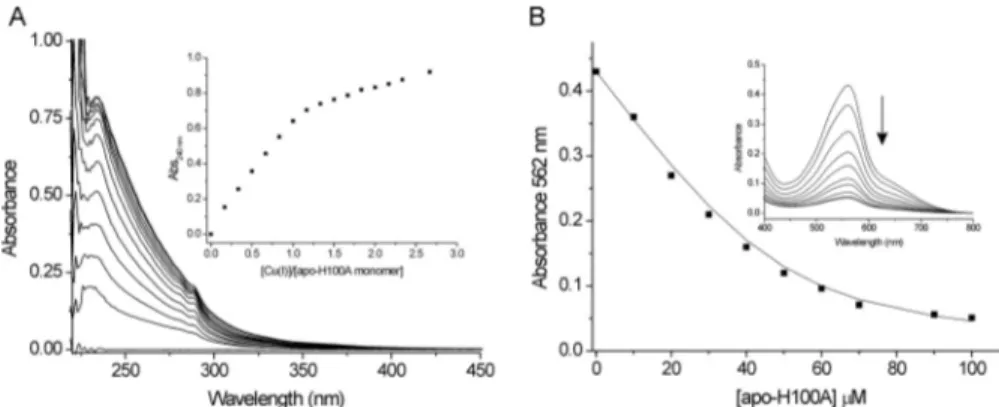
Effect of Mutating Residues Cys-75 and His-100 in CsoRSl— Size-exclusion chromatography and AUC measurements veri-fied that the mutants do not disrupt the tetramer assembly state, and AAS indicated the absence of stoichiometric copper in the purified proteins. Both mutants showed an absorbance increase at 240 nm upon addition of Cu(I) under anaerobic conditions, saturating at ⬃1 mol eq of Cu(I) (Fig. 7A and supplemental Fig. S9), and for the H100A mutant, the absor-bance increase at⬃320 nm that was observed in the wild type protein upon addition of Cu(I) was absent (Fig. 7A). The
KD(Cu
I) values for the mutants were determined using BCA as
a competitive probe (Fig. 7B). Duplicate data sets at pH 7.5 were acquired for both mutants with varying [CuI]
totalor [BCA], and
the data averaged to give a KD(CuI) of 1.3⫻ 10⫺14Mand 9.8⫻
10⫺16Mfor the C75A and H100A variants, respectively. Both
mutants therefore display an increased KD(Cu
I
) compared with wild type CsoRSl.
The mutants were each able to bind to the three DNA oper-ators as indicated by EMSA (Fig. 6D). Addition of Cu(I) to the mutant-DNA operator complexes did not result in the disap-pearance of the low mobility band or a substantial increase in the intensity of the high mobility (free) DNA band (Fig. 6D). This suggests that Cu(I) binding in the mutants does not alter the affinity for the DNA operator to the extent observed for the wild type and suggests that the allosteric mechanism of opera-tor release or exposure is affected. This apparent inability of the CsoR mutants C75A and H100A to disengage in vitro from the DNA operator following Cu(I) binding raises the question of what the effect on growth is when these mutant proteins are expressed in the mycelium. The wild type and mutated CsoRSl
genes were cloned on a low copy plasmid under control of their own promoter. These constructs did not show any effect on growth. Therefore, the CsoRSlwild type gene and the mutant
genes encoding the C75A and H100A variants were cloned under control of a strong constitutive promoter, and the wild type and⌬csoR strain were transformed with these constructs. Fig. 8 clearly shows that the mutant forms of CsoRSlhave a
strong inhibitory growth effect in both strains starting from 100 M Cu(II). The effect of mutant CsoR expression is slightly
FIGURE 6. CsoRSloperator DNA targets and EMSA studies. A, three-locus regulon and the DNA sequences identified by PREDetector (bold) to be target
operator binding sites for CsoRSl. The position of the DNA target sequence in the oligomers used in EMSA is highlighted in bold. B, EMSA of CsoRSlin the presence of the intergenic DNA fragments containing the target sequences for 2730 (232 bp) and 4136 (208 bp). C, EMSA of CsoRSlin the presence of the individual DNA oligomers containing the target sequences shown in A. The gel image to the right is a control EMSA showing the effect on binding CsoRSlto a randomly generated double-stranded DNA oligomer with the sequence 5⬘-GACGAGGACGTCTACGCCATCGACATACTG-3⬘ and the mobility of the 1045 DNA oligomer in the presence of only Cu(I). D, EMSA of the C75A mutant of CsoRSlin the presence of the individual DNA oligomers containing the target sequences shown in A. The components present in each lane of the gel image are indicated with 0.5M[DNA] and 5M[apo-CsoRSlmonomer] and [CuI-CsoRSlmonomer] used.
at UNIV OF ESSEX, on May 21, 2012
www.jbc.org
more pronounced in the⌬csoR mutant than in the wild type because the latter also produces CsoR from the genomic gene copy resulting in a mix of plasmid and genome encoded pro-teins. Upon prolonged incubation, the control transformants harboring the empty vector or the wild type CsoRSlgene
pro-duce confluent growth with 250 and 500MCu(II), although
those expressing the mutant CsoRs do not. These observations support the in silico data indicating that CsoRSlhas control over the expression of genes that encode proteins involved in exporting excess copper ions and its own expression under cop-per stress conditions.
Mapping the Global Response of Wild Type S. lividans to Ele-vated Copper Levels and the⌬csoR Gene by
RNA-seq—RNA-seq is a second generation high resolution RNA-seq—RNA-sequencing tech-nique that enables gene expression profiling in an organism to be analyzed in response to a mutation or an external stimulus (52–54). We have applied RNA-seq to further our insight into the CsoR regulon in S. lividans by analysis of the transcriptome of liquid-grown cultures. The transcriptomes of wild type S.
lividansstrain 1326 grown without and with a 2-h exposure to 400 MCu(II) were compared with that of the⌬csoR strain.
From analysis of the RNA-seq data, both CopZ genes (gene numbers 2730 and 1045) and the 1044 gene encoding for an
uncharacterized secreted protein are all induced by exogenous copper and in the⌬csoR strain (Table 3). These data corrobo-rate the in silico approach and identify these genes as bona fide CsoRSltargets. The CopA-like ATPases (2731 and 1046) are
also up-regulated under elevated copper conditions (Table 3) but less so in the⌬csoR strain. Nevertheless, this suggests reg-ulation by the same promoter as the cognate CopZ. It is notice-able that the RNA-seq data do not provide clear support for the copper induction of gene 4137 or the csoR gene, which was predicted from PREDetector analysis (Table 3 and supplemen-tal Table S2). Therefore, the promoter of csoR was analyzed in a promoter-probing experiment. The data clearly show that transcription originating from the csoR promoter is both cop-per -inducible and CsoR-dependent (Fig. 9A). The low induc-tion by a 2-h exposure to 400MCu(II) seen in the RNA-seq
analysis (1.3-fold, Table 3) is confirmed by the promoter prob-ing experiment (Fig. 9A). It also shows that upon longer expo-sure to Cu(II) the transcription is induced to around 2-fold, a similar level as observed in the⌬csoR strain.
Finally, the transcriptome response to either copper or dele-tion of the csoR gene is not limited to the genes reported in Table 3. The global response can be appreciated visually by the use of Venn diagrams (Fig. 9B). A large number of genes are up-and down-regulated in response to copper up-and deletion of the
csoR, with a significantly greater proportion of genes being down-regulated in the⌬csoR strain. A considerable overlap is present between copper induction and csoR deletion (Fig. 9B andsupplemental Table S6, B–G). Together, these data indicate that the response to changes in copper homeostasis in S.
livi-dansis much more extensive than only the direct CsoRSl
regu-lon reported in Table 3. Aside from the genes up-regulated in Table 3, there is no obvious enrichment of genes that encode proteins known to be directly related to copper homeostasis. However, a gene for a putative copper transporter (3964), part of an operon encoding for two putative copper chaperones, is down-regulated in copper -induced S. lividans but not in the
csoRdeletion (supplemental Table S6, E–G). DISCUSSION
At the molecular level, our understanding of proteins involved in copper detoxification, storage, and trafficking has
FIGURE 7. H100A mutant binds Cu(I) with a reduced affinity. A, changes in the UV region of the apo-H100A mutant base-lined spectrum upon addition of Cu(I)Cl at pH 7.5 and 20 °C. The inset shows the increase in absorbance at 240 nm plotted as a function of [Cu(I)]/[apo-H100A monomer]. B, determination of the KD(CuI) for apo-H100A under copper -limiting conditions imposed by [CuI(BCA)2]3⫺as a competitive probe. The inset shows the decrease in absorbance at 562 nm in the visible region of the spectrum upon increasing [apo-H100A], and Equation 1 was used to determine the KD(Cu
I). The line represents a best fit to the data using KD(Cu
I) 6.3⫻ 10⫺16M. Conditions used are as follows: 10 –100M[apo-H100A], 54M[CuI]
total, and 260M[BCA].
FIGURE 8. Effect of C75A and H100A CsoRSlmutants on growth. S. lividans
1326 and the⌬csoR mutant were transformed with pHJL401 (empty vector) or the same vector harboring the csoR gene (p4136) or the mutants C75A (pC75A) or H100A (pH100A) under control of the constitutive ermE promoter. Spores (1000) were spotted on MM agar plates containing varying [Cu(II)] and incubated at 30 °C. After 50 – 60 h of incubation, a confluent lawn was produced. Images were taken with a Leica M80 stereomicroscope equipped with a DFC295 camera. Note the higher toxicity of exogenous copper supply due to mutations preventing the copper -dependent modulation of CsoRSlDNA binding ability.
at UNIV OF ESSEX, on May 21, 2012
www.jbc.org
advanced considerably in recent times (1, 3, 4). The next chal-lenge is to interface our molecular and mechanistic under-standing of copper homeostasis with the global response of an organism to copper overload. In this study, we have extensively characterized a Cu(I)-CsoR metalloregulator from S. lividans and uncovered the direct and extended CsoR regulon.
Cu(I) Affinity for CsoR Is Unified but How DNA Is Engaged Remains Unresolved—Recent reports have highlighted the need for a unifying approach to accurately determine Cu(I) binding affinities in homeostatic cuproproteins (36, 50). The use of the Cu(I)-specific chelators BCA and BCS has been strongly advocated, and these have been used in this work. Accurate determination of metal affinities of metal sensors is particularly important as these values are considered the thresholds for homeostasis (2). By using BCS, an upper limit
KD(Cu
I) of 1⫻ 10⫺20
Mwas initially reported for CsoRMtb(7)
and later revised to 1⫻ 10⫺19M(55). For CsoRBs, an upper limit
of 1⫻ 10⫺21Mhas been reported using BCS (38) and most
recently a KD(Cu
I
) of 7.9 ⫻ 10⫺19 M for CsoRSa (17). The
KD(Cu
I) value determined for CsoRSlat pH 7.5 is 6.7⫻ 10⫺18M,
an order of magnitude higher than CsoRSaand CsoRMtb, and
thus a comparatively lower affinity for Cu(I). These studies clearly highlight the consistency in using BCS as a probe to determine Cu(I) binding affinity in the attomolar range, ena-bling for cross-species comparisons. The KD(CuI) values
deter-mined using BCS are all consistent with the CsoR sensors being
triggered by [copper]⬃9 orders of magnitude higher than the minimal intracellular copper concentration, consistent with the bacterial cytosol having no free atoms of copper (5). Fur-thermore, this clearly indicates the lengths to which cells go to avoid the potentially deleterious effects of uncomplexed copper ions.
At 1.7 Å resolution, the crystal structure of apo-CsoRSlis the
highest resolution structure to date for a member of the CsoR family. The primary structure of CsoRSlis unique compared
with other characterized CsoR proteins in that it possesses an extended (43 amino acids) N-terminal tail (Fig. 1). Structural organization of this region was not revealed from the crystal structure as electron density was only observed from residue 44 onward. Likewise, the C-terminal extension in CsoRMtb(7) was
not structurally observed, and in the absence of proteolytic cleavage, dynamics outside the “core” structure are likely to be relevant. The core structure of CsoRSlis very similar to that of
CuI-CsoRMtb(7) and has no recognizable DNA structural
bind-ing motif, such as a helix-turn-helix motif. Despite this, apo-CsoRSlhas high specificity and affinity for operator DNA
tar-gets (Fig. 6). The absence of a recognizable DNA-binding motif leaves the question of how CsoR orthologues engage with their operator target unanswered. From our structure of CsoRSl, we
observe that the tetramer assembly creates a large continuous surface area with strong electropositive charge centered in the middle of each face of the tetramer, which may be of
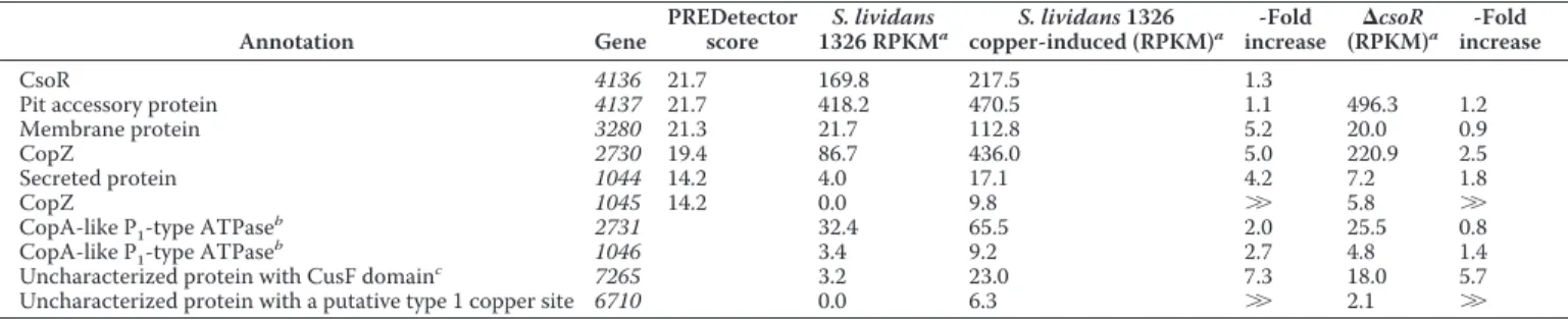
signifi-TABLE 3
Expression levels obtained by RNA-seq of the genes predicted by PREDetector to be under control of CsoRSlalong with other putative copper
proteins up-regulated Annotation Gene PREDetector score S. lividans 1326 RPKMa S. lividans 1326 copper-induced (RPKM)a -Fold increase (RPKM)⌬csoRa -Fold increase CsoR 4136 21.7 169.8 217.5 1.3
Pit accessory protein 4137 21.7 418.2 470.5 1.1 496.3 1.2
Membrane protein 3280 21.3 21.7 112.8 5.2 20.0 0.9
CopZ 2730 19.4 86.7 436.0 5.0 220.9 2.5
Secreted protein 1044 14.2 4.0 17.1 4.2 7.2 1.8
CopZ 1045 14.2 0.0 9.8 ⬎⬎ 5.8 ⬎⬎
CopA-like P1-type ATPase
b
2731 32.4 65.5 2.0 25.5 0.8
CopA-like P1-type ATPase
b
1046 3.4 9.2 2.7 4.8 1.4
Uncharacterized protein with CusF domainc
7265 3.2 23.0 7.3 18.0 5.7
Uncharacterized protein with a putative type 1 copper site 6710 0.0 6.3 ⬎⬎ 2.1 ⬎⬎
aThe selected expression measure is the RKPM. It is defined as the reads/kb of exon model/Million mapped reads (25). bGenes 2731 and 1046 are in the same operon as 2730 and 1045, respectively.
cGene 7265 is the only putative copper-trafficking linked gene that is induced both by copper stress and by csoR deletion (seeTable S6Afor all putative copper proteins).
FIGURE 9. Induction of csoR by promoter probing and global differential gene expression. A, promoter activity in response to the addition of 400MCu(II) in the wild type and the⌬csoR strain are expressed as fold change relative to the wild type strain without addition of Cu(II). Note the copper -induced Red production in the S. lividans wild type background and the constant and copper -insensitive Red production in the S. lividans⌬csoR background. B, Venn diagrams depicting a global overview of the up- and down-regulated genes (⬎2-fold reads/kb of exon model/million) upon Cu(II) induction of wild type S. lividans and in the⌬csoR strain. Thesupplemental Table S6, A–G, reports the lists of genes affected.
at UNIV OF ESSEX, on May 21, 2012
www.jbc.org
cance for DNA binding (Fig. 5B). An alternative possibility is that the absence of a DNA-binding motif is the reason why two tetramers associate with the DNA, as also reported for CsoRBs (38), so as to enhance binding, specificity, or both.
Apo-CsoRSlStructure Provides Insights into the Mechanism of
Allosteric Regulation—An hypothesis for the mechanism of allosteric Cu(I) regulation in the CsoR family has been put for-ward based on the crystal structure of CuI-CsoRMtb(7) and
tested experimentally using unnatural amino acid substitutions of the copper -coordinating His residue (55). These unnatural amino acids were designed to abolish the H-bonding capability of the N⑀2 atom of the imidazole ring so that, on binding Cu(I) to the N␦1 atom, a second coordination sphere H-bond net-work centered on the N⑀2 atom could not form and trigger release of the DNA (Fig. 5E). Cu(I) binding to the non-native His-substituted CsoRMtbdid not significantly affect the DNA
binding affinity, but the allosteric coupling free energy (⌬Gc) was determined to be close to zero (55). This observation was taken to indicate that allosteric switching is initiated upon binding Cu(I) to the N␦1 atom of the His ligand, triggering the formation of a H-bond network to the N⑀2 atom that results in dissociation from the operator DNA sequence. However, this H-bond network is also present in the apo-CsoRSlstructure and
suggests that copper binding is not an essential requirement for its formation (Fig. 5E). One difference in the network found in CsoRSlis the presence of a bridging water molecule between
His-100 and Glu-122 (Fig. 5D). In the CuI-CsoRMtbstructure, no such water molecule is observed, and the H-bond length between the Cu(I) coordinating His and the Glu is⬎4 Å (7). This is indicative of a very weak interaction, which may lead to a destabilization of the DNA-bound structure. Whether or not this water molecule plays a role in the mechanism of allosteric Cu(I) regulation or in DNA binding is presently unclear. From Fig. 5E, it is clear that His-100 and Cys-75⬘ in the apo-CsoRSl
structure are closely aligned to the corresponding residues in the CuI-CsoRMtbstructure. However, Cys-104 in apo-CsoRSl
must undergo a significant movement to complete the Cu(I) coordination sphere (Fig. 5E). It is conceivable that the move-ment associated with the positioning of the Cys-104 side chain to enable Cu(I) coordination may be of significance in alloster-ically regulating the dissociation from the DNA operator.
CsoR Regulon in Streptomycetes—A CsoR-responsive ele-ment prediction in four different Streptomyces species ( supple-mental Tables S2–S5) confirmed a very limited occurrence of highly conserved sequences that have been identified only upstream of one or two sets of orthologues of the copper chap-erone copZ and the efflux ATPase copA, as well as upstream of the copper -sensing repressor csoR itself. EMSA studies (Fig. 6) revealed that CsoRSl recognizes specifically these sequences,
and the effect of Cu(I) on DNA binding supports a mechanism of allosteric regulation most likely similar to other CsoR ortho-logues (7, 17, 38). RNA-seq data corroborate the in silico pre-dictions, revealing clear induction of transcripts for two oper-ons encoding copper efflux systems (2730/2731 and 1045/
1046) (Table 3) and the divergent expression of 1044 (Table 3), a secreted protein of unknown function but not predicted to bind copper. RNA-seq data also indicates that CsoRSltranscript
levels, under normal conditions, are relatively high compared
with the two copZ genes and suggest that a significant propor-tion of CsoRSlis constitutively present (Table 3). The
observa-tion that expression of the mutant csoR genes (C75A and H100A) under control of their own promoter does not lead to growth inhibition at higher [copper] illustrates that CsoRSl
con-trols its own transcription. Failure of the mutants to disengage from the DNA operator has been reported for other copper ligand variants of CsoR orthologues (7, 16, 17) and appears to lead to permanent transcription repression, resulting in too low protein expression levels to block transcription of the other genes with CsoRSl-binding sites. The induction of csoR was not observed from the RNA-seq data (Table 3) but was confirmed after⬎2 h of incubation with exogenous copper (Fig. 9A). This suggests the possibility that a modular system of CsoR repres-sion may be in operation. Interestingly, the in silico analysis in S.
griseuspredicted four other CsoR-responsive elements suggest-ing a somewhat wider regulon in this strain (supplemental Table S5). The SGR3189 (cutC) gene encoding a putative cop-per homeostasis protein (cutC) is located upstream of a copA-like gene but is divergently transcribed. This offers the possibil-ity that the CsoR-responsive elements could act for both copA and cutC in S. griseus. Two other CsoR-binding sites are found at position⫺30 nt from the SGR5260 gene, which could encode for the first member of an operon, including a putative multi-copper oxidase (SGR5259) and at position ⫺36 nt from
SGR1262 (cstR) encoding for a putative non-copper sensing member of the CsoR family (17).
A Model of the Directly Regulated CsoR Regulon in S. lividans— Our findings clearly show that CsoRSldirectly acts to regulate a
three-locus regulon. Under homeostasis conditions, all opera-tor sequences are occupied by two tetramers of CsoRSlresulting
in repression of transcription with some free apo-CsoR and some copper -bound CsoR and CopZ (2730) acting as buffers (Fig. 10). Upon elevated cytosolic copper levels, we suggest, based on our data, a three-step response of the system. Low [copper] can be buffered by free apo-CsoR and CopZ (2730), and no further action is required. Medium [copper] will require a stronger response, achieved by derepressing the 2730 and 1044/1045 operons resulting in expression of CopZ and the CopA P1-type ATPases (Table 3). A further increase in [copper]
will result in copper binding by the apo-CsoRSlstill occupying
the csoR operator. As a consequence, CsoR will be produced at a higher level and will assist in buffering copper in the cyto-plasm. We assume that CsoR can also mediate the trafficking of copper by donating its copper to CopZ, which in turn will have the specificity to deliver the metal to its cognate CopA P1-type ATPase for export. As soon as copper levels are restored, apo-CsoRSlbegins to occupy the operator sequences, and the system
returns to its “ground state.” This model assumes that the up-regulation of CsoRSloccurs later. Quite how this may be
possi-ble is not known, but promoter probing and RNA-seq data are consistent with this phenomenon.
What Can Be Learned About Copper Homeostasis in S. livi-dans from RNA-seq Analysis?—Although RNA-seq analysis clearly provides insight into the response of transcripts under the direct control of CsoRSl(Table 3), it is very much evident
that response to exogenous copper or the deletion of the csoR gene is a complex process (Fig. 9B andsupplemental Table S6,
at UNIV OF ESSEX, on May 21, 2012
www.jbc.org


![FIGURE 10.A model of CsoR regulon and its response to copper stress in S. lividans. Under normal conditions, both csoR and 2730 (CopZ) are expressed at a low CsoR-independent level to ensure an immediate response to an increase in [copper] is present (left](https://thumb-eu.123doks.com/thumbv2/123doknet/6375164.168544/13.891.176.714.89.336/response-lividans-conditions-expressed-independent-immediate-response-increase.webp)
