Lifecycle exposure to perchlorate differentially alters morphology, biochemistry, and transcription as well as sperm motility in Silurana tropicalis frogs
Diana E. K. Campbell1, Robert D. Montgomerie1, and Valerie S. Langlois1, 2,3*
1
Department of Biology, Queen’s University, Kingston, ON, Canada; 2 Department of Chemistry and Chemical Engineering, Royal Military College of Canada, Kingston, ON,
Canada, 3 Institut national de la recherche scientifique, INRS - Centre Eau Terre Environnement, Quebec, QC, Canada
*Author for correspondence and reprint requests (present address): Dr. Valerie S. Langlois
Associate Professor
Institut national de la recherche scientifique - Centre Eau Terre Environnement (INRS-ETE) 490, de la Couronne, Québec (Québec) G1K 9A9, CANADA
Tel 418-654-2547 Fax 418 654-2600
Visual abstract Testes Ovaries Potassium perchlorate (KClO4 ) Hind-limb length Sperm motility KClO4(μg/L) R el a ti v e m R N A l ev el / ref eren ce g en e
Abstract 1
Perchlorate (ClO4–) contamination has been reported in ground and surface waters across North
2
America. However, few studies have examined the effects of prolonged exposure to this thyroid 3
hormone disrupting chemical, particularly at environmentally relevant concentrations in lower 4
vertebrates, such as amphibians. The aim of this study was to examine the effects of a yearlong 5
chronic exposure to ClO4– in in adult male and female Western clawed frogs (Silurana
6
tropicalis). Frogs were spawned and raised from fertilized embryo until sexual maturity in
7
potassium perchlorate (KClO4)-treated water at different concentrations (0, 20, 53, and 107
8
μg/L). Developmental and reproductive indices – including adult morphology, androgen plasma 9
levels, gonadal thyroid hormone- and sex steroid-related transcript levels, and sperm motility – 10
were evaluated in male and female adult frogs. Female growth (e.g., body mass, snout vent 11
length, and hind limb length) was significantly reduced following chronic exposure to 12
environmentally relevant concentrations of KClO4 resulting in females with morphometric
13
indices similar to those of control males – indicating potential sex-specific sensitivities to KClO4.
14
Changes to reproductive indices (i.e., plasma androgen levels, gonadal thyroid hormone- and sex 15
steroid-related transcript levels, and sperm motility) were also observed in both sexes and 16
suggest that KClO4 exposure may also have indirect secondary effects on the reproductive axes
17
in male and female adult frogs. These effects were observed at concentrations at or below those 18
reported in surface waters contaminated with ClO4– suggesting that this contaminant may have
19
developmental and reproductive effects post-metamorphosis in natural amphibian populations. 20
Capsule 21
Developmental and reproductive indices – including adult morphology, androgen plasma levels, 22
gonadal thyroid hormone- and sex steroid-related transcript levels, and sperm motility – were 23
altered in adult Silurana tropicalis frogs following chronic exposure (fertilized embryo to sexual 24
maturity) to environmentally relevant concentrations of potassium perchlorate. 25
26 27
Keywords 28
Perchlorate; Amphibian; Morphology; Gene Expression; Sperm Motility; Western clawed frog 29
1. Introduction 31
Perchlorate (ClO4–) contamination has been reported in aquatic environments in North America
32
as a result of various anthropogenic applications, including solid propellants, munitions, 33
pyrotechnics and fertilizers (GAO 2010; Reviewed in Dasgupta et al. 2006; Reviewed in 34
Trumpolt et al. 2005). The anion can also be introduced to and accumulate in the environment 35
naturally via atmospheric deposition (Jackson et al. 2010; Rajagopalan et al., 2009; Parker et al., 36
2008; Rao et al., 2007; Dasgupta et al., 2006). Since it is highly water soluble, ClO4–
37
accumulates in ground and surface waters (Urbansky, 1998); thereby, placing aquatic vertebrates 38
(e.g., fish, amphibians, and birds) at a high risk of exposure. The majority of surface and ground 39
waters contaminated by ClO4– in the United States of America and Canada are characterized by
40
concentrations less than 100 μg/L (ASTSWMO 2011; Blount et al. 2010; GAO 2010; Parker et 41
al. 2008; Backus et al. 2005; Reviewed in Trumpolt et al. 2005). Therefore, it is important to 42
examine the effects of environmentally relevant concentrations of ClO4– in aquatic species.
43
The effects of ClO4– are mainly mediated through the targeted disruption of thyroid
44
function. Amphibians are highly susceptible to endocrine disruptors that target thyroid function, 45
as metamorphosis is dependent upon thyroid hormones. Exposure to ClO4– has been shown to
46
impede tail reabsorption and hind leg growth in developing tadpoles (Lithobates sylvaticus: 47
Bulaeva et al. 2015; Silurana tropicalis: Flood and Langlois, 2014; Xenopus laevis: Opitz et al. 48
2009; Hu et al. 2006; Tietge et al. 2005; Goleman et al. 2002a, 2002b). Therefore, 49
metamorphosis serves as a critical developmental window for evaluating exposure to thyroid 50
hormone disruptors (Kloas and Lutz, 2006) and the effects of ClO4– have been well studied in
51
this context (L. sylvaticus: Bulaeva et al. 2015; S. tropicalis: Flood and Langlois, 2014; X. laevis: 52
Hu et al. 2006; Opitz et al. 2009; Tietge et al. 2005; Goleman et al. 2002a, 2002b). To date, 53
however, the lasting effects of a developmental exposure (fertilized embryo to sexual maturity) 54
to thyroid hormone-disrupting chemicals on adult amphibians after metamorphosis have received 55
relatively little attention. 56
The primary mechanism of ClO4– is the competitive inhibition of iodide (I–) uptake via
57
the Na+/I– symporter (NIS) limiting the synthesis of the iodine-rich thyroid hormones, 58
tetraiodothyronine (T4) and triiodothyronine (T3), by the thyroid gland (Carr et al. 2008). 59
Thyroid hormones, however, have been shown to integrate with various endocrine axes and the 60
targeted disruption of thyroid hormone synthesis can indirectly mediate the effects of ClO4– on
61
other signalling pathways. For example, thyroid hormone-disrupting chemicals (e.g., ClO4–,
62
methimazole, propylthiouracil, and thiourea) have been shown to alter aspects of the 63
hypothalamus–pituitary–gonad axis (i.e., steroidogenesis, gonadal cellular differentiation, and 64
development (Flood et al. 2013)). Disruption of thyroid function during sexual differentiation 65
can consequently result in observable changes in sex steroid hormone levels, gonadal 66
morphology, and population-level sex ratios in both fish and amphibians (Danio rerio: Sharma 67
and Patiño, 2013; Mukhi et al. 2007; Gasterosteus aculeatus: Bernhardt et al. 2006; Clarias 68
gariepinus: Swapna et al. 2006; Supriya et al. 2005; X. laevis: Goleman et al. 2002a). Transcripts
69
of thyroid hormone-related machinery have moreover been detected in testicular and ovarian 70
tissues of numerous species (Physalaemus pustulosus: Duarte-Guterman et al. 2012; S. 71
tropicalis: Duarte-Guterman and Trudeau, 2011; Scarus iseri: Johnson and Lema, 2011;
72
Oncorhynchus mykiss: Sambroni et al. 2001; Podarcis sicula: Cardone et al. 2000). A direct
73
relationship between thyroid hormone status and sex steroid-related molecular responses in 74
larval gonadal tissues has also been found in S. tropicalis (Duarte-Guterman and Trudeau, 2011). 75
We previously observed that S. tropicalis exposed to KClO4 at environmentally relevant
concentrations ≤100 μg/L from embryo to sexual differentiation (Nieuwkoop–Faber stage 56 and 77
60 (NF); Nieuwkoop and Faber, 1994) induced changes in the transcription of sex steroid-related 78
genes in gonadal and liver tissues (Flood and Langlois, 2014). To further investigate the effects 79
of ClO4– exposure throughout the frog’s lifecycle, a subset of S. tropicalis from the previous
80
experiment were continually exposed to environmentally relevant levels of KClO4 until they
81
reached sexual maturity. Developmental and reproductive indices were assessed, including adult 82
morphology, androgen plasma levels, gonadal thyroid hormone- and sex steroid-related 83
transcript levels, and sperm motility. 84
2. Material and methods 86
2.1. Animals and exposure 87
Larval S. tropicalis (stage NF 10–12) were previously exposed to environmentally relevant 88
concentrations of KClO4 in 1-L glass jars until the climax of metamorphosis (stage NF 60; ∼12
89
weeks post-hatch; for details, refer to Flood and Langlois, 2014). For the present study, a subset 90
of S. tropicalis from the previous study was allowed to develop to sexual maturity (1 year after 91
egg fertilization). Exposure to one of four concentrations of KClO4 of which the average
92
measured concentrations were <1, 20, 53, and 107 μg/L was maintained (Flood and Langlois, 93
2014). Measured concentrations were close to the nominal target concentrations of 0, 25, 50 and 94
100 μg/L (Flood and Langlois, 2014). Studies have confirmed that environmentally relevant 95
concentrations of ClO4‒(≤ 100 μg/L) can have measurable effects on thyroid histology and
96
morphometric indices in developing tadpoles (X. laevis: Hu et al. 2006; Tietge et al. 2005; 97
Goleman et al. 2002a, 2002b), without completely inhibiting metamorphosis – facilitating the 98
study of long-term exposure to KClO4 at sexual maturity. Specifically, with the completion of
99
tail reabsorption (~ 14 weeks after hatch), metamorphs were transferred to glass 10-L treatment 100
tanks where exposure to the same concentrations of reagent-grade KClO4(≥ 99.0%; Sigma
101
Canada Ltd., Oakville, ON, Canada) was continued in dechlorinated, aerated water. Density was 102
maintained at the appropriate body weight per liter for the duration of the experiment (ASTM, 103
1998) and tank size was adjusted as required over the course of the yearlong exposure. We 104
completely replaced water and KClO4 every 3 d, maintaining a water temperature of 25 ± 1 °C
105
and a light:dark regime of 12:12 h (light commencing at 0700 h local time) for the duration of 106
the experiment. Metamorphs were fed once daily with the same amount of commercially 107
available Nasco Xenopus Frog Brittle (Nasco, California, USA) with the essential nutrients for 108
proper Xenopus development including 1.2 ppm of iodine. Animals were housed in the Queen’s 109
University Animal Care Facility (Kingston, ON, Canada) in accordance with the guidelines of 110
the Queen’s University Animal Care Committee and the Canadian Council on Animal Care. 111
One year after fertilization, frogs were anaesthetized by immersion in a 2% w/v solution 112
of ethyl 3-aminobenzoate methanesulfonate (MS-222; Sigma Canada Ltd., Oakville, ON, 113
Canada), after which individual body mass (BM), snout-vent length (SVL), and hind limb length 114
(HLL) was recorded. Animals were then euthanized by decapitation. Blood samples (200-500 115
μL) were collected via exsanguination for sex steroid hormone analyses (1 sample per animal; 8 116
animals per treatment), immediately centrifuged and the plasma fraction (the main medium for 117
sex steroid hormones) was collected and stored at −80 °C. The whole left testis (n = 10 males per 118
treatment) and an ovary section (10-30 mg from each of 10 females per treatment) were 119
dissected, weighed, and stored at −80 °C for further gene expression analysis. The whole right 120
testis of each male was also dissected and weighed, then placed in 2X Simplified Amphibian 121
Ringers (SAR; 113.0 mM NaCl, 1.0 mM CaCl2, 2.0 mM KCl, and 3.6 mM NaHCO3) on ice for
122
immediate sperm analysis. 123
124
2.2 Sex steroid analysis 125
Plasma concentrations of testosterone (T) and 5α-dihydrotestosterone (5α-DHT) were measured 126
using commercially available ELISAs (T: Cayman Chemical, Cedarlane, Burlington, ON, 127
Canada; 5α-DHT: IBL America, Cedarlane, Burlington, ON, Canada). Plasma samples were 128
thawed on ice and diluted in the immunoassay buffer. The quality criteria for the application of 129
commercial kits were verified as instructed by the manufacturer and their immunoassay 130
protocols were followed. All plasma samples were measured in duplicate (2 samples per animal;
6 animals per treatment). The absorbance of samples was measured using an Infinite® M1000 132
PRO plate reader (Tecan, Montreal, QC, Canada) at 405 nm for T and 450 nm for 5α-DHT. The 133
limit of detection according to the manufacturer was 6 pg/mL for both T and 5α-DHT. 134
135
2.3 Gene expression analysis 136
Total RNA from ovary and testis tissue was isolated using TRIzol (Life Technologies, 137
Burlington, ON, CA) following the manufacturer’s protocol and purified using the TURBO 138
DNA-free™ Kit (Ambion; ThermoFisher Scientific, Ottawa, ON). The quantity of RNA was 139
determined on a NanoDrop-2000 spectrophotometer (Thermofisher, Ottawa, ON, Canada). First 140
strand cDNA was synthesized following the GoScript Reverse Transcription kit protocol using 141
random primers (Promega, Madison, WI, USA) in a Mastercycler Pro S Thermocycler (Thermo 142
Fisher, Ottawa, ON, Canada). The cDNA products were diluted 80-fold prior to qPCR 143
amplification. We included negative control reactions for quality control (i.e., no reverse-144
transcriptase (noRT); no-template-controls (NTC)). 145
Primer sequences for aromatase (cyp19), estrogen receptor (erα), androgen receptor (ar), 146
5α-reductase type 2 (srd5α2), deiodinases (dio1, dio2, and dio3), thyroid hormone receptors (trα 147
and trβ), and the reference genes ornithine decarboxylase (odc) and elongation factor-1 alpha
148
(ef1α) were previously designed and validated by Langlois et al. (2010). We performed all qPCR
149
assays using a CFX 96 Real-Time System (Bio-Rad Laboratories Inc, Mississauga, ON) and 150
GoTaq qPCR MasterMix (Promega, Madison, WI, USA). The thermocycler program included an 151
enzyme activation step at 95 °C for 2 min, followed by 40 cycles at 95 °C for 15 s, and 1 min at a 152
gene-specific annealing temperature of 58 °C, 60 °C, or 62 °C, followed by a denaturation step 153
of 1 min at 95 °C. Next, a dissociation curve was generated to confirm the presence of a single 154
amplicon. The threshold for each gene was assessed automatically by the Bio-Rad CFX Manager 155
Software 3.0. Pooled cDNA samples from each treatment were serial diluted (1:4) to produce a 156
standard curve of six points with a starting concentration of 50 ng. Each assay required a reaction 157
efficiency of 100 ± 15% and an R2≥ 0.989. The standard curve, control reactions, and samples 158
were run in duplicate for further quality control. Gene expression data is presented as fold 159
change relative to the mean control treatment. Fold change data of ovary and testis tissue 160
samples were normalized to the mean fold change of the reference genes ef1α and odc, 161
respectively. The expression of reference genes can differ between tissue type. A series of 162
housekeeping genes were therefore profiled for ovary and testis samples (data not shown) and 163
were only considered once the absence of treatment effects was confirmed. 164
165
2.4 Sperm analysis 166
The right testis from each frog was transferred to a clean Kimwipe™ and gently rolled to remove 167
fat bodies and blood vessels from the surface. The cleaned testes were placed in 500 μL of 2X 168
SAR and carefully macerated using long-nosed dissecting scissors to release sperm into solution. 169
The diluted testicular macerate was centrifuged at 1000 rpm for 2 min to remove large cellular 170
debris and the supernatant was collected for sperm video analysis. Placed on ice, the 171
spermatozoa in the supernatant (sperm stock) remained inert until activated with water. 172
The procedure for the analysis of the sperm videos was adapted from Burness et al. 173
(2004). Two sub-samples of the sperm stock were analyzed per testis. A drop of distilled water at 174
room temperature (Morrow et al. 2017; Larroze et al. 2014) was added to two drops of each sub-175
sample of sperm stock to activate motility on a disposable Sperm Count CELL-VU Cytometer 176
(Fisher Scientific, Ottawa, ON). Sperm motility was recorded for 120 s on a high-resolution 177
monochrome CCD camera (Sony model XC-ST50) mounted on a negative phase contrast CH30 178
microscope (Olympus, Tokyo) at 100X. The swimming paths of all spermatozoa were quantified 179
for each sample for 0.5 s sometime between 30 s and 60 s post activation. Five sperm parameters 180
were measured using a CEROS (v.12) video analysis system (Hamilton-Thorne Research, 181
Beverly, Maine, USA): 1) average path velocity (VAP) is the velocity over a smoothed path; 2) 182
straight-line velocity (VSL) is the straight-line distance between the first and last sample point of 183
the sperm’s path divided by the total track time; 3) curvilinear velocity (VCL) is the total 184
distance moved between successive frames on the video recording divided by the time taken for 185
the sperm to move that total distance; 4) straightness (STR) is an estimate of the sperm’s 186
departure from a straight line while swimming, and is calculated by dividing VSL by VAP; and 187
5) amplitude of lateral head displacement (ALH) is the average value of the extreme side-to-side 188
movement of the sperm head in each flagellar beat cycle. 189
An additional sub-sample (15 μL) was taken from the sperm stock to calculate the total 190
number of sperm in the right testis from each male. At high magnification (400X), all 191
spermatozoa in the four large corner squares and the large center square on a haemocytometer 192
were counted (five squares total). The sperm density of the sperm stock was then calculated by 193
dividing by the total spermatozoa count by the volume under these five squares (0.02 µL). 194
195
2.5 Statistical analysis 196
Statistical analyses were performed using Prism 6 (GraphPad Software Inc., San Diego, CA,
197
USA) and JMP (Version 12; SAS, Cary, NC, USA). Data and residuals were tested for normality 198
and homoscedasticity using the Shapiro–Wilk and Levene tests, respectively. Data were log 199
transformed when necessary to improve the fit to normality. Outlier analysis was performed 200
using the Grubbs Test. Morphometric data and plasma sex steroid data are presented as means 201
(least squares means [95% CL]) calculated from a two-way ANOVA (linear model) for each 202
variable (with sex, treatment and their interaction as predictors). Sperm motility data are 203
presented as means (least squares means [95% CL]) and comparisons were calculated from a 204
restricted maximum likelihood method, including male identity as a random factor to account for 205
multiple measurements per male. Testis and ovary gene expression data are presented as 206
standardized means ± 95% CL relative to odc and ef1α expression, respectively. Treatments were 207
compared to controls using one-way ANOVAs and Dunnett’s post hoc tests or post hoc contrast 208
analysis for models that include random effects. 209
3. Results 210
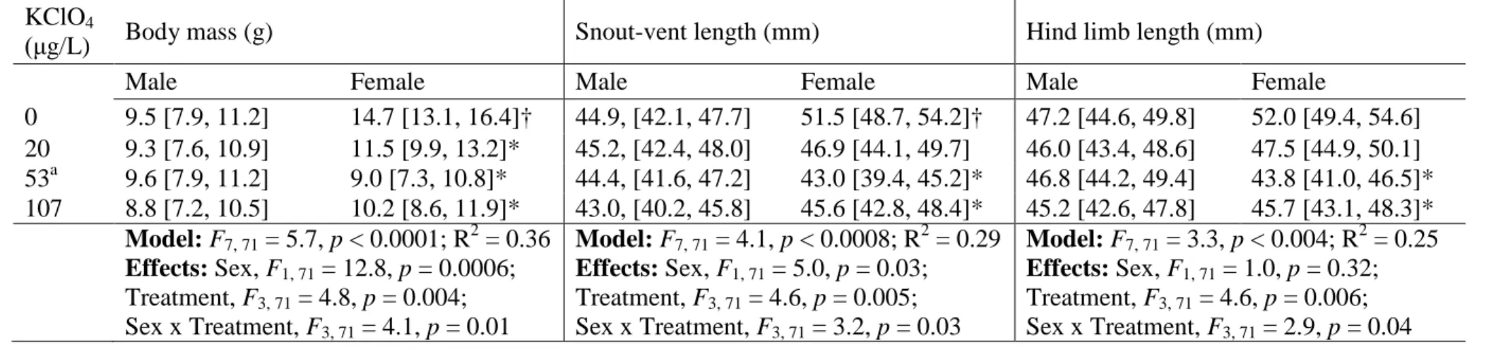
3.1 Morphometric indices 211
Chronic exposure to KClO4 during development generally resulted in smaller adult female frogs
212
(Table 1). For example, all treatments significantly reduced female BM (p < 0.05) and the 53 and 213
107 µg/L treatments significantly decreased both SVL and HLL of females (p < 0.05). The 214
magnitudes of the effects of all treatments on females were similar, with an approximately 40% 215
reduction in BM, and a 10% reduction in both SVL and HLL. Developmental exposure to 216
KClO4; however, did not affect either HSI or GSI in females or males (Dunnett’s tests, p > 0.05;
217
data and analyses not shown). The statistically significant interaction terms in all linear models 218
(Table 1) suggests that KClO4 exposure during development has a different effect on male and
219
female size, for example reducing the degree of sexual size dimorphism compared to frogs 220
developing without such exposure. In one case, sexual dimorphism was reversed such that adult 221
males were larger than females (e.g., at 53 μg KClO4/L; Table 1).
222 223
3.2 Plasma sex steroid hormone levels 224
Plasma androgen content (T and 5α-DHT concentrations) was significantly different between the 225
sexes of S. tropicalis in every treatment (Table 2). Males produced 7–15 times as much T and 2– 226
3 times as much 5α-DHT as females across treatments, as expected in normal conditions. In 227
males, exposure to 53 and 107 μg/L KClO4 produced a slight decrease in T, though the data are
228
very variable and the 95% CL are overlapping. Levels of 5α-DHT production by female and 229
male frogs were not affected by chronic exposure to KClO4.
230 231
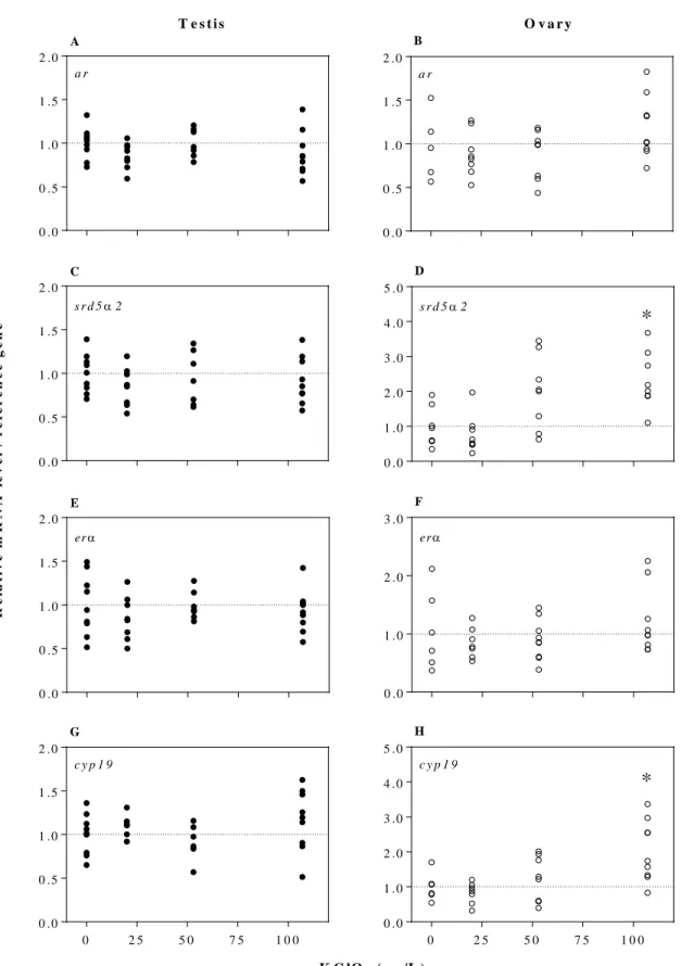
3.3 Gene expression 232
A yearlong exposure to KClO4 resulted in distinct thyroid hormone- and sex steroid-related gene
233
expression patterns in the reproductive tissues of male and female S. tropicalis frogs. Differences 234
in the mean values for the reference gene transcripts did not vary with treatment. Expression of 235
dio1 decreased with increasing KClO4 concentrations (F1, 32 = 9.3, p = 0.005; Fig. 1E). Exposure
236
to 107 μg/L of KClO4 decreased the expression of dio1 by 40% in the testis of males compared
237
to control males (Dunnett’s test, p < 0.05). In female frogs, expression of both srd5α2 (F1, 29 =
238
16.9, p = 0.0003; Fig. 2D) and cyp19 (F1, 29 = 15.9, p = 0.0004; Fig. 2H) in ovarian tissue were
239
positively related to KClO4 concentration. Exposure to 107 μg/L KClO4 increased by twofold, on
240
average, both ovarian srd5α2 and cyp19 transcripts (Dunnett’s tests, p < 0.05). Transcript levels 241
of the remaining thyroid hormone- or sex steroid-related genes did not change in male and 242
female frogs (p > 0.05; Fig. 1 and 2). 243
244
3.4 Sperm motility 245
Sperm motility parameters were differentially affected between treatments (Table 3). In 246
comparison to control males the spermatozoa of KClO4-exposedmales were characterized by a
247
lower VAP (107 µg/L; p < 0.05) and a higher VSL (53 µg/L; p < 0.05) resulting in an increase in 248
the STR of spermatozoa (20 and 53 µg/L; p < 0.05). The ALH was also reduced in spermatozoa 249
of males from all KClO4 treatments (20, 53, and 107 µg/L; p < 0.05) compared to control males.
250
Other parameters – including VCL and sperm count – were unaffected by chronic exposure to 251
KClO4. Sperm counts were however variable within treatments and thus the effect of KClO4 on
252
sperm count might be worth further study with larger sample sizes. 253
4. Discussion 254
Perchlorate contamination has been reported in ground and surface waters across North 255
America (ASTSWMO 2011; Blount et al. 2010; GAO 2010; Parker et al. 2008; Backus et al. 256
2005; Reviewed in Trumpolt et al. 2005) and the anion has been found to compete with iodide at 257
the sodium-iodide symporter in the thyroid (Carr et al. 2008). However, few studies have 258
examined the effects of prolonged exposure to the thyroid hormone disrupting chemical 259
particularly at environmentally relevant concentrations in lower vertebrates, such as amphibians. 260
This study examined the effects of a yearlong chronic exposure to KClO4 in S. tropicalis.
261
Developmental and reproductive indices – including adult morphology, androgen plasma levels, 262
gonadal thyroid hormone- and sex steroid-related transcript levels, and sperm motility – were 263
evaluated in female and male adult frogs. 264
The frogs used in this study were sacrificed once they were one year old, which 265
facilitated the study of potential sex specific differences in developmental and reproductive 266
indices. During the period of metamorphosis, male and female frogs are the same size – sex 267
differences in body size emerge only 10 to 20 weeks after metamorphosis in S. tropicalis 268
(Olmstead et al. 2009). Circulating sex steroid levels moreover develop sexually-dimorphic 269
patterns as males and females differentiate in body size (Olmstead et al. 2009). Both sex steroid- 270
and thyroid hormone-related gene expression are characterized by sexually dimorphic patterns in 271
testicular and ovarian tissues of adult S. tropicalis (Duarte-Guterman and Trudeau, 2011). 272
Disruption of sex-specific morphological, biochemical, and transcriptional dimorphisms would 273
therefore not likely be evident until after the completion of metamorphosis. Previous studies 274
examining the developmental effects of ClO4‒ on aquatic species often focused on a single sex,
275
pooled male and female individuals, or simply did not examine gender differences. 276
Developmental and reproductive data on the differential effects of ClO4‒ as a function of gender
277
are lacking in the literature. 278
Female control S. tropicalis were significantly larger (BM and SVL) than control males – 279
as expected in normal conditions (in the absence of KClO4) – but that difference was reduced at
280
every concentration of KClO4 tested. At 53 μg KClO4/L, this pattern was even reversed where
281
the males were larger than the females. However, since this does not follow a concentration-282
dependent response and the values are within the 95% CI, this difference may be a result of 283
biological variability. We previously documented that the HLL of female stage NF 60 tadpoles 284
exposed to 107 µg/L of KClO4 were shorter than control females prior to the completion of
285
metamorphosis (S. tropicalis: Flood and Langlois, 2014). Other short-term studies have 286
confirmed that ClO4‒at concentrations ≤ 100 µg/L can alter BM, hind leg growth, as well as tail
287
resorption in developing tadpoles (X. laevis: Hu et al. 2006; Goleman et al. 2002a, 2002b). The 288
morphometric data of the present study highlights, for the first time, both the possible 289
permanency of KClO4-induced developmental effects in amphibian ontogeny and the potential
290
for sex differences in the developmental effects of ClO4‒.
291
Amphibian development and growth is dependent on thyroid hormones, and thus we 292
examined the effects of environmentally relevant concentrations of KClO4 on thyroid
hormone-293
related gene expression. ClO4– competitively inhibits the uptake of I– via the NIS limiting the
294
synthesis of the iodine-rich thyroid hormones (T4 and T3) by the thyroid gland (Carr et al. 2008). 295
A direct relationship between thyroid hormone status and thyroid hormone-related gene 296
expression (e.g., trα, trβ, dio1, dio2 and dio3) in larval and adult gonadal tissues has been 297
demonstrated in S. tropicalis (T3, iopanoic acid: Flood and Langlois, Under review; KClO4:
298
Flood and Langlois, 2014; T3: Duarte-Guterman and Trudeau, 2011). Among the five thyroid 299
hormone biomarkers examined in the present study, transcript levels of dio1 decreased by 40% 300
in testicular tissue following exposure to KClO4. Thyroid hormones have been shown to play an
301
important role in testicular development and function (Reviewed in Flood et al. 2013; Wagner et 302
al. 2008; Maran 2003), with thyroid hormone-related genes demonstrating a male-biased pattern 303
of expression in reproductive tissues of adult S. tropicalis (Duarte-Guterman and Trudeau, 304
2011). The activation or deactivation of thyroid hormones are mediated by dios. The dio1 305
enzyme can activate T4 to produce T3 via outer (5′)-ring deiodination as well as inactivate T4 or 306
T3 via inner (5)-ring deiodination. As plasma thyroid hormone levels were not monitored in the 307
present study, the functional significance of changes to this biomarker of peripheral thyroid 308
hormone metabolism in the testicular tissues of S. tropicalis remains to be determined. 309
Chronic exposure to ClO4‒ has been reported to disrupt embryonic androgen synthesis and
310
the subsequent reproductive development of threespine stickleback (G. aculeatus) without 311
changing whole-body levels of thyroid hormones (Petersen et al. 2014). In the present study, 312
KClO4 altered sex steroid-related gene expression in ovary tissue, possibly indicating that KClO4
313
may have indirect secondary effects on the sex steroid axis. The targeted disruption of thyroid 314
hormone synthesis has been shown to indirectly mediate the effects of ClO4– on other endocrine
315
pathways, including the hypothalamus–pituitary–gonad axis (reviewed in Duarte-Guterman et al. 316
2014; Flood et al. 2013). A direct relationship between thyroid hormone status and sex steroid-317
related molecular responses in larval and adult gonadal tissues has moreover been established in 318
S. tropicalis (T3, iopanoic acid: Flood and Langlois, Under review; T3: Duarte-Guterman and
319
Trudeau, 2011). The feminizing effects of ClO4‒ and other thyroid hormone disrupting chemicals
320
have been extensively reported in a wide range of vertebrate species (Vertebrates: Reviewed in 321
Duarte-Guterman et al. 2014; teleost fish: Reviewed in Habibi et al. 2012; Oryzias latipes: Liu et 322
al. 2011; D. rerio: Mukhi et al. 2007; X. laevis: Goleman et al. 2002a). We therefore examined 323
potential estrogenic modes of KClO4 action, and a two-fold increase in cyp19 expression was
324
observed with increasing KClO4 concentrations in ovary tissue. The enzyme cyp19 is responsible
325
for the conversion of T to estradiol. Several studies have previously shown that exposure to 326
KClO4 (Flood and Langlois, 2014) or T3 (Duarte-Guterman and Trudeau, 2011) does not affect
327
cyp19 mRNA levels in gonadal mesonephros tissue of pre-metamorphic S. tropicalis. Exposure
328
to thyroid hormone disruptors have however been reported to increase cyp19 expression in ovary 329
tissue of adult fish (thiourea, C. gariepinus: Rasheeda et al. 2005) and mammals 330
(propylthiouracil, rats: Hapon et al. 2010). In further support of KClO4 mediated changes to
331
cyp19 expression, exposure to T3 has been shown to significantly decrease cyp19 mRNA levels
332
and activity in ovary tissues of a wide range of adult vertebrate species (chicken: Sechman 2013; 333
rat: Hatsuta et al. 2004; mouse: Cecconi et al. 1999; pig: Gregoraszczuk et al. 1998; Chan and 334
Tan 1986). The present study demonstrated that the expression of srd5α2 increased two-fold in 335
ovary tissue of females exposed to 107 μg/L of KClO4. The srd5α2 enzyme converts T to the
336
more potent and non-aromatizable androgen, 5α-DHT – actively competing with cyp19 for T as a 337
substrate. We first documented a KClO4-mediated increase in srd5α2 mRNA levels in hepatic
338
tissue (an important tissue for androgen metabolism) of S. tropicalis tadpoles treated during 339
sexual differentiation, earlier in development (Flood and Langlois, 2014). In support of KClO4
340
mediated changes to srd5α2 expression, exposure to the thyroid hormone triiodothyronine (T3) 341
was shown to decrease srd5α2 transcripts by 50% ex vivo in ovary tissue (Campbell and 342
Langlois, Under review). Taken together these findings indicate that chronic exposure to 343
environmentally relevant concentrations of KClO4 can induce long-term increases in cyp19 and
srd5α2 expression in female S. tropicalis, but the functional significance of these KClO4
345
mediated transcriptional modifications requires further investigation. 346
Male-biased traits such as plasma androgen content and sperm motility were differently 347
impacted by long-term exposure to environmentally relevant concentrations of KClO4. Androgen
348
concentrations (T and 5α-DHT) were significantly higher in male control and KClO4-exposed
349
frogs than their female counterparts. The testosterone concentrations for male and female S. 350
tropicalis in the present study fall proportionately at the low end of the range of mean values
351
reported for S. tropicalis by Olmstead et al. (2009). Studies however have observed plasma 352
testosterone levels as low as 0.5 ng/L in X. laevis (Lee and Veeramachaneni, 2005; Kang et al. 353
1995). Sperm motility was affected by chronic exposure to KClO4. It is noteworthy that sperm
354
motility (measured as progressiveness (VAP, VSL), vigor (VCL), and straightness (STR)) was 355
comparable to that of Larroze et al. (2014), a study on the validity of computer-assisted sperm 356
analysis (CASA) for S. tropicalis. The spermatozoa of KClO4-treated males were characterized
357
by a slower swimming speed (< VAP) and less lateral head displacement (< ALH) than the 358
sperm of control males. Inhibition of flagellar motility could produce these motility patterns. The 359
observed increase in VSL and STR further suggest a decrease in flagellar bending. Romano et al. 360
(2017) observed a significant decrease in mitochondrial activity in spermatozoa of hypothyroid 361
male Wistar rats; less energy would be generated for the flagellum and movement would be 362
impaired. Sperm density and sperm velocity are considered to be primary determinants of male 363
fertility in externally fertilizing aquatic species, thus in vivo exposure to KClO4 may negatively
364
affect male reproductive success however further study would be required. The sperm of 365
externally-fertilizing amphibians may also come in direct contact with ClO4‒ ions in aquatic
366
environments during spawning. Since sperm activity is strongly influenced by the surrounding 367
aqueous chemical conditions, the effect of direct ClO4‒ exposure on sperm motility and
368
fertilization success should also be investigated in future studies. 369
This is the first study to examine the effects of chronic exposure to environmentally 370
relevant concentrations of KClO4 in frogs from fertilized embryo to sexual maturity. Responses
371
in thyroid hormone-sensitive metamorphic processes (i.e., body size, SVL and HLL) indicate 372
targeted disruption of the thyroid hormone axis caused by prolonged exposure KClO4. Moreover,
373
exposed females had morphometric indices similar to those of control males indicating a possible 374
loss of natural sexual dimorphism and highlights for the first-time potential sex-specific 375
sensitivities to KClO4. Changes in reproductive indices (i.e., androgen plasma levels, gonadal
376
thyroid hormone- and sex steroid-related transcript levels, and sperm motility) possibly indicate 377
that KClO4 exposure may also have indirect secondary effects on the reproductive axes in male
378
and female adult frogs. Whether these changes are functionally important to higher-level 379
processes (population, community) remains to be elucidated. This study nonetheless provides a 380
framework for future investigations to examine the effects of chronic exposure to ClO4‒ in
381
natural amphibian populations. 382
Acknowledgements: We thank Almira Siew, Colin Campbell, and Sarah Wallace for their 383
contributions to the project. This work was supported by Discovery Grants from the Natural 384
Sciences and Engineering Research Council (NSERC) of Canada to RDM (RGPIN 2349-08) and 385
VSL (RGPIN 418576-2012), a Canada Research Chair (CRC 950-230442) to VSL, and an 386
NSERC-PGS (PGS D2-460059-2014) to DEKC. 387
References 388
389
ASTM. ASTM E1439-98: Standard guide for Conducting the Frog Embryo Teratogenesis Assay 390
– Xenopus (FETAX). ASTM, Philadelphia. 1998. 391
ASTSWMO. Perchlorate Policy Update. Washington, DC: Federal Facilities Research Center 392
Emerging Issues Focus Group. 2011. 393
Backus, S. M., Klawuun, P., Brown, S., D’sa, I., Sharp, S., Surette, C., Willams, D. J. 394
Determination of perchlorate in selected surface waters in the Great Lakes Basin by 395
HPLC/MS/MS. Chemosphere 2005, 61 (6), 834–843; DOI 396
10.1016/j.chemosphere.2005.04.054. 397
Bernhardt, R. R., von Hippel, F. A., Cresko, W. A., Perchlorate induces hermaphroditism in 398
threespine sticklebacks. Environ. Toxicol. Chem., 2006, 25 (8), 2087–2096; DOI 399
10.1897/05-454R.1 400
Blount, B. C., Alwis, K. U., Jain, R. B., Solomon, B. L., Morrow, J. C., Jackson, W. A. 401
Perchlorate, nitrate, and iodide intake through tap water. Env. Sci. Technol. 2010, 44 (24), 402
9564–9570; DOI 10.1021/es1025195. 403
Bulaeva, E., Lanctôt, C., Reynolds, L., Trudeau, V. L., Navarro-Martín, L. Sodium perchlorate 404
disrupts development and affects metamorphosis-and growth-related gene expression in 405
tadpoles of the wood frog (Lithobates sylvaticus). Gen. Comp. Endocrinol. 2015, 222 (1), 406
33–43; DOI 10.1016/j.ygcen.2015.01.012. 407
Burness, G., Casselman, S. J., Schulte-Hostedde, A. I., Moyes, C. D., Montgomerie, R. Sperm 408
swimming speed and energetics vary with sperm competition risk in bluegill (Lepomis 409
macrochirus). Behav. Ecol. Sociobiol. 2004, 56 (1), 65–70; DOI
10.1007/s00265-003-410
0752-7. 411
Campbell, D. E. K., Langlois, V. S. L. Thyroid hormones and androgens differentially regulate 412
gene expression in testes and ovaries of sexually mature Silurana tropicalis. Gen. Comp. 413
Endocrinol. Under Review.
414
Cardone, A., Angelini, F., Esposito, T., Comitato, R., Varriale, B. The expression of androgen 415
receptor messenger RNA is regulated by tri-iodothyronine in lizard testes. J. Steroid 416
Biochem. Mol. Biol. 2000, 72 (3), 133–141; DOI 10.1016/S0960-0760(00)00021-2.
417
Carr, D. L., Carr, J. A., Willis, R. E., Pressley, T. A. A perchlorate sensitive iodide transporter in 418
frogs. Gen. Comp. Endocrinol. 2008, 156 (1), 9–14; DOI 10.1016/j.ygcen.2008.01.005. 419
Cecconi, S., Rucci, N., Scaldaferri, M. L., Masciulli, M. P., Rossi, G., Moretti, C., D’Armiento, 420
M., Ulisse, S. Thyroid hormone effects on mouse oocyte maturation and granulosa cell 421
aromatase activity. Endocrinology 1999, 140 (4), 1783–1788; DOI 422
10.1210/en.140.4.1783. 423
Chan, W. K., Tan, C. H. Inhibition of follicle-stimulating hormone induction of aromatase 424
activity in porcine granulosa cells by thyroxine and triiodothyronine. Endocrinology 425
1986, 119 (5), 2353–2359; DOI 10.1210/endo-119-5-2353. 426
Dasgupta, P. K., Dyke, J. V., Kirk, A. B., Jackson, W. A. Perchlorate in the United States. 427
Analysis of relative source contributions to the food chain. Env. Sci. Technol. 2006, 40 428
(21), 6608–6614; DOI 10.1021/es061321z. 429
Duarte-Guterman, P., Ryan, M. J., Trudeau, V. L. Developmental expression of sex steroid-and 430
thyroid hormone-related genes and their regulation by triiodothyronine in the gonad– 431
mesonephros of a Neotropical frog, Physalaemus pustulosus. Gen. Comp. Endocrinol. 432
2012, 177, 195–204; DOI 10.1016/j.ygcen.2012.03.011. 433
Duarte-Guterman, P., Trudeau, V. L. Transcript profiles and triiodothyronine regulation of sex 434
steroid-and thyroid hormone-related genes in the gonad–mesonephros complex of 435
Silurana tropicalis. Mol. Cell. Endocrinol. 2011, 331 (1), 143–149; DOI
436
10.1016/j.mce.2010.09.004. 437
Duarte-Guterman. P., Navarro-Martín, L., Trudeau, V. L. Mechanisms of crosstalk between 438
endocrine systems: regulation of sex steroid hormone synthesis and action by thyroid 439
hormones. Gen. Comp. Endocrinol. 2014, 203 (1), 9–85; DOI 440
10.1016/j.ygcen.2014.03.015. 441
Flood, D. E. K., Fernandino, J. I., Langlois, V. S. Thyroid hormones in male reproductive 442
development: evidence for direct crosstalk between the androgen and thyroid hormone 443
axes. Gen. Comp. Endocrinol. 2013, 192 (1), 2–14; DOI10.1016/j.ygcen.2013.02.038. 444
Flood, D. E. K., Langlois, V. S. Crosstalk between the thyroid hormone and androgen axes 445
during reproductive development in Silurana tropicalis. Gen. Comp. Endocrinol. 2014, 446
203 (1), 232–240; DOI 10.1016/j.ygcen.2014.03.037.
447
GAO (U.S. Government Accountability Office). Perchlorate: Occurrence is widespread but at 448
varying levels; Federal agencies have taken some actions to respond to and lessen
449
releases. 2010. http://www.gao.gov/products/GAO-10-769 (accessed 17 November
450
2015). 451
Goleman, W. L., Carr, J. A., Anderson, T. A. Environmentally relevant concentrations of 452
ammonium perchlorate inhibit thyroid function and alter sex ratios in developing 453
Xenopus laevis. Environ. Toxicol. Chem. 2002a, 21 (3), 590–597; DOI
454
10.1002/etc.5620210318. 455
Goleman, W. L., Urquidi, L. J., Anderson, T. A., Smith, E. E., Kendall, R. J., Carr, J. A. 456
Environmentally relevant concentrations of ammonium perchlorate inhibit development 457
and metamorphosis in Xenopus laevis. Environ. Toxicol. Chem. 2002b, 21 (2), 424–430; 458
DOI 10.1002/etc.5620210227. 459
Gregoraszczuk, E. L., Slomczynska, M., Wilk, R. Thyroid hormone inhibits aromatase activity in 460
porcine thecal cells cultured alone and in coculture with granulosa cells. Thyroid 1998, 8 461
(12), 1157–1163; DOI 10.1089/thy.1998.8.1157. 462
Habibi, H. R., Nelson, E. R., Allan, E. R. O. New insights into thyroid hormone function and 463
modulation of reproduction in goldfish. Gen. Comp. Endocrinol. 2012. 175 (1), 19–26; 464
DOI 10.1016/j.ygcen.2011.11.003. 465
Hapon, M. B., Gamarra-Luques, C., Jahn, G. A. Short term hypothyroidism affects ovarian 466
function in the cycling rat. Reprod Biol Endocrinol. 2010, 8 (1), 14; DOI 10.1186/1477-467
7827-8-14. 468
Hatsuta, M., Tamura, K., Shimizu, Y., Toda, K., Kogo, H. Effect of thyroid hormone on CYP19 469
expression in ovarian granulosa cells from gonadotropin treated immature rats. J. 470
Pharmacol. Sci. 2004, 94 (4), 420–425; DOI 10.1254/jphs.94.420.
471
Hu, F., Sharma, B., Mukhi, S., Patiño, R., Carr, J. A. The colloidal thyroxine (T4) ring as a novel 472
biomarker of perchlorate exposure in the African Clawed Frog Xenopus laevis. Toxicol. 473
Sci. 2006, 93 (2), 268–277; DOI 10.1093/toxsci/kfl053.
474
Jackson, W. A., Böhlke, J. K., Gu, B., Hatzinger, P. B., Sturchio, N. C. Isotopic composition and 475
origin of indigenous natural perchlorate and co-occurring nitrate in the southwestern 476
United States. Env. Sci. Technol. 2010, 44 (13), 4869–4876; DOI 10.1021/es903802j. 477
Johnson, K. M., Lema, S. C. Tissue-specific thyroid hormone regulation of gene transcripts 478
encoding iodothyronine deiodinases and thyroid hormone receptors in striped parrotfish 479
(Scarus iseri). Gen. Comp. Endocrinol. 2011, 172 (3), 505–517; DOI 480
10.1016/j.ygcen.2011.04.022. 481
Kang, L., Marin, M., Kelley, D. Androgen biosynthesis and secretion in developing Xenopus 482
laevis. Gen. Comp. Endocrinol. 1995, 100 (3), 293–307; DOI 10.1006/gcen.1995.1160.
483
Kloas, W., Lutz, I. Amphibians as model to study endocrine disrupters. J. Chromatogr. A. 2006, 484
1130 (1), 16–27; DOI 10.1016/j.chroma.2006.04.001.
485
Langlois, V. S., Duarte-Guterman, P., Ing, S., Pauli, B. D., Cooke, G. M., Trudeau, V. L. 486
Fadrozole and finasteride exposures modulate sex steroid-and thyroid hormone-related 487
gene expression in Silurana (Xenopus) tropicalis early larval development. Gen. Comp. 488
Endocrinol. 2010, 166 (2), 417–427; DOI 10.1016/j.ygcen.2009.11.004.
489
Larroze, S., Pickford, D. B., Holt, W. V. Validation of computer-assisted sperm-motility analysis 490
in the amphibian Silurana tropicalis. Reprod. Fertil. Dev. 2014, 27 (7), 1049–1056; DOI 491
10.1071/RD14015. 492
Lee, S. K., Veeramachaneni, D. R. Subchronic exposure to low concentrations of di-n-butyl 493
phthalate disrupts spermatogenesis in Xenopus laevis frogs. Toxicol. Sci. 2005, 84 (2), 494
394–407; DOI 10.1093/toxsci/kfi087. 495
Liu, C., Zhang, X., Deng, J., Hecker, M., Al-Khedhairy, A., Giesy, J. P., Zhou, B. Effects of 496
prochloraz or propylthiouracil on the cross-talk between the HPG, HPA, and HPT axes in 497
zebrafish. Environ. Sci. Technol. 2010, 45 (2), 769–775; DOI 10.1021/es102659p. 498
Maran, R. R. M. Thyroid hormones: their role in testicular steroidogenesis. Arch. Androl. 2003, 499
49 (5), 375–388; DOI 10.1080/01485010390204968.
500
Morrow, S., Gosalvez, J., Lopez-Fernandez, C., Arroyo, F., Holt, W. V., Guille, M. J. Effects of 501
freezing and activation on membrane quality and DNA damage in Xenopus tropicalis and 502
Xenopus laevis spermatozoa. Reprod. Fertil. Dev. 2017, 29 (8), 1556–1566. DOI
503
10.1071/RD16190. 504
Mukhi, S., Torres, L., Patiño, R. Effects of larval–juvenile treatment with perchlorate and co-505
treatment with thyroxine on zebrafish sex ratios. Gen. Comp. Endocrinol. 2007, 150 (3), 506
486–494; DOI 10.1016/j.ygcen.2006.11.013. 507
Nieuwkoop, P. D., Faber, J. Normal Table of Xenopus laevis (Daudin). New York, NY: Garland, 508
1994. 509
Olmstead, A. W., Korte, J. J., Woodis, K. K., Bennett, B. A., Ostazeski, S., Degitz, S. J. 510
Reproductive maturation of the tropical clawed frog: Xenopus tropicalis. Gen. Comp. 511
Endocrinol. 2009, 160 (2), 117–123; DOI 10.1016/j.ygcen.2008.10.025.
512
Opitz, R., Schmidt, F., Braunbeck, T., Wuertz, S., Kloas, W. Perchlorate and ethylenethiourea 513
induce different histological and molecular alterations in a non-mammalian vertebrate 514
model of thyroid goitrogenesis. Mol. Cell. Endocrinol. 2009, 298 (1), 101–114. 515
Parker, D. R., Seyfferth, A. L., Reese, B. K. Perchlorate in groundwater: a synoptic survey of 516
“pristine” sites in the coterminous United States. Env. Sci. Technol. 2008, 42 (5), 1465– 517
1471; DOI 10.1021/es7021957. 518
Petersen, A. M., Dillon, D., Bernhardt, R. R., Torunsky, R., Postlethwait, J. H., von Hippel, F. 519
A., Buck, C. L., Cresko, W. A. Perchlorate disrupts embryonic androgen synthesis and 520
reproductive development in threespine stickleback without changing whole-body levels 521
of thyroid hormone. Gen. Comp. Endocrinol. 2015, 210 (1), 130–144; DOI 522
10.1016/j.ygcen.2014.10.015. 523
Rajagopalan, S., Anderson, T. A., Fahlquist, L., Rainwater, K. A., Ridley, M., Jackson, W. A. 524
Widespread presence of naturally occurring perchlorate in high plains of Texas and New 525
Mexico. Env. Sci. Technol. 2006, 40 (10), 3156–3162. DOI 10.1021/es052155i. 526
Rao, B., Anderson, T. A., Orris, G. J., Rainwater, K. A., Rajagopalan, S., Sandvig, R. M., 527
Scanlon, B. R., Stonestrom, D. A., Walvoord, M. A., Jackson, W. A. Widespread natural 528
perchlorate in unsaturated zones of the southwest United States. Env. Sci. Technol. 2007, 529
41 (13), 4522–4528. DOI 10.1021/es062853i.
530
Rasheeda, M. K., Sreenivasulu, G., Swapna, I., Raghuveer, K., Wang, D. S., Thangaraj, K., 531
Gupta, A. D., Senthilkumaran, B. Thiourea-induced alteration in the expression patterns 532
of some steroidogenic enzymes in the air-breathing catfish Clarias gariepinus. Fish. 533
Physiol. Biochem. 2005, 31 (2-3), 275–279; DOI 10.1007/s10695-006-0036-z.
534
Romano, R. M., Gomes, S. N., Cardoso, N. C. S., Schiessl, L., Romano, M. A., Oliveira, C. A. 535
New insights for male infertility revealed by alterations in spermatic function and 536
differential testicular expression of thyroid-related genes. Endocrine 2017, 55 (2), 607– 537
617; DOI 10.1007/s12020-016-0952-3. 538
Sambroni, E., Gutieres, S., Cauty, C., Guiguen, Y., Breton, B., Lareyre, J. J. Type II 539
iodothyronine deiodinase is preferentially expressed in rainbow trout (Oncorhynchus 540
mykiss) liver and gonads. Mol. Reprod. Dev. 2001, 60 (3), 338–350; DOI
541
10.1002/mrd.1096. 542
Sechman, A. The role of thyroid hormones in regulation of chicken ovarian steroidogenesis. 543
Gen. Comp. Endocrinol. 2013, 190, 68–75; DOI 10.1016/j.ygcen.2013.04.012.
Sharma, P., Patiño, R. Regulation of gonadal sex ratios and pubertal development by the thyroid 545
endocrine system in zebrafish (Danio rerio). Gen. Comp. Endocrinol. 2013, 184 (1), 546
111–119; DOI 10.1016/j.ygcen.2012.12.018. 547
Supriya, A., Raghuveer, K., Swapna, I., Rasheeda, M. K., Kobayashi, T., Nagahama, Y. Thyroid 548
hormone modulation of ovarian recrudescence of air-breathing catfish Clarias 549
gariepinus. Fish. Physiol. Biochem. 2005, 31 (2), 267–270; DOI
10.1007/s10695-006-550
0034-1. 551
Swapna, I., Rajasekhar, A., Supriya, A., Raghuveer, K., Rasheeda, M. K., Majumdar, K. C., 552
Thiourea-induced thyroid hormone depletion impairs testicular recrudescence in the air-553
breathing catfish, Clarias gariepinus. Comp. Biochem. Physiol. Part A. 2006, 144 (1), 1– 554
10; DOI 10.1016/j.cbpa.2006.01.017. 555
Tietge, J. E., Holcombe, G. W., Flynn, K. M., Kosian, P. A., Korte, J. J., Anderson, L. E. 556
Metamorphic inhibition of Xenopus laevis by sodium perchlorate: effects on development 557
and thyroid histology. Environ. Toxicol. Chem. 2005, 24 (4), 926–933; DOI 10.1897/04-558
105R.1. 559
Trumpolt, C. W., Crain, M., Cullison, G .D., Flanagan, S. J. P., Siegel, L., Lathrop, S. 560
Perchlorate: sources, uses and occurrences in the environment. Remed. J. 2005, 16 (1), 561
65–89; DOI 10.1002/rem.20071. 562
Urbansky, E. T. Perchlorate chemistry: implications for analysis and remediation. Bioremediat. 563
J. 1998, 2 (2), 81–95. DOI 10.1080/10889869891214231. 564
Wagner, M. S., Wajner, S. M., Maia, A. L. The role of thyroid hormone in testicular 565
development and function. J. Endocrinol. 2008, 199 (3), 351–365; DOI 10.1677/JOE-08-566
0218. 567
Table 1. Effects of chronic KClO4 treatments (for one year from fertilization) on mean body mass (g), snout-vent length (mm), and
569
hind limb length (mm) of adult S. tropicalis. 570
KClO4
(μg/L) Body mass (g) Snout-vent length (mm) Hind limb length (mm)
Male Female Male Female Male Female
0 9.5 [7.9, 11.2] 14.7 [13.1, 16.4]† 44.9, [42.1, 47.7] 51.5 [48.7, 54.2]† 47.2 [44.6, 49.8] 52.0 [49.4, 54.6] 20 9.3 [7.6, 10.9] 11.5 [9.9, 13.2]* 45.2, [42.4, 48.0] 46.9 [44.1, 49.7] 46.0 [43.4, 48.6] 47.5 [44.9, 50.1] 53a 9.6 [7.9, 11.2] 9.0 [7.3, 10.8]* 44.4, [41.6, 47.2] 43.0 [39.4, 45.2]* 46.8 [44.2, 49.4] 43.8 [41.0, 46.5]* 107 8.8 [7.2, 10.5] 10.2 [8.6, 11.9]* 43.0, [40.2, 45.8] 45.6 [42.8, 48.4]* 45.2 [42.6, 47.8] 45.7 [43.1, 48.3]* Model: F7, 71 = 5.7, p < 0.0001; R2 = 0.36 Effects: Sex, F1, 71 = 12.8, p = 0.0006; Treatment, F3, 71 = 4.8, p = 0.004; Sex x Treatment, F3, 71 = 4.1, p = 0.01 Model: F7, 71 = 4.1, p < 0.0008; R2 = 0.29 Effects: Sex, F1, 71 = 5.0, p = 0.03; Treatment, F3, 71 = 4.6, p = 0.005; Sex x Treatment, F3, 71 = 3.2, p = 0.03 Model: F7, 71 = 3.3, p < 0.004; R2 = 0.25 Effects: Sex, F1, 71 = 1.0, p = 0.32; Treatment, F3, 71 = 4.6, p = 0.006; Sex x Treatment, F3, 71 = 2.9, p = 0.04 571
Means (least squares means [95% CL]) and comparisons were calculated from a linear model for each morphological variable with sex, 572
treatment, and their interaction as predictors. ANOVA statistics for each model are presented below the least squares statistics. 573
574
a
Sample size for each sex per treatment is 10 except for females in this treatment where n = 9 575
* treatment is significantly different (p < 0.05; Dunnett’s tests) from the control (KClO4 = 0 µg/L)
576
† sexes are significantly different within treatment (p < 0.05; Tukey HSD tests) 577
Table 2. Effects of chronic KClO4 treatments (for one year from fertilization) on mean T (pg/mL) and 5α-DHT (pg/mL) plasma levels in male and 579 female S. tropicalis. 580 KClO4 (μg/L) T (pg/mL)a 5α-DHT (pg/mL)
Male Female b Male Female
0 991.3 [607.1, 1618.8]† 63.3 [36.6, 109.5] 1277.9 [975.5, 1580.2]† 577.6 [275.2, 879.9] 20 1096.3 [671.3, 1790.2]† 79.3 [45.8, 137.2] 1301.2 [998.8, 1603.6]† 487.6 [185.2, 790.0] 53 716.5 [438.8, 1170.0]† 82.8 [47.9, 143.3] 1026.7 [724.3, 1329.1]† 407.1 [104.7, 709.4] 107 540.8 [331.1, 883.0]† 71.4 [41.3, 123.6] 1099.4 [797.1, 1401.8]† 347.2 [44.8, 649.6] Model: F7, 28 = 26.2, p < 0.0001; R2 = 0.87 Effects: Sex, F1, 28 = 177.2, p < 0.0001; Treatment, F3, 28 = 0.86, p = 0.47; Sex x Treatment, F3, 28 = 0.97, p < 0.42 Model: F7, 32 = 7.3, p < 0.0001; R2 = 0.61 Effects: Sex, F1, 32 = 47.2, p < 0.0001; Treatment, F3, 32 = 1.12, p = 0.36; Sex x Treatment, F3, 28 = 0.15, p < 0.93 581
Means (least squares means [95% CL]) and comparisons were calculated from a linear model for each androgen with sex, treatment, 582
and their interaction as predictors. ANOVA statistics for each model are presented below the least squares statistics. 583
584
a
T was log-transformed to normalize residuals. 585
b
For each sex per treatment n = 6, except n = 5 for levels of T in females in all treatments. 586
† sexes are significantly different within treatment (p < 0.05; Tukey HSD tests) 587
Fig. 1. Relative expression of trα, trβ, dio1, dio2 and dio3 in testis (A, C, E, G, and I, 589
respectively) and ovary (B, D, F, H, and J, respectively) tissues of Silurana tropicalis frogs 590
chronically exposed to different aqueous concentrations of KClO4 (0, 20, 53, and 107 μg/L; n =
591
7-9 frogs per treatment) for one year after fertilization. Testis and ovary gene expression data are 592
normalized to average expression of the reference genes odc and ef1α, respectively, and 593
presented as fold change relative to the control treatment. Note that the scales of the y-axes vary. 594
Figure 1. 596 597 0 .0 0 .5 1 .0 1 .5 2 .0 trα A 0 .0 1 .0 2 .0 3 .0 trα B 0 .0 0 .5 1 .0 1 .5 2 .0 trβ C 0 .0 1 .0 2 .0 3 .0 trβ D 0 .0 1 .0 2 .0 3 .0 d io 1 * E 0 .0 1 .0 2 .0 3 .0 d io 1 F 0 .0 0 .5 1 .0 1 .5 2 .0 d io 2 G 0 .0 1 .0 2 .0 3 .0 d io 2 H 0 2 5 5 0 7 5 1 0 0 0 .0 0 .5 1 .0 1 .5 2 .0 d io 3 I 0 2 5 5 0 7 5 1 0 0 0 .0 1 .0 2 .0 3 .0 d io 3 J R el a ti v e m R N A l ev e l / r ef e ren ce g en e K C lO (µ g /L ) T e s t i s O v a r y
Fig. 2. Relative expression of ar, srd5α2, erα, and cyp19 genes in testis (A, C, E, and G, 598
respectively) and ovary (B, D, F, and H, respectively) tissues of Silurana tropicalis frogs 599
chronically exposed to KClO4 (0, 20, 53, and 107 μg/L; n = 7-9 frogs per treatment) for one year
600
after fertilization. Testis and ovary gene expression data are normalized to average expression of 601
the reference genes odc and ef1α, respectively, and presented as fold changes relative to the 602
control treatment. Note that the scales of the y-axes vary. 603
Figure 2. 605 606 0 .0 0 .5 1 .0 1 .5 2 .0 B a r 0 .0 1 .0 2 .0 3 .0 4 .0 5 .0 D
*
s r d 5α 2 0 .0 1 .0 2 .0 3 .0 F erα 0 2 5 5 0 7 5 1 0 0 0 .0 1 .0 2 .0 3 .0 4 .0 5 .0 H*
c y p 1 9 0 .0 0 .5 1 .0 1 .5 2 .0 A a r 0 .0 0 .5 1 .0 1 .5 2 .0 C s r d 5α 2 0 .0 0 .5 1 .0 1 .5 2 .0 E erα 0 2 5 5 0 7 5 1 0 0 0 .0 0 .5 1 .0 1 .5 2 .0 G c y p 1 9 R el a ti v e m R N A l ev e l / r ef e ren ce g en e K C lO4(µ g /L ) T e s t i s O v a r y607
Table 3. Effects of chronic KClO4 exposure on mean sperm swimming speed (VAP, VSL, and VCL), linearity (STR), head
608
displacement (ALH), and sperm count of male S. tropicalis. 609 KClO4 (μg/L) VAP (μm s -1 ) F3, 550.7 = 3.16 p = 0.03, R2 = 0.04 VSL (μm s-1) F3, 211.2 = 3.74 p = 0.01, R2 = 0.004 VCL (μm s-1) F3, 557.7 = 0.64 p = 0.59 STR (%) F3, 548.6 = 4.17, p = 0.006, R2 = 0.04 ALH (μm) F3, 483.8 = 6.05 p = 0.0005, R2 = 0.03 Sperm countb (x 106 mL–1) F3, 26.1 = 0.24 p = 0.87 0 23.9 [21.7, 26.0] 7.8 [7.0, 8.6] 46.2 [42.1, 50.3] 34.6 [30.9, 38.3] 3.5 [3.2, 3.8] 6.3 [4.5, 8.2] 20a 21.8 [19.7, 23.9] 8.9 [8.1, 9.7] 44.1 [40.1, 48.1] 40.9 [37.3, 44.4]* 3.0 [2.7, 3.3]* 5.9 [4.0, 7.7] 53 22.2 [20.0, 24.3] 9.1 [8.3, 9.9]* 44.5 [40.4, 48.5] 41.3 [37.7, 44.9]* 2.6 [2.3, 2.9]* 6.6 [4.6, 8.6]b 107 19.8 [17.5, 22.1]* 7.1 [6.2, 8.1] 42.8 [38.4, 47.2] 38.8 [34.9, 42.8] 2.8 [2.4, 3.1]* 5.5 [3.5, 7.5]b 610
Abbreviations: VAP, average path velocity; VSL, straight-line velocity; VCL, curvilinear velocity; STR, straightness; ALH, lateral head 611
displacement. Means (least squares means [95% CL]) and comparisons were calculated from a linear model for each sperm parameter with 612
treatment as predictor (statistics for each of the fixed effects shown at top of each column) and male identity as a random effect (to account 613
for several sperm being measured from each male). 614
615
Sample size for each sperm parameter per treatment is six unless noted otherwise. Sample size for sperm count per treatment is 10 unless 616
noted otherwise (a n = 7; b n = 9). 617
* treatment is significantly different (p < 0.05; post hoc contrast analyses) from the control (where KClO4 = 0 µg/L)
618 619