Université de Sherbrooke
Cytokine Priming Enables Triggering of Naïve Auto-Reactive CDS·T Cells by Weak Agonist Ligands of the TCR
by
Stephanie Dubois
Department of Pediatrics, Division oflmmunology Thesis examinors:
Dr. Sheela Ramanathan Dr. Subburaj Ilangumaran
Dr. Gilles Dupuis Dr. Brian Talbot
Masters thesis presented to the Faculty of Medicine and Health Sciences in partial fulfillment for the degree ofMaster of Science (M.Sc.) in Immunology
••••••••• •• •••••••••••••• •••• •• •• ••••••• . ..
,_,,-NOTICE:
Library and Archives Canada
Published Heritage Branch
395 Wellington Street Ottawa ON K1A ON4 Canada
The author has granted a
non-exclusive license allowing Library and Archives Canada to reproduce, publish, archive, preserve, conserve, communicate to the public by
telecommunication or on the Internet, loan, distrbute and sell theses
worldwide, for commercial or non-commercial purposes, in microform, paper, electronic and/or any other formats.
The author retains copyright ownership and moral rights in this thesis. Neither the thesis nor substantial extracts from it may be printed or otherwise reproduced without the author's permission.
ln compliance with the Canadian Privacy Act some supporting forms may have been removed from this thesis.
While these forms may be included in the document page count, their removal does not represent any loss of content from the thesis.
C d ...
ana.a
Bibliothèque et Archives Canada Direction du Patrimoine de l'édition 395, rue Wellington Ottawa ON K1A ON4 CanadaAVIS:
Your file Votre référence ISBN: 978-0-494-90998-0 Our file Notre référence ISBN: 978-0-494-90998-0
L'auteur a accordé une licence non exclusive permettant à la Bibliothèque et Archives Canada de reproduire, publier, archiver, sauvegarder, conserver, transmettre au public par télécommunication ou par l'Internet, prêter, distribuer et vendre des thèses partout dans le monde, à des fins commerciales ou autres, sur support microforme, papier, électronique et/ou autres formats.
L'auteur conserve la propriété du droit d'auteur et des droits moraux qui protege cette thèse. Ni la thèse ni des extraits substantiels de celle-ci ne doivent être imprimés ou autrement
reproduits sans son autorisation.
Conformément à la loi canadienne sur la protection de la vie privée, quelques formulaires secondaires ont été enlevés de cette thèse.
Bien que ces formulaires aient inclus dans la pagination, il n'y aura aucun contenu manquant.
Abstract
The activation of naive CD8+ T cells by an antigen requires two different signais. The first signal is mediated via the T cell receptor (TCR) following its interaction with the peptide presented on class-I major histocompability complex (MHC-I) molecules. The second signal is delivered via the co-stimulatory receptors upon recognition of their ligands on antigen presenting cells. However, in response to homeostatic pressure, as in lymphopenia, naive T cells undergo proliferation without antigenic stimulation through a process referred to as lymphopenia-induced proliferation (LIP). LIP of naïve CD8+ T cells requires IL-7 and a self peptide presented by MHC-I, which implies that TCR signaling is needed for LIP of naive CD8+ T cells. Recent work from our laboratory has shown that with the homeostatic cytokines IL-7 and IL-15 synergize with IL-21 to induce antigen-independent proliferation of naive CD8+ T cells. Moreover, this cytokine-driven, antigen-independent proliferation "sensitizes" or "primes" naive CD8+ T cells to undergo robust proliferation in response to limiting concentrations of their cognate antigens. Cytokine-primed CD8+ T cells also abundantly produce effector cytokines, such as TNFa and
IFNy and display a potent CTL activity following stimulation by antigen when pre-stimulated. In
my project, I have investigated whether cytokine-induced priming could be an important mechanism by which potentially autoreactive naïve CD8+ T cells are stimulated to cause autoimmune disease using a TCR transgenic mouse model of autoimmune type 1 diabetes (TlD). These mice harbor CD8+ T cells that express transgenic P14 TCR (P14 cells), which recognizes an antigenic peptide derived from the glycoprotein antigen (GP33) of lymphocytic choriomeningitis virus (LCMV). We show that, following priming with IL-21 in the presence of IL-7 or IL-15, P14 cells gain the ability to respond robustly to modified peptide ligands that possess weak agonistic activity towards unprimed P 14 cells. Furthermore, cytokine-primed P 14 cells stmulated with weak TCR ligands displayed potent effector functions such as cytotoxicity and production of effector cytokines, TNFa and IFNy. These cells also induced TlD when adoptively transferred to mice that expressed the LCMV GP antigen under the control of the insulin promoter in the islets. Collectively, our findings show that IL-15 and IL-21 produced during chronic inflammatory conditions could cause priming of potentially autoreactive CD8+ T cells, leading to autoimmune diseases.
Résumé
L'activation des lymphocytes T CD8+ naïfs par un antigène nécessite deux signaux. Le premier signal est médié par le récepteur des cellules T (TCR) à la suite de son interaction avec le complexe majeur d'histocompatibilité (CMH) de classe 1. Le deuxième signal est délivré par l'interaction des molécules de co-stimulation avec leur récepteur spécifique retrouvé sur les cellules présentatrices d'antigène professionnelles. Toutefois en réponse à la pression homéostatique, comme dans le cas de la lymphopénie, les lymphocytes T naïfs sont soumis à une prolifération sans stimulation antigénique par un processus appelé «prolifération induite par la lymphopénie (LIP)». LIP des lymphocytes T CD8+ naïfs nécessite IL-7, et un peptide du soi présenté pas le CMH de classe 1. Des travaux récents de notre laboratoire ont montré que les cytokines homéostatiques IL-7 et IL-15 peuvent agir en synergie avec IL-21 et induire une prolifération antigène-indépendante des cellules CD8+ naïves. Cette activation "sensibilise" les cellules à proliférer en réponse à une concentration sub-optimale de l'antigène specifique au TCR. Également les cellules pré-stimulées avec des cytokines produisent davantage de cytokines effectrices telles que le TNFa et l'IFNy; et possèdent un potentiel plus élevé de cytotoxicité après stimulation avec un antigène spécifique. Dans mon projet de maîtrise, j'ai étudié la possibilité que la pré-stimulation par les cytokines pouvait être un mécanisme important par lequel des cellules T CD8+ naïves qui sont potentiellement autoréactives soient stimulées pour provoquer une maladie auto-immune telle que le diabète de type 1. J'ai démontré cela en utilisant un modèle de souris transgéniques. Ces souris produisent des cellules T CD8+ qui expriment le TCR transgénique Pl4 (cellules Pl4) , lesquelles reconnaissent un peptide antigénique dérivé de l'antigène glycoprotéine (GP33) du virus de la chorioméningite lymphocytaire (LCMV). Nous avons montré qu'une pré-stimulation avec l'IL-21 en présence de l'IL-7 ou l'IL-15, les cellules P 14 acquièrent la capacité de répondre vigoureusement à des ligands peptidiques modifiés qui montrent une faible activité agoniste envers les cellules P14 naïves. Les cellules P14 pré-stimulées qui sont re-pré-stimulées avec des ligands peptidiques modifiés montrent des puissantes fonctions effectrices telles que la cytotoxicité et la production des cytokines effectrices TNFa et IFNy. Ces cellules induisent le diabète de type 1 lorsqu'elles ont été transférées dans des souris qui expriment l'antigène GP LCMV sous le contrôle du promoteur de l'insuline dans les îlots de Langerhans. Dans l'ensemble, nos résultats montrent que l'IL-15 et l 'IL 21 produites durant une réponse inflammation chronique pourraient pré-stimuler des cellules T CD8+ potentiellement autoréactives, conduisant ainsi à des maladies auto-immunes.
Mots clés: cellules T CD8+, IL-21, IL-15, pré-stimulation avec des cytokines, diabète auto-1mmun.
Table of Contents
List of figures ... .IV List of tables ... V List of abbreviations ... VI Abstract ... . Résumé ... . 1. Introduction ... 3 1.1. Development of CD8+ T cells ... .4
1.2. Antigen-mediated activation of CD8+ T cells ... 8
1.3. Interactions between TCR and petide:MHC complex ... 14
1.4. Basal homeostatic proliferation of naïve and memory CD8+ T cells ... 15
1.5. T Lymphopenia and acute homeostatic proliferation of CD8+ T cells ... 16
1.6. LIP and autoimmunity ... 19
1. 7. Type 1 diabetes and T ce Ils ... 19
1.8. RIP/GP Transgenic mouse model of auto immune type 1 diabetes ... 21
1.9. Molecular mimicry in autoimmunity ... 22
1.1 O. Low affinity autoreactive T cells and altered peptide ligands (APL) ... 23
1.11 Antigen non-specific activation of CD8+ T cells ... 26
1.12. Corn mon gamma chain family of cytokines ... 29
1.12.1. IL-7 and the IL-7 receptor in T cell biology ... 29
1.12.2. IL-7 and autoimmunity ... 30
1.12.3. IL-15 and the IL-15 receptor complex ... 31
1.12.4. Effect ofIL-15 on dendritic cells ... 32
1.12.5 IL-15 and autoimmunity ... 32
1.12.6 IL-21 and its receptor ... 33
1.12.7. Effects ofIL-21 on T cells ... 34
1.12.8. IL-21 in anti-tumor immunity ... 35
1.12.9. IL-21 and autoimmunity ... 35
2. Hypothesis / Objectives ... 38
3. Material and Methods ... 39
3.1. Animals ... 39
3.3. Isolation of mononuclear cells ... 39
3.4. Purification of CD8+T cells subsets ... .40
3.5. Flow cytometry ... 40
3.6. Intracellular staining protocol for granzyme B ... .41
3. 7. Cell proliferation assay ... 41
3.8. Cytokine priming ... 42
3.9. Cytotoxicity assays ... 42
3.10. ELISA ... 43
3.11. Induction of diabetes ... 43
3 .12. Histological and immunohistochemical analysis of pancreas ... .43
3.13. Statistical Analysis ... 44
4. Results ... 45
4.1 Cytokine-priming enables naïve P 14 CD8+T cells to proliferate robustly following stimulation with weakly agonistic altered peptide ligands (APL) ... .45
4.2. Cytokine-primed P14 cells show enhanced antigen-specific cytolytic activity following stimulation with a weak agonist.. ... 50
4.3. Cytokine-priming of P14 CD8+T cells enhances TNFa and IFNy production following stimulation with weak agonists ... 53
4.4. Cytokine-priming increased sensitivity of TCR to weakly agonist peptides is not specific to the P14 CD8+T cells as it is also observed in the NOD mode] ... 55
4.5. Cytokine priming enables naïve P14 cells to induce diabetes in RIP-GP recipients following stimulation with a weak TCR ligand ... 59
4.6. Diabetogenic potential of cytokine-primed P14 cells stimulated with a weak TCR agonist is dependent on continuo us availability ofIL-15 in vivo ... 60
4. 7. Cytokine-priming modulates the expression of cell surface molecules that could increase the response to weak agonist. ... 63
5. Discussion and conclusion ... 65
6. References ... 72
List of figures
Figure 1: Development of lymphocytes in the thymus: pos1t1ve and negative selection
... 5
Figure 2: Antigen specific TCR dependent activation of CD8+ T cells ... 10
Figure 3: Relative number and phenotype of CD8+ T cells during and after an acute infection ... 11
Figure 4: LIP and its possible outcomes ... 16
Figure 5: Antigen non-specific TCR independent activation of naïve CD8+ T cells ... 25
Figure 6: Cytokines priming concept of naïve CD8+T cells ... 26
Figure 7: Lymphopenia may lead to autoimmune disease a " Two Hit" mode!.. ... 34
Figure SA: Cytokine priming naïve CD8+T cels increases their proliferation against their cognate antigen GP33 ... .45
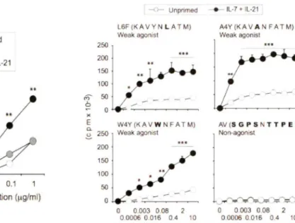
Figure SB: Cytokine priming of naïve CD8+ T cells increases their proliferation to their weak TCR agonists L6F, A4Y and W4Y ... .46
Figure SC: The cytokine priming of naïve CD8+ CD4410 cells also increases the proliferation to weakly TCR agonist W 4 Y ... .4 7 Figure 9: Cytokine pre-stimulated CD8+T cells display increase cytolytic activity following antigen re-stimulation to their cognate antigen as well as their weak altered peptide ... 49
Figure 10: Cytokine-primed Pl4 cells show an increase in the expression of granzyme B in response to weak agonist. ... 50
Figure 11: Cytokine priming of naïve CD8+ T cells results in increased production of TNFa and IFNy following stimulation with weak agonists ... 51
Figure 12: Cytokine-primed NOD 8.31g CD8+T cells respond to weak TCR ligand ... 54
Figure 13: Cytokine priming of NOD 8.3 cells leads to an increase in IFNy production ... 55
Figure 14: Granzyme Bis elevated in NOD 8.3 TCR1 g cells after cytokines stimulation ... 56
Figure 15: Cytokine priming of Pl4 cells with IL-15 in combination with IL-21 followed by stimulation with weak agonists enables them to infiltrate and destroy the beta cells of the islets of the pancreas in the RIP-GP autoimmune diabetes model. ... 60
Figure 16: Cytokine-priming modulates the expression of cell surface molecules that could increase the response to weak TCR ligands ... 62
List of tables
Table 1: Altered peptide ligands for the Pl4 TCR ... 23 Table 2: Cytokine priming with IL-15 in combination with IL-21 enables naïve Pl4 cells to
induce diabetes in a RIP-GP autoimmune diabetes model following a weak TCR stimulation ... 59
List of abbreviations AICD APC: APL: Ag: CD: cDNA: cDC: CpG: CSF: CTL: DC: DP: DN: DNA: ELISA: ER: FasL: GC: GITR: GP: GVHD: HIV: HPE: HSP: IFN: IL: Ig: l.p.: l.V. JAK: kDa: KO: Lck:
Activation-induced cell death Antigen presenting cell Altered peptide ligand Antigen
Cluster of differentiation Complementary DNA Conventional DC
Cytosine-guanine oligodeoxynucleotide Colony stimulating factor
Cytotoxic T lymphocyte Dendritic cell
Double positive Double negative Deoxyribonucleic acid
Enzyme-linked immunosorbent assay Endoplasmic reticulum
Fas ligand Germinal center
Glucocorticoid-induced TNFR-related protein Glycoprotein
Graft versus host disease
Human immunodeficiency virus Homeostatic peripheral expansion Heat shock protein
Interferon Interleukin Immunoglobulin Intraperitoneal injection Intravenous injection Janus Tyrosine Kinase kiloDalton
Knockout
LCMV: LIP: MAPK: mm: MHC: MS: NK: NKT: NOD: pDC: PAMP: PE: PFA: PI3-K: pMHC: PRR: RIP: RA: RT: RTE: SCID: SP: Src: TID: TCR: tg: TNF: TSLP: Treg: yc:
Lymphocytic choriomeningitis virus Lymphopenia-induced proliferation Mitogen-activated protein kinases minutes
Major histocompatibility complex Multiple sclerosis
Natural killer
N atural killer T cells Non-obese diabetic Plasmacytoid DCs
Pathogen associated molecular pattern Phycoerythrin
Paraformaldehyde
Phosphoinositide 3-kinase
Peptide major histocompatibility complex Pathogen recognition receptor
Rat insulin promoter Rheumatoid arthritis Room temperature Recent thymie emigrants
Severe combined immunodeficiency Single positive
Sarcoma kinase Type 1 diabetes T cell receptor Transgenic
Tumor necrosis factor
Thymie stromal lymphopoietin T regulatory cells
Introduction
CDS+ T lymphocytes constitute an important arsenal of the adaptive immune system that confers protection against viruses, intracellular pathogens and cancer cells (Harty et al., 2000). CDS+ T cells are also important mediators of tissue destruction in autoimmune diseases such as autoimmune type 1 diabetes (TlD), multiple sclerosis and rheumatoid arthritis (Walter and Santamaria, 2005). In order to exert their functions, CDS+ T lymphocytes must first become activated by way of their antigen receptor (TCR), which recognizes antigenic peptides presented by class I major histocompatibility complex (MHC-I) molecules captured and cross-presented by professional antigen-presenting cells (APC) such as dendritic cells (DC) along with the costimulatory ligands expressed on these cells (Banchereau and Steinman, 199S; Kurts, 2000). This activation process occurs in draining lymph nodes with help provided by CD4+ T cells, and results in proliferatioq of the activated CDS+ T cells and their differentiation into cytotoxic T lymphocytes (CTL). The effector functions of CTLs are directed against pathogen-infected cells, tumors and, in some instances, healthy tissues (Harty et al., 2000; Walter and Santamaria, 2005). Once the infection is cleared, the expanded CDS+T cell pool undergoes a contraction phase that eliminates 90% to 95% of the effector cells through apoptosis. A small proportion of the activated cells (~5%) differentiates into long-term memory CDS+ T cells (Murali-Krishna et al.,
199S).
Under certain circumstances, na1ve antigen-inexperienced CDS+ T cells can undergo antigen-independent proliferation in vivo. Specifically, under lymphopenic conditions, T lymphocytes undergo lymphopenia-induced proliferation (LIP) in order to restore T cell numbers (Jameson, 2002). This process is essentially driven by cytokines, specifically, IL-7 and IL-15. It has been proposed that homeostatic proliferation may lead to inadvertent activation of potentially
autoreactive CD8+ T cells. However this process may require additional contributing factors (Krupica et al., 2006). One of these contributing factors could be IL-21, which has been genetically linked to many autoimmune diseases including autoimmune TlD (Sarra et al., 2010). Previous work from our laboratory has shown that IL-21 synergizes with IL-7 or IL-15 to induce a robust antigen-independent proliferation and enhances antigen responsiveness of naïve CD8+ T cells (Gagnon et al., 2010b; Gagnon et al., 2008; Gagnon et al., 2007). This Master's thesis describes the results of experiments designed to investigate the implication of this cytokine-driven antigen-independent proliferation of potentially autoreactive naïve CD8+ T cells in a mouse model ofTlD.
1.1. Development of CDS+ T cells
Bone marrow stem cells mature and become lymphoid progenitors that migrate to the thymus. There, these progenitors develop into distinct lineages of cells, namely T cells expressing the aB or the
yô
TCR, Natural Killer (NK) cells and NK T cells. In order to become a committed T cell expressing the aBTCR, the lymphoid progenitor must go through distinct developmental stages, broadly distinguished as double negative (DN, CD4-CD8-), double positive (DP, CD4+CD8+) and single positive (SP, CD4+ or CD8+) stages (Takahama, 2006; Zuniga-Pflucker and Lenardo, 1996) (Fig. 1). The DN stage is subdivided into four sub-stages identified by the differential expression of CD44 and CD25, during which period thymocytes in the CD25+CD44- DN-111 stage expand in number, mainly under the influence of IL-7 and colony stimulating factor (CSF) produced by the stroma of the thymus. In this stage, thymocytes rearrange their TCRB
genes (Germain, 2002). If the TCRB
chain rearrangement occurs successfully, it is expressed on the surface with the pre-TCR a to form the pre-TCR complex.This step is required for the passage through subsequent differentiation steps (von Boehmer and Fehling, 1997). If rearrangement of the ~ chain is unsuccessful, the cell undergoes apoptosis. Signaling through the pre-TCR complex leads to proliferation and transition to the double positive (DP) stage, where CD4 and CDS co-receptors are expressed (Zuniga-Pflucker, 2004).
DP cells express low levels of a~ TCR. If the TCR is able to recognize, with a low affinity, MHC class I or class II molecules which display self-peptides, T cell are positively selected and survive. If there is an absence of recognition, DP cells die by neglect. During this process, DP cells that recognize MHC class I retain their CDS co-receptor to become CDS+ SP T cells. In contrast, if the developing cells recognize a self-peptide presented by MHC class II, they retain their CD4 co-receptor to become CD4+ SP T cells (Sebzda et al., 1999). SP thymocytes are then subjected to negative selection in the cortex at the cortico-medullary junction, where cells that recognize self peptide:MHC complex with high affinity are deleted. Deletion occurs because, if allowed to emigrate to the periphery, these cells would cause autoimmune reactions. Only a very small fraction of the DP thymocytes makes it through the positive and negative selection processes to become SP thymocytes (Goldrath and Bevan, 1999; Takahama, 2006).
The clonal deletion process does not eliminate all potentially self-reactive T cells within the thymus (Goodnow et al., 2005). Sorne T cells that bear TCRs that are potentially self reactive, but whose affinities are insufficient to induce negative selection do escape the thymus and enter the peripheral circulation. Moreover, T cells that recognize proteins found exclusively in immune-privileged sites of the body, such as the lens of the eye, and those expressed specifically during development, such as aft:er puberty, could also become autoaggressive T cells. These potentially autoreactive cells are regulated by peripheral tolerance mechanisms (Ohashi, 2002; Walker and Abbas, 2002).
SP thymocytes go through further maturation. The late-stage mature thymocytes, phenotypically identified as Qa2hiCD62L hiCD241°CD6910 cells, are resistant to apoptosis and proliferate in response to TCR crosslinking (Jin et al., 2008; McCaughtry et al., 2007). These T cells exit the thymus and enter the periphery as recent thymie emigrants and undergo further maturation as naive T cells (Takahama, 2006). In the periphery, mature naive T cells express IL-7 receptors and require IL-IL-7 for survival (Aspinall, 2006; Jacobs et al., 2010). It has been shown that absence oflL-7 signaling in mature T cells compromises their survival (Peschon et al., 1994; von Freeden-Jeffry et al., 1995). In addition, naïve T cells require IL-7 and basal signaling through the TCR by the peptide MHC complex for their survival (Brown et al., 2005; Marrack and Kappler, 2004).
Hematopoietic precursor
\
ON1 X lymphoid precursor Oeath by neglect ON2 / ON3 \ ON4 OPMHC~
MHC Il C08 committed OP C04 committed OP negative / selection/j
emigration to the periphery
negative
~ lection
Cortex
Medulla
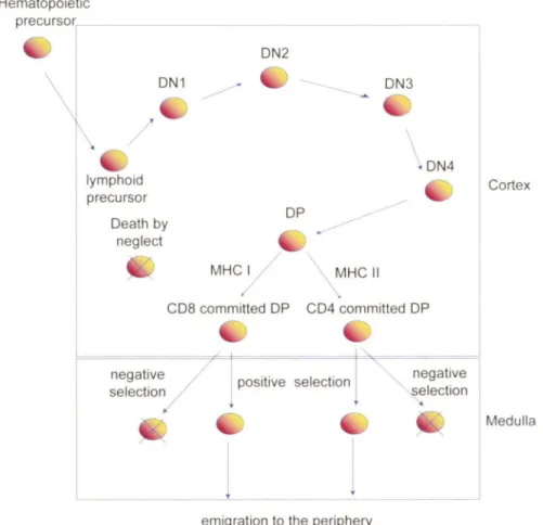
Figure 1: Development of T cells in the thymus: Positive and negative selection.
Bone marrow stem cells mature and become lymphoid progenitors that migrate to the thymus. Early committed T cells lack expression of the T cell receptor (TCR), C04 and C08, and are termed double-negative (ON) thymocytes. ON thymocytes can be further subdivided into four stages of differentiation: DNI , C044+ CD25; DN2, CD44+ C025+; ON3, CD44-CD25+; and DN4, C044-CD25-. The rearrangement of the TCR
p
chain of DN thymocytes depends on IL-7. At the double positive stage (DP), T cells rearrange the a chain of the TCR. OP ce lis go through a process called positive selection to become single positive (SP) T cells (either C04+ or CD8+), depending on whether the TCR recognizes MHC-11 or MHC-1 respectively. If T cells possess a low affinity for MHC:self-peptide, they die by neglect. lf the TCRs of SP cells possess a high affinity towards MHC:self-peptide, they die by apoptosis. This T cel l selection process is known as negative selection. The survival of the newly developed single positive T ce lis depends on IL-7. (Illustration adapted from (Germain, 2002; Zuniga-Ptlucker, 2004)).1.2. Aotigen mediated activation of CDS+ T cells
Mature SP T cells migrate out of the thymus to the secondary Iymphoid organs and remain in a resting state while they circulate throughout the body. These cells patrol the body to ward off microbial pathogens. Most of the infectious agents gain entry through the epidermis, gastro-intestinal tract or respiratory tract. These environments are Iined by a continuous epithelium, which functions as a physical barrier against infections. These "barriers" contain sentine} cells composed of dendritic cells and macrophages that function as professional antigen presenting cells (APC) (Abbas and Lichtman, 2006). Epithelial dendritic cells are considered immature because of their inability to stimulate T cells. However, dendritic cells and macrophages possess membrane receptors known as pathogen recognition receptors (PRR), such as Toll-Iike receptors (TLRs) and nucleotide binding oligomerization domain proteins (NOD)-Iike receptors, which recognize pathogens' terminal mannose residues and other residues which are collectively referred to as pathogen-associated molecular patterns (P AMP). Upon recognition of PAMPs, DCs become activated, endocytose microbial pathogens, upregulate costimulatory molecules and process and present antigenic peptides (Banchereau and Steinman, 1998). Activated DCs lose their adhesiveness to epithelia and express surface receptors for chemokines produced in the T cell zone of the lymph-nodes. These chemokines induce DCs emigration to draining Iymph-nodes. DCs maturation is reflected by the increased stability in expression of the MHC molecule on the cell surface (Abbas and Lichtman, 2006; Banchereau and Steinman,
1998).
Processing of protein antigens varies for peptides that become Ioaded by MHC class 1 or class II molecules. For example, peptide presentation by the MHC class 1 requires endogenous, cytosolic antigens (e.g., microbial proteins) are unfolded and degraded by the
ubiquitin-immunoproteasome system (Sijts and Kloetzel, 2011) and other cytosolic proteases to peptides of eight or nine amino acid residues in length. The peptides are then transported from the cytosol to the endoplasmic reticulum (ER) by members of the TAP transporter family where they are assembled in the trans Golgi network with the nascent MHC class I molecules and beta 2 microglobulin. These complexes are eventually transported, generally as vesicles, to the cell membrane (Cresswell, 2000; Purcell and Elliott, 2008; Van Kaer, 2002). Naïve CD8+ T cells are activated following interaction with DCs that provide two essential signais (Fig. 2). One signal is delivered via the TCR following the recognition of the specific antigenic peptide presented on the restricting MHC molecule of the activated DC. In addition to the TCR MHC:peptide complex interaction, the CD8 co-receptor interacts with the MHC class-I molecule simultaneously (Kerry et al., 2003). The second signal is delivered through interaction of co-stimulatory molecules, such as CD80 or CD86 with its respective ligand CD28 expressed on CD8+ T cells (Acuto and Michel, 2003). Naïve CD8+ T cells require at least few hours of interaction with the APC for efficient activation. In absence of co-stimulation, TCR engagement results in unresponsiveness to subsequent antigen stimulation by a process is called anergy (Schwartz, 2003).
In addition to signais delivered via the TCR and the constimulatory receptors, a third signal is needed for optimal activation of CD8+ T cells and display of effector functions (Haring et al., 2006). Using chemically cross-linked MHC protein peptide complex on microspheres, Curtsigner's group discovered that CD8+ T cells require IL-12, or type 1 IFNa and IFN~ to be
optimally activated to undergo clonal expansion. Stimulation of CD8+ T cells in the absence of this third signal led to proliferation but clonal expansion was limited by poor cell survival. In
addition, CD8+ T cell effector functions did not develop (Curtsinger et al., 2003; Curtsinger et al., 1999; Curtsinger et al., 2005).
Clonai expansion of activated CD8+ T cells is maintained by cytokines that trigger the common gamma chain (yc) family of receptors. Effector CD4+ and CD8+ T cells secrete IL-2, which controls the clonai expansion in a paracrine and autocrine manner to promote naïve CD8+ differentiation into effector CD8+ T cells (D'Souza and Lefrancois, 2003). CD8+ T cell expansion can occur independently of the TCR, but relies on IL-2, IL-7 or IL-15 for survival. IL-7 is produced by bone marrow-derived stromal cells, whereas IL-15 is produced by macrophages and monocytes (Kaech and Ahmed, 2001; van Stipdonk et al., 2001; Wong and Pamer, 2001 ).
CD8+ effector T cells, which gain the abilty to kill target cells displaying cognate antigenic peptides, are also known as cytotoxic T lymphocytes (CTL). CTLs enter the circulation and disperse throughout the body to clear pathogens. When CTL recognize infected cells, they induce cytolysis by releasing granzyme B and perforin (Harty et al., 2000). CTLs also cause apoptosis oftarget cells following interaction of endogenous Fas with its ligand (FasL) expressed on target cells. CD8+ effector T cells also produce effector cytokines such as IFNy and TNFa, which can induce an acute phase reaction (Haring et al., 2006).
Following elimination of pathogens, effector CD8+ T cells enter a programmed contraction phase (Badovinac et al., 2002). Approximately 90% to 95% of effector CD8+ T cells die due to absence of antigenic stimulation and increased competition for survival cytokines. During the contraction phase, CD8+ T cells are eliminated by apoptosis through the Fas mediated pathway following a process referred to as activation-induced cell death (AICD) (Vella et al.,
Following antigen clearance, approximately 5% of effector CD8+ T cells become memory T cells, which confer long-term immunity (Murali-Krishna et al., 1998). There are two types of CD8+ memory T cells (Sallusto et al., 2004; Sallusto et al., 2000). One type is effector memory T cells (T EM) which reside in peripheral tissues and which are characterized by the CCR?1°, CD62L10 and IL-7Rhi phenotype. The other type is the long-lived central memory T cells (TcM) of the CCR7hi and CD62Lhi and IL-7Rhi phenotype and which circulate through lymph nodes. Upon re-infection with the same pathogen, central memory cells proliferate rapidly to generate new effector cells, whereas effector memory cells are immediately able to produce effector cytokines and to become CTL (Seder and Ahmed, 2003). Survival of both types of memory cells is under the influence of IL-7 and IL-15 in both humans and mice (Kennedy et al., 2000; Lodolce et al., 1998; Sallusto et al., 2004).
Path<>gen..s
•
,~,,
pattern . _ ~APC recog n it:ion ~ recepto rs / ./
.
'CD4
./~
: .Signal 2 • ... I/ i
'O. 1 Signal 1 \ I 'O. I I I I ~Naïvecoa
IL-7\
... ~ Th IL-12. 1 1 1...
l
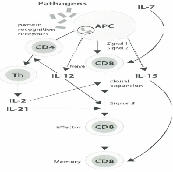
IL-15 clonal ~ e~pansion \ IL-2 IL-21 Effect:or CDS Mem<>ry CDSFigure 2: Antigen-specific TCR-dependent activation of CDS+ T cells. During an immune stimulus, such as microbial infection, activation of naive CD8+ T cells requires two initial signals provided by triggering TCR and CD28 activation, and a third signal provided by cytokines (e.g., IFN type 1, IL-2 or IL-21) for efficient clonal expansion. Activated CD8+ cells undergo clonal expansion to boost the efficiency of the CD8+ T cell response. Activated CD8+ T cells become CTL. Most of these CTL die upon elimination of the antigen. IL-7 from bone marrow-derived stomal cells induces approximately 5% of effector cells to become memory cells, which are kept
(/) (]) (.) 1-co 0 ü '+-0 ::t:t: (]) > :.;::; CU (]) ...
Î
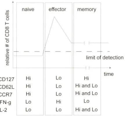
1 naive CD 127 Hi CD62L Hi CCR? Hi IFN-g 1 Lo IL-2~
effectorl
memory
--
-
-limit o detection Lo Hi time Lo Hi and Lo Lo Hi and Lo Hi Lo Lo Hi and LoFigure 3: Relative numbers and phenotype of CDS+ T cells during and after an acute infection. After an infection, indicated by the black arrow, antigen-specific naive CD8+ T cells undergo rapid and vigorous proli feration known as the expansion phase. Most effector CD8+ T cells generated by this expansion die during the contraction phase. Approximately 5% of the expanded cells differentiate and develop into memory CD8+ T cells. The phenotypic and functi onal changes that occur during the transition of naive to memory CD8+ T cells are menti oned in the text. Dashed line represents the limit of detection (LOD). (Illustration adapted from (Badovinac and Harty, 2006)).
1.3. Interaction between TCR and petide:MHC complex
The TCR is a complex cell surface structure that defines the T cell lineage and fulfills two basic functions. The primary function of the TCR is to recognize a peptide fragment of foreign antigens in the context of self MHC molecules. The second is to trigger the immune response needed for elimination of foreign antigens (Rojo et al., 2008). The TCR is composed of two disulfide-linked class I membrane glycosylated polypeptides named a and p, which mediate antigen recognition (Clevers et al., 1988; Kuhns et al., 2006). The TCR ap chains interact with the subunits of the CD3 complex namely, CD3yE and CD3è3E, chains, as well the Ç homodimer. CD3y, CD3ô and CD3E chains contain one immunoreceptor tyrosine-based activation motif (IT AM), whereas the TCRÇ chain contains three IT AM motifs. IT AM motifs are sites of Src (Lck, Fyn)-dependent tyrosine phosphorylation. They serve as anchors for recruitment of components of the machinery of signal transduction in response to antigen recognition (Palacios and Weiss, 2004; Smith-Garvin et al., 2009). TCRap recognizes specific short antigenic peptides bound to MHC expressed on APC (Rudolph et al., 2006). MHC class I molecules that activate CD8+ T cells have a peptide binding groove in their polymorphie heavy chains that is closed at both ends, imposing a constraint on the length of the peptide. Binding of the peptide to the groove formed by the al, a2 binding domain of the MHC leaves the upward pointing peptide chain available for interaction with the TCR (Madden et al., 1993; Stern and Wiley, 1994).
The crystal structure of the TCR/ peptide-MHC (pMHC) complex revealed that only two to five peptide residues made contact with the TCR. On MHC class I molecules these interactions are dominated by the amino acids that bulge out and therefore represent "functional hot spots" (Garboczi et al., 1996; Garcia et al., 1996). (Degano et al., 2000) for nanomeric and/or
octameric peptides. The hot spots of the peptide are predominantly residues P5, P7 and P8 for the nonamer and P4, P6 and P7 for the octamer (Rudolph et al., 2006; Rudolph and Wilson, 2002).
1.4. Basal homeostatic proliferation of naïve and memory CDS+ T cells
The thymus undergoes involution at puberty and thymie output becomes significantly reduced. T cell numbers in the secondary lymphoid organs are maintained by a slow turnover of a small fraction of T cells by a process called homeostatic proliferation (Rocha et al., 1989). The turnover of naive and memory ce lis occurs independently of each other and their respective niche is maintained (Surh and Sprent, 2002; Tanchot and Rocha, 1998). The basal homeostatic proliferation maintains the polyclonality and the proportion of naive and memory cells relatively constant (Tanchot and Rocha, 1998).
Studies using mice that do not express the MHC molecules, specific cytokines or cytokine receptors have contributed to the knowledge of the factors that regulate T cell homeostatic turnover in mice. It has been found that naive, but not memory CD8+ T cells, require pMHC:TCR interactions for survival. In addition, both naive and memory T cells require signais initiated by occupation of the IL-7 receptor, whereas memory T cells require signais through the IL-7 and IL-15 receptors (Murali-Krishna et al., 1999; Schluns and Lefrancois, 2003; Surh and Sprent, 2005; Tan et al., 2001). It has also been shown that memory cells can proliferate in response to an abundance of IL-7 (Murali-Kishna et al., 1999; Judge et al., 2002; Kieper et al., 2002; Tan et al., 2001).
1.5. T Lymphopenia and acute homeostatic proliferation of CDS+ T
ceIIs
An organism is considered lymphopenic in the T cell compartment when there is a reduction in T cell numbers in the secondary lymphoid organs. There are different types of lymphopenia. Transient lymphopenia occurs as a sequel to some viral or microbal infections whereas chronic lymphopenia is observed in patients who are infected with HIV. Iatrogenic lymphopenia occurs following radiation or chemotherapy for the treatment of a cancer. Decreases in T cell numbers during lymphopenia may have additional devastating consequences in elderly individuals under treatments since they already have a low output of recent thymie emigrants (Jameson, 2002).
The rebuilding of the T cell compartment by homeostatic proliferation may be the last effort of the immune system to replenish the number of immune cells to provide protection against harmful and potentially life threatening infections (Jameson, 2002). In the absence of a functional thymus, an expansion of the remaining cells occurs. This process is called lymphopenia-induced proliferation (LIP). There are two possible outcomes during LIP,. The first possibility is that all the remaining T cells proliferate equally to replenish the T cell compartment. This situation leads to an overall general reduction of TCR diversity but a "full" compartment. The second possibility is T cell recovery as a result of LIP, but some T cells may possess a proliferative advantage over others. These conditions would also lead to oligoclonality and important reduction in TCR diversity. Oligoclonality may be responsible for autoimmunity if T cell proliferation targets self-antigens, as shown in Fig. 4 (Khoruts and Fraser, 2005).
LIP is driven by interaction of the TCR with MHC self-peptide complex and by the action of IL-7 which is available in abundance under lymphopenic conditions (Fry and Mackall, 2001; Surh and Sprent, 2005). As a consequence of rapid homeostatic expansion, naive T cells
may gain expression of certain memory markers such as CD122hi, CD132h\ CD44hi and Ly6C, but do not gain expression of the activation markers CD25 and CD69 (Cho et al., 2000; Goldrath et al., 2000; Kieper et al., 2002; Murali-Krishna and Ahmed, 2000). The CD44hi CD122hï Ly6C+ phenotype is similar to the phenotype of memory T cells generated by an antigenic response. These cells are very distinct because they do not display effector functions. However, when these cells are stimulated with the cognate antigen they gain high levels of effector functions in a manner similar to genuine memory cells (Ramanathan et al., 2009).
0
&
$
@
y
*
#
*
#
&
Lymphopenia inducingj
insult@f
@y
*
0*
&
#
$
&
Recovery via LI P with diversity @y
~ ~ 0*
&
#
$
'
f@f
@y
*
0#
*
&
#
$
&
Recovery in the presence of functional thymus
Steady state
normal T cell population size Great TCR diversity
/r1
#
y
tt#
y y
...
*
*
*
@f
#
y
*
Recovery via LIP with limited diversit y
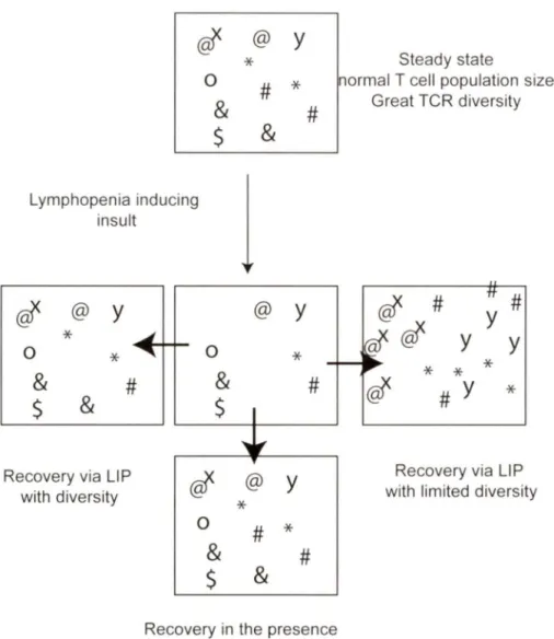
Figure 4: Lymphopenia-induced proliferation (LIP) and its possible outcomes. The normal
T cell population displays a vast TCR diversity, which is diminished during T lymphopenia. However, a fui 1 recovery of T cell diversity occurs in the presence of a functional thymus. In its absence, LIP helps the replenishment of the T cell pool, although with limited TCR diversity. In this situation, T cells whose TCR is at the limit of threshold level of activation by self antigens may have an advantage over others. This situation can lead to their preferential expansion, and possibly account for autoimmunity associated with lymphopenia (Illustration adapted from (Khoruts and Fraser, 2005).
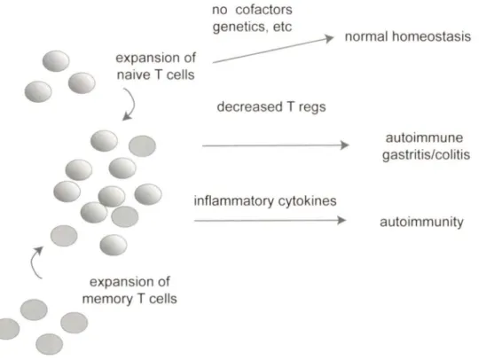
1.6. LIP and autoimmunity
Homeostatic proliferation of naïve CD8+ T cells in response to antigen stimulation is beneficial to the host during infections (microbial or viral). In addition, it is needed for restoring the T cell compartment following lymphopenia resulting from oncotherapy (radiation therapy or chemotherapy). However, there are increased risks of susceptibility to infections and occurrence of malignant disease due to the fact that T cell replenishment is usually slow. An additional risk of LIP is the expansion autoreactive CD4+ and CD8+ T cells, as mentioned above (Gleeson et al., 1996; Jameson, 2002; Khoruts and Fraser, 2005). However, LIP alone is not sufficient to trigger autoimmune responses. For instance, in the case of genetically susceptible individuals, one compounding susceptibility factors could be cytokines that modulate T cell responses. It has suggested that there is a link between autoimmune type 1 diabetes in the Non Obese Diabetic (NOD) mouse model and lymphopenia-associated homeostatic expansion and IL-21 production (King et al., 2004). These reported improvement of mild lymphopenia in these mice by injection of T cells and prevention of autoimmune diabetes. Furthermore, genetic analysis of the disparity between congenic NOD and NOD.B6 Idd3 mice that do not develop diabetes, revealed an increase in IL-21 production and IL-21 receptor expression on T cells in NOD mice with respect to NOD.B6 Idd3 mice (King et al., 2004; McGuire et al., 2009). Consistent with the requirement of IL-21 for induction of autoimmune diabetes, NOD mice lacking IL-21Ra or IL-21 do not develop diabetes (Spolski and Leonard, 2008; Sutherland et al., 2009).
1.7. Type 1 diabetes and T cells
Autoimmune type 1 diabetes (TlD) or diabetes mellitus type 1 is an organ-specific autoimmune disease. TlD is a multifactorial disease. Susceptibility to TlD involves complex
mechanistic interplays of environmental and genetic factors (Maier and Wicker, 2005; Todd, 2010; von Herrath, 2009). The rate of TlD incidence has been rising steadily in developed countries from the 1950s to the present day, and it has been predicted that the incidence of TlD will double in children under the age of five by 2020 (Patterson et al., 2009). Clinical symptoms of T 1 D usually become apparent only after 90% of the insulin-producing pancreatic ~ ce lis have been destroyed by a T cell-mediated cytotoxic process that leads to profound diminution of insulin production (Itoh and Maki, 1996). Clinical symptoms include polyuria, polydipsia, polyphagia, fatigue and weight loss. The pathology of this disease reveals a lack of insulin-producing cell in the pancreas, but cell secreting glucagon, somatostatin and pancreatic polypeptides are fully functional.
The study of TlD has been greatly facilitated by animal models such as the Biobreeding (BB) rat and the NOD mouse. Development of TlD in these models is quite similar to human TlD (Delovitch and Singh, 1997; Wicker et al., 1995). T cells play a critical role in the pathogenesis of TlD in both models (Gepts and Lecompte, 1981; Miyazaki et al., 1985). Different groups have shown that depletion of T cells in NOD mice and BB rats prevents the disease (Christianson et al., 1993; Ikehara et al., 1985) and that the adoptive transfer of T cells from diabetic donors leads to the development of TlD (Christianson et al., 1993; Whalen et al., 1994). The pathogenic process is believed to be dependent on the presence of autoreactive CD8+ T cells. CD8+ T cells are more important in the early phase of the disease, whereas CD4+ and CD8+ T cells are required throughout its progression (DiLorenzo et al., 1998; Groen et al., 2003; Walter and Santamaria, 2005).
1.8. RIP/GP transgenic mouse model of autoimmune type 1 diabetes
Transgenic mouse models have contributed significantly to understand the mechanism of activation of potentially autoreactive CD8+ T cells. In the case of the RIP-GP model, the glycoprotein (GP) antigen of the lymphocytic choriomengitis virus (LCMV) is expressed under regulation of the rat insulin promoter (RIP) (Ohashi et al., 1991 ). Peripheral T cells specific for the GP antigen ignore the antigen expressed in the islets. The GP-specific CTLs can be detected in the periphery of RIP-GP mice following antigen stimulation in vitro. However, CTLs do not cause diabetes in this case because the self antigen that is expressed at a very low levels in the islets is ignored. However, following LCMV infection, the CTL response directed towards viral GP antigen destroys the islets within eight to fourteen days in these mice. lt has been shown that the T cells that infiltrate the islet are specific for the viral gene product of the glycoprotein and lyse the infected target cells in vitro.
CD8+ T cells bearing transgenic P14 TCR (P14 cells) recogn1ze the GP33 peptide spanning positions 33-41 of the glycoprotein (GP) antigen (Ag) of the LCMV presented by the MHC of class 1 molecule H-2Db (Ohashi et al., 1991). In RIP-GP mice expressing transgenic P14 TCR, P 14 cells are not deleted during thymie development and are able to circulate in the periphery without attacking the islets due to low levels of GP antigen expression in the islets (Oldstone, 2005). Similarly, when naive P14 cells are transferred into RIP-GP recipients, they do not damage the islets or cause TlD (Ohashi et al., 1991). P14 cells fail to cause TlD in RIP-GP/P14 double transgenic mice, or in RIP-GP recipients, because the cells remain 'ignorant' of the antigen in the islets. However, LCMV infection will activate P14 cells and induce TlD, presumably as a result of inflammation-induced exposure to the Ag in the islets (Oldstone, 2005). lmmunization of RIP-GP/P14 mice with GP33 peptide activates P14 cells, but fails to
induce TID. To induce TID in the RIP-GP/P14 model, GP33 immunization must be combined with induction of an inflammatory response by using the TLR3 ligand poly(I:C) or activation of DCs by cross-linking CD40 or using HSP70 (Garza et al., 2000; Lang et al., 2005; Millar et al., 2003). Poly(I:C) provides innate immune stimulation to upregulate MHC-I on pancreatic beta cells to make them susceptible to autoreactive CTLs (Lang et al., 2005).
1.9. Molecular mimicry in autoimmunity
Environmental factors are thought to be important for induction of autoimmune diseases (Fujinami et al., 2006). One of the environmental etiologic factors that may trigger autoreactive T lymphocyte activity are viral pathogens. They have long been proposed to play a role in the initiation, progression and exacerbation of autoimmune diseases: including TlD (Fujinami and Oldstone, 1989; Fujinami et al., 2006; von Herrath et al., 2003). A mechanism suggested to explain autoimmunity associated with microbial infections is molecular mimicry (Fujinami and Oldstone, 1989). According to this hypothesis, autoimmune diseases are triggered by the initial activation of lymphocytes that recognize a microbial derived epitope that shares homology with a self-peptide. These cross-reactive lymphocytes or autoreactive cells can then migrate to the target organ that expresses the self antigen and induce autoimmunity (Fujinami and Oldstone, 1989; Ryan et al., 2007). T cells exhibit cross reactivity at the TCR level, meaning they are able to recognize similar peptides that may corne from different antigens. This molecular flexibility seems to expand the functional reactivity of the TCR, which is estimated to posssess a repertoire of 108 different variations. On the othe hand, antigenic peptides are estimated to be in the range of 1012 to 1015
• The scenario proposed to provide an effective immuno-surveillance is that each T cell should be able to specifically recognize 106 different but similar peptides (Anderton and
Wraith, 2002; Mason, 1998; Sospedra and Martin, 2006). According to this model, molecular mimicry could probably break tolerance to autoantigens and cause autoimmunity. However, there is limited experimental evidence to support this notion (Benoist and Mathis, 2001).
1.10. Low affioity autoreactive T cells and altered peptide ligands (APL)
During thymie selection, low-avidity T cells that escape negative selection may become autoreactive T cells (Goodnow et al., 2005). Zehn and Bevan have demonstrated that low-avidity T cells, although harboring self-destructive potential, might routinely escape both central and peripheral tolerance mechanisms (Zehn and Bevan, 2006). The question arises as to how these low avidity autoreactive T cells cause disease? Zehn and Bevan proposed that although self-reactive T cells that can be primed with endogenous levels of self antigen are eliminated, the remaining low avidity cells remain in the periphery and can be activated by excess of cross-reactive foreign antigen and cause autoimmunity. These authours presented evidence for this mechanism by challenging RIP-ovalbumin mice (where OV A-specific high affinity T cells have been deleted during selection) with a recombinant bacterium expressing the self antigen (Lm-OV A). Lm-(Lm-OV A primed low-avidity T cells (in a RIP-m(Lm-OV A recipient), and the animals rapidly developed autoimmune diabetes. In essense, this study demonstrated that potentially autoreactive clones that escape the negative selection process in the thymus (due to lower reactivity of their TCR towards self antigens) could be stimulated by higher amounts of self or cross-reactive antigens in the periphery (Zehn and Bevan, 2006).
The amino acid composition of the peptide ligand recognized by TCR can be modified to alter its affinity for the TCR, without affecting its binding properties towards the restricting MHC molecules. Such altered peptide ligands (APL) display a range of physical properties and
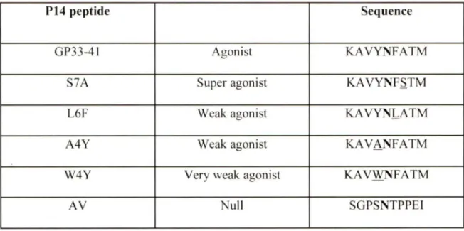
induce a host of effector phenotypes in T cells expressing the same TCR (Sloan-Lancaster and Allen, 1996). Holmberg et al. have characterized a series of APLs with differing abilities to activate the P14 TCR of the RIP-GP/P14 model of TlD used in our experiments (Table 1) (Holmberg et al., 2003). APLs with weak agonist activity (L6F, A4Y, W4Y) towards P14 TCR were generated by mutation of the TCR contact residues of the cognate peptide ligand GP33 (KA VYNFATC), (Holmberg et al., 2003). The peptide:MHC complexes of L6F, A4Y and W4Y displayed faster dissociation rates with P14 TCR compared to GP33 peptide (Holmberg et al., 2003). The weak TCR ligands of P14 TCR are less efficient in inducing calcium flux, proliferation or effector functions compared to cognate GP33 peptide or superagonist APL S7 A (Bachmann et al., 1997; Sebzda et al., 1999). These low reactivity APLs have been used to test whether they have the potential to activate autoreactive P14 cells in the RIP-GP/P14 model (Gronski et al., 2004).
P14 peptide Sequence
GP33-41 Agonist KAVYNFATM
S7A Super agonist KA VYNF.S.TM
L6F Weak agonist KAVYNL_ATM
A4Y Weak agonist KAVANFATM
W4Y Very weak agonist KAVWNFATM
AV Nuit SGPSNTPPEI
Table 1: Altered peptide ligands used to investigate responses of transgenic P14 TCR. The
Table shows the sequence of the P 14 TCR APL. The sequence of the super (S7 A), weak (L6F, A4Y) and very weak (W4Y) peptides differ in one amino acid residue (underlined) from cognate peptide GP33 (aa33-41 ). The am ino ac id substitution occurs in a position where the peptide makes contact with the TCR, but does not change the affinity for the MHC. The A V peptide is a nul] peptide which has no affinity for the TCR, but it binds to MHC because the important anchor points are maintained, which is illustrated in bold (Holmberg et al., 2003).
1.11. Antigen non-specific activation of CDS+ T cells
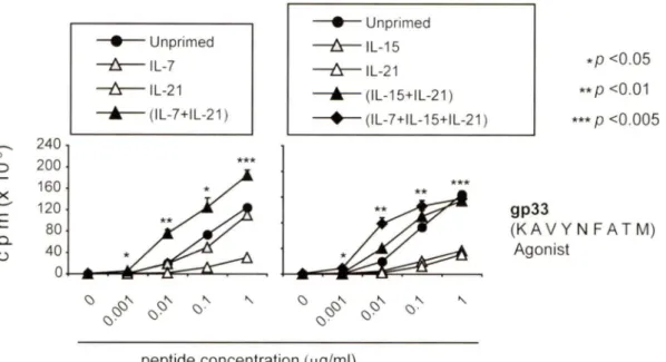
A few years ago, different groups including our own, have reported that T cells can be activated to proliferate in vitro in absence of stimulation through the TCR using various combinations of cytokines (Gagnon et al., 2007; Zeng et al., 2005). These reports showed that naïve CD441°CD8+ T cells could be induced to proliferate in the presence of homeostatic cytokines, such as IL-7 or IL-15, in combination with an inflammatory cytokine, such as IL-6 or IL-21 (Fig. 5). IL-21 boosted survival and proliferation of naïve and memory CD8+ T cells induced by IL-7 or IL-15, respectively (Gagnon et al., 2008). Furthermore, naïve P14 TCR transgenic CD8+ T cells that had been pre-stimulated with a synergistic combination of cytokines showed an increase in TCR sensitivity to low concentrations of cognate antigen, GP33, as revealed by increased proliferation and 112 and lfng gene expression (Gagnon et al., 2008). These cells also produced increased amounts of granzyme B and displayed a more efficient antigen-specific cytotoxicity towards target cells.
ln light of the above findings, it has been proposed that a combination IL-15 and IL-21 can stimulate naïve CD8+ T cells in an antigen-independent manner and would sensitize them to low concentration of antigens. This process is referred to as cytokine priming (Fig. 6) (Ramanathan et al., 2008). Cytokine priming proposes that, during an immune response, CD8+ T cells are recruited to the site of inflammation. At the site of inflammation, cytokines produced by cells of the innate immune system sensitize these CD8+ T cells and enable them to respond to low affinity antigenic peptides. It has been shown that "cytokine-primed" cells can up-regulate IL-2R
a
andB
as well as CD62L and CD44, which may allow them to continue to migrate in search of antigen (Gagnon et al., 2008).lnflammat:ion Transie nt:
j
~
~
Lymphopenia CDS Naive1
1
l
iL- 7 11
CDS
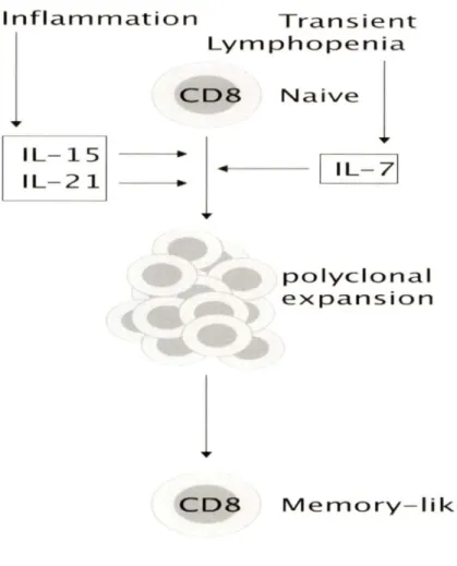
polyclonal expansion Memory- likeFigure 5: Antigen non-specific activation of naïve CDS+ T cells by cytokines. During an
intlammatory response, DCs, NK and macrophages of the innate immune system produce the IL-15 and IL-2 1 cytokines. The availability of IL-7 increases because of the down regulation of its receptor (Park et al., 2004). Sorne combinati ons of cytokines, such as IL-7 with IL-2 1 or IL- 15 with IL-21 , lead to a stimulation of naïve CD8+ T cells in absence of antigen or co-stimulation. These cells become "memory-like", gain effector functions and respond to a limited concentration of antigen (Illustration adapted from (Ramanathan et al., 2008)).
Na ive CDS T cell Antigen IL-15 + IL-21 IL- 7 + IL- 21 Cytokin synergy IL- 15 + IL- 21 IL- 7 + IL- 21 Proliferation
IL- 2 or IFNy secretion Ag -s pecific cytolytic activity
Signficant proliferation No IL- 2 or IFNy secretion
Negligible Ag-specific cytolytic activity
Primed CDS T cell Limiting Antigen High AG sensitivity lncreased proliferatio lncreased transcriptio
oftl2 andlfng genes lncreased secretion of
of IL-2 and IFNy lncreased expression
of granzyme B Elevated Ag-specific
cytolytic activity
Figure 6: The cytokine priming concept of naïve CDS+ T cells. Cytokine synergy induces
antigen-independent proli feration of naive CD8+ T cells and primes them to acquire potent effector functions upon subsequent antigen stimul ation. During an inflammatory response, cells of the innate immune system produce IL- 15 and IL-2 1, while IL-7 is constantly available. In synergy with IL-2 1, IL-1 5 or IL-7 can stimulate the proliferation of na ive CD8+ T cells in absence of its cognate antigen. These cytokine-pri med CD8+ T cells display a "memory-like" phenotype and have an increase TCR sensitivity to sub-optimal concentrations of cognate peptides. These cells also secrete high amounts of IL-2 and IFNy and display increased CTL response (Illustration adapted from (Ramanathan et al., 2009).
1.12. Common gamma chain family of cytokines
The IL-2 family of cytokines is also known as the common gamma chain family of cytokines, because their receptors share the gamma chain (CD132) of the IL-2 receptor. Besides IL-2, IL-7, IL-15 and IL-21, the other members of this family are IL-2, IL-4, IL-9 and TSLP (Alpdogan and van den Brink, 2005; Kovanen and Leonard, 2004). The absence of the gamma chain is associated with X linked SCID (X-SCID) in humans, which is characterized by a reduction in T-lymphocytes, B-lymphocytes and NK cells (Noguchi et al., 1993).
1.12.1. IL-7 and the IL-7 receptor in T cell biology
IL-7 is a hematopoietic growth and survival factor for T cells. IL-7 is a 25 kDa glycoprotein secreted by fetal liver cells, stromal cells from the bone marrow and, epithelial cells, including keratinocytes (Jiang et al., 2005). The IL-7 receptor is a heterodimer, composed of the IL-7Ra chain (CD127) and the CD132 chain. The binding of IL-7 toits receptor induces heterodomerization of CD127 and CD132, allowing the CD132-ssociated JAK3 to transphosphorylate the CD127-associated JAKl and to initiate the canonical JAK-STAT signaling pathway (Leonard and Lin, 2000; Rochman et al., 2009). Activated JAK3 and JAKl kinases phosphorylate the IL-7 receptor chains, leading to recruitment and phosphorylation of STAT3 and STAT5, their homo- and/or hetero- dimerization, translocation to the nucleus, binding to target gene promoters and expression of genes that promote cell survival (Jiang et al., 2005). IL-7 is a growth and maintenance factor for immature and mature thymocytes (Morrissey et al., 1994). IL-7 promotes the differentiation and survival of CD4+ and CD8+ SP thymocytes and proliferation, and is also a cofactor for the rearrangement of the TCR
p
gene (Muegge et al., 1993). IL-7 or IL-7Ra knockout mice displayed severely impaired T and B cell development(Peschon et al., 1994; von Freeden-Jeffry et al., 1995). In humans, absence of IL-7Ru leads to a genetic disorder known as severe combined immunodeficiency syndrome, where there is a complete lack of T cells and both arms of the adaptive immune system are disabled (Puel et al., 1998). In the periphery, IL-7 has a pro-survival function for naive and memory T cells, by up-regulating the anti-apoptotic factors Bcl-2 and the Kruppel-like factor (Schluns and Lefrancois, 2003; Schober et al., 1999). It has been shown that IL-7 is not needed in the antigenic response, but after the expansion phase, IL-7 is needed in the generation of memory CD4+ T cells (Kondrack et al., 2003; Li et al., 2003).
1.12.2. IL-7 and autoimmunity
IL-7 is a contributing factor in rheumatoid arthritis (RA), a systemic intlammatory autoimmune disease characterized by chronic inflammation of the joints, eventually resulting in the destruction of cartilage and bone (Feldmann et al., 1996). IL-7 may promote the immunopathology in RA by activating T cells, B cells, or monocytes/macrophages (van Roon et al., 2007). Patients suffering from RA show elevated levels of IL-7 as compared to healthy individuals. There is also an increase in the levels of IL-7Ru in inflamed joints, which could induce an immune response and recruitment of the immune cells to the joints (Hartgring et al., 2009). As discussed before, IL-7 may promote the expansion of naive autoreactive T cells under conditions of lymphopenia by inducing homeostatic expansion of naive T cells (Krupica et al., 2006).
1.12.3. IL-15 and the IL-15 receptor complex
15 was discovered in 1994 by two different groups, based on its ability to "mimic" IL-2-dependent T cell proliferation (Burton et al., 1994; Grabstein et al., 1994). IL-15 is a pleiotropic, four
a
helix bundle cytokine that belongs to the yc family of cytokines. The IL-15 receptor is a hetero-trimer composed of a specific IL-15Ra chain, a IL-2P chain (CD 122) and the common yc (CD132) chain. IL-15Ra is responsible for binding of the IL-15 cytokine. IL-15Ra is a type 1 transmembrane protein with a short cytoplasmic domain related structurally to IL-2Ra but it is specific to the IL-15 receptor (Giri et al., 1995). The binding of IL-15 to its receptor leads to the activation of the JAK/STAT pathway. Binding of IL-15 to the hetero-trimeric complex activates JAKl and JAK3, leading to phosphorylation of STAT3 and STAT5 (Alpdogan and van den Brink, 2005; Lin et al., 1995). Phosphorylated. STAT5 forms homodimers or hetero-dimers with phospho-STAT3 that translocates to the nucleus to bind target DNA regulatory elements, leading to the activation of gene expression (Alpdogan and van den Brink, 2005; Johnston et al., 1995). The IL-15 signaling pathway also induces phosphorylation of lymphocyte-specific protein tyrosine kinase (Lck) and the Syk kinase (Ratthe and Girard, 2004; Uhlin et al., 2005), even though the detailed mechanisms remain unknown. IL-15 promotes long-term survival and proliferative renewal of CDS+ memory T cells (Becker et al., 2002; Lodolce et al., 1998). IL-JS1- and IL-15Ra_1_ mice are lymphopenic, and 1ack CD8+mem:ory T cells (Kennedy et al., 2000; Lodolce et al., 1998). IL-15 enhances the survival of CD8+ T cells by up-regulating Bcl-2 (Schluns and Lefrancois, 2003; Wu et al., 2002).
1.12.4. Effect of IL-15 on dendritic cells
IL-15 stimulation increases DC's ability to stimulate Ag specific CD8+ T cells, and to activate NK cells (Jinushi et al., 2003; Mattei et al., 200la). IL-15 is important for antigen presentation. Ohteki's group has demonstrated that mice deficient in IL-2B or the common
y
chain showed a decrease in IL-12, IFNy and nitric oxide (NO) production (Ohteki et al., 2001), suggesting that IL-15 signaling is important for the initial activation of APC. Ohteki's group also showed that IL-15 on conventional DC ( cDC) induced an increased expression of CD40, which enabled interaction with CD40L expressed on plasmacytoid DCs, facilitating IL-12 production (Kuwajima et al., 2006). IL-12 functions as signal 3 for acquisition of CD8+ T cells full effector functions (Curtsinger et al., 1999; Curtsinger et al., 2005).
1.12.5. IL-15 and autoimmunity
IL-15 has been shown to be involved in the pathogenesis of some autoimmune diseases. For instance, IL-15 has been detected in the serum of patients with ulcerative colitis (Kirman and Nielsen, 1996), in the mucosa of patients with inflammatory bowel disease (Sakai et al., 1998) and in psoriatic patients. The psoriasis xenograft model in mice showed an anti-IL-15 therapy reduced the symptoms of the disease (Villadsen et al., 2003). Another autoimmune disease in which IL-15 is involved is multiple sclerosis (MS). MS patients show an increase in the number of peripheral blood mononuclear cells that expressed mRNA of IL-15 in comparison with bealthy subjects (Kivisakk et al., 1998). It was also observed that T cells had increased levels of membrane bound IL-15 and IL-15 receptor, which may the cause of increased release of IFNy (Vaknin-Dembinsky et al., 2008). IL-15 is also found in the synovial fluid ofrheumatoid arthritis patients and targeting IL-15 or its receptor is being evaluated as promising therapeutic option for
treating arthritis patients (Baslund et al., 2005; Mclnnes and Liew, 1998). Among these,
neutralizing human IL-15 Ab (HuMax-IL15 and AMG714) are in phase II clinical trials with
promising results in patients refractory to anti-TNF therapy (Asquith and Mclnnes, 2007;
Mclnnes and Schett, 2007)
1.12.6. IL-21 and its receptor
IL-21 is the latest member of the common gamma chain family of cytokines to be
discovered (Parrish-Novak et al., 2000; Spolski and Leonard, 2008). The IL-21 receptor was
identified prior to discovery of IL-21 because of its similarity to cytokine receptors of type 1
family (Ozaki et al., 2000; Parrish-Novak et al., 2000; Parrish-Novak et al., 2002). IL-21 was
cloned from a bank of cDNA of activated CD4+ T lymphocytes (Parrish-Novak et al., 2000).
IL-21 is produced by activated CD4+ T cells, specifically Th 17 and NKT cells (Monteleone et al.,
2009).
Itis structurally similar to IL-15 and shows homology to IL-2 (29% identity and 46%
similarity) (Parrish-Novak et al., 2000). The exons and introns of
1121 gene resemble those of the
112 gene, suggesting that they may have arisen from a gene duplication event (Parrish-Novak et
al., 2002).
Many Iymphoid cells, such as natural killer cells, T cells and B cells express the IL-21
receptor, depending on their activation state. Keratinocytes and cells of the myeloid linage, such
as DC, also express IL-21R (Collins et al., 2003; Distler et al., 2005). The IL-21 receptor is a
hetero-dimer composed of an IL-21Ra chain and CD132. IL-21 activates JAKl and JAK3.
Binding of ligand to the IL-21R results in the phosphorylation of JAKl that is associated with
the IL-21Ra chain, and JAK3 that is associated with the IL-21Ry chain. Activated JAKI/3
phosphorylate STATl and STAT3 and to a lesser extent STAT4, STAT5a and STAT5b (Habib
et al., 2003; Spolski and Leonard, 200S; Zeng et al., 2007). IL-21 has been shown to activate the MAPK pathway (Spolski and Leonard, 200S).
1.12. 7. Effects of IL-21 on T cells
Signaling via the IL-21 receptor can have multiple effects on hematopoietic cells including proliferation, differentiation, activation, changes in cytokines and chemokines production and effector functions (Davis et al., 2007a). Earlier studies have implicated IL-21 in antibody responses (Ozaki et al., 2002). Mice that are deficient in the IL-21 receptor showed normal B cell numbers, but generated more IgE and a significant defect in the antibody response to antigenic stimulation. Recent studies have shown an indispensable role for IL-2 l in the development of T follicular helper cells (Tfh) (Vogelzang et al., 200S). Tfh cells, which produce high amounts of autocrine IL-21, regulate the development of germinal centers. In synergy with IL-6, IL-21 plays a key role in T-helper cell differentiation into Thl 7 cells and this process is dependent on STAT 3 (Korn et al., 2007; Nurieva et al., 2007; Zhou et al., 2007). During Th17 differentiation process, IL-21 inhibits TGFP-driven differentiation of naïve T helper cells into Foxp3+ cells. In the Treg subset CD4+ CD25+ Foxp3+ IL-
?1°,
IL-21 induces both cell proliferation and suppressive functions.IL-21 alone has no discernible effects on CDS+ T cells. However, in synergy with IL-7 or IL-15, IL-21 induces proliferation of both CD4410 naïve and CD44hi as well as memory CDS+ T cell subsets (Gagnon et al., 2007; Zeng et al., 2005). As discussed earlier, naive CDS+ T cells stimulated with IL-21 in the presence of IL-7 or IL-15 show increased reactivity to cognate antigens and showed a gain of effector functions (Gagnon et al., 200S).