Title: Subcellular partitioning of metals and metalloids (As, Cd, Cu, Se and Zn) in liver and gonads of wild white suckers (Catostomus commersonii) collected downstream from a mining operation
Authors: Nastassia Urien, Sophie Cooper, Antoine Caron, Helga Sonnenberg, Lisa Rozon-Ramilo, Peter G.C. Campbell, Patrice Couture
PII: S0166-445X(18)30378-3
DOI: https://doi.org/10.1016/j.aquatox.2018.07.001
Reference: AQTOX 4974
To appear in: Aquatic Toxicology
Received date: 24-4-2018
Revised date: 27-6-2018
Accepted date: 1-7-2018
Please cite this article as: Urien N, Cooper S, Caron A, Sonnenberg H, Rozon-Ramilo L, Campbell PGC, Couture P, Subcellular partitioning of metals and metalloids (As, Cd, Cu, Se and Zn) in liver and gonads of wild white suckers (Catostomus
commersonii) collected downstream from a mining operation, Aquatic Toxicology
(2018), https://doi.org/10.1016/j.aquatox.2018.07.001
This is a PDF file of an unedited manuscript that has been accepted for publication. As a service to our customers we are providing this early version of the manuscript. The manuscript will undergo copyediting, typesetting, and review of the resulting proof before it is published in its final form. Please note that during the production process errors may be discovered which could affect the content, and all legal disclaimers that apply to the journal pertain.
Subcellular partitioning of metals and metalloids (As, Cd, Cu, Se and Zn) in liver and gonads of wild white suckers (Catostomus commersonii) collected downstream from a mining operation.
Nastassia Urien1, Sophie Cooper1, Antoine Caron1, 2, Helga Sonnenberg3 ỻ, Lisa Rozon-Ramilo3 ỻ, Peter G. C. Campbell1,
Patrice Couture1*
1 Institut national de la recherche scientifique, Centre Eau Terre Environnement (INRS–ETE), 490 de la Couronne,
Québec, QC G1K 9A9, Canada
2 Département des Sciences Biologiques, Université de Montréal, C.P. 6128, Montréal, QC H3C 3J7, Canada
3 Stantec Consulting Ltd., Guelph, ON N1G 4P5, Canada
ỻ Now at Ecological and Regulatory Solutions Inc., 17 Kirkland St., Guelph, ON N1H 4X7, Canada
* Corresponding author: Patrice Couture
Institut national de la recherche scientifique, Centre Eau Terre Environnement (INRS–ETE) 490 de la Couronne, Québec, QC G1K 9A9, Canada
Tel.: +1 418 559 3825 Fax: +1 418 654 2600
E-mail address: [email protected]
Highlights
Liver and gonads of exposed white suckers were markedly more contaminated in Se and Cd than those of reference fish.
Metal-handling strategies were overall similar between liver and female gonads.
Metal-handling strategies in ovaries and testes did not always follow the same pattern, likely due to their different reproductive roles.
Cadmium was reasonably well detoxified in liver and gonads by binding to metallothioneins.
Selenium accumulated in potentially metal-sensitive subcellular fractions, suggesting that fish may be subject to Se-related stress.
Abstract
In the present study, we examined the subcellular distribution of metals and metalloids (As, Cd, Cu, Se and Zn) in the liver and gonads of wild white suckers (Catostomus commersonii) collected downstream from a metal mining
operation (exposure area) and in a reference area. Metal partitioning among potentially metal-sensitive fractions (heat-denatured proteins (HDP), mitochondria and microsomes) and potentially biologically detoxified fractions (heat-stable proteins (HSP) and metal-rich granules) within cells was determined after differential centrifugation, NaOH digestion and heat-denaturation steps. Metal-handling strategies between liver and gonads, and between sexes, were examined. Hepatic metal concentrations were significantly higher in exposed compared to reference fish, especially for Se (14x), Cd (5x) and Cu (3x), and did not vary between sexes. In contrast, gonadal Cd, Cu, Se and Zn concentrations were consistently lower in testes than in ovaries; marked differences in Cd and Se concentrations between exposed and reference fish were observed for both sexes. Overall, metal-handling strategies were similar in both liver (male and female pooled) and female gonads, but differed from those in male gonads, likely due to the different functions assigned to ovaries and testes. Subcellular partitioning of As, Cd and Cu showed that the HSP fraction was most responsive to increased metal exposure, presumably reflecting Cu regulation, and possibly Cd and As detoxification. Zinc concentrations were tightly controlled and mainly found in the HDP fraction. Interestingly, changes in Cd-handling strategy in female gonads were particularly evident, with Cd shifting dramatically from the metal-sensitive HDP fraction in reference fish to the metal-detoxified HSP fraction in exposed fish. It seems that Cd detoxification in female gonads was not fully induced in the less contaminated fish, but became more effective
above a threshold Cd concentration of 0.05 nmol/g dry weight. Partitioning of Se was different, with the largest contributor to the total liver and gonad Se burdens being the putative metal-sensitive HDP fraction, suggesting that excess Se in this fraction in exposed fish may lead to Se-related stress. The present subcellular partitioning results demonstrate that metal handling strategies vary among metals, between organs and (in some cases) as a function of metal exposure. They also show promise in identifying metals of potential concern in a risk assessment context.
Keywords
Selenium; Cadmium; Fish; Subcellular distribution; Reproductive toxicity; Detoxification
1. Introduction
In Canada, governmental regulations on discharges and emissions from mining and smelting operations have contributed to decrease metal inputs into aquatic environments (Gunn et al., 1995). However, fish inhabiting lakes near metal mining and smelting operations are often contaminated by various metals, which, once they enter cells, can bind to biological sensitive components such as cytosolic molecules (e.g., enzymes and RNA) and organelles (e.g., mitochondria and nuclei), interfere with cell function, and cause deleterious effects (Wallace et al., 2003).
Fortunately, aquatic organisms are equipped with subcellular defence mechanisms, designed to prevent metals from exerting their toxic effects. For example, metals can be sequestered in insoluble metal-rich granules (MRG) or bound to cytosolic ligands that are able to complex metals, such as metallothioneins (MT) and metallothionein-like peptides (MTLP) (Mason and Jenkins, 1995).
It is generally recognized that bioaccumulated metal concentrations in wild fish are useful for evaluating metal exposure and risk of toxicity (Adams et al., 2011). However, given that some of the metal may have been detoxified, knowledge of metal subcellular partitioning within fish or specific fish organs may yield better estimates of the risk of metal toxicity than the current approach of using total tissue metal accumulation. Moreover, in aquatic ecosystems metals are usually present as mixtures, and determining the subcellular partitioning of different metals
could also help identify potential metals of concern. The partitioning of metals among subcellular fractions can be determined by successive differential centrifugation and heat-denaturation steps, leading to the separation of operational subcellular fractions that can be grouped into two categories: (i) “metal-sensitive fractions or MSF” (i.e., heat-denaturable proteins (HDP); mitochondria; and lysosomes and microsomes) where metals are thought to be bound to potentially metal-sensitive sites leading to deleterious effects, and (ii) “biologically detoxified metals or BDM” (i.e., MRG and heat-stable proteins (HSP), containing MT and MTLP) where metals are considered to have been handled in order to limit their toxic effects (Wallace et al., 2003).
Subcellular partitioning of Cd, Cu and Zn in fish has been well documented (Eyckmans et al., 2012; Giguère et al., 2006; Kraemer et al., 2005; Rosabal et al., 2015). Thus, it is now well established that a large majority of
accumulated hepatic Cd is normally found in the HSP fraction and is assumed to be mainly detoxified by binding to MT (and MTLP), for which it has a high affinity (Brown et al., 1990; Campbell et al., 2005; Giguère et al., 2006). Similarly, Cu and Zn, which are known to be regulated in part by MT and MTLP, are also found in the HSP fraction, almost exclusively in the case of Cu but distributed between the cytosolic HSP and HDP fractions for Zn (Giguère et al., 2006). In contrast, studies assessing the subcellular partitioning of As and Se are scarce (Barst et al., 2016; Ponton et al., 2016; Rosabal et al., 2015). Moreover, in fish, the liver is usually the chosen target organ for assessing the link between the subcellular partitioning of metals and toxicity, mostly because it is a key organ for energy storage and metal detoxification. Several studies have also reported the potential of metals to cause reproductive impairment in fish (Cazan and Klerks, 2015; ECC HC, 2017), but subcellular metal partitioning in fish gonads remains unexplored.
In this context, the present study had two objectives: (1) to assess the subcellular partitioning of arsenic (As), cadmium (Cd), copper (Cu), selenium (Se) and zinc (Zn) in the liver and gonads of mature wild white suckers
(Catostomus commersonii) collected in an exposure lake and in a reference lake; and (2) to compare metal-handling strategies in different organs (liver, ovaries and testes). White suckers were chosen as sentinel species because previous monitoring surveys (Environmental Effects Monitoring (EEM) studies conducted under the Metal Mining Effluent Regulations) have shown that in the exposure lake, the condition of the exposed white sucker population
was affected (liver and gonad-related indices being the most affected). Moreover, white suckers are well distributed and representative of the sampling region, abundant and sedentary (Harrison and Klaverkamp, 1990).
2. Materials and methods
2.1. Study areas
Water samples and fish were collected in October 2014 from two large lakes: one lake exposed to a mining effluent, referred to as “exposure area” hereafter, located on the Manitoba-Saskatchewan border (Canada), and one
reference lake, referred to as “reference area”, located 60 to 65 km west-northwest of the exposure area in
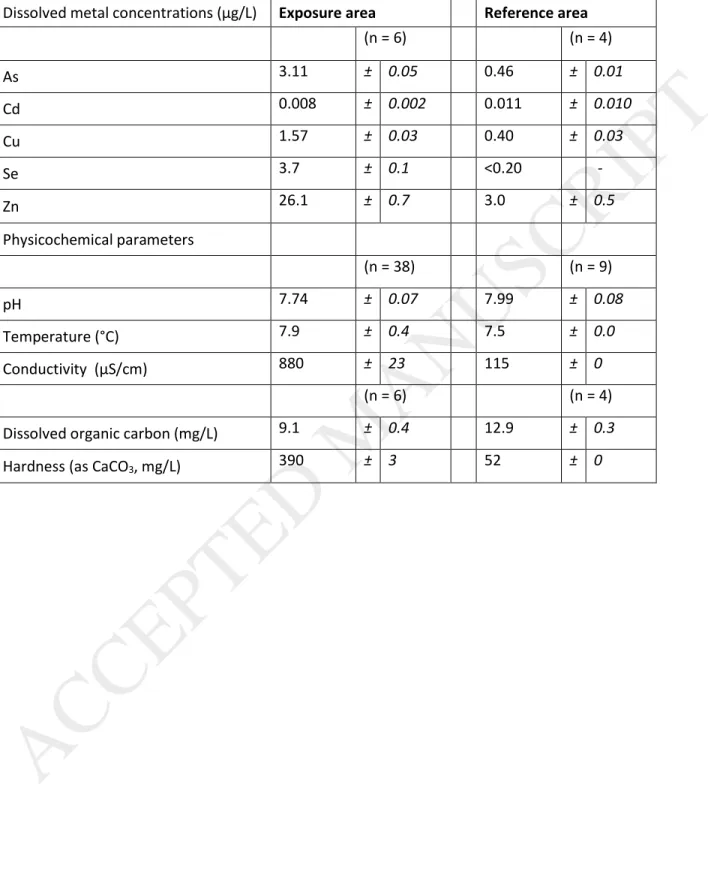
Saskatchewan. Both lakes are situated on the Precambrian Shield, where the surface waters are typically acidic, with high dissolved organic matter concentrations and low hardness (Environment Canada, 2000). The exposure area is situated downstream from a metal mining complex and has been subjected to inputs of various trace metals from mining and concentrating activities, and from a former smelting facility that ceased operations in 2010. The reference lake was of similar size compared to the exposure lake but has not been subjected to inputs from mining activities. The reference lake was also located upwind of the former smelter. Physicochemical parameters and dissolved metal concentrations in the water of the two study areas are presented in Table 1. For dissolved metal concentration measurements, water samples were collected just below the lake surface at the location where fish were caught and field-filtered using a hand pump and sterile Nalgene container fitted with a filter (0.45 μm porosity, Fisher Scientific). Samples were kept at 4°C until analyses were performed by ICP-MS. Other parameters were measured using a YSI™ water quality multiprobe (model 650-03 MSDS, Hoskin Scientific, Burlington, ON). Note that, because of the high volume discharge and the addition of lime during treatment processes, the conductivity and hardness values for the exposure area are much higher than in the reference area.
ACCEPTED MANUSCRIPT
2.2. Fish collection
White suckers are sedentary fish and were present in both lakes in shallow waters (< 10 m) in habitats harbouring aquatic plants. White suckers were caught from each lake using short-set gill nets, checked at intervals of less than 120 minutes to minimize the stress on the fish. Eight male and 10 female white suckers were collected from the exposure area, whereas 9 males and 9 females were collected from the reference area. Upon capture, white suckers were euthanized quickly by concussion and placed in a cooler on ice until processing on the shore. The capture and sampling protocol was approved by the INRS animal-care committee.
Once fish length (± 1 mm) and weight (± 0.01 g) had been recorded, fish were dissected to collect liver and gonads, and sex was determined. Organs were weighed and placed in pre-labelled Whirlpak bags, put on dry ice and shipped to the laboratory where they were immediately stored at -80˚C until analysis. To minimize the potential influence of fish age on the liver metal concentrations (Giguère et al., 2004; McFarlane and Franzin, 1980), attempts were made to collect fish of the same size class (36 – 46 cm). ). Scales and the first pectoral fin ray were collected for ageing (North Shore Environmental Services, Thunder Bay, ON). Both males and females were similar between areas in terms of age; overall, white sucker collected from the exposed and reference area were between 7 and 13 years old.
2.3. Subcellular metal partitioning
2.3.1. General subcellular fractionation procedure
To determine the subcellular distribution of metals in fish, one must first isolate the various metal-binding ligands and then determine the amount of metal associated with each ligand pool. In the present study, a differential centrifugation method was adapted from protocols previously described for yellow perch by Giguère et al. (2006) and Lapointe and Couture (2009) to separate biological samples into up to six operationally-defined subcellular fractions: “debris+nuclei” including nuclei, cell membranes, intact cells and connective tissue; NaOH-resistant debris, referred to as “granule-like structures”; “mitochondria”; “lysosomes+microsomes”; heat-denatured proteins termed
“HDP”, including cytosolic enzymes; and heat-stable proteins and peptides, termed “HSP”, including metallothionein (MT) and metallothionein-like peptides (MTLP). In the present study, we have assigned the granule-like structure and HSP fractions to the biologically detoxified metal category (BDM), and the HDP, mitochondria and
lysosomes+microsomes fractions to the metal-sensitive category (MSF) (Error! Reference source not found., see Supplementary data), while admitting that this classification is an oversimplification (Wallace et al., 2003) and may well not apply to all metals and metalloids (Caron et al., 2018). The “debris+nuclei” is an ambiguous fraction, in that it does not properly fall into either the BDM or MSF categories as we don’t yet know to what extent metals present in this fraction are associated with the nuclei, considered as sensitive to metals, or with cellular debris, not
considered as metal-sensitive. Generally, the presence of metals in this fraction is not interpreted and simply ignored (Cain et al., 2004; Rosabal et al., 2014).
2.3.2. Subcellular fractionation procedure: liver
Liver samples (≈ 1 g) were manually homogenized on ice in a Tris-sucrose buffer (25 mM/250 mM, pH 7.4) at a dilution of 1:3 (wet weight tissue:volume of buffer) using a glass grinder (4 x 5 turns of the pestle with 20-s intervals on ice). The tissue homogenate was centrifuged at 1500 x g for 15 min at 4°C. The supernatant (S1) was transferred to a microcentrifuge tube for further separations. The pellet (P1) was suspended in ultrapure water, heated at 96°C for 5 min and digested with NaOH (1 M) at 80°C for 60 min. Centrifugation at 10,000 x g for 30 min at room
temperature (≈ 20°C) was performed to separate the NaOH-resistant fraction (“granule-like structures”, P2) from the “debris+nuclei” (S2). The supernatant from the original homogenization step (S1) was centrifuged at 15,000 x g for 45 min at 4°C to collect the mitochondrial fraction (“mitochondria”, P3). A thin intermediary layer of fat was present between the supernatant and the pellet after this centrifugation step. This intermediate layer was not observed in earlier work on juvenile fish (Giguère et al., 2006), probably because in the present study the fish were mature and correspondingly richer in lipids than juvenile fish. In order to collect the best pellet for the mitochondria fraction (P3), the initial supernatant was collected and kept at 4°C, and the remaining pellet (and the intermediary layer) was
washed by re-suspension with 500 µL Tris-sucrose buffer and again centrifuged at 15,000 x g for 45 min at 4°C. The supernatant was collected and added to the supernatant resulting from the first centrifugation; the combined supernatant (S3) was centrifuged at 180,000 x g for 60 min at 4°C, giving a pellet containing the lysosomes and the microsomes (“lysosomes+microsomes”, P4), and the cytosolic fraction in the supernatant (S4). Separation of the heat-denaturable proteins (HDP) and the heat-stable proteins (HSP) was performed by heating the cytosolic
supernatant (S4) at 85°C for 30 min, cooling it on ice for 60 min and centrifuging at 50,000 x g for 10 min at 4°C. The supernatant (S5), which includes MT and MTLP, was collected as the HSP fraction. For each differential centrifugation step, both the resulting supernatant and the pellet (before and after lyophilization) were weighed.
2.3.3. Subcellular fractionation procedure: gonads
White sucker gonads were homogenized on ice in the Tris-sucrose buffer at a dilution of 1:4 (wet weight
tissue:volume of buffer), using five low-speed passages of a motorized Potter–Elvehjem homogenizer equipped with a Teflon pestle (Fisher Scientific, Whitby, ON, Canada). The procedure was the same for ovaries and testes, and similar to the one used for liver samples, except that the supernatant from the original homogenization step (S1) was directly centrifuged at 180,000 x g for 60 min at 4°C, so as to obtain a pellet containing the mitochondria and the lysosomes and microsomes together (“mitochondria+lysosomes+microsomes”, P4) since the mitochondria fraction was very small.
2.3.4. Metal concentrations in the fractions and in whole tissue
All microcentrifuge tubes and other labware were acid washed in 15% nitric acid (v/v; ACS grade, SCP Science) for at least 24 h, rinsed 5 times with distilled water, then twice with ultrapure water and dried under a laminar-flow fume to prevent inadvertent trace element contamination. Aliquots taken from the whole liver and gonad homogenates, as well as all pellets obtained from the subcellular fractionation procedure, were freeze-dried and then weighed to determine dry weights (dw). The freeze-dried samples were digested in 100 µL of nitric acid (70%, v/v, Optima grade,
Fisher Scientific, Whitby, ON, Canada) per mg of sample (dw) for 5 days at room temperature before addition of H2O2 (30%, v/v; Optima grade, Fisher Scientific, Whitby, ON, Canada) at a ratio of 48 µL per mg of sample (dw), and then heated at 60°C for 2 h. The supernatants (“HSP” and “debris+nuclei” fractions) were directly digested by adding 1 mL nitric acid for 5 days at room temperature, then 480 µL of H2O2 were added to the sample, which was then heated as described for the pellet. Cooled digests were then diluted 10-fold with ultrapure water, except for the
“debris+nuclei” fraction, for which a 100-fold dilution was applied. Certified reference material (lobster
hepatopancreas reference material for metals, TORT-3, National Research Council Canada, Halifax, NS, Canada) and digestion blanks were subjected to the same digestion procedure.
Total metal concentrations were also measured directly on raw organ tissues. Samples (≈ 0.5 g) were freeze-dried for 4 days, weighed and digested overnight at room temperature in 1 mL of nitric acid. The samples were heated at 80°C for 2 h before addition of 500 µL of H2O2 and heated again at 80°C for 2 h. Digests were then completed with 8.5 mL ultrapure water. As in the case for the subcellular fractions, TORT-3 and digestion blanks were subjected to the same digestion procedure.
Concentrations of As, Cd, Cu, Se, and Zn in aliquots of whole homogenates, pellets, supernatants and raw tissue samples were determined with an inductively coupled plasma–mass spectrometer (ICP–MS; Thermo Elemental X Series, Winsford, England, UK). Analytical procedural blanks and standard reference waters (one synthetic water [900-Q30-100, SCP Science] and one natural water [TMDA-64.2] from the Environment Canada Proficiency Testing Program #101 (2013), used to check the compliance of the calibration curve and to control for any drift of the calibration curve during the many hours of analyses), were analyzed during each run (every twenty samples). Aqueous certified reference samples were quantitatively recovered and analytical procedural blanks indicated no appreciable contamination. The recovery of TORT-3 reference samples (n = 6) was within 20% of the certified values for As (96 ± 18 %), Cd (91 ± 11 %), Cu (87 ± 4 %), Se (99 ± 16 %), and Zn (90 ± 11 %). No correction was applied.
ACCEPTED MANUSCRIPT
2.3.5. Subcellular fractionation procedure quality control
To assess the performance of the differential centrifugation procedures applied on liver and gonad tissues, a mass balance calculation was carried out by comparing total metal contents (nmol), estimated from the aliquots of whole liver and gonad homogenates, with the sum of metal contents measured in the various subcellular fractions as follows: [sum of metal burden in all the fractions/metal burden in the aliquot of whole liver and gonad homogenates aliquot] x 100. Regarding subcellular metal distribution assessment and interpretation, only samples for which the recovery was between 60% and 140% were included (detail of the number of samples included in Error! Reference
source not found.). Overall, mean recoveries were satisfactory for each metal in both tissues. In the gonads, the
number of samples for which the recovery for Cd was between 60% and 140% was less satisfactory than that observed for liver, likely because of the very low Cd levels observed in the gonads.
One of the biggest challenges in performing a subcellular fractionation protocol is homogenizing the biological tissue strongly enough to disrupt cell membranes, without damaging the organelles. Centrifugation force and duration can then be adapted to optimize the separation of organelles as a function of their settling velocities. Before applying the subcellular fractionation protocol to all liver and gonad samples, the efficacy of the procedure was assessed by determining the distribution of some marker enzymes located specifically in the cytosol (lactate dehydrogenase) and mitochondria (cytochrome C oxidase) (Rosabal et al., 2014). The final procedure represented the best compromise between having complete disruption of the tissue and maintaining organelle integrity. Although the ambiguity regarding the “debris+nuclei” fraction and the metal forms therein prevents us from assigning it to either the BDM or MSF categories, the relative contribution from this fraction to the total metal burden can be used as an indication of the efficacy of the homogenization procedure; a low and constant percentage contribution indicates that the homogenization step was both effective and reproducible (Rosabal et al., 2014). All these considerations have been taken into account in the interpretation of the subcellular data.
ACCEPTED MANUSCRIPT
2.4. Data analyses and statistics
Significant differences in total metal concentrations in fish between male and female fish, and between areas, were determined by analysis of variance (ANOVA, p < 0.05) if the conditions of normality and homoscedasticity were respected (respectively tested by the Shapiro-Wilkinson and Levene tests). If these conditions were not met (even after a log10-transformation), differences in metal concentration were tested using a non-parametric test performed on ranks (Wilcoxon-Mann-Whitney test, p < 0.05). Total metal concentrations are expressed in nmol/g dry weight (dw) and mean concentrations are given with their standard error (se). Metal concentrations in subcellular fractions were also expressed as total metal burden divided by total tissue dry weight (nmol/g dw). The relative contribution of each subcellular fraction to the total metal burden was estimated as: [metal burden in each fraction/sum of the metal burdens in all fractions] x 100, with results expressed in percentage (%).
Relationships between total metal concentrations in both organs and (i) metal concentrations in each subcellular fraction, or (ii) relative metal contributions (%), were tested by simple correlation analysis (Pearson correlation matrix if the condition of normality was respected, or, if not, with a Spearman test (p < 0.05)). For percentages, the data were arcsine-transformed before statistical analysis.
3. Results
3.1. Total metal concentrations
3.1.1. White sucker livers
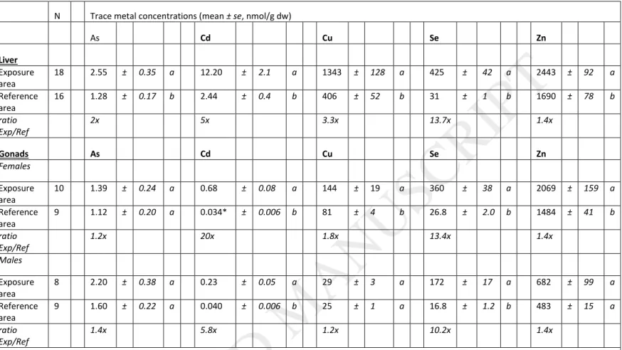
Total hepatic metal concentrations in white suckers from both the exposure and the reference areas were first compared between sexes. No significant differences in metal concentrations between male and female livers within a given area were observed (except for slight differences for Se and Zn in the reference area; 34 ± 2 vs. 29 ± 1.4 nmol/g dw (mean ± se) for Se in females and males, respectively, and 1908 ± 121 vs. 1520 ± 61 nmol/g dw for Zn). Accordingly, total metal concentration data from males and females were pooled together for the inter-site
comparison. For all metals studied, total concentrations were significantly higher in exposed compared to reference fish (Table 2). The highest differences between areas were observed for Se (13.7-fold) > Cd (5-fold) > Cu (3.3-fold). Arsenic and Zn liver concentrations in exposed fish were equal or less than 2-fold higher than in reference fish (2- and 1.4-fold higher, respectively).
3.1.2. White sucker gonads
For gonads, total metal concentrations in white sucker females were significantly higher in fish collected in the exposure area than in the reference area for Cd (20-fold) > Se (13.4-fold) > > Cu (1.8-fold) ≈ Zn (1.4-fold), whereas total metal concentrations in males were significantly higher in the exposure area for Se (10.2-fold) > Cd (5.8-fold) (Table 2). Total metal concentrations were systematically lower in male gonads than in female gonads for the essential elements Cu, Se and Zn in both the exposure and reference areas, and for Cd in the exposure area (ANOVA, p < 0.05). Overall, metal concentrations measured in gonads tended to be lower than hepatic concentrations.
3.2. Subcellular metal partitioning
To evaluate differences in metal partitioning in white suckers between the exposure and reference areas in liver and gonads, we plotted the metal concentrations (nmol/g dw) measured in each fraction against the total metal
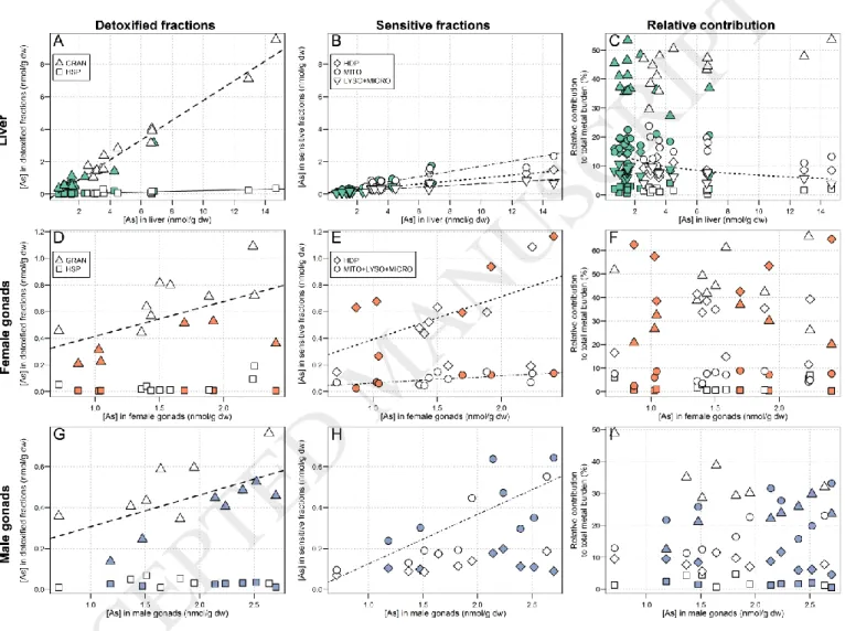
concentrations of the respective organ (nmol/g dw) for all individuals from the two areas together. In presenting the data this way, we can examine how metal partitioning varies over a relatively wide contamination gradient while taking into consideration the variability observed in total hepatic and gonadal metal concentrations. Results for subcellular partitioning in livers are presented with male and female fish pooled together, whereas results for female and male gonads are presented separately. We also plotted the relative contributions of each subcellular fraction to the total metal burden (%) against the total metal concentrations (nmol/g dw) in order to observe any changes in the metal-handling strategy. In the following figures presenting the results of subcellular partitioning for each element,
will always follow the same layout: plots A, B and C are attributed to the liver, plots D, E and F are attributed to the ovaries, and the plots G, H and I are attributed to the testes. Results regarding the fractions considered to be part of the BDM category are presented in the first column and fractions thought to belong to the MSF category are
presented in the second column. Results concerning the relative contributions of each subcellular fraction to the total metal burden (%) are presented in the third column.
The relative contribution from the “debris+nuclei” fraction to the total metal burden can be used as an indication of the efficacy of the homogenization procedure. In the present study, for the liver and female gonads, the contribution from the “debris+nuclei” fraction was reasonably low for all metals (on average ≤ 30%). In contrast, for male gonads, the relative contribution from the “debris+nuclei” fraction was higher (≥ 35% and up to 60%),
indicating that the homogenization step was less effective in male than in female gonads. Given this result,
comparisons of metal-handling strategies between male and female gonads, or between the liver and male gonads are compromised. Discarding this fraction (debris+nuclei) and re-calculating the relative contribution of each remaining fraction would imply that the metal burden measured in the “debris+nuclei” fraction is entirely “debris”, and that if it were completely homogenized the metal distribution among the other fractions would not change. This assumption could not be verified, but as a test the relative contribution of each fraction was re-calculated without considering the “debris+nuclei”; since no substantial changes were observed for interpretation of the data, we chose to retain the “debris+nuclei”.
In addition, a change in the relative contribution of the “debris+nuclei” fraction for one or some metals along the metal contamination gradient may reflect the association of the metal with nuclei rather than with debris
(Rosabal et al., 2015). Therefore, for a given organ, if the relative contribution of the “debris+nuclei” fraction varied as a function of the total metal concentrations, we assumed that this metal could be associated with nuclei and considered the fraction as potentially metal-sensitive (MSF); in these cases, the “debris+nuclei” fraction was plotted.
ACCEPTED MANUSCRIPT
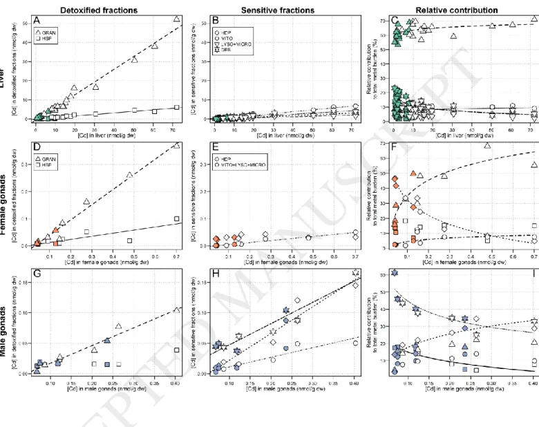
3.2.1. Non-essential metal (Cd)
Liver Cd concentrations increased significantly in all subcellular fractions with increasing total Cd concentrations, the strongest increase being observed for the HSP fraction (slope: 0.67, r2 = 0.98; slopes of the other fractions < 0.10)
(Figure 1, A and B). In fish from both the reference and exposure areas, the relative contribution of the HSP fraction was dominant (min – max: 53-71%) and slightly increased with increasing hepatic Cd concentrations (≈ 61% 64%; relationship best described by a logarithmic curve, r2 = 0.33). In comparison, the relative contributions of all other
fractions were low (≤ 20%). Note, however, that the relative contribution from the granule-like structure fraction increased with increasing hepatic Cd concentrations (≈ 5% 9%; logarithmic relationship, r2 = 0.16) while the
relative contribution from debris+nuclei decreased (≈ 17% 9%, logarithmic relationship, r2 = 0.57) (Figure 1, C).
In female gonads, for which total Cd concentrations were about 20 times higher in exposed compared to reference fish, Cd concentrations in all subcellular fractions increased significantly with increasing total Cd
concentrations, with the exception of the putative metal-sensitive HDP fraction that remained low and stable (p = 0.11, Spearman test). As was the case for the liver, the strongest increase was observed for the HSP fraction (slope: 0.56, r2 = 0.98) (Figure 1, D and E). In reference fish, the HDP fraction contributed the most to the total Cd burden
(mean contribution: 41%), whereas in exposed fish, the contribution from HDP dropped (HDP: 41% 20%,
logarithmic relationship, r2 = 0.96) in favour of the HSP fraction, for which the relative contribution dramatically rose
along the bioaccumulation gradient and became dominant (HSP: 28% 47%, logarithmic relationship, r2 = 0.80). A
slight increase in the contribution from the mitochondria+lysosomes+ microsomes fraction was also observed (logarithmic relationship, r2 = 0.49) but this change appeared negligible compared to changes in the HSP and HDP
fractions (Figure 1, F). No changes in the contribution from the debris+nuclei fraction were observed.
In male gonads, the Cd-handling strategy differed from that in liver and female gonads (Figure 1, G and H). Cadmium concentrations in each subcellular fraction increased significantly with increasing total Cd concentrations, except for the granule-like structures fraction (p = 0.37, Spearman test). The strongest increases were observed for the HDP and debris+nuclei fractions (slope: 0.44, r2 = 0.93 and slope: 0.35, r2 = 0.91, respectively), followed by the
HSP fraction, in third position only (slope: 0.29, r2 = 0.96). The relative contribution from the granule-like structures
and debris+nuclei fractions declined with increasing Cd concentrations (logarithmic relationship, r2 = 0.80 and power
relationship, r2 = 0.70, respectively). In contrast, the relative contribution from the HDP fraction increased with
increasing Cd concentrations (logarithmic relationship; r2 = 0.79) (Figure 1, I). If we exclude the debris+nuclei fraction,
the contribution from the HDP fraction becomes dominant. Note, however, that compared to females, total Cd in male gonads was 3- to 5-fold lower.
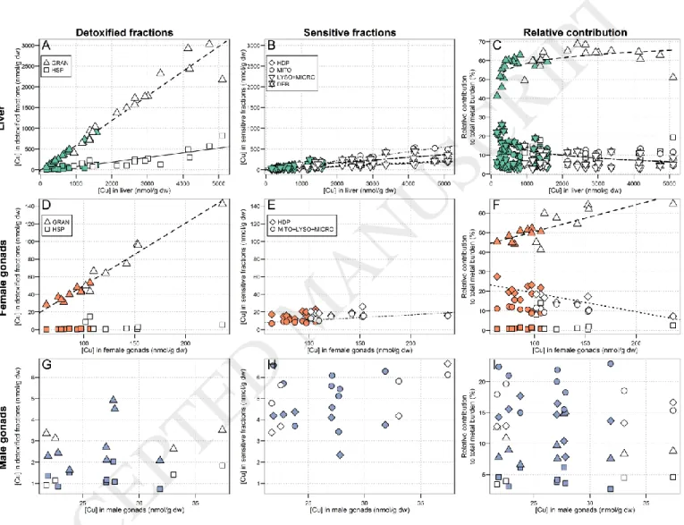
3.2.2. Essential metals (Cu and Zn)
As observed in Figure 2 (plots A and B), subcellular Cu partitioning in white sucker livers was very similar to that of Cd. Copper concentrations in all subcellular fractions increased significantly with increasing hepatic Cu levels, with the strongest increase being observed for the HSP fraction (slope: 0.60, r2 = 0.95; slopes of the other fractions ≤
0.11). Similar to Cd, the contribution of the HSP fraction to the total Cu burden was dominant (min – max: 41 - 69%) and it significantly increased with increasing hepatic Cu concentrations (≈ 56% 62%, adjustment best described by a logarithmic curve, r2 = 0.42). The contribution from the other fractions did not change, except for a slight decrease
for the debris+nuclei fraction (≈ 15% 9%, adjustment best described by a logarithmic curve, r2 = 0.44) (Figure 2, C).
Copper partitioning in female gonads was also similar to that of Cd. A significant increase in the HSP fraction with increasing Cu concentrations was observed (slope: 0.71, r2 = 0.96), as well as in the
mitochondria+lysosomes+microsomes fraction, but to a much lesser extent (slope: 0.06, r2 = 0.58) (Figure 2, D and E).
The contribution from the HSP fraction was dominant in reference and exposed fish and significantly increased with increasing Cu concentrations in whole gonads (≈ 49% 56%, r2 = 0.61). In contrast, the HDP fraction, the
contribution of which was only about half that of the HSP contribution, significantly decreased with increasing Cu concentrations in whole female gonads (≈ 21% 15%, r
ACCEPTED MANUSCRIPT
2 = 0.67, Figure 2, F).In male gonads, total Cu concentrations were similar between areas, and no changes in subcellular Cu partitioning were observed (p > 0.05, Pearson test). The mitochondria+lysosomes+microsomes fraction contributed the most to the total Cu burden (Figure 2, G, H and I).
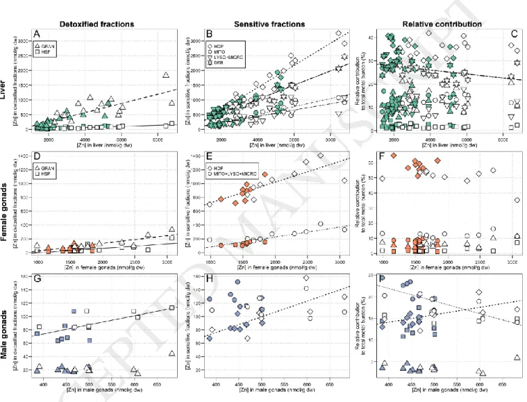
As total hepatic Zn concentrations increased, a significant increase in Zn concentrations was observed in all subcellular fractions. The importance of these increases is as follows: HDP (slope: 0.37, r2 = 0.95) > debris+nuclei
(slope: 0.24, r2 = 0.93) > HSP (slope: 0.16, r2 = 0.67) > mitochondria (slope: 0.14, r2 = 0.85) > lysosomes+microsomes
(slope: 0.11, r2 = 0.58) > granule-like structures (slope: 0.02, r2 = 0.68) (Figure 3, A and B). The HDP fraction afforded a
major contribution in both reference and exposed fish, and this proportion remained constant with increasing total hepatic Zn concentrations (p = 0.40, reference: 34%; exposed: 35%, on average). The contribution from the
debris+nuclei fraction slightly decreased with increasing hepatic Zn concentrations (29% 24%, r2 = 0.17) (Figure 3,
C).
In the gonads, Zn partitioning varied between sexes. For both males and females, no marked differences in total Zn concentrations were observed between areas (1.4-fold higher in exposed compared to reference fish) and the relative contribution of each fraction to the total Zn burden remained constant in proportion, except for the HDP and mitochondria+lysosomes+microsomes fractions in male gonads, which respectively increased and decreased with increasing Zn concentrations (r2 = 0.37 and 0.51, respectively). In females, the contribution of the HDP fraction
was dominant (≈ 54%, on average, Figure 3, F), whereas in male gonads the mitochondria+lysosomes+microsomes, HDP and granule-like structures fractions contributed more or less equally to the total Zn burden (Figure 3, I). In both cases, the relative contribution from the HSP fraction remained negligible, in distinct contrast to the results for Cd
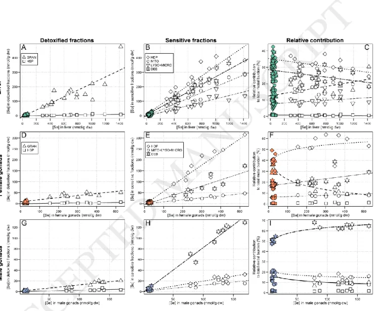
3.2.3. Metalloids (Se and As)
Selenium concentrations in each subcellular fraction increased significantly with increasing total hepatic Se
(concentrations about 14x higher in exposed compared to reference fish). The strongest increase was observed for the HDP fraction (slope: 0.33, r2 = 0.94), followed by the debris+nuclei (slope: 0.26, r2 = 0.95) > HSP (slope: 0.22, r2 =
0.84) > mitochondria (slope: 0.20, r2 = 0.94) > lysosomes+microsomes (slope: 0.12, r2 = 0.89) (Figure 4, A and B). The
increase in the granule-like structure fraction was negligible compared to the changes in the other fractions (slope: 0.007, r2 =0.80). As observed for Zn, the contribution of the HDP fraction was dominant in reference and exposed fish
but slightly decreased with increasing total hepatic Se (≈ 35% 31%, r2 = 0.35). The relative contribution from the
debris+nuclei fraction also slightly decreased with increasing hepatic Se (≈ 29% 24%, r2 = 0.21), whereas the
relative contribution from the HSP fraction to the total Se burden increased (≈ 12% 19%, r2 = 0.30). The relative
contributions from the lysosomes+microsomes and the granule-like structure fractions also significantly increased with increasing hepatic Se concentrations but remained negligible. No changes in the contribution from the mitochondria fraction were observed (Figure 4, C).
In female gonads, Se concentrations in each subcellular fraction increased significantly with increasing total Se concentrations. As observed for the liver, the strongest increases were observed for the HDP (slope: 0.60, r2 =
0.94) and nuclei+debris fractions (slope: 0.28, r2 = 0.89) (slopes for the other fractions < 0.10) (Figure 4, D and E). The
relative contribution of each fraction to the total Se burdens significantly varied with increasing total Se concentrations, except for the granule-like structure fraction. The most important changes in the relative
contribution of the fractions occurred for the HDP and HSP; an increased contribution from the HDP fraction was observed, accentuating its dominant contribution in exposed fish (≈ 41% 54%, logarithmic relationship, r2 = 0.46),
whereas a drop in the HSP relative contribution was observed (≈ 35% 13%, logarithmic relationship, r2 = 0.86)
(Figure 4, F). Overall, the subcellular Se partitioning pattern resembled that of Zn.
In male gonads, Se concentrations in all subcellular fractions also increased significantly with increasing Se concentrations, and this trend was particularly marked for the debris+nuclei (slope: 0.85, r2 = 0.98; slopes of the
other fractions ≤ 0.2). Moreover, the relative contribution from the debris+nuclei to the total Se burden slightly increased with increasing Se concentrations (≈ 51% 64%, logarithmic relationship, r2 = 0.88). The contributions
from the HSP and HDP fractions slightly decreased with increasing Se concentrations (logarithmic relationships, r2 =
0.73 for both HSP and HDP). When excluding the debris+nuclei fraction, the HDP fraction contributed the most to the total Se burden, but only slightly more than the HSP and mitochondria+lysosomes+microsomes fractions (Figure 4, G, H and I).
Hepatic As concentrations increased significantly with increasing As concentrations in all subcellular fractions, with the largest increase being observed for the HSP fraction (slope: 0.61, r2 = 0.96; slopes for the other
fractions ≤ 0.3) (Figure 5, A and B). The relative contribution from the HSP fraction to the total As burden was dominant and did not change with increasing As concentrations (42% on average), the relative contributions of the other fractions falling below 25%. Overall, the relative contribution of each fraction to the total As burden did not significantly vary with increasing As concentrations, except for the contribution from the HDP fraction that slightly decreased with increasing As concentrations (logarithmic curve adjustment, r2 = 0.44) (Figure 5, C).
In male and female gonads, total As concentrations did not vary between areas, and no changes in the contribution from each fraction to the total As burden were observed, except for a slight increase in the
mitochondria+lysosomes+microsomes fraction in males (ρ = 0.043, Spearman test, regression curve not shown). In females, subcellular fractions that contributed the most to the total As burden were the HSP and HDP, together, whereas in males, the HSP and the mitochondria+lysosomes+microsomes fractions were dominant (Figure 5, F and I).
4. Discussion
4.1. Subcellular metal partitioning
All statistically significant changes have been systematically reported in the Results section. In the following discussion, only results that have a high ecotoxicological relevance and importance will be discussed.
4.1.1. Non-essential metal (Cd)
In the present study, Cd accumulated in organs of exposed fish was 5 to 20 times higher (for liver and ovaries, respectively) than in reference fish. In a previous study conducted on white suckers in the same region, Harrison and Klaverkamp (1990) reported similar hepatic Cd concentrations (0.28 µg/g ww in exposed white suckers, i.e., ≈ 12 nmol/g dw).
Liver
The majority of hepatic Cd was found in the HSP fraction (≈ 60%), which is consistent with most other studies that have assessed subcellular Cd partitioning in fish livers (Brown et al., 1990; Campbell et al., 2008; Rosabal et al., 2015). In fish, the heat-stable proteins (HSP) fraction has been shown to contain mainly metallothionein (MT) and metallothionein-like proteins (MTLP) (Caron et al., 2018), which are rich in cysteine and exhibit a high affinity for Cd, resulting in its sequestration and detoxification (Mason and Jenkins, 1995). Thus, in the present study, we suggest that white suckers were reasonably able to cope with hepatic Cd accumulation, likely by involving MT and MTLP found in the HSP fraction. Note, however, that in our study, detoxification seems to be incomplete as increasing Cd concentrations in the liver were accompanied by significant increases in Cd concentrations in the organelles and HDP fraction (approximately 10% of the hepatic Cd burden). Incomplete Cd detoxification has been reported in the past in field-collected fish such as yellow perch (Perca flavescens) and eels (A. anguilla and A. rostrata) (Giguère et al., 2006; Rosabal et al., 2015).
Gonads
In female gonads, although Cd concentrations were much lower than those observed in the liver (≈7x less in reference fish and ≈50x less in exposed females), ovarian Cd concentrations in exposed fish were 20 times higher than in reference fish and Cd-handling strategies differed markedly between areas. In reference fish, the relative contribution from the HDP fraction was higher than that of the HSP fraction; the opposite trend was observed in exposed fish, with a dramatic increase in the relative contribution from the HSP fraction (~28% 47%, Figure 1) and
a concurrent drop in the contribution from the HDP fraction (~41% 20%, Figure 1). These results point to the clear involvement of MT in Cd detoxification in white sucker ovaries and suggest that above a threshold Cd concentration, Cd detoxification becomes more effective. The same pattern of response was also observed in a field study by Rosabal et al. (2014), who reported that in the aquatic invertebrate, Chaoborus punctipennis, there was a threshold body concentration of about 50 nmol/g dw below which the invertebrate “[did] not ‘turn on’ its detoxification machinery to the fullest extent”. In our study, the observation that reference white suckers “allowed” the presence of a small amount of Cd in the HDP fraction, and apparently did not induce detoxification defences, suggests that at internal Cd concentrations below 0.05 nmol/g dw in female gonads, a trade-off occurs between the cost of Cd detoxification and the induction of cell repair mechanisms initiated by Cd spillover into metal-sensitive fractions. This possibility has been previously discussed by Campbell et al. (2005) in the case of bivalves chronically exposed to Cd in the environment.
Cadmium concentrations in male gonads were very low (0.23 nmol/g dw in exposed fish, Table 2), and as previously discussed, Cd detoxification in male gonads does not seem to have been fully induced, since the proportion of Cd in the HDP fraction increased as the total gonadal Cd burden increased, and this fraction contributed more than the HSP fraction. Nevertheless, the declining relative contribution from the debris+nuclei fraction that was noted with increasing total gonadal Cd concentrations suggests a partial protection of the nuclei. To our knowledge, there are no previous studies on subcellular Cd partitioning in fish gonads with which to compare our results.
Besides the reasonable detoxification of Cd in fish organs (liver and gonads) observed in the present study, it is worth mentioning that it has been suggested that Se could prevent Cd toxicity by protecting organisms from oxidative stress (Ponton et al., 2016). Moreover, a recent laboratory study has also reported a reduced accumulation of Cd in the liver and kidney of rainbow trout (Oncorhynchus mykiss) chronically exposed to Cd and fed a diet
supplemented with selenomethionine (SeMet) (Jawmal et al., 2018). Although it was not possible to verify this in our
study, we speculate that the high amount of Se accumulated in the liver and gonads may have reduced Cd accumulation.
4.1.2. Essential metals (Cu and Zn)
Liver
Subcellular Cu partitioning in white sucker livers was very similar to that of Cd, with the Cu burden being mostly associated with the HSP fraction both in reference and exposed fish (≥ 50%, Figure 2). Zinc was mainly found in the HDP fraction (relative contribution to the total Zn burden ≈ 30-40%, Figure 3), and was also distributed in all of the other subcellular fractions, which is consistent with its essential role in many metabolic reactions, particularly as a co-enzyme and as a key constituent of zinc fingers (Mason and Jenkins, 1995). The higher contribution from the HSP fraction to the total Cu burden compared to that of Zn could be explained by the stronger affinity of MT for binding Cu (and Cd) than Zn (Hogstrand et al., 1991). Moreover, the relative proportion of Cu associated with the HSP increased with increasing Cu concentrations in liver, indicating that MT are involved in Cu regulation. In a previous study on white suckers inhabiting a metal-contaminated environment, Klaverkamp et al. (1991) observed that 47% of the hepatic Cu burden was bound to MT. Similar responses were also observed in wild juvenile yellow perch (Perca
flavescens) collected or transplanted in metal-contaminated areas; Cu and Cd were mainly associated to the HSP
fraction and MT, whereas Zn was mostly found in the HDP fraction (Giguère et al., 2006; Kraemer et al., 2005). Despite elevated dissolved Zn concentrations in the exposure area (9 times higher than in the reference lake, Table 1), no marked differences in hepatic Zn concentrations between exposure and reference fish were observed (only 1.4-fold higher in exposed fish compared to reference fish), indicating that Zn was well regulated by white suckers. This observation is consistent with the essential nature of this metal and the homeostatic control on its concentrations exerted by aquatic organisms (Rainbow, 2002).
Gonads
For male and female gonads, no consistent differences in Cu and Zn concentrations between lakes were observed (Exposed/Reference ratios, see Table 2), which suggests tight homeostatic control. Subcellular distributions of Cu and Zn in female gonads were similar to the patterns observed for the liver; Cu was found in the HSP fraction, whereas most of the cellular Zn was found in the HDP fraction. In contrast, in males both Cu and Zn were found bound to the mitochondria+lysosomes+microsomes fraction to a greater extent than in the HDP or HSP fractions. Note, however, that gonadal Cu and Zn concentrations were about 3-times lower in males than in females, likely due to the higher needs for these essential metals for ovary maturation than in spermatogenesis. These results suggest that in the absence of Cu or Zn overloads, these metals were mainly accumulated in organelles for metabolic purposes.
4.1.3. Metalloids (Se and As)
Se
Compared to the other metals and arsenic, Se was the most accumulated element in white sucker livers along the bioaccumulation gradient (≈ 14x, Table 2). In an earlier study assessing metal contamination in white suckers collected in 1982 in lakes near our exposure area, Harrison and Klaverkamp (1990), observed lower hepatic Se concentrations in exposed white suckers (2.20 µg/g ww, i.e. ≈133 nmol/g dw) and similar Se levels in reference fish (≈33-51 nmol/g dw).
Selenium is an essential nutrient for animals, required for normal biochemical processes such as the action of glutathione peroxidases and thioredoxin reductase (Arnér and Holmgren, 2000; Pappas et al., 2008). However, compared to other essential elements, Se is an element with a relatively narrow concentration range between the threshold for physiological requirements and the onset of toxicity (Lemly, 1993a). In the literature, it has been reported that food was the main source of Se accumulation for fish, in the form of organo-selenium compounds (Hamilton 2004; Stewart et al. 2010).
Liver
As hepatic Se concentrations increased in white suckers, Se consistently increased in all subcellular fractions, the strongest increase being observed for the HDP fraction, followed by the debris+nuclei, HSP and mitochondria fractions (Figure 4). The presence of significant amounts of Se in almost every fraction, and particularly in the HDP pool, is consistent with the observation that, in vertebrates, Se is mainly found in organic forms, usually incorporated into proteins as selenoamino acids (selenocysteine, SeCys; selenomethionine, SeMet) (Janz et al., 2014, 2010). Note that SeMet is not thought to confer heat-stability to proteins and thus proteins containing SeMet would be expected to report to the HDP fraction (Ponton et al., 2016). In the literature, it has been suggested that when present in excess, Se that incorporates non-specifically into proteins may prevent proteins from folding correctly, causing subsequent alteration of their functions (Janz, 2012; Maier and Knight, 1994).
In exposed white suckers, the relative contributions of the HSP fraction increased with increasing total hepatic Se concentrations (from approximately 10% to 20%), whereas the contribution from the HDP and debris+nuclei fractions decreased. The proportion of Se in the HSP fraction is consistent with the relative
contribution of ≈15% reported in yellow perch by Ponton et al. (2016). The overall trends with increasing hepatic Se observed in the present study suggest partial sequestration of Se in the HSP fraction, contributing to protection of the HDP and nuclei. However, unlike other essential metals such as Cu or Zn, for which many studies have
demonstrated the clear involvement of MT in their regulation, suggesting that Se is also associated with or
incorporated into MT is speculative. Indeed, recent investigations into the metal-binding ligands present in the HSP fraction in white suckers showed no evidence for an association of Se with MT (N. Urien, unpublished data). At this stage, we can only speculate as to the proper classification of the Se found in the HSP fraction (i.e., BDM or MSF).
Gonads
Most of the Se found in the female gonads was accumulated in the putative metal-sensitive HDP fraction and the relative contribution of this fraction to the total Se burden increased with increasing gonadal Se concentrations. Concurrently, the contribution from the HSP fraction dropped from approximately 40% to 10%. The overall trends
with increasing hepatic Se observed in female gonads suggest reduced Se-handling in the HSP fraction, which may reflect differences in Se-handling strategies in ovaries and liver (assuming that HSP is a detoxified fraction for Se). The presence of high amounts of Se in the putative metal-sensitive HDP fraction could be partly explained by the high demand for enzymes, including enzymes, needed for oogenesis. Maternal transfer of Se and seleno-proteins in yolk during oogenesis has been well documented in the past (Janz, 2012; Lemly, 1993b; Unrine et al., 2006). However, in egg-laying organisms, high amounts of internal Se have often been associated with teratogenic effects, such as higher offspring deformities, reduced offspring survival, and also with oxidative stress
(Covington et
al., 2018; Janz, 2012; Janz et al., 2010; Lemly, 1993b)
. In white suckers collected downstream from uranium mining and milling operations in Saskatchewan, Muscatello et al. (2009) observed fish egg Se concentrations two- to threefold higher than in reference fish (4.9 µg/g dw versus 1.9 µg/g dw), and also reported a slightly higher frequency (≈ 3%) of deformities (edema) in larvae from exposed fish compared to those from the reference area. In another study, performed on northern pike from the same area (uranium mining operations in Saskatchewan; Muscatello et al., 2006), a 10-, to 15-fold increase in Se concentrations in eggs (mean concentration in exposed fish of about 40 µg/g dw) was associated with a significant increase in the frequencies of larval deformities. Recently, threshold values in freshwater fish eggs for reproductive toxicity, ranging from 11.8 µg/g dw to 20 µg/g dw (i.e., 149 to 253 nmol/g dw), have been proposed (DeForest et al., 2012; ECC HC, 2017; US EPA, 2016). In our study, Seconcentrations in mature ovaries from exposed females were above these thresholds (360 nmol/g dw on average, i.e., 28.4 µg/g dw).
Finally, the presence of Se in the putative metal-sensitive HDP fraction suggest that Se detoxification was limited (assuming that HSP is a detoxified fraction for Se) and that white suckers may be subjected to stress.
As (liver and gonads)
In the present study, arsenic contamination of liver and gonads was generally low (< 3 nmol/g dw) and few differences between exposed and reference fish were observed (Table 2). In comparison, hepatic As concentrations
measured in eels (immature A. anguilla and A. rostrata) collected in the Saint-Lawrence and Gironde estuaries ranged on average from 30 to 150 nmol/g dw (Pannetier et al., 2016). This marked difference presumably reflects the fact that white suckers reside in freshwater environments, which are naturally lower in As than are coastal marine environments.
In the liver, and to a lesser extent in the gonads, As was mainly associated with the HSP fraction and no significant changes in its relative contribution to the total As burden were observed as As concentrations increased, suggesting that no consistent defence mechanisms were triggered, and also probably reflecting a partial protection of the metal-sensitive fractions. High proportions of As were also reported in the HSP fraction of livers of the European eels (A. anguilla), and to a lesser extent in the American eels (A. rostrata) (Rosabal et al., 2015).
Nevertheless, in our study, non-negligible proportions of the As were found associated with metal-sensitive fractions, HDP and organelles (≈ 20% for the liver; and up to 40% for the gonads), which could potentially lead to toxicity since As has no known essential role in fish.
4.2. Organ comparison
Comparison between the livers and female gonads revealed no marked differences in metal-handling strategies (HSP dominant for As, Cd and Cu; HDP dominant for Zn and Se). However, in the case of Se, female gonads seem to be less effective in protecting the presumably metal-sensitive HDP fraction than is the liver (decreased contribution from the HSP in favour of the HDP in ovaries versus increased contribution from the HSP and decrease of the HDP in liver). This difference could be explained by the fact that liver and ovaries have distinct functions; the liver is an important organ involved in substance detoxification and energy metabolism, whereas the ovaries are dedicated to egg production. Nevertheless, these results highlight the overall similarity of the metal-handling strategies between these two organs that have substantially different functions.
Comparison between liver and testes was more difficult, given the high proportions of metals found in the debris+nuclei fraction of testes. Nevertheless, differences between liver and testes were more frequent than for liver
versus ovaries: HSP was dominant in liver for Cd and Cu but in testes HDP was dominant for Cd and organelles for Cu;
HDP was dominant in liver for Zn but organelles were dominant in testes. These differences are likely related to the lower metal concentrations in the testes compared to those observed in the liver, where the need to trigger detoxification mechanisms was presumably greater.
Metal-handling strategies in ovaries and testes did not always follow the same pattern, probably because of the sex-related differences of these two reproductive organs; testes are dedicated to spermatogenesis and ovaries have not only the function to produce eggs but also to transfer enough reserves (including essential metals) to allow the eggs to develop once fertilized.
5. Conclusions
Subcellular metal partitioning was evaluated in white sucker liver and gonads in order to understand their behaviours and how they were regulated by fish once internalized, and to eventually identify potential metals of concern in a mixture. Subcellular metal distributions were overall similar in both liver and female gonads, but differed from those of male gonads. Except for Se, white suckers were overall able to cope with all elements, As, Cd and Cd being mainly present in the HSP fraction and presumably sequestered by MT and MTLP, and Zn exerting a homeostatic control on its concentrations. In the case of Se, given that Se concentrations differed markedly between exposed and reference fish in both livers and gonads, and that the sequestration of this metalloid by cytosolic, thermostable ligands (HSP fraction) was weak in livers and quasi-absent in gonads, our results suggest that fish could be subjected to Se-induced stress. Cadmium detoxification in female gonads seemed to be more efficient than in the liver, with a majority of Cd found in the HSP fraction despite a Cd concentration up to 20 times higher in exposed compared to reference females. Interestingly, it appears that detoxification of Cd in female gonads was not fully induced in the less contaminated fish, “allowing” Cd to spill over into metal-sensitive fractions, but above a threshold Cd
concentration (0.05 nmol/g dw), Cd detoxification became more effective. Lastly, the subcellular metal partitioning
approach has made it possible to make inferences about the behaviour of metals once internalized and has allowed us to identify potential metals of concern within a mixture of bioaccumulated metals.
6. Acknowledgments
The authors thank the industrial partner, Stantec Consulting Ltd., and the Natural Sciences and Engineering Research Council of Canada (NSERC) for co-funding this study. We would also like to thank Stantec staff members who
participated in the field work, and J. Perrault for her assistance in metal measurements in the INRS-ETE laboratory. PGCC was supported by the Canada Research Chairs program.
7. References
Adams, W.J., Blust, R., Borgmann, U., Brix, K.V., DeForest, D.K., Green, A.S., Meyer, J.S., McGeer, J.C., Paquin, P.R., Rainbow, P.S., Wood, C.M., 2011. Utility of tissue residues for predicting effects of metals on aquatic organisms. Integr. Environ. Assess. Manag. 7, 75–98. https://doi.org/10.1002/ieam.108
Arnér, E.S., Holmgren, A., 2000. Physiological functions of thioredoxin and thioredoxin reductase. FEBS J. 267, 6102– 6109.
Barst, B.D., Rosabal, M., Campbell, P.G.C., Muir, D.G.C., Wang, X., Köck, G., Drevnick, P.E., 2016. Subcellular
distribution of trace elements and liver histology of landlocked Arctic char (Salvelinus alpinus) sampled along a mercury contamination gradient. Environ. Pollut. 212, 574–583.
https://doi.org/10.1016/j.envpol.2016.03.003
Brown, D.A., Bay, S.M., Hershelman, G.P., 1990. Exposure of scorpionfish (Scorpaena guttata) to cadmium: effects of acute and chronic exposures on the cytosolic distribution of cadmium, copper and zinc. Aquat. Toxicol. 16, 295–310. https://doi.org/10.1016/0166-445X(90)90042-N
Cain, D.J., Luoma, S.N., Wallace, W.G., 2004. Linking metal bioaccumulation of aquatic insects to their distribution patterns in a mining-impacted river. Environ. Toxicol. Chem. 23, 1463–1473. https://doi.org/10.1897/03-291 Campbell, P.G.C., Giguère, A., Bonneris, E., Hare, L., 2005. Cadmium-handling strategies in two chronically exposed
indigenous freshwater organisms—the yellow perch (Perca flavescens) and the floater mollusc (Pyganodon
grandis). Aquat. Toxicol. 72, 83–97.
Campbell, P.G.C., Kraemer, L.D., Giguère, A., Hare, L., Hontela, A., 2008. Subcellular distribution of cadmium and nickel in chronically exposed wild fish: inferences regarding metal detoxification strategies and implications for setting water quality guidelines for dissolved metals. Hum. Ecol. Risk Assess. 14, 290–316.
Caron, A., Rosabal, M., Drevet, O., Couture, P., Campbell, P.G., 2018. Binding of trace elements (Ag, Cd, Co, Cu, Ni, and Tl) to cytosolic biomolecules in livers of juvenile yellow perch (Perca flavescens) collected from lakes representing metal contamination gradients. Environ. Toxicol. Chem. 37, 576–586.
Cazan, A.M., Klerks, P.L., 2015. Effects from a short-term exposure to copper or cadmium in gravid females of the livebearer fish (Gambusia affinis). Ecotoxicol. Environ. Saf. 118, 199–203.
https://doi.org/10.1016/j.ecoenv.2015.04.039
Covington, S.M., Naddy, R.B., Prouty, A.L., Werner, S.A., Lewis, M.D., 2018. Effects of in situ selenium exposure and maternal transfer on survival and deformities of brown trout (Salmo trutta) fry. Environ. Toxicol. Chem. (in press). https://doi.org/10.1002/etc.4086
DeForest, D.K., Gilron, G., Armstrong, S.A., Robertson, E.L., 2012. Species sensitivity distribution evaluation for selenium in fish eggs: Considerations for development of a Canadian tissue-based guideline. Integr. Environ. Assess. Manag. 8, 6–12. https://doi.org/10.1002/ieam.245
ECC HC, 2017. Screening assessment selenium and its compounds (No. ISBN 978-0-660-24255-2). Environment and Climate Change Canada Health Canada, Ottawa, ON, Canada.
Environment Canada, 2000. Ecological assessment of the boreal forest ecozone. N. Urquizo, J. Bastedo, T. Brydges and H. Shear (Eds), Ottawa, ON, Canada.
Eyckmans, M., Blust, R., De Boeck, G., 2012. Subcellular differences in handling Cu excess in three freshwater fish species contributes greatly to their differences in sensitivity to Cu. Aquat. Toxicol. 118–119, 97–107. https://doi.org/10.1016/j.aquatox.2012.03.019
Giguère, A., Campbell, P.G.C., Hare, L., Couture, P., 2006. Sub-cellular partitioning of cadmium, copper, nickel and zinc in indigenous yellow perch (Perca flavescens) sampled along a polymetallic gradient. Aquat. Toxicol. 77, 178–189.
Giguère, A., Campbell, P.G.C., Hare, L., McDonald, D.G., Rasmussen, J.B., 2004. Influence of lake chemistry and fish age on cadmium, copper, and zinc concentrations in various organs of indigenous yellow perch (Perca
flavescens). Can. J. Fish. Aquat. Sci. 61, 1702–1716. https://doi.org/10.1139/f04-100
Gunn, J., Keller, W., Negusanti, J., Potvin, R., Beckett, P., Winterhalder, K., 1995. Ecosystem recovery after emission reductions: Sudbury, Canada. Water. Air. Soil Pollut. 85, 1783–1788. https://doi.org/10.1007/BF00477238 Hamilton SJ. 2004. Review on selenium toxicity in the aquatic food chain. Sci Total Environ 326:1–31.
Harrison, S.E., Klaverkamp, J.F., 1990. Metal contamination in liver and muscle of northern pike (Esox lucius) and white sucker (Catostomus commersonii) and in sediments from lakes near the smelter at Flin Flon, Manitoba. Environ. Toxicol. Chem. 9, 941–956. https://doi.org/10.1002/etc.5620090712
Hogstrand, C., Lithner, G., Haux, C., 1991. The importance of metallothionein for the accumulation of copper, zinc and cadmium in environmentally exposed perch, Perca fluviatilis. Pharmacol. Toxicol. 68, 492–501. https://doi.org/10.1111/j.1600-0773.1991.tb01275.x
Jamwal, A., Lemire, D., Driessnack, M., Naderi, M., Niyogi, S., 2018. Interactive effects of chronic dietary
selenomethionine and cadmium exposure in rainbow trout (Oncorhynchus mykiss): A preliminary study. Chemosphere 197, 550–559.
Janz, D.M., 2012. Selenium in homeostasis and toxicology of essential metals. Fish Physiol. 314, 327–374.
Janz, D.M., DeForest, D.K., Brooks, M.L., Chapman, P.M., Gilron, G., Hoff, D., Hopkins, W.A., McIntyre, D.O., Mebane, C.A., Palace, V.P., 2010. Selenium toxicity to aquatic organisms, in: Chapman, P.M., Adams, W.J., Brooks, M.L., Delos, C.G., Luoma, S.N., Maher, W.A., Ohlendorf, H.M., Presser, T.S., Shaw, D.P. (Eds.), Ecological Assessment of Selenium in the Aquatic Environment. CRC Press, Boca Raton, FL, USA, pp. 141–231. Janz, D.M., Liber, K., Pickering, I.J., Wiramanaden, C.I., Weech, S.A., Gallego-Gallegos, M., Driessnack, M.K., Franz,
E.D., Goertzen, M.M., Phibbs, J., Tse, J.J., Himbeault, K.T., Robertson, E.L., Burnett-Seidel, C., England, K., Gent, A., 2014. Integrative assessment of selenium speciation, biogeochemistry, and distribution in a northern coldwater ecosystem. Integr. Environ. Assess. Manag. 10, 543–554.
https://doi.org/10.1002/ieam.1560
Klaverkamp, J.F., Dutton, M.D., Majewski, H.S., Hunt, R.V., Wesson, L.J., 1991. Evaluating the effectiveness of metal pollution controls in a smelter by using metallothionein and other biochemical responses in fish, in:
Newman, M.C., McIntosh, A.W. (Eds.), Metal Ecotoxicology - Concepts and Applications. Lewis Publishers Ltd., Chelsea, MI, USA, pp. 33–64.
Kraemer, L.D., Campbell, P.G.C., Hare, L., 2005. Dynamics of Cd, Cu and Zn accumulation in organs and sub-cellular fractions in field transplanted juvenile yellow perch (Perca flavescens). Environ. Pollut. 138, 324–337. https://doi.org/10.1016/j.envpol.2005.03.006
Lapointe, D., Couture, P., 2009. Influence of the route of exposure on the accumulation and subcellular distribution of nickel and thallium in juvenile fathead minnows (Pimephales promelas). Arch. Environ. Contam. Toxicol. 57, 571–580.
Lemly, A.D., 1993a. Guidelines for evaluating selenium data from aquatic monitoring and assessment studies. Environ. Monit. Assess. 28, 83–100. https://doi.org/10.1007/BF00547213
Lemly, A.D., 1993b. Teratogenic effects of selenium in natural populations of fresh water fish. Ecotoxicol. Environ. Saf. 26, 181–204. https://doi.org/10.1006/eesa.1993.1049
Maier, K.J., Knight, A.W., 1994. Ecotoxicology of Selenium in Freshwater Systems, in: Ware, G.W. (Ed.), Reviews of Environmental Contamination and Toxicology, Reviews of Environmental Contamination and Toxicology. Springer New York, pp. 31–48. https://doi.org/10.1007/978-1-4684-7068-0_2
Mason, A.Z., Jenkins, K.D., 1995. Metal detoxification in aquatic organisms, in: Tessier, A., Turner, D. (Eds.), Metal Speciation and Bioavailability in Aquatic Systems. J. Wiley & Sons, Chichester, UK, pp. 479-608.
McFarlane, G.A., Franzin, W.G., 1980. An examination of Cd, Cu, and Hg concentrations in livers of northern pike,
Esox lucius, and white sucker, Catostomus commersonii, from five lakes near a base metal smelter at Flin
Flon, Manitoba. Can. J. Fish. Aquat. Sci. 37, 1573–1578. https://doi.org/10.1139/f80-203
Muscatello, J.R., Bennett, P.M., Himbeault, K.T., Belknap, A.M., Janz, D.M., 2006. Larval deformities associated with selenium accumulation in northern pike (Esox lucius) exposed to metal mining effluent. Environ. Sci. Technol. 40, 6506–6512. https://doi.org/10.1021/es060661h
Muscatello, J.R., Janz, D.M., 2009. Assessment of larval deformities and selenium accumulation in northern pike (Esox lucius) and white sucker (Catostomus commersoni) exposed to metal mining effluent. Environ. Toxicol. Chem. 28, 609–618. https://doi.org/10.1897/08-222.1
Pannetier, P., Caron, A., Campbell, P.G.C., Pierron, F., Baudrimont, M., Couture, P., 2016. A comparison of metal concentrations in the tissues of yellow American eel (Anguilla rostrata) and European eel (Anguilla anguilla). Sci. Total Environ. 569–570, 1435–1445. https://doi.org/10.1016/j.scitotenv.2016.06.232
Pappas, A.C., Zoidis, E., Surai, P.F., Zervas, G., 2008. Selenoproteins and maternal nutrition. Comp. Biochem. Physiol. B Biochem. Mol. Biol. 151, 361–372. https://doi.org/10.1016/j.cbpb.2008.08.009
Ponton, D.E., Caron, A., Hare, L., Campbell, P.G.C., 2016. Hepatic oxidative stress and metal subcellular partitioning are affected by selenium exposure in wild yellow perch (Perca flavescens). Environ. Pollut. 214, 608–617. https://doi.org/10.1016/j.envpol.2016.04.051
Rainbow, P.S., 2002. Trace metal concentrations in aquatic invertebrates: why and so what? Environ. Pollut. 120, 497–507. https://doi.org/10.1016/S0269-7491(02)00238-5
Rosabal, M., Hare, L., Campbell, P.G.C., 2014. Assessment of a subcellular metal partitioning protocol for aquatic invertebrates: preservation, homogenization, and subcellular fractionation. Limnol. Oceanogr. Methods 12, 507–518.
Rosabal, M., Pierron, F., Couture, P., Baudrimont, M., Hare, L., Campbell, P.G.C., 2015. Subcellular partitioning of non-essential trace metals (Ag, As, Cd, Ni, Pb, and Tl) in livers of American (Anguilla rostrata) and European (Anguilla anguilla) yellow eels. Aquat. Toxicol. 160, 128–141. https://doi.org/10.1016/j.aquatox.2015.01.011 Stewart R, Grosell M, Buchwalter D, Fisher N, Luoma SN, Mathews T, Orr P, Wang XW. 2010. Bioaccumulation and
trophic transfer of selenium. In: Chapman PM, Adams WJ, Brooks ML, Delos CG, Luoma SN, Maher WA, Ohlendorf HM, Presser TS, Shaw DP, editors. Ecological assessment of selenium in the aquatic environment. Boca Raton (FL): CRC. p 93–139.
Unrine, J.M., Jackson, B.P., Hopkins, W.A., Romanek, C., 2006. Isolation and partial characterization of proteins involved in maternal transfer of selenium in the western fence lizard (Sceloporus occidentalis). Environ. Toxicol. Chem. 25, 1864–1867.
US EPA, 2016. Aquatic life ambient water quality criterion for selenium – freshwater 2016 (No. EPA 822-R-16-006). US Environmental Protection Agency, Office of Water, Office of Science and Technology, Washington, DC, USA.
Wallace, W.G., Lee, B.-G., Luoma, S.N., 2003. Subcellular compartmentalization of Cd and Zn in two bivalves. I. Significance of metal-sensitive fractions (MSF) and biologically detoxified metal (BDM). Mar. Ecol. Prog. Ser. 249, 183–197.