DOCTORAT DE L'UNIVERSITÉ DE TOULOUSE
Délivré par :Institut National Polytechnique de Toulouse (Toulouse INP) Discipline ou spécialité :
Sciences des Agroressources
Présentée et soutenue par :
Mme SARA OBEID le lundi 12 novembre 2018
Titre :
Unité de recherche : Ecole doctorale :
Ecodesign process for microalgae fractionation: use of supercritical CO2,
membrane technology and low frequency ultrasounds
Sciences de la Matière (SDM)
Laboratoire de Chimie Agro-Industrielle (L.C.A.) Directeur(s) de Thèse :
M. PIERRE YVES PONTALIER M. ALI ISMAIL
Rapporteurs :
M. KARIM ALLAF, UNIVERSITE DE LA ROCHELLE M. LUC MARCHAL, UNIVERSITE DE NANTES
Membre(s) du jury :
M. MOHAMED GHOUL, UNIVERSITÉ LORRAINE, Président M. ALI ISMAIL, UNIVERSITE LIBANAISE, Membre M. HOSNI TAKACHE, UNIVERSITE LIBANAISE, Membre Mme HELENE GREIGE-GERGES, UNIVERSITE LIBANAISE, Membre
M. PIERRE YVES PONTALIER, INP TOULOUSE, Membre
M. YOUSSEF EL RAYESS, UNIVERSITE SAINT-ESPRIT DE KASLIK, Membre
2
This thesis is dedicated to my husband, mother, father, brothers and sisters. You have been my constant support. I owe you the world for that.
Mama, I just want you to be proud and happy…
3
Acknowledgment
First of all, I want to acknowledge my kind director Mr. Pierre-Yves Pontalier, for his enthusiasm, guidance, and unrelenting support throughout this Ph.D. His observation and comment helped me to establish the overall direction of the research and to move forward with investigation in depth. He has consistently gone beyond his duties to fight my worries and concerns and has worked magically to instill great confidence in the way I worked during my thesis. Thank you for all the advices, ideas, moral support. Your wealth of knowledge in the field of algorefinery is truly inspiring. Thank you for giving me the opportunity to grow in this field of research.
I would like to thank my director and co-director Mr. Ali Ismail and Mr. Hosni Takache, for their limitless help during my research and study at the Lebanese University. The enthusiasm they have for research was very contagious and motivational for me. I appreciate all their contributions of time and ideas in aim to make my experience productive and stimulating. This thesis would not have been possible without your extraordinary support. I owe you lots of gratitude and appreciation for showing me how to be a real scientist, a good supervisor and a kind person.
I also want to thank my committee members, Mr. Mohamed Ghoul, Mr. Karim Allaf, Mr. Luc Marchal, Mrs. Helene Greige-Gerges and Mr. Youssef El Rayess. Thank you for investing time and providing interesting remarks and feedback. I am thankful that you have accepted to be on my committee.
Special mention goes to Mrs. Laurie Barthe and Mrs. Imane Bnehamed at LGC, my work with you has been an amazing experience, thanks for helping me enormously with the ultrasound assisted extraction and for your time and encouragement.
I extend my sincere gratitude to the administrative and technical staff members of LCA. I am grateful also for your hospitality and for always making me feel so welcome. I had a great stay in France, thanks to you.
All my lab friends at the LCA made it a pleasant place to work. In particular, I would like to thank my office-mates. I would like to thank also my colleagues, Bouchra Sayed Ahmad (my daily
4 coffee-break partner with the special ‘galette’), Esaïe Kouassi (the humorous one), Douaa, Pablo, Jesus, Nicholas, Remi et Marian. Assad Mogni thanks for the friendship and beautiful memories. My time in Toulouse was amazing thanks to my friend Alaa Hamdar. I am indebted to you and you were always so helpful in numerous ways. Thank you for your unconditional friendship throughout these years.
Much gratitude goes to Rayan and Fleurine, your solid unwavering support, friendship, sense of humor and your constant encouragement was more valuable than you could ever imagine. I love you so much sisters.
I am extremely grateful to my mom, your love, prayers, patience, caring and sacrifices. I have made it this far because of you my all, thank you for offering me encouragement and support in all my accomplishments and for preparing me to face the challenges with faith and humility. Dad, thank you for believing in me and being always by my side. I love you so much!
I also want to thank my brother Mohammad, whom I can't force myself to stop loving. Thank you for your love, for believing in me and being there for practical and emotional support in all those things of life. I know I always have you to count on when times are rough.
Thank you my beloved sisters: Taghrid “nixi”, Aya “ouways”, Lara “Monti”, and my little Lebanese karate champion Walid “walti”.
Finally, a good husband makes a good wife. My love Bassem, I enjoy spending each second with you. You are not just my husband, you are my rock and my very best friend. You are my best treasure. I appreciate whatever you do for me. I love you much. Techy!
5
Abstract
The development of an integrated and simplified biorefinery process able to produce a wide range of multiple molecules of interest is crucial for the commercialization of microalgae. The objective of this study was to simplify the downstream processes of microalgae valorization by optimizing the selectivity of extraction while respecting the principles of ecodesign. On the one hand, using dry microalgae, supercritical CO2 extraction was carried out for selective recovery of neutral lipids
without cell disruption pretreatment. Results showed that up to 97% of neutral lipids from
Chlorella vulgaris were extracted. Neutral lipids from Nannochloropsis oculata represented most
of the extracts (83%), whereas the proportion of glycolipids and phospholipids did not exceed 12.1% and 5.3%, respectively. Following the lipophilic recovery via supercritical extraction, 67 % of total proteins were released using double stage aqueous extraction. Moreover, up to 84% of the extracted pigments free proteins were obtained using semi-frontal ultrafiltration. On the other hand, a green ultrasound assisted extraction process using water as solvent was performed on wet microalgae for proteins recovery at low frequency (12 kHz). Results of Chlorella vulgaris showed that almost all the proteins content (97%) were released in the aqueous medium allowing a high selective extraction while microalgae cells remained intact. In contrary, total proteins content were co-extracted with impurities from totally disintegrated Spirulina sp. Thus, for proteins clarification, quasi-frontal ultrafiltration process was carried out leading to 67% of total proteins obtained in the permeate. Furthermore, up to 95% of phycocyanin were retained with other impurities like residues and pigments. Since selective extraction of all the molecules of interest in microalgae biomass remains a challenge, additional research must be conducted to alleviate the downstream process.
6
Résumé
Le développement d'un procédé intégré et simplifié de bioraffinerie capable de produire une large gamme de molécules d'intérêt est crucial pour la commercialisation des microalgues. L'objectif de cette étude était de simplifier le processus de valorisation des microalgues en optimisant la sélectivité de l'extraction tout en respectant les principes de l'écoconception. Dans un premier temps, les travaux se sont focalisés sur la récupération sélective de lipides neutres sans désintégration préalable des cellules par extraction au CO2 supercritique de microalgues
lyophilisées. Les résultats ont montré que près de 97% des lipides neutres de Chlorella vulgaris ont été extraits. Pour Nannochloropsis oculata, les lipides neutres représentaient la majorité de l’extrait (83%), tandis que la proportion de glycolipides et de phospholipides ne dépassait pas 12,1% et 5,3% respectivement. Après la récupération de la fraction lipophile par extraction supercritique, 67% des protéines totales ont été libérées par une double extraction aqueuse. En outre, plus que 84% des protéines, exemptes de pigments ont été obtenues, en utilisant l’ultrafiltration semi-frontale. Ensuite, en utilisant des microalgues humides, un procédé d'extraction assistée par ultrasons à basse fréquence a été étudié pour la récupération des protéines. Les résultats sur Chlorella vulgaris ont montré que presque toute la teneur en protéines (97%) était libérée dans la phase aqueuse, permettant une extraction hautement sélective tout en conservant l’intégrité cellulaire. En revanche, la totalité des protéines a été co-extraite avec des impuretés à partir de cellules de Spirulina sp. entièrement désintégrées. Ainsi, pour la clarification des protéines, un procédé d'ultrafiltration semi-frontale a conduit à 67% des protéines totales obtenues dans le perméat. En outre, jusqu'à 95% de phycocyanine ont été retenus avec d'autres impuretés comme les résidus et les pigments. Les essais montrent l’intérêt de l’extraction sélective des molécules, mais que l’efficacité du procédé dépend de la structure des cellules. Des travaux complémentaires devraient encore être réalisés pour mieux comprendre les mécanismes impliqués dans la libération des molécules.
7
Table of content
Dedication ... 2 Acknowledgment ... 3 Abstract ... 5 Résumé ... 6 Table of content ... 7 List of abbreviations ... 11 General introduction ... 13Chapter 1: state of the art on microalgal biomass: algorefinery defies ... 17
1.1. Generalities on microalgae ... 17
1.2. Diversity of groups and species ... 18
1.2.1. Green algae (Chlorophyceae) ... 19
1.2.2. Cyanobacteria or blue-green algae (Cyanophyceae) ... 19
1.2.3. Coccoid (Eustigmatophyceae) ... 20
1.3. Microalgal cell wall ... 21
1.4. Microalgae biochemical composition ... 22
1.4.1. Pigments ... 22
1.4.2. Proteins ... 24
1.4.3. Lipids ... 27
1.4.4. Sugars ... 28
1.5. Microalgae cultivation ... 31
1.5.1. Open ponds culture ... 31
1.5.2. Closed culture or tubular photobioreactor ... 32
1.6. Microalgae harvesting... 33 1.6.1. Centrifugation ... 33 1.6.2. Flocculation ... 34 1.6.3. Sedimentation ... 34 1.6.4. Filtration ... 34 1.6.5. Flotation ... 34
1.7. Effects of the medium on the growth and biochemical composition of microalgae ... 35
1.7.1. Carbon ... 36
8
1.7.3. Phosphorus... 37
1.7.4. Sulfur ... 38
1.7.5. Salts ... 38
1.8. Extraction processes for component recovery ... 39
1.8.1. Extraction using organic solvents ... 39
1.8.2. Cell disintegrating techniques ... 40
1.9. Supercritical extraction ... 46
1.9.1. Influence of extraction parameters ... 48
1.9.2. Supercritical CO2 and microalgae: benefits and drawbacks ... 50
1.10. Membrane filtration ... 51
1.10.1 Limitations ... 52
1.10.2. Influence of operating conditions ... 55
1.10.3. Membrane efficiency ... 56
1.11. Algorefinery: advantages and difficulties ... 56
1.12. Conclusion and thesis action plan ... 57
Chapter 2: Materials and methods ... 61
2.1. Microalgal biomass ... 61
2.2. Biomass quantification ... 62
2.3. Extraction methods ... 62
2.3.1. Total lipids content: Conventional extraction according to modified Bligh and Dyer method ... 62
2.3.2. Supercritical CO2 extraction ... 63
2.3.3. Ultrasound ... 65
2.3.4. Total pigments extraction ... 67
2.4. Purification technique: ultrafiltration ... 67
2.5. Analytical techniques ... 69
2.5.1. Proteins analysis ... 69
2.5.2. SDS-PAGE Analysis ... 70
2.5.3. Size exclusion chromatography ... 70
2.5.4. Lipids analysis ... 71
2.5.5. Pigment analysis ... 72
References ... 75
Chapter 3: Fractional extraction ... 88
9
3.2. Supercritical carbon dioxide extraction and fractionation of lipids from freeze-dried
microalgae Nannochloropsis oculata and Chlorella vulgaris ... 89
3.2.1. Introduction ... 89
3.2.2. Materials and methods ... 91
3.2.3. Results and discussion ... 94
3.2.4. Conclusion ... 104
3.2.5. References ... 104
3.3. Supercritical fluid extraction and solid phase fractionation of pigments from Chlorella vulgaris and Nannochloropsis oculata ... 107
3.3.1. Introduction ... 108
3.3.2. Materials and methods ... 109
3.3.3. Results ... 114
3.3.4. Conclusion ... 121
3.3.5. References ... 122
3.4. Extraction and purification of soluble proteins from Nannochloropsis oculata by pH-shifting and ultrafiltration ... 124
3.4.1. Introduction ... 125
3.4.2. Materials and methods ... 126
3.4.3. Results and discussion ... 130
3.4.4. Conclusion ... 138
3.4.5. References ... 138
3.5. Chapter conclusion ... 141
Chapter 4: Extracts purification ... 142
4.1. Introduction ... 142
4.2. Proteins and pigments extraction from microalgae Spirulina sp. assisted by ultrasound irradiation: effect of variable low power and pre-treatment conditioning .. 144
4.2.1. Introduction ... 145
4.2.2. Material and methods ... 146
4.2.3. Results and Discussion ... 148
4.2.4. Conclusion ... 152
4.2.5. References ... 153
4.3. Clarification of non-pigmented proteins from Spirulina sp. using ultrasound assisted extraction coupled with ultrafiltration ... 155
4.3.1. Introduction ... 155
4.3.2. Materials and Methods ... 157
10
4.3.4. Conclusion ... 169
4.3.5. References ... 169
4.4. Selectivity of low-frequency ultrasound-assisted extraction for proteins recovery from the microalga Chlorella vulgaris ... 173
4.4.1. Introduction ... 173
4.4.2. Methods ... 175
4.4.3. Results and discussion ... 178
4.4.4. Conclusion ... 183
4.4.5. References ... 183
4.5. Chapter conclusion ... 187
General Conclusionta ... 188
Scientific production ... 191
Résumé générale en français ... 193
Chapitre 1: Etat de l'art... 193
Chapitre 2 : Méthodes et matériels ... 198
Chapitre 3 : Extraction fractionnée ... 200
Publication # 1 ... 200
Publication # 2 ... 201
Publication # 3 ... 202
Chapitre 4 : Purification des extraits ... 203
Publication # 4 ... 203
Publication # 5 ... 204
Publication # 6 ... 205
11
List of abbreviations
APC Allophycocyanin
BSA Bovine serum albumin
β-car β-carotenes
CPC C-phycocyanin
Chl a Chlorophyll a
Chl b Chlorophyll b
Cpi Concentration of component i in the permeate,
Cri Concentration of component i in the retentate
DW Dry weight
DAG Diacylglycerol
EPS Exopolysaccharides
FFA Free fatty acids
GL Glycolipids
Lut Lutein
MAG Monoacylglycerol
MWCO Molecular weight cut-off
NL Neutral lipids
NTP Nitrogen-to-protein factor
OD Optical density
PEF Pulsed electric fields
P Partition-coefficient
12 PE Phycoerythrin PES Polyethersulfone PC Phosphatidyl choline PG Phosphatidyl glycerol PBR Photobioreactor PE Phosphatidyl ethanolamine
Rf Retention factor in%.
ScCO2 Supercritial carbon dioxide
SCF Supercritical fluids
SFE Supercritical fluid extraction
SPE Solid Phase Extraction
TN Total nitrogen
TAG Triacylglycerol
TMP Trans-membrane pressure
UF Ultrafiltration
VCR Volume concentration rate
13
General introduction
To date, more than 20,000 microalgae species have been identified. These organisms afford considerable reservoir of interesting biomolecules like lipids, proteins, pigments, polysaccharides, vitamins and so on [1]–[5]. Recently, scientists, researchers and also entrepreneurs have begun to recognize the potential of this biomass mainly as promising source of sustainable energy. Thus, microalgae have been investigated for the production of a number of different biofuels including biodiesel, bio‐oil, bio‐syngas, and bio‐hydrogen [6]. Besides, development in photobioreactor design, biomass harvesting, drying and extraction processes have begun to lower the overall cost of the biofuel production from microalgae [6]. However, the production dedicated to bioenergy is still uncompetitive in the market. Hence, scientists found that, considering all the benefits this biomass can provide, it is inconvenient to limit the outputs of the microalgae to bioenergy purposes, and this biomass should be optimally exploited into a variety of valuable co-products that could be used in several fields such as pharmaceuticals, nutraceuticals, chemicals, cosmetics, biomaterials, feed and food [2][4][6][7]. In this view, the collaboration between research and private sector commenced to play an important role and a variety of valuable chains started to be developed and established in aim to apply the algorefinery concept by valorizing different co-products from the algae biomass fractions. Scientists are convinced that a sequence of unit operations to achieve the whole fractionation of microalgae would make the industrial sector profitable [7]–[9].
The dominating species of microalgae in commercial production includes Nannochloropsis,
Chlorella and many other green microalgae which are known to have rigid cell wall [9] [10]–[12].
However, the extraction of valuable components outside this rigid wall requires a specific pretreatment for maximum recovery yield [13]–[18]. Various chemical, physical, physico-chemical, and biological methods have been applied for lipids, proteins and pigments recovery [19][16][17][18][19][23][24][25]. Pre-treatments such as high pressure homogenizers, bead mills, enzymatic or chemical treatments, thermal or osmotic shocks (repeated freezing/thawing) are frequently applied allowing a dramatic cell disintegration, while many other components (undesirable) are also co-released [18] [19] [23] [24] [25]. Thus, in absence of a selective extraction, this could lead to complex extracts of hydrophilic and hydrophobic components.
14 Consequently, additional costly stages in downstream processing for phase separation and compounds purification are mandatory complicating then the downstream processes [26]. Particular challenge in this regard is that the cell integrity has to be maintained while high extraction yield is needed. Thus, since cell wall plays an important role as barrier to hinder the passage of inner compounds, it could be well considered for its importance on extraction selectivity.
The challenge is not only in the selective separation of targeted components to simplify the downstream processing stages, but also in taking into consideration the environmental aspect of the applied extraction procedures. In fact, extraction of lipids from microalgal biomass is generally achieved using chemical solvents like hexane, methanol and other flammable and toxic solvents [27]. For that, this technique is currently being phased out for their environmental, health and safety impacts.
Recently, supercritical carbon dioxide (ScCO2) extraction [28] has received attention as an
alternative to solvent extraction of microalgal lipids. Similarly, ultrasound-assisted extraction (UAE) as an environmental friendly mechanical method, is able to be universally applied to any type of microalgae cell for valuable component like proteins [29]. However in major cases, prior to ScCO2 extraction, mechanical pretreatment was carried out and high amount of organic
co-solvent was used to increase lipids recovery yield all-in sharply decreasing selective extraction. Also, many scientists investigated the addition of toxic solvent during ultrasonic treatment for proteins release resulting in high extraction yield with low extraction selectivity [30]. Furthermore, all UAE studies conducted for microalgae components release (like proteins) have been performed with frequencies comprised between 20 and 100 kHz [31], frequencies known as hazardous and dangerous for human health. However, to our best of knowledge, UAE treatments for microalgal proteins extraction at audible frequencies (12 kHz) have never been reported in the literature. Concerning the purification/concentration techniques, membrane processes, particularly ultrafiltration, have begun to find new applications in the field of marine bioresources [15] [16][33][34]. Actually, limited studies have investigated the use of membrane technology to purify components from microalgae [33]–[38]. Ultrafiltration technology is considered as an efficient technique with relatively moderate capital and operating costs [32]. Contrary to other separation techniques, membrane processes are isothermal and involve no phase change or chemical reagents
15 [39] [40], [41]. These separation techniques are particularly suited to the purification of water-soluble biomolecules without neither thermal denaturation nor chemical solvents use.
Hence, in this context, this study proposes to evaluate the selective extraction of neutral lipids, proteins and pigments from Chlorella vulgaris, Nannochloropsis oculata and Spirulina sp. First, after reducing the water content below 2%, an extraction with ScCO2 without any previous
cell wall disintegration has been performed on dry biomass to selectively recover reserve lipids (neutral lipids), proteins and pigments from C. vulgaris and N. oculata.
Second, in the absence of any drying treatment, a sequence of purification operations implemented on humid biomass of C. vulgaris and Spirulina sp. has been applied in order to obtain separated enriched fractions of biomolecules like proteins and pigments. New innovative parameters (low power, low frequency) to achieve some intensification and optimization of UAE techniques have been investigated. Furthermore, water as green solvent, to comply with the criteria of the green-chemistry concept, has been evaluated for proteins selective extraction and high recovery yield. Ultrafiltration has been used as concentration/purification technique.
Our challenge is to effectively separate each component with minimum process stages (enhancing selective extraction) and minor environmental footprint. This study should be harmonious with industrial realities. This challenge represents a part of the global algorefinery concern and could contribute to achieve this goal.
***
The manuscript is composed of four chapters accessorized with six publications (published or ready to be submitted) that reveal the obtained results:
Chapter one presents a general idea on microalgae, their composition and extraction processing for component recovery. This literature review is followed by a discussion of the algorefinery challenges and concludes on the implemented plan of our work.
Chapter two is composed of materials and methods applied during this research. Chapter three is composed of three articles which investigated the impact of bypassing the cell disruption step on the extraction selectivity and the recovery yield. It also deals with the concept of fractional extraction.
16 Chapter four contains three publications that deal with the extracts purification and the role of cell wall characteristic on extraction selectivity.
Finally a general conclusion recapitalizes the main results and opens new perspectives in the algorefinery concept.
17
defies
1.1. Generalities on microalgae
Microalgae are unicellular aquatic organisms, vary from 10 to 100 microns (figure 1). They most often multiply non-sexually. There are between 200,000 and 1,000,000 species [42] of which 30,000 are known and studied today [43]. Microalgae can develop mainly in suspension in aqueous solutions [44]. They are able to colonize all types of environments, including salty, soft, brackish waters and even wastewater through their survival skills. They can also grow on rigid surfaces, such as tree trunks, submerged structures or walls [45]. A range of microalgae species can nowadays be produced in artificial environment called photobiorectors (PBR) where they are grown under desired light exposure, specified concentration, nutrients and vital factors.
Microalgae are photosynthetic organisms that use sunlight as a source of energy to fix carbon dioxide then to multiply by cell division, therefore having a very rapid growth rate: the fastest in the world of plants. This rate of growth varies from one species to another and some reproduce more quickly. This biomass colonizes its environment by dividing by mitosis quickly and actively if the physicochemical and nutritive conditions are favorable (Table 1). The size of cell decreases until they can no longer divide. Rate of growth, dry and fresh weight, cells number, size of clusters (colonies), and number of microalgal cells per cluster significantly vary in terms of growth conditions like light, nutritive elements in the media, temperature, and CO2/O2 ratio [46].
Table 1- Growth rate of different microalgae [47]
Maximal growth rate (day-1) Duplication time (day)
Chlorella vulgaris 1.84 0.37
Nannochloropsis oculata 0.48 0.8
Spirulina platensis 0.5 1.38
Chlamydomonas rheindardii 3.8 0.18
18 Figure 1- Daphnia sp. (water flea) and Volvox sp. (green microalgae) [48].
1.2. Diversity of groups and species
There are two groups of microalgae, those belonging to the group of eukaryotes, that is to say having cell organelles (chloroplasts, mitochondria and its nucleus surrounded by its envelope, etc.) necessary for the functioning of the biomass and its metabolism, and those belonging to the group of prokaryotes possessing no cellular organelles (Figure 2).
Figure 2- (A) Schematic ultrastructure representing a typical prokaryotic cell [49], (B) different organelles of eukaryotic microalga [50].
By microscopic observation, microalgae present different forms: often spherical (Porphyridium), droplet (Chlamydomonas), filament (Spirulina), spiral (Arthrospira), and even star (Staurastrum) [51]. In aim to distinguish them, scientists divide them into several classes according to general criteria. From a taxonomic point of view, the classification of the microalgae is based on the diversity of their properties, morphology, pigmentation, size, colors, cell wall characteristics and
19 biochemical composition. Thus the species are classified into 11 divisions and 29 classes [52]. The most common classes are green algae or Chlorophyceae, Eustigmatophyceae and cyanobacteria or
Cyanophyceae.
1.2.1. Green algae (Chlorophyceae)
Chlorophycea are very abundant in freshwater. They can develop in unicellular mode or in
colonies that can become very dense. They are able to tolerate several types of conditions and are adapted to varied environments. Some species are also able to live on the ground or on wet surfaces exposed to light. Several strains are promising and contain highly valuable components.
Chlorella vulgaris is a famous Chlorophycea, not much larger than a red blood cell. The name of this single-celled water plant comes from the Greek chloros = green or yellow-green and –
ella = small (Figure 3). The microalgae are spherical in shape and appear everywhere worldwide.
They have been discovered in19th century by M. Beijerinck, a Dutch biologist [53] [54].
Chlorella is among the most known and produced for many purposes and one of the most
thoroughly studied plants due to its nutritional physiological properties. For example, this biomass contains 100 μg of total vitamin B12 per 100 g of product, i.e. the recommended daily dose of 3 g
of Chlorella represents 120 % of the daily requirement.
Figure 3- Photography of Chlorella vulgaris (Chlorophyceae) [55].
1.2.2. Cyanobacteria or blue-green algae (Cyanophyceae)
This family of microalgae has about 2,000 species in various habitats. Their structure is similar to that of bacteria. These “prokaryote” organisms are not linked to any other group of algae and are
20 able to absorb and fix nitrogen directly from the atmosphere. They essentially require only four vital sources: water, light, nitrogen and CO2. They can be found in aquatic or terrestrial habitats.
Spirulina sp. are among the most known and produced in the world primarily for human
consumption [56]. A French phycologist Dangeard (1940) reported that a substance called Dihé in the local language (Kanembu) was eaten by the native population, and was obtained by sun drying mats of microscopic algae harvested from the surface of small lakes or ponds around Lake Chad (Orio, 1983). The alga was identified as Spirulina (Arthrospira) platensis (Figure 4). Early interest focused mainly on its rich content of proteins, vitamins, essential amino acids, minerals, and essential fatty acids and lots of medical useful properties [57].
Figure 4- Photography of Spirulina sp. (personal picture).
1.2.3. Coccoid (Eustigmatophyceae)
Microalgae belonging to this class are called coccoid with polysaccharide cell walls as reported by Barsanti and Gualtieri [58]. Only a few species are known, from both fresh and marine waters. These coccoid cells are microalgae whose cells are non-mobile, spherical in shape, measure about few micrometers in diameter, non-toxic, and acclimate quickly and easily to large variation in salinity. They are easy to experience at different salinities and contain large amount of fatty acids present in the cells which make many of them regularly used in aquaculture [58].
Nannochloropsis oculata (Figure 5) are among the most produced species in the world. It is being
studied as an example of algae's biofuel potential for its high lipids content [59]. It offers advancements not only for biofuels but also for agricultural applications and medicine.
21 Figure 5- Photography of Nannochloropsis oculata (Eustigmatophyceae) [60].
1.3. Microalgal cell wall
The chemical substances composing the cell surface are the main agent for indicating the complexity of the wall/ surface structures. Microalgae are showing differences in the distribution of components like phospholipids, glycolipids, glycoproteins, sugars in algal cell membranes (Figure 6) [61].
Phospholipids (PPLs) and glycolipids (GLs) are relatively impervious to the passage of most water-soluble molecules. It is therefore a very effective barrier, even though hydrophobic molecules such as alcohol can easily pass through it. The double layer structure of the plasma membrane is directly due to the amphiphilic properties of phospholipids which have a hydrophilic end, that is to say water-loving, and a hydrophobic end, which on the contrary fears water.Major phospholipids were phosphatidyl choline (PC), phosphatidyl glycerol (PG) and phosphatidyl ethanolamine (PE) [62] [63]. Glycoproteins are embedded in the cell-surface membrane with attached carbohydrate (sugar) chains of varying lengths and shapes. Glycoproteins play major role in cell adhesion and as receptors for chemical signals
Essential proteins that are included between phospholipids and glycolipids are extremely variable from one biomass to another in terms of quantity and properties. These proteins are associated with the membrane in various ways and have many functions:
Transport of molecules through the membrane;
Adhesion on surfaces;
22
Signaling in the cell;
Microalgae are considered as solid particles in suspension in the aqueous medium, from which they use the essential elements required for their development. Like any solid, these microalgal surfaces can interact with dissolved compounds [64] through a process named sorption. The sorption process is complex and depends on the nature of the compounds in interaction with cell surfaces and the nature of the cell wall surface itself. When talking about microalgae, several types of cell surface exist. Some microalgae have directly their plasma membrane in contact with the environment. Normally this type of surface are rich in lipids, proteins and carbohydrates. Other microalgal species possess cellular wall with extracellular material [65]. Euglenophyceae have cell walls containing intracellular and extracellular materials and a very resistant compound called algaenans [66] [67] [68]. According to the microalgae, the cell surface can be composed of either inorganic or organic materials.
Figure 6- Schematic ultrastructure of cell wall composition [69].
1.4. Microalgae biochemical composition
Microalgae differ mainly from other plants in their richness in lipids, proteins, polysaccharides, vitamins, pigments and antioxidants. They are mainly exploited for their primary components. Lipids, proteins, polysaccharides and pigments.
1.4.1. Pigments
Microalgae have a wide range of pigments which can act as antioxidants. In addition to chlorophyll (a, b, c, d and e), which is the primary photosynthetic pigment in all photosynthetic algae, there is
23 a range of additional yellow-orange lipid pigments as carotenoids. They serve key roles in microalgae in absorbing light energy for use in photosynthesis and protecting chlorophyll from photo-damage [70]. They split into two classes: xanthophyll (which contain oxygen) and carotenes (which are purely hydrocarbons and contain no oxygen) (Figure 7).
Figure 7- Chlorella vulgaris pigments (personal picture).
In addition, phycobiliproteins (phycoerythrin and phycocyanin) are among the mainly exploited microalgal pigments. Phycobiliproteins (PBP) are hydrophilic pigments of a protein (Figure 8). The subunits of these pigments consist of a proteins chain associated with a tetrapyrrolic chain. They are widely used in immunofluorescence techniques as biomarkers [71].
24 The availability of large quantities of highly purified pigments (chlorophylls, carotenoids, phycobiliproteins) at reasonable cost, is therefore a key step in pigments industry [71]. To date, the production of some pigments such as fucoxanthin is not industrialized, despite their benefits to public health [73]. In fact, the cost of purified xanthophyll remains very high (more than 8000 € per kilogram for fucoxanthin). Therefore, many researchers are working on the development of new methods of extraction or eco-extraction, and on purification processes of the main promising pigments [16], [19].
1.4.2. Proteins
Most microalgae exhibit a high proteins content depending on the growing conditions. Proteins are of central importance in the chemistry of microalgae. They are playing a major role in growth, repair, and maintenance of the cell. Proteins are found to be bound to the cell wall, internal cytosol, internal organelles or in and out of the cell [74]. Spirulina, Chlorella spp. and Dunaliella spp. are among the richest photosynthetic microorganisms in proteins. The high content of proteins, peptides and amino acids (between 12 and 75% dry matter) of several species of microalgae, make them an unconventional source of proteins in human and marine animal nutrition [2]. Spirulina
platensis, is well known in developed countries and could be a solution to the problems of
malnutrition. The production of spirulina makes it possible to obtain nearly 50 tons of proteins per hectare and per year. This is why its culture is promoted by WHO and FAO. Spirulina is an interesting food for many reasons: it’s rich in iron and proteins and could be given to children without any risk [75].
The nutritional quality of proteins is determined according to its amino acid profile. Like the majority of microalgae, the amino acid profile seemed to be favorable to the standard profile for human nutrition proposed by WHO because this biomass contained essential and non-essential amino acids like eggs, meats and milk (Table 2). It is mainly consumed by people with proteins deficiency and as a dietary supplement by athletes and vegetarians for its proteins intake.
The proteins portion is generally underestimated compared to products such as unsaturated fatty acids or pigments. Today, there are still no significant demands for purified microalgal proteins extracts because the presence of non-proteins components generally leads to undesirable changes
25 in the color or taste of proteins. Nevertheless, efforts are now turning towards the impressive potential activities of microalgae proteins.
26 Table 2- Amino acid profile of different microalgae as compared with conventional proteins sources (g per 100g biomass).
Source Ile Leu Val Lys Phe Tyr Met Cys Trp Thr Ala Arg Asp Glu Gly His Pro Ser Ref
Egg 6.6 8.8 7.2 5.3 5.8 4.2 3.2 2.3 1.7 5.0 - 6.2 11.0 12.6 4.2 2.4 4.2 6.9 [41] Soybean 5.3 7.7 5.3 6.4 5.0 3.7 1.3 1.9 1.4 4.0 5.0 7.4 1.3 19.0 4.5 2.6 5.3 5.8 [41] Chlorella 4.4 9.2 6.1 8.9 5.5 4.2 2.2 0.4 - 4.7 8.3 7.1 9.4 12.9 5.4 2.4 4.8 4.0 [42] Spirulina sp. 5.8 9.0 6.4 5.1 4.8 4.8 2.9 0.3 - 5.1 7.4 7.6 10.2 16.1 4.6 2.0 3.3 4.8 [42] Dunaliella sp. 4.5 9.3 6.0 6.2 6.0 4.0 2.5 4.0 - 5.0 7.8 6.6 10.5 13.6 5.7 2.5 4.9 4.4 [42] Aphanizomenon sp. 2.9 5.2 3.2 3.5 2.5 - 0.7 0.2 0.7 3.3 4.7 3.8 4.7 7.8 2.9 0.9 2.9 2.9 [41] Scenedesmus sp. 4.7 9.4 6.0 6.8 5.5 4.0 2.4 0.1 - 4.9 6.8 6.0 9.2 13.8 5.2 2.6 8.3 4.2 [42]
27
1.4.3. Lipids
Microalgae can accumulate more than 80% of their dry weight in lipids [76]. Marine lipids can be classified according to two different categories based on their polarity: on the one hand, neutral lipids (NL) including monoacylglycerol (MAG), diacylglycerol (DAG), triacylglycerol (TAG) and free fatty acids (FFA). On the other hand, polar lipids divided into two groups: glycolipids (GLs) and phospholipids (PLs).
For neutral lipids, MAGs and DAGs come from the degradation of TAGs (Figure 9). TAGs complexes are hydrophobic, meaning they are non-dissolvable in water, a property which affects their transport through the water-rich media. Their common feature is their insolubility in water (= hydrophobic) and their solubility in non-polar organic solvents (ether, benzene, chloroform ...).
Figure 9- Breakdown of triacylglycerol into diacylglycerol and one free fatty acid [77]. Neutral lipids are difficult to classify. We can classify them according to their chemical structure, their role, their charge, etc. Due to their hydrophobic nature, most lipids droplets are stabilized within the water rich environment by amphiphilic lipids and proteins. Amphiphilic meaning that the molecule has a water-soluble group attached to a nonpolar group, [78]. FFA and MAGs are more hydrophilic, making them easier to dissolve in water comparing to DAGs and TAGs. TAGs molecule consists of a glycerol unit having three hydroxyl functional groups on which three fatty acid chains are esterified.
28 The PPL molecule has a phosphate group on one of the three hydroxyl functions (figure 10). PLs consist of glycerol (an alcohol with three OH groups) to which two hydrophobic fatty acids residues are bonded via an ester bond. The remaining OH group is connected to a hydrophilic phosphate group which is attached to a polar alcohol. In algae, the most common classes of phospholipids are phosphatidyl inositol, phosphatidyl choline, phosphatidyl glycerol, phosphatidyl ethanolamine and diphosphatidyl glycerol [79]. Whereas, GLs are lipids containing a carbohydrate group and found on the surface of cell membrane. They extend usually from the phospholipids bilayer into the extracellular environment.
Figure 10- Phospholipids and Glycolipids chemical structure [77].
In microalgae, neutral lipids are accumulated and constitute a reserve element, located mainly in the cytoplasm and also in the form of compounds in the membranes, whereas the polar lipids are called lipids of constitution [43]. The lipids composition of microalgae can in some cases be regulated by the addition or removal of certain substances from their culture medium. Therefore the different nutritional stresses (nitrogen or silicate) can increase the production of lipids [80]. For example, a macronutrient-deficient cell in nitrogen will store the carbon flux from photosynthesis as TAGs neutral lipids [3].
1.4.4. Sugars
Sugars or carbohydrates are composed of carbon, hydrogen and oxygen according to the formula Cn (H2O)n. Simple carbohydrates or monosaccharides are the basic unit of saccharides. They carry
29 (-CHO) or ketone (-C = O)), and sometimes an amine (-NH2) or carboxylic function.
Polysaccharides are either holosides composed solely of monosaccharides, or glycosides composed of monosaccharides and a non-osidic part called aglycone. Polysaccharides can have several cellular functions. They can play the role of reserve molecules for the storage of energy (starch and glycogen) by the cells (Figure 11). They have an important role in the storage of information and cellular communication. This is the case of nucleic acids (polyriboses and polydesoxyriboses), glycosylations of proteins (glycoproteins) or lipids (lipopolysaccharides), glycosaminoglycans. They may also have a role in the maintenance and resistance of tissues or cells. Cellulose, chitin or peptidoglycan can play also a major role in cell resistance and rigidity [81].
Figure 11- Microalgal saccharides [82].
In microalgae, the degree of polymerization of starch is variable and its cellular localization varies according to the division of the algae. Microalgae accumulate starch as reserve. Starch consists of amylose (20-30%) and amylopectin (70-80%) (Figure 12). This accumulation is not identical for all species of microalgae. There are microalgae that accumulate large quantities of starch such as
Chlorella vulgaris which can accumulate up to 38-48% of its dry mass (under nitrogen-deficient
culture), Chlamydomonas reinhardtii can store 35-58% of starch (dry mass) and Tetraselmis
subcordiformis which can accumulate up to 35% starch (dry mass). The structure of the starch
30 Figure 12- Structure of starch components: (A) amylose and (B) amylopectin [84].
The potential of marine polysaccharides is still underdeveloped and not much exploited, and their structural and functional diversities still little and poorly known. Also their current uses are still limited, whereas the first experiments exploring their biological or medical properties are more than promising. For example, hydrolysates and fractions of large polysaccharides may exhibit biological activities (antiviral, antibiotic) in humans [85].
Microalgae aside with macroalgae and crustaceans, are the most exploited sources today of these marine polysaccharides. Scientists realized the emergence of studies on exopolysaccharides (EPS) especially originated from biomasses (adapted or not to extreme environments) like Rhodella
violacea (red microalga) [86]. There has been a growing interest in recent years for the discovery and selection of this microalgae producing EPS. Marine polysaccharides have been marketed for many years and are mainly used as texture agents (they are hydrocolloids which, dispersed in an aqueous medium, increase its viscosity and give an agaric texture).
Also several studies have highlighted their applications in the plant domain. For example, in agriculture, marine poly- and oligosaccharides are already used as active ingredients to stimulate the natural defense mechanisms (NDM) of plants in agriculture (Figure 13). In animal sector, marine oligo- and polysaccharides also have interesting prebiotic activities for the nutrition-animal health. Several journals summarize researches conducted in laboratory about the beneficial effects of sulphated polysaccharides on animal health [87].
31
Figure 13- Microalga protection in vineyards to reduce or even replace copper fungicides. Inhibition of the sporulation of grapevine downy mildew (Plasmopara viticola) after use of the protection product based on microalgae sugars [88].
1.5. Microalgae cultivation
The production of microalgae is increasing strongly around the world. The annual production is estimated at 6,000 tons dry matter [89]. Large scale culture can be conducted in two modes, using open ponds “the raceway” or in an enclosure transparent closed medium, photobioreactors, using natural or artificial light.
1.5.1. Open ponds culture
These systems are closed-loop recirculation ponds with a depth of a few tens of centimeters. Mixing and circulation of the medium is possible due to paddle wheel: the flow is guided by the speed of rotation of this wheel. The temperature of the environment fluctuates according to the day/night and seasonal cycles. However, few species of microalgae can be grown in an open environment. For any microalgae growing, the main constraint is the risk of contamination by other species of microalgae or other micro-organisms such as bacteria [90]. This is why current open cultivation concerns microalgae species growing in very selective environments such as
Dunaliella salina which develops in a hypersaline environment [91] or Arthrospira platensis
growing in alkaline medium [92].
The yields achieved with these systems are not optimal because of the difficulty to control the environmental factors such as evaporation, wind blowing dust particles into ponds, and rain causing changes in salinity and pH which affect growth of algae. The biomass concentration for
32 this type of culture is generally low because the agitation of the medium is low and the existence of unstirred areas. For the most part, algae cultivation in open ponds has been pioneered in the following shapes [90] (Figure 14):
Round.
Raceway.
Unstirred.
Closed pond system.
Closed pond systems idea came as a variation of the open pond system [8]. The pond is close off with a greenhouse-like covers of plastic or other similar clear material. It is an alternative to open ponds built in order to avoid the negative point discussed in the open pond. Normally, with closed ponds the control over the environment can be regulated better than open ponds.
Figure 14- Examples of different types of open pond systems. (A) Unstirred pond (B) Circular pond (C) Raceway pond [93].
1.5.2. Closed culture or tubular photobioreactor
The tubular photobioreactor is a combination of many transparent tubes (plastic or glass). The light source can be natural or artificial. This closed environment helps avoiding external contamination [89]. The injection of air can provide carbon in the form of CO2. Carbon dioxide helps in stirring
the medium and evacuating pockets of toxic oxygen. In the case of natural light, the arrangement of the tubes is often from north to south to ensure maximum sunlight and parallel or horizontal so that each tube can receive the same rate of sunshine [94].
33 The artificial light of PBR is technically feasible but it adds a significant cost compared to natural light. Temperature control is an important element to consider. Production costs using PBR are much higher than in open basin (Figure 15).
The main benefits of PBRs:
Exploitation of a wider range of microalgae.
Achieving better yields.
Overcoming problems of contamination.
Reserved for microalgae with high added values.Figure 15- Geometries of closed photo-bioreactor [93]
1.6. Microalgae harvesting
Harvesting of microalgae is not an easy step and it is considered as a limiting step because this biomass have a size most often of the order of few microns. This step can be costly and represents a significant economic part of the cost of an industrial-scale process as reported by Mata et al. [7] [95]. Harvesting can be done by many ways: centrifugation, filtration, sedimentation, flotation and flocculation.
1.6.1. Centrifugation
In general, this technique is 90% efficient, fast, and one of the most used methods regarding the industrial scale as reported by Park et al.[96]. However, this method has some negative sides
34 which could be summarized by the fact of being expensive in energy which makes it difficult to achieve economically [97] and could exposure cells to damage due to its centripetal force [95].
1.6.2. Flocculation
It is a process in which the microalgae in solution join together to form aggregates called "flocs" [98]. When flocculation is combined with filtration, the efficiency is significantly increased [97]. For example, a study conducted by Gerde et al. [99] to flocculate cells of Scenedesmus spp., Chlamydomonas reinhardtii, and Schizochytrium limacinum using aluminum sulfate (Al2 (SO4)3) and two cationic starches as flocculants, showed that cationic
starches provide an efficient and ecologically friendly way to harvest microalgae. A flocculation efficiency >98 % was achieved at both pilot-scale by Ndikubwimana et al. [100] using a simple, effective, economic, and environmentally friendly bio-flocculant “broth of Bacillus
licheniformis”.
1.6.3. Sedimentation
Microalgae of large size and high density can be harvested by sedimentation such as Spirulina sp. The sedimentation rate can be significantly improved by the addition of flocculants as reported by several authors [95] [98] [101] [99].
1.6.4. Filtration
In this technique, the microalgal suspension passes through filtering membranes to retain only the cells. There are different forms of filtration: microfiltration, vacuum filtration, pressure filtration, ultrafiltration and tangential or frontal flow filtration. The choice of the molecular weight cut-off (MWCO) of the membrane depends on the size of the species to be harvested [102].
1.6.5. Flotation
A process in which gas bubbles are bound to microalgal cells allowing them to float on the surface. Flotation system for microalgae harvesting is an effective method for small size cells such as
Nannochloropsis oculata [97] [95]. In the dissolved air flotation process, the air bubbles are
reduced in size from 10 to 100 μm [95]. The air bubbles pass through the medium, adhere to each other, bind to the particles and thus increase their buoyancy, and create "flocs" on the surface
35 where a "compacting" zone is formed [96]. In the dispersed air flotation process, air injection and strong agitation form 700 to1500 μm bubbles inside the medium. These bubbles will "react" with the negative charges of the microalgae cells [95] (Figure 16).
Figure 16- A laboratory model showing electro-coagulation–flocculation of microalgae [103].
The technique can be improved with the addition of surfactants that will bring a positive charge to the environment [97]. For example, Kurniawati et al. investigated the harvesting of Chlorella
vulgaris and Scenedesmus obliquus using natural bio surfactant saponin as the collector and
chitosan as the flocculent [104].
1.7. Effects of the medium on the growth and biochemical composition of
microalgae
The nutritional requirements of microalgae are similar to those of higher plants [105]. The culture medium must satisfy the needs in major elements (or macro-elements) C, H, N, O, P, S and micro-elements called traceable micro-elements. Table 3 indicates the major constituent micro-elements of microalgae. Microalgae (like most cells) are composed mainly of carbohydrate proteins and lipids. The culture medium must be able to support the needs of the organisms, however, cells could be adapted in order to continue their development under certain conditions of stress or deficiency.
36 Table 3- Major constituent elements of microalgae [106].
Element Cell composition
μg / mg dry weight
Element Cell composition
μg / mg dry weight C 176-650 Mg 0.5-75 H 205-330 Fe 0.2-34 O 29-100 Zn 0.005-1 N 10-140 Mn 0.02-0.24 Na 0.4-47 Si 0.2-30 K 1-75 B 0.001-0.25 1.7.1. Carbon
Carbon is the main constituent of microalgae [107]. Most microalgae have photoautotrophic metabolism (use of light as a source of energy and of inorganic carbon), others have heterotrophic metabolism (use of organic carbon in the absence of light) or mixotrophic (conjugated photoautotrophic and heterotrophic metabolisms), simultaneously or sequentially. The main source of carbon for microalgae is atmospheric carbon dioxide. Heterotrophic culture is inappropriate for most microalgae. However some of those belonging to the genus Chlorella can develop in heterotrophy. Heterotrophic culture is of great interest because it allows to produce biomass (lipids, proteins ...) with a higher yield than in autotrophy [108].
Microalgae cultures in autotrophy use inorganic carbon for the production of carbohydrate molecules. The origin and content of CO2 have an impact on the efficiency of photosynthesis. The
percentage of CO2 in the air needed to feed the photosynthetic microorganism cultures varies
according to the genus to which this microorganism belongs. Many research discussed about the phenomenon "CO2 inhibitory effect" on the growth starting from a critical concentration (threshold
concentration tolerated by the cells). According to Yang and Gao [109], the value of the critical concentration depends on several parameters: the contribution of CO2, the affinity of the cells for
it, the rate of growth, the size of the cells (the faster the cells growth is, the higher is the requirement in CO2 concentrations) [110].
The carbon source used for heterotrophic cultures and the most effective organic substrate is glucose, the presence of which induces a phenomenon of adaptation characterized by important physiological changes, accompanied by a phenomenon of storage of the molecules. Indeed, the
37 cells increase the intracellular synthesis of polysaccharides, lipids, proteins, chlorophylls, RNA and vitamins, which leads to an increase in their volume according to Perez-Garcia et al. [111].
1.7.2. Nitrogen
For the development of microalgae, nitrogen is essential. Indeed, it is a constituent element of the amino acids that make up the proteins. The nitrogen sources are mainly mineral (NH4+, NO3- and
NO2-), but there are also organic sources such as urea and amino acids.
When microalgae culture medium is deficient in nitrogen, the growth rate of microalgae decreases. Cells enter the stationary phase and accumulate lipids (triacylglycerol) and / or carbohydrates, to the detriment of proteins and pigments [112] [113]. This phenomenon induces an increase in cell volume [113]. Nitrogen deficiency is responsible for a quantitative and qualitative change in the production of molecules by microalgae. Microalgae belonging to the genus Chlorella accumulate neutral lipids in response to nitrogen substrate stress as reported by Lin et al. [114]. Nitrogen deficiency is known as an effective tool for the production of algal lipids. In addition, researches show a connection between the cellular lipids accumulation (observed in nitrogen deficiency) and autophagy. In the green alga Chlamydomonas reinhardtii, Perez-Perez et al. [115] identified that autophagy was active in this alga in stress conditions, including nitrogen deficiency, oxidative stress, or the presence of mis-folded proteins in the endoplasmic reticulum.
Other than autophagy and lipids accumulation, nitrogen deficiency can also result in a change of pigmentation of microalgae. To protect against nitrogen deficiency and maintain an available source of nitrogen, microalgae catabolize their constitutive pigments and in particular the supernumerary pigments of collecting antennae such as phycoerythrin [113].
However, the concentration of certain pigments increases during a nitrogen deficiency. This is the case of the intracellular beta-carotene of the microalga Dunaliella salina [116]. The same phenomenon has been described, in Eustigmatos cf. polyphem for which the intracellular concentration of beta-carotene increases by a factor of 3 following a nitrogen deficiency.
1.7.3. Phosphorus
Phosphorus is not widespread in nature. It is mainly found in the form of phosphate salts (PO43-).
38 metabolic processes, notably through energy exchanges (ATP), in the formation of nucleic acids and phospholipids [117]. Like nitrogen, phosphorus deficiency causes a decrease in cell growth. In Chlorella vulgaris, the same deficiency causes a specific decrease in the amount of intracellular inorganic polyphosphate [118]. The affinity of microorganisms for the substrate (phosphate) varies depending on the species. It is 4-5 μM for the genus Chlorella as reported by Martinez Sancho et
al. [119].
1.7.4. Sulfur
Sulfur is also important for the development of organisms. It is used in certain amino acids, such as cysteine and methionine. It is used by photosynthetic organisms mainly in the form of sulphate (SO42-) as reported by Prescott et al., [117]. Sulfur deficiency leads to a decrease in growth by a
factor of 2 to 2.5 in Chlorella vulgaris and Chlorella sorokiniana, and leads to an increase in starch reserves.
1.7.5. Salts
Some microalgae are adapted to fresh water, others to marine environments and some are halophiles. Marine microalgae can adapt to media with salt concentrations (NaCl) of 10% to 50% (w/v) depending on the microalgae.
Beyond these threshold values, cell growth is slowed down or even halted as cited by Eggert et al. [120]. When the salt concentration becomes too important for the marine microalgae, osmotic stress is created which induces changes in its metabolism. Excess salt has no lethal effect on the cells of marine microalgae. Microalgae of the genus Scenedesmus under salt stress conditions accumulate lipids (triacylglycerols) and carotenoids. The morphology of the microalgae is also modified and the cells round out. All of this adaptation is at the expense of cell growth as reported by Vidyashankar et al. [121]. During osmotic stress, some microalgae produce an exo-polysaccharide that forms a mucilage around the cells. This carbohydrate capsule limits ion exchange and protects cells.
39
1.8. Extraction processes for component recovery
The potential of microalgae is now well established. They arouse the interest and hope of researchers. The general processing scheme involves the step of production / cultivation of micro-algal biomass, harvesting / concentration and extraction of components.
The components extraction stage remains a critical step for which significant efforts must be made. Given the size and nature of the components (hydrophilic or hydrophobic), many of the techniques used in the field are more or less effective.
The valuable compounds of microalgae are stored within the cells that can be protected by a thick wall. Their extraction can be done using specific organic solvents and often requires a step to break the cell walls to make them accessible. To do this, several treatments are possible: grinding, ultrasound, microwaves, osmotic shocks, enzymatic lysis, etc. The choice of the bursting technique depends mainly on the cellular characteristics and the dry matter content of the algal biomass [122].
1.8.1. Extraction using organic solvents
The lipids and pigments can be generally extracted with a water-immiscible organic solvent such as n-hexane, chloroform, petroleum ether, or a mixture of solvents on the basis of the method developed by Bligh and Dyer [123]. This general principle of dissolution is often expressed by the expression "the like dissolves in the like".
The extraction can also be optimized by an intensification of the temperature and pressure conditions thus making it possible to increase the solvating power of the solvent used [95]. The ideal extraction solvent for marine lipids should have a low boiling point to facilitate its removal. For example, chloroform/methanol (1/2: v/v) is the most commonly used solvents mixture for extracting lipids from living tissue. This procedure is known as the Bligh and Dyer method (Bligh and Dyer, 1959), which today constitutes the reference method in the literature. The Soxhlet extraction technique is also a widely used method [124].
There is, moreover, a process for extracting lipids from fresh biocompatible microalgae which makes it possible to keep the cells alive during the extraction step. In this case, the algae are brought into contact with an organic solvent, n-decane, and proceed to a liquid/liquid extraction
40 operation, followed by phase separation. The treated algae are then put back into cultivation, with in some cases survival rates close to 100%. This process is called "milking" [125]. However, this procedure has limitations because it requires the use of large volume of toxic solvent for human health.
Currently, "green" solvents constitute a potential way of substitution of n-hexane from renewable raw materials. They have the advantage of offering an alternative to fossil resources and can be produced from biomass such as wood, starch, vegetable oils, fruits or even aromatic plants. They are classified according to 3 categories, esters, alcohols and terpenes. Some of these have already been shown to be effective for oilseed oil extraction [126]. However, very little work has been done on the extraction of lipids from microalgae using these solvents. Zbinden et al. [127] has used ethyl acetate as an alternative for the extraction of lipids from Ankistrodesmus falcatus.
1.8.2. Cell disintegrating techniques
The efficiency of the extraction of microalgal valuable components like lipids, proteins, pigments, polysaccharides is closely linked to the level of cells disintegration. When the intact cells are disintegrated, the intracellular molecules are released from the cell structures and then diffuse into the surrounding medium. The methods of disintegrating cells are classified according to their mechanical or non-mechanical mode of action (Figure 17).
Mechanical and physical methods include bead mill, ultrasound, autoclave, freeze-drying and microwaves, while non-mechanical methods involve cell lysis by enzymes, acidic or basic treatments, or osmotic shock. These different techniques can be used to assist the solvent extraction or can constitute a pretreatment before the actual extraction step.
41
Figure 17- The processes of microalgal cell disintegration.
1.8.2.1. Non-mechanical cell wall disintegration techniques
These methods are labelled as environmental friendly. The first method involves the use of enzymes which allow to break the cell wall with a release of the cellular content without the alteration of the present molecules (Figure 18). However, the difficulty is to choose the appropriate enzyme to hydrolyze the microalgae cells. For this reason, it is desirable to know the composition of the cell wall to be treated. Enzymes like chitinase, lysozyme, pectinase, sulfatase, β-glucuronidase, and laminarinase had the widest effect across Nannochloropsis strains. Chlorella strains are typically more sensitive to chitinases and lysozymes which degrade the outer surface of the cell wall and removes hair-like fibers protruding from the surface [11].
Figure 18- Disruption of cell walls using degrading enzymes for enhanced lipid recovery [128].
Cell disintegration methods
Mechanical/Physical Microwaves Pulsed electric field Bead beating Autoclave Ultrasound Chemical/Biological Acid or base Enzymes Osmotic shock
42 A second non-mechanical method is “Osmotic shock”. The increase in salinity in an aqueous medium is at the origin of a hypertonic medium which constitutes an osmotic shock for marine microalgae. As a result, the cells will contract, thus promoting the diffusion of cellular content to the outside. This technique proved to be effective for the extraction of lipids from wet
Chlamydomonas reinhardtii with NaCl or sorbitol as osmotic agents at different concentrations as
reported by Yoo et al. [129].
1.8.2.2. Mechanical cell wall disintegration techniques 1.8.2.2.1 The “bead beating"
Bead beating, also known as bead mill, is a very simple technique of cell degradation. It consists of a very strong agitation of the medium comprising the solution of microalgae and quartz or metal beads [130]. The collision and the friction of these beads with the cells cause a disintegration of the latter. It is a very advantageous technique in terms of efficiency and energy consumption and can be applied on an industrial scale (Figure 19).
For the extraction of lipids from Botryococcus braunii, among all the techniques tested, bead beating was very effective in pretreatment followed by chloroform / methanol (2/1: v / v) extraction [131].
Figure 19- Cell disruption by Bead Milling [132].
Over-disruption may impact and cause denaturation of the desired component. The time to disrupt the cells and the reproducibility of the method become more important factors for production scale processes. It is usually necessary to establish the minimum force of the disruption method that will
43 yield the best product [133]. Additionally, once the cells are disrupted, it is often essential to protect the desired product from normal biological processes and from oxidation or other chemical events.
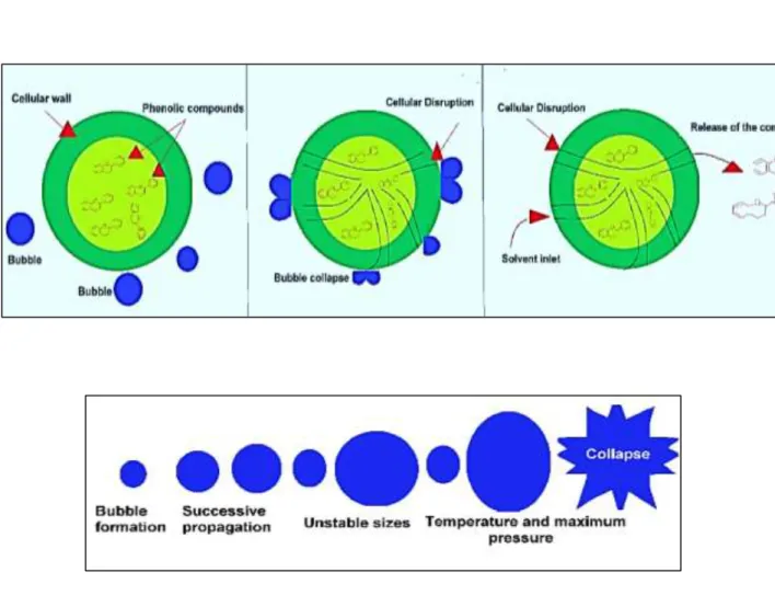
1.8.2.2.2 Microwaves
Microwaves have the particularity of instantaneously heating polar molecules and especially water molecules. Since microalgae cells are made up of 65-85% of water, following exposure to microwaves, the cells are subject to significant constraints that result in cells disintegration and thus favoring the release of molecules of interest (Figure 20). The phenomenon can therefore lead to increase the efficiency of microalgae lipid extraction procedures. However, microwaves require working from wet biomass. The study of Lee et al [27] also highlighted the effectiveness of microwaves compared to other techniques (such as bead beating, osmotic shock and autoclave) on different microalgae such as Botryococcus sp., Chlorella vulgaris and Scenedesmus. This process can reduce the production costs of microalgae biodiesel as reported by Yong-Ming Dai et al, [15].
Figure 20- Microalgae extraction mechanism using microwave irradiation [134].
1.8.2.2.3. Pulsed electric fields (PEF)
PEF is another potential technique in which cells are exposed to strong electric fields for very short periods of time. Electrical pulses make the cells permeable, thus implying an increase in the transfer of material through the membranes. This very promising technique can be applied in the
![Figure 6- Schematic ultrastructure of cell wall composition [69]. 1.4. Microalgae biochemical composition](https://thumb-eu.123doks.com/thumbv2/123doknet/2991975.83352/22.918.212.712.494.745/figure-schematic-ultrastructure-cell-composition-microalgae-biochemical-composition.webp)
![Figure 11- Microalgal saccharides [82].](https://thumb-eu.123doks.com/thumbv2/123doknet/2991975.83352/29.918.167.749.461.668/figure-microalgal-saccharides.webp)

![Figure 14- Examples of different types of open pond systems. (A) Unstirred pond (B) Circular pond (C) Raceway pond [93]](https://thumb-eu.123doks.com/thumbv2/123doknet/2991975.83352/32.918.179.736.485.763/figure-examples-different-types-systems-unstirred-circular-raceway.webp)
![Figure 18- Disruption of cell walls using degrading enzymes for enhanced lipid recovery [128]](https://thumb-eu.123doks.com/thumbv2/123doknet/2991975.83352/41.918.258.662.773.998/figure-disruption-walls-using-degrading-enzymes-enhanced-recovery.webp)
![Figure 19- Cell disruption by Bead Milling [132].](https://thumb-eu.123doks.com/thumbv2/123doknet/2991975.83352/42.918.162.767.705.913/figure-cell-disruption-bead-milling.webp)
![Table 4- Critical coordinates and dipole moment of some supercritical fluids [147]. Fluids Critical temperature (° C) Critical pressure (bar)](https://thumb-eu.123doks.com/thumbv2/123doknet/2991975.83352/48.918.142.776.231.453/critical-coordinates-supercritical-fluids-critical-temperature-critical-pressure.webp)
![Figure 25- Schematic diagram of the continuous supercritical flow system [162]. 1.10](https://thumb-eu.123doks.com/thumbv2/123doknet/2991975.83352/51.918.274.654.241.554/figure-schematic-diagram-continuous-supercritical-flow.webp)
![Figure 26- Membrane separation process based on the approximate MWCO range – required/used pressure [165]](https://thumb-eu.123doks.com/thumbv2/123doknet/2991975.83352/52.918.134.787.380.856/figure-membrane-separation-process-based-approximate-required-pressure.webp)
