O
pen
A
rchive
T
OULOUSE
A
rchive
O
uverte (
OATAO
)
OATAO is an open access repository that collects the work of Toulouse researchers and
makes it freely available over the web where possible.
This is an author-deposited version published in :
http://oatao.univ-toulouse.fr/
Eprints ID : 18552
To link to this article : DOI:
10.1007/s00330-015-4065-9
URL :
http://dx.doi.org/10.1007/s00330-015-4065-9
To cite this version : Brucher, Nicolas and Vial, Julie and Baunin,
Christiane and Labarre, David and Meyrignac, Olivier and Juridic,
Michel and Bouali, Ourdia and Abbo, Olivier and Galinier, Philippe
and Sans, Nicolas and Faruch, Marie Non-contrast-enhanced MR
angiography using time-spin labelling inversion pulse technique for
detecting crossing renal vessels in children with symptomatic
ureteropelvic junction obstruction: comparison with surgical findings.
(2016) European Radiology, vol. 26 (n° 8). pp. 2697-2704. ISSN
0938-7994
Any correspondence concerning this service should be sent to the repository
administrator:
[email protected]
Non-contrast-enhanced
MR angiography using time-spin
labelling
inversion pulse technique for detecting crossing renal
vessels
in children with symptomatic ureteropelvic junction
obstruction:
comparison with surgical findings
Nicolas Brucher1 & Julie Vial1 & Christiane Baunin1 & David Labarre1 & Olivier Meyrignac3 & Michel Juricic4 & Ourdia Bouali2 & Olivier Abbo2 & Philippe Galinier2 & Nicolas Sans1
Abstract
Objectives Investigate the feasibility and evaluate the ac-curacy of non-contrast-enhanced MR angiography (NC-MRA) using time-spin labelling inversion pulse (time-SLIP)to identify crossing renal vessels (CRVs) in children requiring surgical treatment of ureteropelvic junction (UPJ) obstructionand compare to laparoscopic findings. Materials and methods Nineteen children ranging from 6 to 16 years of age underwent NC-MRA using the time-SLIP technique before surgery. Two independent readers analysed the MRA images. Number of renal arteries and presence or absence of CRVs were identified and com-pared with surgicalfindings. Image quality was assessed, as well as the presence of CRVs and measurement of renal pelvis diameter. Intra and inter-reader agreement
was calculated using Cohen’s kappa coefficient and Bland–Altman plots.
Results The overall image quality was fair or good in 88% of cases. NC-MRA demonstrated CRVs at the level of the ob-struction in 10 children and no CRV in 9 children. All were confirmed intra-operatively except in one of the nine children. Sensitivity, specificity, NPV, PPV for predicting CRVs were 92%, 100%, 100% and 87.5%, respectively, for both readers. Conclusion NC-MRA is a good alternative to contrast-en-hanced MRA and CT scanning for identifying CRVs in chil-dren with symptomatic UPJ.
Key points
• Time-SLIP technique offers acceptable imaging quality for identifying crossing renal vessel.
• Time-SLIP technique is easy to apply to the renal MRA examination.
• Time-SLIP technique is an alternative to contrast-enhanced MRA and CT scanning.
Keywords Ureteropelvic junction . Crossing vessel . MR angiography . Non-contrast-enhanced . Children
Abbreviations
CE-MRA Contrast-enhanced magnetic resonance angiography
NC-MRA Non contrast-enhanced magnetic resonance angiography
Time-SLIP Time spin labelling inversion pulse CRV Crossing renal vessel
UPJ Uretero pelvic junction
* Nicolas Brucher
1
Department of Radiology, University Hospital Centre of Toulouse Purpan, Place du Docteur Baylac, TSA 40031,
31059 Toulouse, France
2
Department of Paediatric Surgery, University Hospital Centre of Toulouse Purpan, Hôpital des enfants. 330 avenue de
Grande-Bretagne, TSA 70034, 31059 Toulouse, France
3 Department of Radiology, University Hospital Centre of Toulouse
Rangueil, 1 avenue du Pr Jean Poulhès, TSA 50032, 31059 Toulouse, France
4
Department of Paediatric Surgery, Clinique Sarrus Teinturiers, 49 Allée Charles de Fitte, 31300 Toulouse, France
CDS Colour Doppler sonography
bSSFP Balanced steady state free procession MAG3 Mercaptoacetyltriglycine
FSE Fast spin echo TSE Turbo spin echo
BBTI Black blood inversion time MIP Maximum intensity projection ICC Intra-class correlation coefficient
Introduction
Obstruction of the ureteropelvic junction (UPJ) is frequently associated with lower-pole renal vessels crossing at the UPJ, particularly in older children with intermittent obstruction [1]. UPJ obstruction is frequently associated with lower pole renal vessels crossing at the UPJ, particularly in older children with intermittent obstruction [2]. The presence of crossing vessels (CRV) may influence surgical management. Open dismem-bered or laparoscopic pyeloplasty, via a retroperitoneal ap-proach, remains the gold standard management of UPJ ob-struction in children [3,4].
In 1949, Hellstrom first described [5] the superior reloca-tion of a lower pole crossing vessel in UPJ obstrucreloca-tion. Inher-ent to the success of this technique were the absence of intrin-sic ureteral obstruction, and the presence of extrinintrin-sic ureteral compression causing renal pelvic herniation and obstruction at the UPJ. Classically, the technique consists of exposure of the CRV using a transperitoneal approach with the patient in a lateral position. The lower pole vessels are dissected free of the UPJ and pexed in a cephalad position well away from the region of the UPJ by suturing the pelvis on either side of the vessels [6,7]. Precise knowledge of the vessel’s course at the
kidneys will help to shorten surgical time and minimize the risk of bleeding complications.
Colour Doppler sonography (CDS), supplemented or not by contrast-enhanced sonography, CT angiography, and MR angiography with intravenous contrast media are available as non-invasive methods for CRV detection [8–10]. CDS ap-pears to have poor specificity and to be highly operator de-pendant [11]. CT angiography has been found to be a very efficient technique to identify crossing vessels [12]. The lim-itations and disadvantages include radiation exposure and the need for intravenous contrast medium, with potential compli-cations from anaphylaxis and nephrotoxicity. Contrast-enhanced MR angiography (CE-MRA) appears to be an ac-curate technique in the identification of CRV [13,14], but exposes one to nephrogenic systemic fibrosis [15,16] espe-cially in patients with renal impairment.
Some studies have reported promising data on using non-contrast magnetic resonance angiography (NC-MRA) to eval-uate the renal arteries and especially spin labelling inversion pulse (Time-SLIP) technique on 1.5 T [17,18]. NC-MRA
Time-SLIP technique uses inversion recovery prepulse and three-dimensional (3D) balanced steady-state free precession (bSSFP) under respiratory gating. To the best of our knowl-edge, no published study has evaluated the identification of CRV in children with NC-MRA.
The purpose of this study was to prospectively investigate the feasibility of NC-MRA with the Time-SLIP technique at 1.5 T in the identification of CRV in children with symptom-atic UPJ obstruction compare to the surgical findings.
Material and methods
Subjects
This prospective single-centre study included 19 children be-tween August 2011 and November 2014. Informed consent was obtained from each participant or parents of the partici-pant. Children were referred for MRI and considered eligible for the study if they satisfied three criteria:
1. UPJ obstruction defined by renal pelvic dilatation on ul-trasound (US) with evidence of impaired drainage on ex-cretory diuretic renography (Tc-99 m-MAG3 with furosemide).
2. No history of antenatal hydronephrosis, with current fea-tures suggestive of intermittent obstruction such as inter-mittent pain or hydronephrosis.
3. A decision made to perform surgery based on clinical and imaging findings.
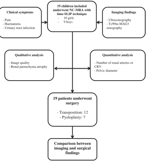
Ten girls and nine boys (age 4-16, median 9 years) who met these criteria underwent NC-MRA (Fig.1).
Left kidneys were more often affected [11] than right ones [8]. Hydronephrosis on US was present in all children. All preoperative Tc-99 m-MAG3 renography were performed at our institution and showed evidence of impaired or absent drainage of renal pelvis.
MR Protocol
All MR images were acquired using a 1.5 T MR system (Toshiba Vantage Titan, Japan) and performed prior to sur-gery. All scans were performed without sedation or intrave-nous cannulation. The child was positioned supine on the imaging table. After scout images acquisition to determine the position of the kidneys, coronal T2-weighted (T2-W) Fast Spin Echo (FSE) and axial T2-W Turbo Spin Echo (TSE) with respiratory gating were performed. NC-MRA used a fat-suppressed 3D TSE inversion pulse with free breath under the respiratory-triggered method (Table1). Both kidneys were covered in the coronal plane. Slice-selective inversion recov-ery prepulse was applied to suppress the background signal.
Targeted flow was visualised when non-inverted inflow blood came within the spatially selective inversion recovery volume by waiting for the optimised inversion time (Black Blood Inversion Time or BBTI)). The inflow delay time specified the interval between labelling the inflowing blood and image acquisition.
In general, a longer delay time allowed stronger distal vas-culature signal. However, when the BBTI was too long, less background suppression occurred because of T1 relaxation and the label intensity was attenuated. A few studies reported the optimal BBTI for renal arteries, around 1200 ms [17–19].
Maximum intensity projection (MIP) images were manually generated at the end of Time-SLIP imaging.
Image analysis
All MR images were independently analysed by two readers, one junior and one senior (JV and NB who had 7 years and 1 year of paediatric MR imaging experience, respectively). The readers were blinded to patient identity. A second analysis and measurements were performed 6 weeks later by the junior radiologist. On T2-W images, the maximum axial diameter of
19 children included underwent NC-MRA with
time-SLIP technique - 10 girls - 9 boys Imaging findings - Ultrasonography - Tc99m-MAG3 renography 19 patients underwent surgery - Transposition: 12 - Pyeloplasty: 7 Quantitative analysis
- Number of renal arteries or CRV
- Pelvic diameter
Comparison between imaging and surgical
findings
Clinical symptoms
- Pain - Haematuria - Urinary tract infection
Qualitative analysis
- Image quality
- Renal parenchyma atrophy
Fig. 1 Flow chart of study design and methodology
Table 1 Imaging parameters
Coronal T2 FASE (Fast-Spin Echo) Axial T2 TSE (Turbo-Spin Echo) Axial time-SLIP (3D SSFP) Repetition time (ms) 12600 3300 5,2 Echo time (ms) 80 90 2,6 Thickness (mm) 5 5 3 Matrix 256×256 256×256 256×256 Number of sections 20 30 50
Acquisition time 30 s 3 min 5 min 30 s
the pelvis were determined according to the specifications of the society of foetal urology (SFU). On NC-MRA imaging using Time-SLIP technique, the number of renal arteries and presence of CRV were evaluated. To assess the image quality, a 3 point scale was used as follows: 1: good, visualisation of whole main renal artery with segmental arteries, 2: fair, visu-alisation of whole main renal artery from the ostium to the hilum, and 3: poor, faint visualisation of main renal artery (Figs.2,3,4).
Surgical methods
Four surgeons (MJ, PG, OB and OA who had 20, 15, 6 and 4 years of paediatric surgery experience, respectively) had access to the NC-MRA images and to the routine radiology report prior to surgery. In those with CRV on NC-MRA, laparoscopy with a view to performing vessel transposition was scheduled. Consent was also obtained for Anderson-Hynes pyeloplasty in the event that intrinsic obstruction was identified at surgery. Initially described by Hellstrom in 1949 [5], the vessel transposition procedure was as fol-lows: the surgeon recorded the presence of CRV, which were then dissected away from the UPJ. Absence of intrin-sic obstruction was confirmed by careful examination of the UPJ following an IV fluid bolus.
Provided there was no evidence of intrinsic obstruc-tion, the vessels were then sutured superiorly away from the UPJ, typically by fixation into a tunnel created from the anterior wall of the dilated renal pelvis. Those with-out CRVs at NC-MRA were scheduled for laparoscopic pyeloplasty.
Statistical analysis
The sensitivity and specificity for predicting the number of renal arteries and the presence of CRVs were assessed on
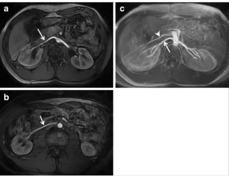
Fig. 3 A 10-year-old boy with right loin pain. (a) Coronal T2W-FASE. Right hydronephrosis with an abrupt transverse cut-off caused by a lower pole vessel crossing at the UPJ (arrow). (b) Coronal maximal intensity projection (MIP) using Time-SLIP technique depicts the main renal artery (arrow) and the CRV (arrowhead)
Fig. 2 A 9-year-old girl with intermittent left back pain. (a) Axial T2W-TSE showing Grade 3 pyelectasis with major pelvic dilatation (arrow). (b) Axial image of NC-MRA using Time-SLIP technique with main left renal artery (arrow). (c) Axial image of NC-MRA using Time-SLIP technique demonstrates a crossing lower pole artery (arrow). (d) Laparoscopic view shows the crossing lower pole artery (arrow) and the normal UPJ (arrowhead)
NC-MRA using surgical findings data as a standard reference. Cohen’s kappa coefficient was used to evaluate the agreement and intraobserver and interobserver variability in the identifi-cation of CRV, estimating the image quality and parenchyma atrophy. Intraclass correlation coefficients (ICCs) and Bland Altman plots were used to evaluate the agreement of renal pelvis diameter measurement. Statistical analyses were per-formed using commercial MedCalc software, version 12.0 (MedCalc Software, Inc., Mariakerke, Belgium). A P value of less than 0.05 was considered significant.
Results
In 17 of 19 children, dilatation was found in single system kidneys. Duplex with dilatation of the lower system and
dilatation of a horseshoe kidney were seen in the two other children (Table2).
Qualitative analysis
In both readers, 88 % of subjects (17/19) had good or fair image quality, and 12 % (2/19) had poor image quality [2].
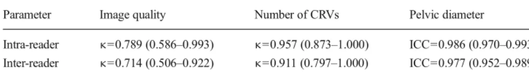
Intrareader and interreader agreements were good for the image quality, with Cohen’s kappa=0.789 and 0.714, respectively (Table3).
Quantitative analysis
NC-MRA demonstrated CRV at the UPJ in 11 children, one early branching vessel from the main renal artery crossing UPJ and no crossing vessel in 7 children. For the 11 preoperatively
Fig. 4 A 14-year-old boy with intermittent right loin pain and one episode of pyelonephritis. Axial images of NC-MRA using Time-SLIP technique with main right renal artery (a) and a crossing lower pole artery (b). Axial maximal intensity projection (MIP) using Time-SLIP technique demonstrates the main renal artery (arrow) and the CRV (arrowhead) (c)
Table 2 Characteristics of the
study population Parameter Average
Age 9 years 2 months (4 years 8 months to 16 years)
Female/Male 10/9
Loin pain 18
Urinary tract infection 4
Haematuria 0
Lithiasis 0
Right UPJ 8
Left UPJ 11
Modal vascularisation (1 main renal artery) 7
Lower pole crossing vessel 11
diagnosed CRV and the one with early branching vessel, all vessels (except one false negative case) were confirmed intraop-eratively, yielding a sensitivity of 92 % [95 % CI: 64 % to 99 %], a specificity of 100 % [95 % CI: 54 % to 100 %], a negative predictive value of 100 % [95 % CI: 73 % to 100 %] and a positive predictive value of 87.5 % [95 % CI: 42 % to 99 %] for predicting the presence of CRVs. All visualised CRV were sol-itary arterial vessels originating from the aorta. In one case, CRV could only be detected in the T2-W sequence because of the poor signal on Time-SLIP sequence. In one case with a poor image quality, a CRV was not seen on NC-MRA but confirmed intraoperatively. In 11 children with CRV and the one with early branching vessel, there was no evidence of intrinsic UPJ ob-struction on direct inspection during surgery, and all of these children proceeded to a vessel transposition. This technique was performed too in the remaining child with a CRV not seen on NC-MRA but intraoperatively. All children with single renal arteries required an open or laparoscopic trans-peritoneal dis-membered pyeloplasty.
Intrareader and interreader agreement was excellent for the identification of CRV, with Cohen’s kappa=0.957 and 0.911, respectively.
When measuring the pelvic diameter, ICC was excellent for intraobserver variability (0.986) and interobserver variabil-ity (0.977). The average difference between reader 1 (junior) and reader 2 (senior) for the pelvic diameter measurement was −0.03 mm (95% CI: −0.93 to 0.86). One value did not fall within the predefined test limits (−4.57 to +4.50; Fig5).
Over a period of 3 to 24 months of clinical followup, all 12 children who underwent transposition alone showed an im-provement in their symptoms. Pain either resolved completely or lessened in frequency and severity. All 12 patients showed an improvement of hydronephrosis on ultrasound with main pelvic diameter of 11.75 mm ± 4.32 (range 6 to 29) after surgery versus 29.52 mm ± 9.71 (range 13 to 44) preoperatively.
Discussion
UPJ obstruction due to CRV is a distinct entity separate from antenatal hydronephrosis, and manifests later in childhood. CRVs are found in children with ureteropelvic junction steno-sis in 49% of cases [11,20,21]. Before performing surgery or
Table 3 Intrareader and interreader agreements and variability with NC-MRA
Parameter Image quality Number of CRVs Pelvic diameter
Intra-reader κ=0.789 (0.586–0.993) κ=0.957 (0.873–1.000) ICC=0.986 (0.970–0.993)
Inter-reader κ=0.714 (0.506–0.922) κ=0.911 (0.797–1.000) ICC=0.977 (0.952–0.989)
Fig. 5 Bland–Altman plots for
maximum pelvic diameter show similar measurements from reader 1 and reader 2
interventional procedures in the kidney, the renal artery anat-omy should be accurately assessed. Preoperative awareness of a CRV or an early branching vessel may influence surgical management. The most widely performed therapeutic proce-dures for UPJ obstruction in children are open and laparoscop-ic pyeloplasty. Endopyelotomy is a widely performed tech-nique in adults and occasionally in older children [22,23]. CRV represent a relative contraindication to endopyelotomy, as they lower the success rate, in addition to presenting a potential bleeding risk [24]. Pesce et al. reported a successful outcome in 60 of 61 children treated with vessel transposition alone [25]. At our institution, laparoscopic vessel transposi-tion is offered to children with a clinical presentatransposi-tion of inter-mittent obstruction and CRV at laparoscopy, provided intraop-erative inspection of the UPJ reveals no evidence of intrinsic obstruction [26]. The procedure offers the advantage of not slitting the upper urinary tract and is a considerably shorter procedure than laparoscopic pyeloplasty.
The goal of our study was to evaluate whether NC-MRA is capable of detecting CRV in children with ureteropelvic junc-tion obstrucjunc-tion, and whether it is ideally also possible to evaluate the obstructive effect of these vessels on the UPJ without functional MR urography.
In this study, the use of NC-MRA at 1.5 T using the Time-SLIP technique allowed acceptable overall image quality (fair or greater image quality in 88 %) for visualising renal arteries. Of the 19 children who underwent surgery, a CRV could be diagnosed preoperatively in 11 children and an early branching vessel crossing UPJ in one case. There was no CRV in seven children. All preoperatively diagnosed CRVs were found intraoperatively and a negative MRA finding was confirmed by surgery in seven cases. A CRV that was not detected on NC-MRA was found in one child. To predict the presence or absence of CRVs, both sensitivity and specificity ranged from 92 % to 100 %. Interreader agreement on NC-MRA was good or excellent for identifying number of renal arteries. These findings indicate that the use of NCMRA at 1.5 T with Time-SLIP technique may be a feasible alternative to CDS, CT angiography, and CE-MRA for preoperative eval-uation of renal artery anatomy.
Parienty et al. [17] recently reported that the image quality of NC-MRA using the 3D bSSFP technique was good in 87 % and moderate in 13 % of images using a 3-point scoring sys-tem. Another previous study reported good image quality in 88 % of NC-MRA images taken using the 3D bSSFP tech-nique to evaluate the overall number of renal arteries [18]. These previous results are in agreement with our current study using Time-SLIP technique.
In adults, a number of imaging techniques have been employed to identify CRVs, typically as a means of excluding their presence prior to endopyelotomy. Contrast-enhanced CT examination is a popular technique owing to its relatively non-invasive nature, rapid image acquisition and excellent spatial
resolution. A number of studies have demonstrated sensitivi-ties and specificisensitivi-ties in excess of 90 % for CRV detection [12,
27]. However, the radiation dosage is considerable particular-ly in the evaluation of children, and there has been no system-atic evaluation of CT techniques in children. Non-contrast-enhanced colour Doppler ultrasound has been evaluated but appears to have poor specificity and to be highly operator dependent [11]. Ultrasound with endoscopic or contrast-enhanced methods and CE-MRA appears to be accurate tech-niques [8,14].
NC-MRA using Time-SLIP technique is well suited to this purpose. It can be performed reasonably rapidly (about 15– 20 minutes in our experience) and uses non-ionizing radiation or intravenous contrast medium administration. Despite its potential advantages, NC-MRA has not been widely applied to the detection of CRV in either adult or children and to our knowledge there are no reports of systematic evaluation of NC-MRA in this setting in the literature.
The image quality was poor in two cases in our study. This might be attributed to the specific paediatric population. In-deed the parameters, especially BBTI, are defined for an adult population [17,18] and may not be optimal for children, with many variations of the respiratory frequency, the heart rhythm, and the velocity of arterial circulation.
Despite the fact that hydronephrosis may be more impor-tant in the CRV group, we cannot conclude that this is a main rule. We did not perform NC-MRA studies during episodes of pain and clinically suspected obstruction. In all our patients, the MRI examination identified hydronephrosis of the symp-tomatic kidney, which may have led to the assumption that any CRV would have been the cause of the obstruction.
Our study has several limitations. Firstly, the surgeons were not blinded to the NCMRA findings; however, we do not believe that foreknowledge of the NC-MRA findings compro-mised accuracy of the surgical findings in practice. Secondly, the study cohort was relatively small. Further prospective multicentre studies with a large population are necessary. The follow-up period for the patients who underwent vessel transposition was relatively short. This study does not allow us to draw conclusions as to the effectiveness of the surgical technique, although the results to date are encouraging. How-ever, the focus of this study was to determine the accuracy of the MR imaging with Time-SLIP technique for identifying the presence of CRV.
Conclusion
We demonstrated that NC-MRA using Time-SLIP technique at 1.5 T appears to be useful and accurate in the identification of CRV in older children with intermittent UPJ obstruction. In this selected paediatric population, the incidence of CRV is high and their presence may influence surgical management.
Acknowledgments The scientific guarantor of this publication is Pr Nicolas Sans. The authors of this manuscript declare no relationships with any companies, whose products or services may be related to the subject matter of the article. The authors state that this work has not received any funding. Olivier Meyrignac kindly provided statistical advice for this manuscript. Institutional Review Board approval was obtained. Written informed consent was obtained from all subjects (patients) in this study. Methodology: Prospective, diagnostic or prognostic study, performed at one institution.
References
1. Coley BD (2013) Caffey’s Pediatric Diagnostic Imaging. Elsevier
Health Sci
2. Hoffer FA, Lebowitz RL (1985) Intermittent hydronephrosis: a
unique feature of ureteropelvic junction obstruction caused by a
crossing renal vessel. Radiology 156(3):655–658
3. Peters CA, Schlussel RN, Retik AB (1995) Pediatric laparoscopic
dismembered pyeloplasty. J Urol 153(6):1962–1965
4. Braga LHP, Lorenzo AJ, Bägli DJ et al (2010) Comparison of flank,
dorsal lumbotomy and laparoscopic approaches for dismembered pyeloplasty in children older than 3 years with ureteropelvic
junc-tion obstrucjunc-tion. J Urol 183(1):306–311
5. Hellstrom J, Giertz G, Lindblom K (1951) Pathogenesis and
treat-ment of hydronephrosis. J Belge Urol 20(1):1–6
6. Stern JM, Park S, Anderson JK, Landman J, Pearle M, Cadeddu JA
(2007) Functional assessment of crossing vessels as etiology of ureteropelvic junction obstruction. Urology 69(6):1022–1024
7. Godbole P, Mushtaq I, Wilcox DT, Duffy PG (2006) Laparoscopic
transposition of lower pole vessels–the Bvascular hitch^: an alter-native to dismembered pyeloplasty for pelviureteric junction ob-struction in children. J Pediatr Urol 2(4):285–289
8. Frauscher F, Janetschek G, Helweg G, Strasser H, Bartsch G,
Nedden zur D (1999) Crossing vessels at the ureteropelvic junction: detection with contrast-enhanced color Doppler imaging. Radiology 210(3):727–731
9. Morita S, Masukawa A, Suzuki K, Hirata M, Kojima S, Ueno E
(2011) Unenhanced MR Angiography: techniques and clinical ap-plications in patients with chronic kidney disease. Radiographics 31(2):E13–E33
10. Glockner JF, Vrtiska TJ (2007) Renal MR and CT angiography:
current concepts. Abdom Imaging 32(3):407–420
11. Veyrac C, Baud C, Lopez C, Couture A, Saguintaah M, Averous M
(2003) The value of colour Doppler ultrasonography for identifica-tion of crossing vessels in children with pelvi-ureteric juncidentifica-tion
ob-struction. Pediatr Radiol 33(11):745–751
12. Rouvière O, Lyonnet D, Berger P, Pangaud C, Gelet A, Martin X
(1999) Ureteropelvic junction obstruction: use of helical CT for
preoperative assessment–comparison with intraarterial
angiogra-phy. Radiology 213(3):668–673
13. Mitterberger M, Pinggera GM, Neururer R et al (2008) Comparison
of contrast-enhanced color Doppler imaging (CDI), computed
tomography (CT), and magnetic resonance imaging (MRI) for the detection of crossing vessels in patients with ureteropelvic junction
obstruction (UPJO). Eur Urol 53(6):1254–1260
14. Calder AD, Hiorns MP, Abhyankar A, Mushtaq I, Olsen OE (2007)
Contrast-enhanced magnetic resonance angiography for the detec-tion of crossing renal vessels in children with symptomatic ureteropelvic junction obstruction: comparison with operative
find-ings. Pediatr Radiol 37(4):356–361
15. Bhave G, Lewis JB, Chang SS (2008) Association of gadolinium
based magnetic resonance imaging contrast agents and nephrogenic
systemic fibrosis. J Urol 180(3):830–835
16. Yang L, Krefting I, Gorovets A et al (2012) Nephrogenic systemic
fibrosis and class labeling of gadolinium-based contrast agents by the Food and Drug Administration. Radiology 265(1):248–253
17. Parienty I (2008) Angio IRM, sans injection de produit de contraste
pour l’exploration des arteres renales. J Radiol
18. Park SY, Kim CK, Kim E, Park BK (2015) Noncontrast-enhanced
magnetic resonance renal angiography using a repetitive artery and venous labelling technique at 3 T: comparison with contrast-enhanced magnetic resonance angiography in subjects with normal
renal function. Eur Radiol 25(2):533–540
19. Glockner JF, Takahashi N, Kawashima A et al (2010) Non-contrast
renal artery MRA using an inflow inversion recovery steady state free precession technique (Inhance): comparison with 3D
contrast-enhanced MRA. J Magn Reson Imaging 31(6):1411–1418
20. Rigas A, Karamanolakis D, Bogdanos I, Stefanidis A,
Androulakakis PA (2003) Pelviureteric junction obstruction by crossing renal vessels: clinical and imaging features. BJU Int 92(1):101–103
21. Rooks VJ, Lebowitz RL (2001) Extrinsic ureteropelvic junction
obstruction from a crossing renal vessel: demography and imaging.
Pediatr Radiol 31(2):120–124
22. Motola JA, Badlani GH, Smith AD (1993) Results of 212
consecutive endopyelotomies: an 8-year followup. J Urol
149(3):453–456
23. Baldwin DD, Dunbar JA, Wells N, McDougall EM (2003)
Single-center comparison of laparoscopic pyeloplasty, Acucise
endopyelotomy, and open pyeloplasty. J Endourol 17(3):155–
160
24. Van Cangh PJ, Wilmart JF, Opsomer RJ, Abi-Aad A, Wese FX,
Lorge F (1994) Long-term results and late recurrence after endoureteropyelotomy: a critical analysis of prognostic factors. J Urol 151(4):934–937
25. Pesce C, Campobasso P, Costa L, Battaglino F, Musi L (1999)
Ureterovascular hydronephrosis in children: is pyeloplasty always
necessary? Eur Urol 36(1):71–74
26. Abbo O, Patard P-M, Mouttalib S et al (2015) Laparoscopic
trans-position of lower polar vessels for pyelo-ureteral junction
obstruc-tion: preliminary experience. Prog Urol 25(2):96–100
27. El-Nahas AR, Abou-El-Ghar M, Shoma AM, Eraky I, El-Kenawy
MR, El-Kappany H (2004) Role of multiphasic helical computed tomography in planning surgical treatment for pelvi-ureteric