Université de Montréal
ANTI-VEGFA THERAPY REDUCES TUMOR GROWTH AND EXTENDS SURVIVAL IN A MURINE MODEL OF OVARIAN GRANULOSA CELL TUMOR
par MAYRA TSOI
Département de biomédecine vétérinaire Faculté de médecine vétérinaire
Mémoire présenté à la Faculté de médecine vétérinaire en vue de l’obtention du grade de maître ès sciences (M.Sc.)
en sciences vétérinaires option reproduction
Septembre 2012
!" "
Résumé
Les tumeurs des cellules de la granulosa (GCTs) sont des tumeurs avec un potentiel malin ayant tendance à récidiver, provoquant ainsi la mort dans 80% des cas de stade avancé consécutif à une rechute. Bien que les GCTs représentent 5% des tumeurs ovariennes, peu d’études ont évalué les protocoles de traitement adjuvant pour la maladie avancée ou récurrente. Notre but était d’évaluer l’efficacité de la voie de signalisation du facteur de croissance de l’endothélium vasculaire A (VEGFA) comme cible pour le traitement de la GCT utilisant le modèle murin transgénique Ptentm1Hwu/tm1Hwu; Ctnnb1tm1Mmt/+; Amhr2tm3(cre)Bhr/+ (PCA) qui reproduit le stade avancé de la maladie humaine. Un anticorps anti-VEGFA a été administré une fois par semaine par voie intrapéritonéale (IP) à partir de 3 semaines d’âge. La thérapie anti-VEGFA a permis une réduction de la taille des tumeurs à 6 semaines d’âge (p<0.05) et une prolongation de la survie des animaux traités, lorsque comparé aux animaux contrôles. L’analyse des GCTs a montré une réduction significative de la prolifération cellulaire (p<0.05) et de la densité microvasculaire (p<0.01) mais aucune différence
significative n’a été détectée dans l’apoptose cellulaire. p44/p42 MAPK, un effecteur de la signalisation pour le récepteur 2 de VEGFA (VEGFR2) associé à la prolifération cellulaire, était moins activé dans les tumeurs traitées (p<0.05). Par contre, l’activation d’AKT, un effecteur impliqué dans la survie cellulaire, était similaire d’un groupe à l’autre. Ces résultats suggèrent que l’anticorps anti-VEGFA réduit la prolifération cellulaire et la densité
microvasculaire chez les souris PCA par inhibition de la voie de signalisation VEGFR2-MAPK, inhibant ainsi la croissance tumorale. En conclusion, l’efficacité de la thérapie anti-VEGFA mérite d’être évaluée en essais contrôlés randomisés pour le traitement des GCTs chez l’homme.
!!" "
Mots clés : tumeur des cellules de la granulosa, thérapie anti-VEGFA, angiogenèse, traitement adjuvant, modèle murin préclinique, signalisation AKT, signalisation MAPK
!!!" "
Abstract
Ovarian granulosa cell tumors (GCTs) are potentially malignant tumors that have a tendency for late recurrence and cause death in 80% of women with advanced GCT due to recurrent disease. Although GCTs represent 5% of ovarian tumors in women, few studies have evaluated adjuvant treatment protocols for advanced or recurrent disease. Our goal was to determine the potential of targeting the vascular endothelial growth factor A (VEGFA) signaling pathway for the treatment of GCT. We used a genetically engineered mouse model, Ptentm1Hwu/tm1Hwu; Ctnnb1tm1Mmt/+; Amhr2tm3(cre)Bhr/+ (PCA), which imitates the advanced human disease. A monoclonal anti-VEGFA antibody was administered by intra-peritoneal injection once a week beginning at 3 weeks of age. Anti-VEGFA therapy significantly decreased tumor weights by 6 weeks of age (p<0.05) and increased survival in treated animals in comparison to controls. Significant decreases in tumor cell proliferation (p<0.05) and microvessel density (p<0.01), but no significant difference in apoptosis was found in PCA tumors. p44/p42 MAPK, a VEGFA receptor 2 (VEGFR2) signaling effector associated with cell proliferation, was significantly less activated in anti-VEGFA-treated tumors (p<0.05). In contrast, AKT activation, a VEGFR2 signaling effector associated with cell survival was similar among all groups. These results suggest that anti-VEGFA therapy effectively reduces cell proliferation and microvessel density in PCA mice by inhibition of the VEGFR2-MAPK pathway, resulting in inhibition of GCT growth. We conclude that anti-VEGFA therapy merits further
investigation in the form of controlled randomized trials for the treatment of human GCT.
Keywords: granulosa cell tumor, anti-VEGFA therapy, angiogenesis, adjuvant therapy, preclinical mouse model, AKT signaling, MAPK signaling
!#" "
Table of contents
Résumé ... i
Abstract ... iii
List of tables ... viii
List of figures ... viii
List of acronyms and abbreviations ... ix
Acknowledgements ... xii
Introduction ... 1
Literature review: ... 3
Chapter 1: Ovarian cancer ... 3
1.1. Ovarian tumors in women ... 3
1.2. Granulosa cell tumors in women ... 3
1.2.1. Forms ... 3
1.2.2. Clinical presentation ... 4
1.2.3. Diagnosis ... 5
1.2.4. Serum tumor markers ... 6
1.2.4.1. Inhibin ... 6 1.2.4.2. Estradiol ... 7 1.2.4.3. Anti-Müllerian hormone ... 7 1.2.5. Current treatment ... 8 1.2.5.1. Chemotherapy ... 8 1.2.5.2. Radiation therapy ... 9
#" "
1.2.5.3. Hormonal therapies ... 9
1.2.6. Recurrence and survival ... 10
Chapter 2: Pathophysiology of GCT ... 11 2.1. Etiology ... 11 2.1.1. Cytogenetic abnormalities ... 12 2.1.2. Molecular genetics of GCT ... 12 2.1.2.1. FSH signaling pathway ... 12 2.1.2.2. Estrogen receptors ... 13 2.1.2.3. GATA-4 expression ... 13
2.1.2.4. Inhibin expression and synthesis ... 14
2.1.2.5. Wnt/!-catenin signaling pathway ... 14
2.1.2.6. Oncogenes and tumor-suppressor genes ... 15
2.1.2.7. FOXL2 mutation ... 15
2.2. Angiogenesis ... 17
2.2.1. Regulation of angiogenesis ... 17
2.2.2. Vascular endothelial growth factor ... 18
2.2.3. Other roles for VEGF in tumorigenesis ... 19
2.2.4. VEGF and GCT ... 20
Chapter 3: Transgenic mouse models ... 21
3.1. Importance ... 21
3.2. Existing models of GCT ... 21
3.2.1. Inhibin "-subunit knockout ... 21
#!" "
3.2.3. Simian virus 40 T-antigen ... 22
3.2.4. Estrogen receptor ! knockout ... 23
3.2.5. SMAD knockouts ... 23
3.2.6. Bone morphogenetic protein receptor knockouts ... 24
3.3. Transgenic GCT models relevant to the present study ... 24
3.3.1. The Ctnnb1tm1Mmt/+; Amhr2tm3(cre)Bhr/+ model ... 24
3.3.2. The Ptentm1Hwu/tm1Hwu; Amhr2tm3(cre)Bhr/+ model ... 26
3.3.3. The Ptentm1Hwu/tm1Hwu; Ctnnb1tm1Mmt/+; Amhr2tm3(cre)Bhr/+model ... 27
3.3.4. The KRASG12D; Ctnnb1tm1Mmt/+; Amhr2tm3(cre)Bhr/+ model ... 28
3.3.5. Conclusions from the GCT models ... 29
Chapter 4: Targeting angiogenesis ... 29
4.1. Classes of angiogenic inhibitors ... 31
4.1.1. VEGFR antibodies ... 31
4.1.2. Soluble VEGFRs ... 31
4.1.3. Aptamers ... 32
4.1.4. Small-molecule VEGFR tyrosine kinase inhibitors ... 32
4.1.5. Monocloncal anti-VEGFA antibodies ... 33
4.1.5.1. Dosage and administration ... 33
4.1.5.2. In epithelial ovarian cancer ... 34
4.3.2.3. In GCT ... 35
4.2. Resistance ... 35
4.3. Adverse events ... 36
#!!" "
Hypothesis and objectives ... 38
Publication as first author ... 39
General discussion ... 71
Conclusion ... 77
#!!!" "
List of tables Publication
Supplemental Table I. Masses of granulosa cell tumors used for analyses ... 70
List of figures Figure 1: FSH receptor downstream signaling effectors ... 13
Figure 2 : VEGFR2 signaling pathway in endothelial cells ... 19
Figure 3 : WNT/!-catenin signaling pathway and dominant stable !-catenin mutant ... 25
Figure 4 : PI3K/AKT pathway and conditional targeting of PTEN ... 27
Figure 5 : Classes of VEGF inhibitors ... 30
Publication Figure 1. Anti-VEGFA antibody reduces tumor burden and improves survival of PCA mice with GCTs ... 64
Figure 2. Anti-VEGFA antibody reduces cell proliferation in GCTs from PCA mice ... 65
Figure 3. Anti-VEGFA antibody has no significant effect on apoptosis or tumor necrosis in 6GCTs from PCA mice ... 66
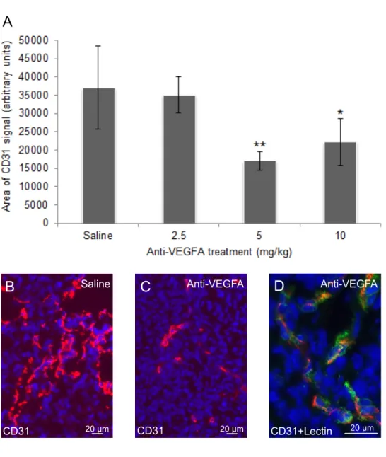
Figure 4. Anti-VEGFA antibody significantly reduces microvessel density in GCTs from 6 week-old PCA mice ... 67
Figure 5. Anti-VEGFA antibody significantly reduces MAPK activation in GCTs from 6 week-old PCA mice ... 68
Figure 6. Anti-VEGFA antibody has no significant effect on AKT activation in GCTs from 6 week-old PCA mice ... 69
!$" "
List of acronyms and abbreviations AC: adenylyl cyclase
AKT: protein kinase B
AMH: anti-Müllerian hormone APC: adenomatous polyposis coli
BCL2L2: beta-cell leukemia/lymphoma-2-like protein 2 BEP: bleomycin/etoposide/cisplatin
BMP: bone morphogenetic protein
c-erbB2 : human epidermal growth factor receptor 2 CA: Ctnnb1tm1Mmt/+; Amhr2tm3(cre)Bhr/+
CT: computed tomography
CYP19A1: cytochrome P450 aromatase DSH: disheveled
EGFR: epidermal growth factor ERK: extracellular regulated kinase ESR: estrogen receptor
FDA: US Food and Drug Administration
FIGO: International Federation of Gynecology and Obstetrics Flk-1: fetal liver kinase 1
Flt-1: FMS-like tyrosine kinase 1 FOXL2: forkhead box L2
FSH(R): follicle-stimulating hormone (receptor) FZD: frizzled receptor
$" ""
GCT: granulosa cell tumor
GnRH: gonadotropin releasing hormone GSK3!: glycogen synthase kinase 3 beta HIF: hypoxia inducible factor
HPF: high power field
HPS: hematoxylin phloxine saffron
IGF1(R): insulin-like growth factor-1 (receptor) IP: intraperitoneal
IV: intravenous
KDR: kinase insert domain receptor LH: luteinizing hormone
LRP: low-density lipoprotein receptor-related protein mAb: monoclonal antibody
MAPK: mitogen-activated protein kinase MEK: ERK kinase
MRI: magnetic resonance imaging NSCLC: non-small-cell lung cancer PA: Ptentm1Hwu/tm1Hwu; Amhr2tm3(cre)Bhr/+
PCA: Ptentm1Hwu/tm1Hwu; Ctnnb1tm1Mmt/+; Amhr2tm3(cre)Bhr/+ PDGFR: platelet-derived growth factor receptor
PDK: 3-phosphoinositol-dependent protein kinase PI3K: phosphoinositide 3-kinase
$!" "
PTEN: phosphatase and tensin homolog RTK: receptor tyrosine kinase
SCST: sex cord-stromal tumor SFK: SRC family protein kinase
TCF/LEF: transcription factor, T-cell specific/lymphoid enhancer factor 1 TGF: transforming growth factor
TKI: tyrosine kinase inhibitor
$!!" "
Acknowledgements
I would like to take this opportunity to thank those who made this work possible. To my research director Dr. Derek Boerboom, thank you for answering my endless questions and helping me discover the wonderful world of research. To Dr. Marie-Ève Nadeau, thank you for sparking my interest in oncology. To Dr Marilène Paquet, thank you for your excellent analyses. To my colleagues Marie-Noëlle, Evelyne, Alexandre, Aurore and Charlène, thank you for all your help. Thank you to the members of my advisory committee and to the members of the jury who graciously agreed to assist me in this endeavor. Also, thank you to all those who assisted in the management of the mouse colony. Finally, a special thank you to my boyfriend, family and friends for their endless patience and constant support.
Introduction
Cancer is a comprehensive term that encompasses over one hundred diseases that all share in common the transformation of normal human cells into malignant (cancer) cells (1, 2). Initially, a succession of genetic changes occurs that confer growth advantages to given cells, leading to their conversion to cancer cells and ultimately tumor development (2). In contrast to normal cells, cancer cells are able to sustain proliferation, evade tumor suppression, resist apoptosis, replicate indefinitely, induce angiogenesis, and invade and form metastases (3). These acquired alterations in cell physiology are what dictate tumor growth in
combination with signals from the tumor microenvironment and inflammatory mediators (2, 4).
Many different types of cancer affect the female reproductive system, with ovarian cancer being the leading cause of death among gynecological cancers and representing 3% of all cancers in women (1). Three categories of ovarian tumors exist with each one named according to its derived cell type (5). 80-90% of ovarian tumors are epithelial ovarian tumors, followed by sex cord-stromal tumors (SCSTs), which make up 7-8%, and the remaining 1-3% are germ cell tumors (5, 6). Within the category of SCSTs, a given tumor will contain sex cord and stromal components of the developing gonad that may include one or a combination of granulosa cells, theca cells, lutein cells, Sertoli cells, Leydig cells, or fibroblasts (7). The majority are granulosa cell tumors (GCTs), accounting for 70-90% of SCSTs and
approximately 5% of all ovarian tumors (6, 8-10). GCTs are considered as potentially malignant tumors characterized by insidious growth and late recurrence (6, 8, 11, 12).
Although some studies have investigated GCT pathogenesis, the mechanisms
component of GCT disease is angiogenesis, as these are highly vascularized tumors that express high levels of vascular endothelial growth factor (VEGF), a key mediator of tumor angiogenesis (11, 13, 14). Based on this, angiogenesis was proposed as a therapeutic target for the adjuvant treatment of GCT (11, 13-16).
Animal models can serve as useful tools to evaluate the efficacy of novel therapeutic agents and targets. A genetically engineered mouse model that develops GCT that imitates the advanced disease in women was generated and served as a preclinical model for investigating a novel therapeutic agent that targets VEGF.
This thesis will present the evaluation of an anti-angiogenic therapy for the treatment of GCT in our mouse model. First off, a review of ovarian and GCTs in women will be presented, followed by current knowledge of GCT pathophysiology. Next, descriptions of the existing transgenic mouse models of GCT will be given, followed by a presentation of
angiogenic inhibitors. Finally, the findings from this study will be presented and discussed.
Literature Review
Chapter 1: Ovarian Cancer
1.1. Ovarian tumors in women
The American Cancer Society estimates that 22,280 women will be newly diagnosed with ovarian cancer in 2012, of which 15,500 will die from it in the United States alone (1). Ovarian cancer is the leading cause of death among gynecological cancers, accounting for 3% of all cancers in women (1).
Ovarian tumors are classified into three major groups based on histological patterns and cell types; the most common and best described are epithelial ovarian tumors (80-90% of cases), followed by SCSTs (7-8%), and finally germ cell tumors (1-3%) (5, 6). Accounting for 70-90% of SCSTs are GCTs, which represent approximately 5% of all ovarian tumors and are derived from the hormonally active component of the ovarian stroma, the granulosa cell (6, 8-10, 17). The reported incidence of GCT is 0.58 to 1.6/100,000/year (18).
1.2. Granulosa cell tumors in women
1.2.1. Forms
Although GCTs are considered to be rare, they are potentially malignant, tend to recur over long periods, and are therefore of clinical importance (8, 11, 12). They are divided into the adult (95%) and juvenile forms (5%) based on clinical presentation and histological characteristics (8, 18). The adult form mostly occurs in post-menopausal women with the
median age at presentation being 50-54 years old (17) and is characterized by well and poorly differentiated histological patterns with coffee-bean grooved nuclei (6). On the other hand, the juvenile form mostly affects women less than 30 years of age (mean 13-17 years) and is characterized by large luteinized cells with hyperchromatic nuclei lacking nuclear grooves (6, 8, 18). Both the adult and juvenile form have been reported in children and adults,
respectively (8, 17, 18).
1.2.2. Clinical presentation
GCT cells share morphological, biochemical, and hormonal features of normal proliferating pre-ovulatory granulosa cells (19, 20). Approximately 70% of GCTs produce hormones that include estradiol, inhibin, and anti-Müllerian hormone (AMH) that are responsible for a variety of nonspecific symptoms such as abdominal pain, distension, bloating, and abnormal uterine bleeding (6, 8,18). Other symptoms may include menstrual irregularities, post-menopausal bleeding, abdominal discomfort, weight loss, presence of a pelvic or abdominal mass, and breast enlargement or tenderness (18). Abnormal uterine bleeding and abdominal pain have been reported as the most common presenting symptoms (18, 21). The tumor mass may compress adjacent organs and cause dysuria and constipation (8). Ascites occurs in 10-28.8% of cases (8, 18, 22). Complications, such as ovarian torsion, haemorrhage, or cystic rupture, often manifest as acute abdominal pain and distension (8, 18). Unfortunately, 12.5-14.3% of patients are asymptomatic and are only diagnosed during routine examinations (21, 22).
Specific to the juvenile form, 80-90% of patients 8 years of age or less present with isosexual precocious pseudopuberty, characterized by precocious breast development, increased pubic hair, vaginal bleeding, and advanced growth (6, 8, 18).
Some pathological conditions have been associated with GCT and include endometrial hyperplasia in 25-50% of women (due to estrogen stimulation of the endometrium),
endometrial adenocarcinoma in 8-13% of women over the age of 45, breast cancer in 3% of women, and infertility in 22% of women of childbearing age (8, 18). The juvenile form has been associated with Potter’s syndrome, multiple congenital abnormalities, Ollier’s disease, and Maffucci’s syndrome (18).
1.2.3. Diagnosis
Initial diagnosis is based on clinical features and pelvic examination (8). As the majority of GCTs produce oestrogens that are responsible for various symptoms, this often allows for early detection (17). At diagnosis, many GCTs have already attained a considerable size (average diameter of 12 cm), 95% of cases are unilateral, 85-95% are palpable, and 78-91% are at stage I/IV (i.e. confined to the ovary(ies); according to FIGO classification) (8, 17, 18, 23).
The tumors may appear as a solid mass with hemorrhagic or fibrotic changes, as completely cystic, or with multilocular cystic lesions (18). One classification type categorizes adult-type GCT based on morphologic patterns as either multilocular cystic, thick-walled unilocular cystic, or thin-walled unilocular cystic, homogenously solid or heterogeneously solid (24).
Diagnostic tools include ultrasound, which provides qualitative information about the mass, such as size, echogenicity, and consistency however, CT and MRI are more sensitive exams (8, 17). Uterine biopsy is indicated in cases of postmenopausal bleeding in order to exclude adenocarcinomas (17).
Final diagnosis of GCT is based on histopathology examination (8). Malignant GCTs resemble normal granulosa cells but with round to oval nuclei and coarsely stippled chromatin. Cytoplasm is often faintly eosinophilic and vacuolated. GCTs demonstrate a high degree of cellular polymorphism and a high mitotic rate with focal areas of necrosis and hemorrhage, and evidence of local invasion (25).
1.2.4. Serum tumor markers
Many serum tumor markers have been proposed for the management and surveillance of GCT, although very few are specific (8). The most relevant markers include: inhibin, estradiol, and anti-Müllerian hormone (6, 8, 17, 26).
1.2.4.1. Inhibin
Inhibin is a member of the transforming growth factor (TGF)-! family that is
synthesized by normal granulosa cells. It is mostly secreted during the follicular phase of the menstrual cycle and acts by suppressing the synthesis and secretion of follicle-stimulating hormone (FSH) (17, 18). Patients with GCT tend to have elevated inhibin serum
concentrations (mostly inhibin B), which has been positively correlated with tumor size (26) and therefore serve to detect GCT and estimate tumor bulk (6). As inhibin levels drop after tumor removal, they also serve to predict and detect recurrence (6, 26, 27). Serum inhibin
levels also drop in postmenopausal women (secondary to the depletion of ovarian follicles), which may serve as a comparative baseline in these women (6, 28). It is important to note, however, that elevated inhibin levels are not specific for GCT, and may be elevated in epithelial ovarian tumors (17).
1.2.4.2. Estradiol
Estradiol is mostly produced by granulosa cells during the reproductive period in women (6). In the majority of GCTs, estradiol is secreted in large amounts and is responsible for many of the tumor-related symptoms (6). Although estradiol might seem like a logical tumor marker for GCT, no correlation between estradiol levels and GCT progression or recurrence has been found (29, 30). This could be due to the fact that estradiol production depends on its precursor, androstenedione, produced by theca cells and in the absence of theca cells in some GCTs, no estradiol is secreted (6, 17, 29). On the whole, estradiol is not
considered to be a reliable tumor marker for GCT, especially in comparison to inhibin B and AMH (6, 17, 29).
1.2.4.3. Anti-Müllerian hormone
AMH is another member of the TGF! superfamily that is expressed by granulosa cells and controls the formation of primary follicles during reproductive life, but is virtually
undetectable after menopause (17, 28). Serum AMH levels were found to be elevated in the majority of patients with GCT, undetectable in patients with clinical remission, and elevated several months prior to clinical recurrence (30, 31). In a recent review, inhibin B and AMH
were both found to be useful serum markers for diagnosis and surveillance of patients with GCT (32).
1.2.5. Current treatment
Initial management of GCT involves cytoreductive surgery, which allows for staging, definitive diagnosis, and tumor debulking (8, 17, 18). For women beyond childbearing age, total hysterectomy and bilateral salpingo-oophorectomy are attempted (8, 18). For women of childbearing age who wish to preserve fertility, a unilateral salpingo-oophorectomy is
performed, provided that the disease is contained within one ovary (8). In these instances, conservative surgery is often complemented by careful staging and endometrial biopsy to rule out uterine cancer (17, 18). Staging normally includes a partial omentectomy, evaluation of para-aortic and pelvic lymph nodes, peritoneal biopsies, and peritoneal washing cytology (8, 17). If the biopsies are disease free, no further treatment is required.
Adjuvant treatment is recommended for patients with recurrent or advanced disease; essentially for patients with stages II-IV disease or stage I disease associated with large tumor size, high mitotic index, or tumor rupture (8, 17). Standardized adjuvant treatment protocols, however, have yet to be established. Adjuvant therapies include: chemotherapy, radiation therapy, hormonal therapy, and more recently, anti-angiogenic therapy.
1.2.5.1. Chemotherapy
GCT is generally responsive to chemotherapy, which is mostly platinum-based, with bleomycin/etoposide/cisplatin (BEP) being the most frequently used regimen (8, 17, 19). Chemotherapy is indicated for advanced, inoperable, or recurrent disease, when surgery or
radiotherapy is not a viable option (18). In a retrospective study by Park et al (19), 13 patients diagnosed with advanced GCT had undergone cytoreductive surgery followed by 6 cycles of adjuvant chemotherapy with BEP. None of the patients who received the full 6 cycles of BEP had recurrent disease with a 5-year disease-free survival rate of 100% (19). Secondary effects to the BEP regimen are non-negligible however, and include myelotoxicity and a risk of secondary acute myelogenous leukemia (18).
1.2.5.2. Radiation therapy
Radiation therapy is indicated as an adjuvant treatment or for recurrent disease (18). Although it has been associated with improved survival in a few studies (33-35), other studies have found no survival advantage (36-38). In a retrospective study by Hauspy et al (34), 31 patients diagnosed with GCT had received adjuvant radiotherapy. They found a significantly longer disease free survival in patients who had received radiation therapy (251 months) in comparison to patients who had not (112 months, p=0.02) (34). In contrast, Malmström et al (36) found no survival benefit in 48 GCT patients treated with radiation therapy. Due to the retrospective nature of these studies, too many variables could have potentially confounded the real efficacy of radiation therapy for the treatment of GCT; large-scale prospective trials would be required in order to elucidate its role in the treatment of recurrent disease or as an adjuvant treatment for GCT.
1.2.5.3. Hormonal therapies
Hormonal therapies include gonadotropin releasing hormone (GnRH) agonists, GnRH antagonists, progestogens, and anti-oestrogens (8). A limited number of case reports have
evaluated their efficacy in the adjuvant treatment of GCT and most often describe last resort attempts in cases of recurrent disease (6). While some studies have shown hormonal therapy to result in stabilized disease, prolonged remission, partial response, and no major side effects (18, 39-42), others show no response whatsoever (43, 44). Although these therapies may indeed be effective in slowing down the progression of GCT disease, it is not possible to draw any reliable conclusions from a handful of case reports. In addition, the fact that the majority of patients were in the final stages of disease and several other treatment options had
previously been attempted, it is difficult to attribute the precise contribution of hormonal therapy to the observed effects.
1.2.6. Recurrence and survival
As GCT is often diagnosed at an early stage, survival is considered excellent (17). Still, 80% of cases diagnosed with advanced-stage GCT will die from recurrent disease (45).
GCTs can spread locally by direct extension and intraperitoneal seeding to ovarian serosa, omentum, fallopian tube, and pelvic walls or by hematogenous or lymphatic routes to reach the lungs, liver, brain, skeleton, diaphragm, abdominal wall, and adrenal glands to form metastases (8, 18).
GCT has a reputation for recurrences over long periods, with 40 years being the longest time period of recurrence ever recorded (46). In one study (22), recurrence rate was 5.4% for stage I, 21.1% for stage III, and 40% for stage IV disease; overall recurrence rates vary between 11.2-21% (8, 9, 21). Recurrences are thought to originate from peritoneal seeds, which are points of contact between the primary tumor and abdominal or pelvic structures that remain after removal of the primary tumor and grow very slowly. For this reason, recurrences
most often occur in the abdomen or pelvis and tend to occur between 5 to 10 years after initial diagnosis and treatment (8, 18).
Many predicting factors for recurrence and poor survival have been identified and include: advanced stage at diagnosis, high mitotic index (>10 mitoses/10 HPF), bilaterality, large tumor size (>5cm), tumor rupture, lymphatic system invasion, nuclear atypia, absence of call-exner bodies, presence of residual disease, DNA ploidy, and age (50+) (8, 17-19, 47). Stage is universally accepted as the most important prognostic factor (17, 22, 28) with a 5-year survival of 90-100% for stage I disease, 55-75% for stage II, and 22-50% for stage III and IV, and a 10-year survival of 84-95% for stage I, 50-65% for stage II, and 17-33% for stage III/IV (18). VEGF has also been identified as a prognostic factor. Brown et al (48) showed that increased microvessel density and overexpression of VEGF correlated with the presence of distant metastasis and shorter survival in patients with SCSTs. Many other prognostic factors are mentioned in the literature, but have not been consistently reported.
Following diagnosis and treatment, regular examination is recommended every three months for the first two years, and then every four to six months for five years. Afterwards, examinations should be continued for life, as late recurrences tend to occur, and normally include a physical examination, ultrasound, CT scan, and evaluation of tumor markers (8).
Chapter 2: Pathophysiology of GCT
2.1. Etiology
Little is known about the etiology of GCT despite the fact that a number of studies have attempted different approaches to elucidate this. A few studies have examined the GCT
karyotype in order to identify abnormalities (6, 27, 49-51). Another approach has been to focus on pathways involved in normal folliculogenesis, as GCTs display many features of proliferating pre-ovulatory granulosa cells, such as FSHR expression, FSH binding, estrogen synthesis, GATA-4 expression, and inhibin expression and synthesis (6, 20, 27). Others have studied pathways, oncogenes and tumor-suppressor genes that have frequently been implicated in many cancer types (6, 12, 27, 52-57).
2.1.1. Cytogenetic abnormalities
A distinctive pattern of chromosomal aberrations has been detected in 5-20% of aneuploid GCTs (6, 49). Trisomy 12 has been observed in 14-33% of cases, trisomy 14 in 25-33%, and monosomy 22 in 35-40% (6, 27, 49-51). Although several important genes have been identified on chromosome 12 such as KRAS 2, KRAG, MDM2 and on chromosome 14 such as FOS, BCL2L2, TGR!3, their precise role in GCT pathogenesis is unclear (49).
2.1.2. Molecular genetics of GCT
2.1.2.1. FSH signaling pathway
Although GCTs express high levels of the FSHR gene, no activating mutations in the FSHR or its associated G proteins have been identified (6, 27). Instead, it is suspected that activating mutations in genes encoding signaling effectors downstream of the FSHR, involving extracellular regulated kinases (ERK) or phosphoinositide 3-kinase (PI3K)/AKT signaling cascades might be involved (Figure 1) (6, 27, 58). Although mutation of the PI3K
subunit genes or inactivation of tumor suppressor phosphatase and tensin homolog (PTEN) has been implicated in epithelial ovarian tumors, Bittinger et al (59) found no association in GCT.
2.1.2.2. Estrogen receptors
Estrogen synthesis is a crucial part of folliculogenesis with expression of estrogen receptor genes (ESR1 and 2) occurring during the proliferative phase in human granulosa cells (27). ESR2 was found to be predominantly and abundantly expressed in GCTs, in comparison to ESR1 (27, 60). As approximately 70% of GCTs produce estrogens, it is suspected that these receptors might play a role in stimulating granulosa cell growth and differentiation (20).
2.1.2.3. GATA-4 expression
Another feature of proliferating granulosa cells is GATA-4 expression. GATA-4 is a member of the GATA family of transcription factors that regulates the expression of many
Figure 1
FSH receptor downstream signaling effectors. FSH binding to its receptor
FSHR activates both the ERK1/2 and PI3K/AKT intracellular signaling pathways in granulosa cells. AKT = protein kinase B, ERK = extracellular regulated kinase, FSH(R) = follicle stimulating hormone (receptors), MEK = ERK kinase, PDK = phosphatidylinsitol-dependant kinase, PI3K = phosphoinositide 3-kinase, PTEN = phosphatase and tensin homolog.
Adapted from Jamieson & Fuller 2012 and Laguë et al 2008.!
important genes in the gonads, whose expression was found to be activated in primary follicles in conjunction with the activation of granulosa cell proliferation (61). Anttonnen et al (61) reported high levels of GATA-4 in 44% of primary GCTs and a significant positive correlation between GATA-4 expression and clinical stage and risk of recurrence. This group later
reported a correlation between GATA-4 expression and Bcl-2, an anti-apoptotic gene
controlled by GATA-4 (62). The authors suspect that an increase in GATA-4 expression up-regulates Bcl-2 activation thereby promoting granulosa cell survival and inhibiting apoptosis in GCT tumorigenesis (62).
2.1.2.4. Inhibin expression and synthesis
Serum inhibin levels are elevated in women with GCT and have been associated with suppressed FSH levels, indicating a biologically active form of inhibin (6, 63). In contrast, in an inhibin-deficient mouse model, SCSTs developed in 100% of animals (64). Matzuk et al (64, 65) explained this apparent contradiction by the development of resistance to inhibin in the human disease, whose normal role may be tumor suppression specifically in the gonads.
2.1.2.5. WNT/!-catenin signaling pathway
Another pathway that has been implicated in several cancer types is the WNT/!-catenin signaling pathway (52). Boerboom et al (52, 53) evaluated the WNT/!-WNT/!-catenin pathway that is normally expressed in ovarian granulosa cells and found hyperactivation in a subset of human and equine GCTs. In order to confirm whether activation of the WNT/!-catenin signaling pathway was in fact involved in GCT development, this group developed a mouse model expressing a dominant stable !-catenin mutant (52). The majority of animals did
in fact develop ovarian lesions at an advanced age that were confirmed as GCTs (52). These findings were the first to demonstrate a causal link between altered WNT/!-catenin signaling and GCT tumorigenesis (53), and later served to develop the mouse model that is the focus of this paper.
2.1.2.6. Oncogenes and tumor-suppressors genes
Several oncogenes that have been implicated in a range of tumors have also been examined for their potential role in GCT pathogenesis however, these investigations have yielded few novel insights. No mutation or overexpression was found in the following oncogenes: c-myc, p21-ras, c-erbB2, K-ras, N-ras, H-ras, B-raf, or WT1 (6, 12, 54, 55). Neither mutation nor loss of tumor-suppressor p53 was found to be implicated in GCT (27, 56, 57). The inability to identify oncogenes and tumor-suppressor genes that might be responsible for initiating the development of GCT represents a further delay in the development of
effective treatment agents, as many current cancer drugs specifically target the proteins encoded by oncogenes (66).
2.1.2.7. FOXL2 mutation
The most significant breakthrough in GCT pathogenesis came in 2009 when Shah et al (67) identified a somatic missense point mutation in FOXL2 402C"G (C134W) in 97% of adult GCTs. The FOXL2 gene encodes a member of the forkhead domain/winged-helix family of transcription factors that is specifically expressed in ovarian follicular cells and is crucial for granulosa cell function and follicle development (6). This mutation is almost entirely absent in other cancer types, such as epithelial ovarian, breast, and even juvenile
GCTs (67, 68). Several subsequent studies confirmed the mutation in adult-type GCT with an overall occurrence of 94% (6). These findings strongly suggest that FOXL2 mutation is pathognomonic for adult-type GCT and that it constitutes either an activating or a gain-of-function mutation (6, 67, 69).
In an attempt to explain the mechanism of tumorigenesis driven by the FOXL2 mutation, studies have evaluated FOXL2 targets, such as the aromatase gene CYP19A1 (69). FOXL2 induces CYP19A1 which leads to increased estrogen conversion by granulosa cells (69). Fleming et al (70) confirmed that aromatase is a direct target of FOXL2 (C134W) in adult-type GCT and more specifically, that the mutation altered the regulation of the aromatase promoter (6). As hyperestrogenism is seen in approximately 70% of adult-type GCTs, deregulated activity of FOXL2 via altered aromatase activity might contribute to the development of this condition (6, 69).
In a FOXL2 knockout mouse model, increased follicular loss and oocyte atresia were found, which suggests that FOXL2 plays an anti-apoptotic role (69, 71). This finding was supported by Kim et al (72), who demonstrated that mutant FOXL2 exhibited minimal cell death in comparison to the wild-type. These findings suggest a tumor suppressor role for FOXL2 in normal granulosa cells, which in the mutant form, is impaired and incapable of mediating death ligand-induced apoptosis (72).
In juvenile-type GCT, FOXL2 expression is decreased or absent and has been associated with aggressive or advanced stage disease (6, 68).
2.2. Angiogenesis
In 1971, Judah Folkman (73) hypothesized that tumor growth depends on the
formation of new blood vessels from pre-existing ones, a concept coined as angiogenesis, and proposed angiogenesis as a therapeutic target for cancer treatment. Angiogenesis is now known to be crucial for tumor growth, as small tumor masses are unable to grow beyond 1-2 mm without a blood vessel supply to support their growth (74). Early during tumor
development, an angiogenic switch occurs in which quiescent tumor endothelial cells are activated (75, 76). Tumor cells release pro-angiogenic mediators that bind to adjacent vascular endothelial cells and stimulate the formation of new blood vessel capillaries that infiltrate the tumor mass (74). This infiltration allows the microscopic tumor mass to expand because tumors, just like normal tissues, require oxygen, nutrients, and the evacuation of carbon dioxide and metabolic waste (3). This neovascularization also allows the tumor to eventually form metastases (74).
Tumor blood vessels tend to be aberrant, characterized by precocious capillary sprouting, convoluted and excessive vessel branching, distorted and enlarged vessels, erratic blood flow, micro-hemorrhaging, leakiness, and abnormal levels of endothelial cell
proliferation and apoptosis (3, 77, 78).
2.2.1. Regulation of angiogenesis
The angiogenic process depends on interactions between vascular endothelial cells, tumor cells, inflammatory cells, the surrounding micro-environment, and many auxiliary factors (79). Numerous environmental factors stimulate VEGF expression, a major mediator of angiogenesis, which include hypoxia, low pH, hormones, growth factors, and cytokines (74,
75, 80-84). Mutations in oncogenes and/or tumor suppressor genes also contribute to
upregulating VEGF (74, 85). Negative regulators play essential roles in keeping angiogenesis under control, and include: thrombospondin, endostatin, tumstatin, vasostatin, and vasohibin (84). For the purposes of this study, the remainder of this review will focus on the VEGF pathway, as it represents the major regulatory pathway involved in tumor angiogenesis (83, 84).
2.2.2. Vascular endothelial growth factor
In 1989, Napoleone Ferrara (86) purified VEGF, a key mediator in angiogenesis, vasculogenesis, and lymphangiogenesis during embryonic and post-natal development (82). The VEGF family is made up of five members, with VEGFA, being the best-characterized and the most important activator of vascular endothelial cells (76, 79, 80, 84, 87). VEGFA is a homodimeric glycoprotein made up of two identical 23 kDa subunits (82). Alternative exon splicing of the VEGFA gene generates various isoforms, with VEGF165 being the predominant and most mitogenic isoform, that is frequently overexpressed in many tumor types, including colon, renal cell, and ovarian cancer (79, 80, 82, 83).
VEGFA signals via two cell surface receptor tyrosine kinases (RTK), VEGFR1 (Flt-1) and VEGFR2 (Flk-1 in mice/KDR in humans), which are chiefly expressed in endothelial cells (Figure 2) (13, 14, 76, 80, 82, 83, 88). The receptors possess seven extracellular
immunoglobulin-like domains and an intracellular tyrosine kinase domain (88). VEGFR2 is the dominant pro-angiogenic receptor and upon VEGF binding, VEGFR2 undergoes
dimerization and tyrosine kinase auto-phosphorylation, which activates downstream signaling pathways involved in vascular endothelial permeability, proliferation, migration, and survival
(14, 79, 82, 83, 87). Some of these pathways include ERK1/2 and PI3K/AKT (80). VEGFR2 is also strongly expressed in some tumor cells (e.g. granulosa) (13). VEGFR1, on the other hand, is suspected to be a decoy receptor that sequesters VEGF and prevents binding to VEGFR2 (83, 84, 89).
2.2.3. Other roles for VEGF in tumorigenesis
Although VEGF’s role in tumor growth and metastasis has mostly been attributed to its angiogenic activity, VEGF has recently been implicated in other tumorigenic functions as well. VEGF is suspected of playing an immunosuppressive role in cancer development (74, Figure 2. VEGFR2 signaling pathway in endothelial cells. VEGFA binding to its main
receptor VEGFR2 activates downstream effectors involved in vascular endothelial cell proliferation, migration, permeability, and survival. VEGFR1 may be a decoy receptor that prevents VEGFA binding to VEGFR2. AKT = protein kinase B, ERK = extracellular regulated kinase, PI3K = phosphoinositide 3-kinase.
Adapted from Ferrara and Kerbel 2005. !
90), as well as directly promoting tumor growth by binding to its receptors expressed on tumor cells (91). VEGF increases the permeability of tumor microvasculature, which contributes to reshaping the extracellular matrix (leading to cell migration and proliferation), forming ascites, and forming metastases by allowing tumor cells to disseminate through the leaky capillaries (76).
2.2.4. VEGF and GCT
Angiogenesis is suspected to play an important role in the development and
progression of the highly vascularized GCTs and its key mediator, VEGF, has been identified as an important component of this disease (11, 13, 14). Schmidt et al (92) found VEGF expressed in 94% of GCT specimens and none in the controls. Similarly, Färkkilä et al (13) found VEGF expressed in 93% of GCTs and VEGFR2 highly expressed in 82% of tumors, in both granulosa and endothelial cells of primary and recurrent tumors. VEGF expression was positively correlated to tumor microvessel density and to the expression of VEGFR2 at protein and mRNA level (13). Also, high levels of VEGF were detected in the serum of patients with primary GCT, whose levels significantly dropped following tumor removal (13, 14).
Together, these data demonstrate VEGF’s critical role in GCT disease that extends beyond affecting a single cell population, that is implicated in primary and recurrent disease, and that is in part responsible for tumor microvessel development. This makes VEGF a logical therapeutic target for GCT.
Chapter 3: Transgenic mouse models
3.1. Importance
Several mouse models genetically engineered to develop GCT have been created in an attempt to reproduce the human disease. An animal model that is easily reproducible is especially useful for this disease as the incidence in women is quite low, which limits the possibility of organizing large-scale randomized trials. Although the extent in which a mouse model is able to imitate the human disease is unclear (and varies depending on the given model), it serves as a powerful tool in which etiology, diagnosis, treatment, recurrence and especially novel therapeutic agents can be investigated.
3.2. Existing models of GCT
3.2.1. Inhibin #-subunit knockout
Matzuk et al (64) created an inhibin #-subunit knockout model by using homologous recombination in mouse embryonic stem cells to delete the #-inhibin gene. Mice homozygous for the null allele (or inhibin-deficient) developed early onset (4 weeks), bilateral, multifocal, hemorrhagic, mixed or incompletely differentiated SCSTs with 100% penetrance in both sexes (6, 64). Tumor development was accompanied by a cachexia-like wasting syndrome that ultimately led to their death (6, 93). Based on these findings, #-inhibin was identified as a tumor-suppressor gene and inhibin, as a tumor-suppressor protein with gonadal specificity (64, 93).
3.2.2. Targeted overexpression of luteinizing hormone
Risma et al (94) generated a transgenic model with chronically elevated serum luteinizing hormone (LH) levels. This model expressed a chimeric LH! subunit in
gonadotropes that contained the C-terminal peptide of the human chorionic gonadotropin !-subunit, linked to a bovine LH! !-subunit, and driven by a bovine #-subunit promoter (94). The transgenic females showed chronically elevated LH levels (5-10 fold above controls), were infertile, and (some) developed pathologic ovarian lesions such as cyst formation, ovarian hypertrophy, and GCTs by 4-8 months of age (94). The findings in this study suggested that gonadotropin hyperstimulation may be a mechanism for GCT tumorigenesis (94).
3.2.3. Simian virus 40 T-antigen
Kananen et al (95) produced transgenic mice that expressed the viral oncogene, simian virus 40 T-antigen, driven by a 6-kb fragment of the murine inhibin #-subunit promoter (6, 96). Transgenic females were infertile and developed ovarian granulosa and theca cells tumors by 5-7 months of age with 100% penetrance (6, 96). Tumor growth was gonadotropin-dependant, in a manner similar to inhibin-# knockout mice (95, 97). Mice displayed many similarities to the human disease, such as rapid development of primary tumors, continued folliculogenesis (until advanced disease), depressed serum gonadotropins, elevated serum inhibin levels, and similar histopathologic features and therefore represented a useful animal model for GCT (6, 97).
3.2.4. Estrogen receptor ! knockout
Krege et al (98) generated transgenic mice lacking estrogen receptor ! (ER!-/-) by inserting a neomycin resistance gene into exon 3 of the coding gene by homologous recombination in embryonic stem cells. ER!(-/-) female mice developed large ovarian sex cord (poorly differentiated) and GCTs (differentiated and estrogen secreting) with 100% penetrance by 2 years of age (6, 63). Tumors had high proliferative indices and expressed high levels of nuclear phospho-SMAD3 and LH receptor (6, 63). Fan et al (63) suggested that in the absence of ER!, the FSH/SMAD3 pathway is able to signal and in combination with increased ER# expression, induce proliferation, leading to the development of ovarian tumors (6).
3.2.5. SMAD knockouts
Pangas et al (99) developed a mouse model by genetic deletion of the bone
morphogenetic protein (BMP) signaling transcription factors, SMADs 1, 5, and 8, specifically in ovarian granulosa cells (100). Both female Smad1/5 double knockouts and Smad 1/5/8 triple knockouts were infertile and developed poorly differentiated GCTs with 100% penetrance by 3 months of age (6, 99). Almost 80% of mice developed peritoneal and lymphatic metastases (6, 99). Further analyses of the Smad1/5 double knockout model showed similarities with the human juvenile-type GCT such as activation of TGF!/SMAD2/3 pathway (6, 100). These findings demonstrated a role for the SMAD family in the malignant transformation of granulosa cells, especially in juvenile form of GCT (99, 100).
3.2.6. Bone morphogenetic protein receptor knockouts
Edson et al (101) generated BMP receptor 1A and 1B double-mutant mice that
developed GCT. Similar to SMAD1 and SMAD 5, which showed redundancy in suppressing GCT development in mice, BMPR1A and BMPR1B were found to act in conjunction to prevent GCT tumorigenesis (101). This study in combination with the studies by Pangas and Middlebrook strongly suggest a role for the BMP pathway (including BMP receptors and SMADS) in ovarian tumor suppression (101).
3.3. Transgenic GCT models relevant to the present study
3.3.1. The Ctnnb1tm1Mmt/+; Amhr2tm3(cre)Bhr/+ model
In 2005, Boerboom et al (52) reported that misregulated WNT/!-catenin signaling leads to ovarian GCT development. !-catenin is a key effector of the WNT signal
transduction pathway that is involved in cell proliferation and cell fate during embryogenesis, including embryonic development of the ovary (102-104). Altered regulation of the WNT/!-catenin pathway has previously been implicated in many types of cancer (52, 105, 106).
Subsequently, a transgenic mouse model, Ctnnb1tm1Mmt/+; Amhr2tm3(cre)Bhr/+ (CA), was generated that expressed a dominant stable !-catenin mutant specifically in ovarian granulosa cells (Figure 3) (52). By 6 weeks of age, CA mice developed pretumoral ovarian lesions resembling antral-size follicles, containing solid nests of disorganized and pleiomorphic granulosa cells, few proliferating cells, and no oocytes (52). The lesions were highly vascularized (52). In a subset of older mice, uni- and bilateral ovarian tumors consisting of large blood-filled cysts were discovered and revealed to be GCTs (52). Discrete histological
patterns were found, the most common composed of a solid sheet of disorganized granulosa cells with randomly distributed, fenestrated, antrum-like spaces filled with eosinophilic material (52). In a subset of tumors, well-organized areas of ossification and mineralization were found (52). No dissemination beyond the ovaries was observed (52). The prevalence of GCT increased over time with 0% of tumor formation in mice at 19 weeks of age, 44% at 6 months of age, and 57% at 7.5 months of age, however, no increase in mortality rate was observed (52). These findings suggested that constitutive activation of the WNT/!-catenin pathway in granulosa cells induces a premalignant state but requires additional factors to cause GCT formation (52,102).
Figure 3
WNT/!-catenin signaling pathway and dominant stable !-catenin mutant. In the CA mouse model,
GSK3! is unable to phosphorylate !-catenin (phosphorylation normally leads to degradation of !-catenin), which becomes a dominant stable mutant that translocates to the nucleus and mediates the transcription of target genes. APC =
adenomatosis polyposis coli, DSH = disheveled, FZD = frizzled receptor, GSK3! = glycogen synthase kinase 3 beta, LRP = low-density lipoprotein receptor-related protein, TCF/LEF = transcription factor, T-cell specific/lymphoid enhancer factor 1.
3.3.2. The Ptentm1Hwu/tm1Hwu; Amhr2tm3(cre)Bhr/+ model
Next, the possible involvement of the PI3K/AKT pathway in GCT development was investigated. The PI3K/AKT pathway is known to contribute to the development of many cancer types when dysregulated, as many of its components are tumor-suppressor genes or proto-oncogenes (102, 107). PTEN is a tumor suppressor gene that reduces PI3K/AKT activity by dephosphorylating phosphatidylinositol (3,4,5)-triphosphate and is inactivated in many types of cancer (102, 108).
In 2008, a genetically engineered mouse model, Ptentm1Hwu/tm1Hwu; Amhr2tm3(cre)Bhr/+ (PA), was created by conditional targeting of PTEN, which triggered the derepression of the PI3K/AKT pathway in granulosa cells (Figure 4) (102). In the PAmodel, only $7% of female mice developed aggressive, mostly bilateral, anaplastic GCTs with pulmonary metastases, diagnosed between 7 weeks to 7 months of age (102). GCTs showed either a solid or
trabecular pattern, with two distinct tumor cell populations (differing by degrees of anaplasia), areas of osseous metaplasia, and cystic structures (102). These findings suggested that
dysregulation of the PI3K/AKT pathway contributes to the pathogenesis of GCT and that the
3.3.3. The Ptentm1Hwu/tm1Hwu; Ctnnb1tm1Mmt/+; Amhr2tm3(cre)Bhr/+ model
A mouse model was then generated to determine whether the WNT/!-catenin and PI3K/AKT pathways would interact in GCT development (102, 109-111). In
Ptentm1Hwu/tm1Hwu; Ctnnb1tm1Mmt/+; Amhr2tm3(cre)Bhr/+ (PCA) mice, both PI3K/AKT and WNT/!-catenin pathways were constitutively activated in granulosa cells and resulted in bilateral GCT development with 100% penetrance, perinatal onset, very rapid growth, and pulmonary tumor cell embolisms (by 6 weeks of age) (102, 112). Abdominal distension due to large GCTs (>2cm in diameter) was severe by 7 weeks of age, typically causing death by 8 weeks of age (102, 112). This phenotype resembled that of the PA mice, with solid and trabecular
histological patterns, different cell populations, and areas of ossification (102). In a surgical model of PCA, GCTs were surgically removed from 6 week-old mice that were later sacrificed 6-16 weeks postoperatively (102, 112). 100% of mice showed large lung metastases (102). In additional 6 week-old mice, tumors were removed and tumor cells suspended in saline were injected into the peritoneal cavity after closure of the abdominal wall (102). 6-9 weeks
Figure 4
PI3K/AKT pathway and conditional targeting of PTEN. In the PA mouse
model, inactivated PTEN is unable to antagonize PI3K/AKT, which
consequently remains active. AC = adenyl cyclase, AKT = protein kinase B, FSH(R) = follicle stimulating hormone (receptor), IGR1(R) = insulin-like growth factor-1 (receptor), PDK = 3-phosphoinositide-dependent protein kinase, PI3K =
phosphoinositide 3-kinase, PKA = protein kinase A, PTEN = phosphatase and tensin homolog, SFK = SRC family protein kinases.
Adapted from Laguë, et al 2008. !
postoperatively, the mice were sacrificed and multiple abdominal tumors invading the lungs, mesentery, peritoneum, abdominal muscles, pancreas, adrenal glands, and liver were
discovered (102). This surgical mouse model showed that PCA mice form pulmonary metastases and have the ability to seed into the peritoneal cavity in a manner similar to the advanced disease in women (17, 102).
3.3.4. The KRASG12D; Ctnnb1tm1Mmt/+; Amhr2tm3(cre)Bhr/+ model
In a collaborative laboratory, Richards et al (10) generated a mouse model in which constitutive activation of !-catenin was combined with oncogenic KRASG12D expression selectively in granulosa cells. The RAS pathway is known to play important roles in normal ovarian function (113) including ovulation, oocyte maturation, and the terminal differentiation of granulosa cells (114-117). Mutant KRASG12D has shown anti-proliferative properties, such as inhibition of proliferation, differentiation, and apoptosis in granulosa cells, which manifests as accumulations of small abnormal follicle-like structures in the ovary leading to premature ovarian failure (10, 118, 119). Richards et al (10) generated a KRASG12D; Ctnnb1tm1Mmt/+;
Amhr2tm3(cre)Bhr/+ mutant model in which mice developed early-onset GCTs that displayed increased granulosa cell proliferation, decreased apoptosis, and impaired differentiation, which ultimately resulted in early death. These unexpected observations resembled those found in the PCA model (10). The authors concluded that either KRAS activation or PTEN loss is able to promote GCT progression if initiated by activated !-catenin (10).
3.3.5. Conclusions from the GCT models
A synergistic interaction was found to occur between the PI3K/AKT and WNT/!-catenin pathways that resulted in GCT development in all cases; however the molecular mechanisms mediating the interaction remain unknown (102). The pretumoral ovarian lesions found in the CA model suggest that activation of the WNT/!-catenin pathway is involved in initiating GCT development (102). In contrast, the rarity of GCT formation combined with the aggressive phenotype in the PAmodel suggest that the PI3K/AKT pathway is more involved in GCT progression (102). Therefore, the development of GCTs with 100%
penetrance obtained by combining the two pathways in the PCA model, might be explained by a coordinated effort by both pathways, with tumor growth initiated by one pathway (WNT/!-catenin) followed by tumor progression stimulated by the other (PI3K/AKT) (102). Also, the observed similarities in the PCA model compared to the advanced disease in women, such as formation of lung metastases and the ability to seed into the peritoneal cavity, suggest that similar molecular mechanisms of tumorigenesis might be involved (102, 112).
Chapter 4: Targeting angiogenesis
Angiogenesis is known to play a crucial role in tumor growth and formation of metastases in a variety of cancer types (84, 120, 121). For this reason, angiogenesis is a logical target for cancer treatment, and several agents that specifically inhibit one of the many steps of the angiogenic pathway have been generated (74, 79, 122-125). Interest in novel therapies has also been prompted due to drug resistance and disease recurrence in association with chemotherapy in ovarian and other cancer types (76). Several classes of inhibitors that
target various steps of the VEGF pathway are currently under investigation and include VEGFR antibodies, soluble VEGFRs, aptamers, and small-molecule VEGFR tyrosine kinase inhibitors (TKI) (Figure 5) (74, 79, 84, 122-125). However, the class of angiogenic inhibitors that has shown the most promise and is widely used in the clinical setting is monoclonal antibodies that bind and neutralize the VEGF ligand and consequently, prevent VEGF binding to its receptors (4, 80, 82, 123, 126, 127).
Figure 5. Classes of VEGF inhibitors. The VEGF pathway is inhibited by targeting either the
VEGFA ligand or its main receptor VEGFR2. Anti-VEGF antibodies, soluble VEGF receptors, and aptamers bind VEGFA and prevent binding to its main receptor VEGFR2. Anti-VEGFR antibodies and small-molecule VEGFR TKIs bind VEGFR2 and prevent the transduction of intracellular signaling. TKI = tyrosine kinase inhibitors.
Adapted from Ferrara & Kerbel 2005. !
4.1. Classes of angiogenic inhibitors
4.1.1. VEGFR antibodies
As VEGFR2 is the major receptor involved in transducing the VEGFA signaling cascade, its extracellular domain represents a logical target for the inhibition of tumor
angiogenesis (79, 122). Fully human monoclonal antibody, ramucirumab (IMC-1121B), is the most clinically advanced and specific inhibitor of VEGFR2 (74, 79). Phase I and II trials have demonstrated its efficacy at causing tumor responses and stabilizing tumor size in patients with a variety of tumors who have received prior treatment, including treatment with other anti-angiogenic agents (79). Many phase II and III trials are underway evaluating its efficacy in the treatment of melanoma, gastric, and ovarian cancer, among many others (79).
4.1.2. Soluble VEGFRs
Another approach to inhibiting VEGF signaling is to prevent VEGF from binding to its normal receptors by having it bind instead to decoy-soluble VEGFRs (123). A potent high-affinity VEGF blocker, aflibercept (or VEGF-Trap), is a decoy fusion protein consisting of domain 2 from VEGFR1 fused to domain 3 from VEGFR2 attached to the Fc fragment of IgG1, that binds and neutralizes all VEGFA isoforms (74, 79). Aflibercept has been shown to inhibit tumor growth, suppress angiogenesis, induce tumor necrosis, and reduce tumor
microvessel density in various in vitro and in vivo studies (79, 123, 128-131). Although it hasn’t been approved for use in the clinical setting, it shows good potential, and many studies are currently underway evaluating its use in colorectal, lung cancer, and lymphoma (79, 131).
4.1.3. Aptamers
RNA oligonucleotide ligands (or aptamers) to human VEGF165 potently inhibit VEGF binding to its receptors (124). In the clinical setting, pegaptanib sodium (Macugen), is an aptamer that binds and blocks VEGF165 activity with high specificity and affinity, and was the first anti-VEGF agent to be approved by the FDA in 2004 for the treatment of neovascular age-related macular degeneration (124, 132).
4.1.4. Small-molecule VEGFR tyrosine kinase inhibitors
Small-molecule TKIs act by reversible competition with the ATP binding site of the catalytic (tyrosine) domain of several oncogenic tyrosine kinases, such as VEGFRs, and thus interfere with multiple signaling pathways (125). A long list of inhibitors have shown efficacy in treating various forms of cancer. Of particular interest, are the receptor TKIs that target VEGFRs, which include semaxinib (SU5416), vatalanib (PTK787/ZK222584), sorafenib (BAY 43-9006), sunitinib malate (Sutent, SU11248), cediranib (AZD2171), pazopanib (GW78603), and BIBF 1120, with sunitinib and sorafenib being the most established (84, 125). Sunitinib targets many tyrosine kinases including VEGFR, PDGFR, c-KIT, and FLT-3 (133), and has shown improved progression-free survival and response rate in phase III trials of metastatic renal cell carcinoma (134). This led to its FDA approval for treatment of advanced renal cell carcinoma as well as imatinib-resistant gastrointestinal stromal tumors (82). Sorafenib targets Raf, VEGFR, EGFR, and PDGFR (135) and was shown to inhibit tumor cell proliferation and angiogenesis (125). In a phase III trial, sorafenib improved progression-free survival and overall survival (82, 136) and was subsequently approved by the FDA for treatment of advanced/metastatic renal cell carcinoma and hepatocellular cancer (79).
Unfortunately, these two drugs demonstrated little activity and high toxicity in the treatment of ovarian cancer, so further development into its use in ovarian cancer was halted (76, 137, 138).
4.1.5. Monoclonal anti-VEGFA antibodies
Bevacizumab (Avastin) is a recombinant humanized monoclonal IgG1 antibody (mAb) that binds and neutralizes all biologically active forms of VEGFA (74, 80) and was the first anti-angiogenic drug to be approved for cancer treatment (4). Its activities include inhibiting neovascularization and inducing regression of existing microvessels, leading to inhibition of tumor growth and formation of metastasis (80, 126, 127). Bevacizumab was investigated in two separate phase III trials in combination with chemotherapy for the treatment of metastatic colorectal cancer and advanced non-squamous, non-small-cell lung cancer (NSCLC) (139, 140). Both studies showed significantly improved median survival, median duration of progression-free survival, and response rates (139, 140). In 2004, the FDA approved the use of bevacizumab combined with chemotherapy for the treatment of metastatic colorectal cancer and NSCLC (11, 141, 142). Today, bevacizumab is also approved for the treatment of
metastatic kidney cancer in combination with interferon-#, and glioblastoma as second-line treatment (142).
4.1.5.1. Dosage and administration
Several trials have attempted to establish an optimal bevacizumab dosage and schedule of administration however, no ideal dosing schemes exist that may be applied for all types of cancer (143, 144). The effective and well tolerated dose of bevacizumab for the treatment of
metastatic colorectal cancer in combination with 5-fluorouracil (bolus-IFL) is 5mg/kg by intravenous (IV) injection every 2 weeks (143). For the treatment of other tumor types, recommended doses vary between 5-15mg/kg every 2-3 weeks and are often combined with other chemotherapy drugs (145). Although optimal dosing schemes vary, the same general guidelines for the administration of bevacizumab apply to all types of cancer (145). For the first infusion, bevacizumab is administered slowly over 90 minutes to watch for signs of infusion reactions and is subsequently infused over a shorter period of time (30-60 minutes for subsequent infusions) as long as it is well tolerated (146). Monitoring for signs of toxicity and adverse events (that are well described, see below) is necessary and if such signs occur,
treatment is discontinued (146).
4.1.5.2. In epithelial ovarian cancer
More focus has been placed on investigating the use of bevacizumab in the treatment of the more common epithelial ovarian cancer than on GCT (11). Phase II trials evaluating either single-agent bevacizumab or in combination with chemotherapy have shown improved response rates, median progression-free survival, and median overall survival relative to controls (147-152). Recently, phase III trials have been conducted evaluating bevacizumab in combination with paclitaxel and carboplatin (in comparison to chemotherapy alone) and found significantly longer progression-free survival (153, 154). Many other phase III trials of
bevacizumab in combination with chemotherapy for the treatment of recurrent disease are currently underway (80). Based on the encouraging results obtained in the treatment of epithelial ovarian tumors, bevacizumab has slowly begun to be incorporated into treatment protocols for GCT.
4.1.5.3. In GCT
To date, very few studies have investigated the use of bevacizumab in the treatment of GCT and the results have yielded little insight (11, 15, 16). Tao et al (11) evaluated the clinical efficacy of bevacizumab with or without chemotherapy and found a response rate of 38% and a clinical benefit rate of 63%. It must be noted that this study was limited by its retrospective nature, its small sample size, and the variation of treatments administered (11). In a case report documented by Kesterson et al (15), a patient with refractory GCT was treated with paclitaxel and bevacizumab. A symptomatic improvement of the patient’s malignant ascites was obtained and attributed to bevacizumab (as prior monotherapy with paclitaxel offered no improvement (15). Conversely, a case report documented by Barrena et al (16) found no clinical improvement with bevacizumab therapy in the first-line treatment of adult-type GCT. It is therefore difficult to draw any conclusions from the limited number of small-scale studies evaluating bevacizumab for the treatment of GCT. If standardized treatment regimens incorporating bevacizumab are to be established, then large-scale randomized trials are required.
4.2. Resistance
Although inhibitors of the VEGF pathway are valid treatment options that have shown promising results in the treatment of various cancer types, their benefits can be transitory (84, 155). Many theories have attempted to explain this acquired resistance to anti-angiogenic drugs, some of which include: VEGFA replacement by other angiogenic pathways, the selection of aggressive tumor cells that are resistant to hypoxia and are less dependent on angiogenesis, and vascular remodelling and stabilization that is less responsive to angiogenic
inhibition (4, 84, 156-158). Beyond having a transitory effect, some VEGF inhibitors have even been implicated in accelerating tumor progression and worsening patient prognosis. In a study by Ebos et al (155), short-term treatment with sunitinib was found to accelerate
metastatic tumor growth and decrease overall survival in a mouse model. Similarly, Pàez-Ribes et al (159) observed increased invasiveness and metastasis after treatment with a VEGFR2 inhibitor in mouse models of pancreatic neuroendocrine carcinoma and
glioblastoma. These findings suggest that targeting the VEGF signaling pathway is effective in the short-term (e.g. at reducing tumor burden), but in the long-term, the tumor develops a resistance to treatment, disseminates, and forms metastases (159).
4.3. Adverse events
Although bevacizumab is generally well tolerated, monotherapy with this agent has been associated with a number of side effects, with gastro-intestinal perforation (11.4%), hypertension (9.1-9.7%), proteinuria (1.6-15.9%), and arterial thromboembolisms (1.6-6.8%) occurring most often (147-149). Other adverse events that have been reported include: hematologic, gastro-intestinal, allergic, hepatic, pain, bleeding, dyspnea, pulmonary emboli, and impaired wound-healing (147-149). In a meta-analysis including 16 randomized control trials and a total of 10,217 patients diagnosed with various advanced solid tumors, the overall incidence of fatal adverse events with bevacizumab was 2.5%, with the most common causes being hemorrhage (23.5%), neutropenia (12.2%), and gastrointestinal tract perforation (7.1%) (80, 160). These side effects and the development of resistance found with some VEGF inhibitors are critical factors that must be taken into consideration when evaluating anti-VEGFA therapy in future trials.
4.4. Combinatorial therapies
Given the complexity of signaling pathways involved in carcinogenesis, targeting a single pathway has shown limited clinical benefits. For this reason, combining different treatment modalities that target different pathways tends to be a more effective approach (16). Preclinical studies have shown synergistic effects in suppressing tumor growth by combining anti-angiogenic drugs with chemotherapy or radiation therapy (121); however the mechanisms of action remain theoretical. Browder et al (161) and Klement et al (162) suggest that
metronomic chemotherapy preferentially damages endothelial tumor cells and by
simultaneously blocking VEGFA, endothelial cell survival signal is reduced, which further amplifies endothelial cell targeting by chemotherapy, and consequently, more cancer cells are killed (84). Another hypothesis, by Jain et al (163) suggests that anti-angiogenic agents normalize the tumor vasculature and as a result, excessive endothelial and perivascular cells are pruned, tumor hypoxia and interstitial pressure in tumors are reduced, leading to improved oxygenation, perfusion, and delivery of chemotherapy (82, 84, 87, 164).
Hypothesis and objectives
Few studies have focused on investigating adjuvant treatment protocols for GCT in women as the prevalence is quite low and consequently, large-scale randomized trials are difficult to perform. However, GCTs still represent up to 5% of all ovarian tumors, are potentially malignant with a tendency toward late recurrence, require surveillance for life, and are therefore of clinical importance. Taking into consideration the role of angiogenesis and VEGF in GCT disease, we hypothesized that the VEGFA signaling pathway is a valid and useful pharmacological target for the treatment of GCT. We proposed that the PCA model would be a useful preclinical model for the investigation of this therapeutic approach, as it imitates the advanced disease in women, with early onset and 100% penetrance.
In order to test this hypothesis, this study’s objectives were to evaluate the effect of anti-VEGFA antibody in the PCA model on:
1. Tumor growth and survival 2. Cell proliferation and apoptosis 3. Tumor microvessel density