HAL Id: tel-02181415
https://tel.archives-ouvertes.fr/tel-02181415
Submitted on 12 Jul 2019HAL is a multi-disciplinary open access archive for the deposit and dissemination of sci-entific research documents, whether they are pub-lished or not. The documents may come from teaching and research institutions in France or abroad, or from public or private research centers.
L’archive ouverte pluridisciplinaire HAL, est destinée au dépôt et à la diffusion de documents scientifiques de niveau recherche, publiés ou non, émanant des établissements d’enseignement et de recherche français ou étrangers, des laboratoires publics ou privés.
Mechanics of antigen extraction by B cells
Anita Kumari
To cite this version:
Anita Kumari. Mechanics of antigen extraction by B cells. Human health and pathology. Université Sorbonne Paris Cité, 2017. English. �NNT : 2017USPCB037�. �tel-02181415�
1
UNIVERSITE PARIS DESCARTES
Spécialité Biologie interdisciplinaire
Ecole doctorale Frontières du Vivant
THESE DE DOCTORAT
Soutenue le 11Octobre 2017 par
Anita KUMARI
Pour obtenir le titre de
DOCTEUR EN SCIENCES
DE L’UNIVERSITE PARIS DESCARTES
Thèse codirigée par Paolo PIEROBON et Ana-Maria LENNON DUMENIL
Jury :
Pavel TOLAR Rapporteur Pierre-Henri PUECH Rapporteur Claire HIVROZ Examinateur Martial BALLAND Examinateur Paolo PIEROBON Examinateur Ana-Maria LENNON DUMENIL Directeur de thèse
Mechanics of antigen extraction by B cells
4
To Vijay,
To Arun,
To my parents,
6
Acknowledgements
To jury members
First of all, my sincere thanks to the jury members who kindly accepted my invitation to evaluate my thesis work. Thanks to Dr Pavel Tolar and Dr Pierre-henri Puech for their time spent in reading my manuscript. Sincere thanks to Dr Claire Hivroz and Dr Martial
Balland for reviewing my thesis work . Finally thank you all for being with me at my thesis defense and for the fine discussion we will have together.
To my incredible two bosses
Thank you Ana and Paolo for giving me the opportunity to work with you. Ana, thank you for always pushing me forward. I have learnt a lot from you in past 4 years, not only scientifically but also personally. Discussions with you have always been very inspiring and made us see things clearly. Paolo my “actual boss”, we have walked this difficult yet adventures path of PhD together with our share of “the firsts”. Me being your first PhD student, we have learned to understand each other in the process. You have been a cool boss and I learnt being always curious from you. Because of you I started to like physics (still scared of those equations though). In the end it comes down to only one word for both of you …..THANK you.
To STRAP team
I would like to thank the entire Lennon team for many exchanges, discussions, and hard work that have made my stay in the lab so enriching. I especially thank Pablo SAEZ, who taught me almost everything in the lab. Pablo I met you as a colleague, least I knew you would become a friend for life. Thank you would be an understatement on how you have helped me in not only my professional life but also listening to my endless blabbering. Thank you Danielle Lankar for helping me in my initial years in developing experiments and making me understands the infamous “Bilan”, thank you for your valuable advices and always being so compassionate and available for doing the SEM experiments. Thank you to Isa, Pablo Vargas, Dorian, Marine, Odile, Violaine , Doriane, Zahraa, Camille, Margot, Judith, and Graciela for your unfailing support, for your availability, your advice, thank you for having been there in moments of doubt to
re-7
motivate me. I would like to specially thank Hélène for helping me towards the end in the writing phase; you are a saviour, thank you so much. Special mention to Andrea for listening to me so patiently throughout the last few months, in you I found another Chilean friend, thank you for being there in moment of despair. How can I not mention the “Buddha” Mathieu MAURIN for making me laugh all these years? Thank you Mathieu for just being you and making me look at the bigger picture always. Your advices have always been very helpful and you know it ;), thank you for everything.
To 5th Floor, U932, CRI
A big thank you to all the smiling people on the floor who made these past years so much fun. I would like to thank each one of you individually for creating a very nice environment on the floor. Special thanks to Matteo, Ester, FX, Ahmed, Cecile and rest of the team member. This would not be complete without the support of Silvia, who has been with me in thick and thins in these past years, thank you sister for just being yourself. I would like to add my sincere acknowledgements for the whole unit U932 for the annual feedback in the unit meeting and constantly creating a stimulating environment. Last but not at all the least I would like to thank Paris Descartes and Labex for funding me for my first 3 and last year respectively. I wouldn’t be here without being associated to CRI so a big thank you to my wonderful doctoral school.
To my family and friends
I wouldn’t have imagined being at this point in my life without the constant support of my family and my in-laws. I would like to thank my parents for always encouraging me to follow my dreams with special mention to my elder brother Vijay, whatever I am today is because of you bhai and forever I shall remain indebted to you. I would like to thank my sister Jyoti for her selfless love and for always believing in me. I would not be writing this acknowledgement without the support of my two pillars of strength Sandy and Anuj, thank you everyone…..cheers to life.
I came to France in 2011 and since than I made a family away from home here, who has made it living in Paris so much more worthwhile. Khushbu and Dev has not only made me feel at home but also always have given me the best advice be it personal or professional, you have always challenged me to become a better version of myself,
8
thank you guys for everything. Thank you Pooja and Sanjog for always being so positive and giving the best advices and making it all look so easy, you guys are an inspiration, thank you. I would like to mention my friends back home in India for always giving me courage in least expected times, thank you Sudi, Annu and Chaitanya, you guys are precious. In the end, I would like to thank you Arun for being the coolest husband ever and encouraging me to do this PhD at the first place and being so helpful throughout the journey of not only this thesis but life. You are the best possible thing happened to me, I love you.
10
Abstract
B cells produce antibodies and are therefore essential effectors of adaptive immunity. In vivo, their activation is mostly triggered by the engagement of their B cell receptor (BCR) with antigens exposed at the surface of neighboring antigen presenting cells. This leads to formation of an immune synapse that coordinates the signaling and cytoskeleton rearrangement events that are essential for B cells to extract and process antigens. Two models have been proposed for extraction of surface-tethered antigens by B cells: (1) Membrane spreading followed by cell contraction and (2) direct mechanical pulling on BCR-antigen molecular complexes. According to the first model, specific recognition by the BCR of antigens bound to supported lipid bilayer leads to contraction of the actin cytoskeleton, transporting BCR-bound antigens towards the centre of the synapse. The second model arose from observations made using atomic force microscopy of antigens tethered to plasma membrane sheets, which suggest the actin based motor protein myosin II actively pulls on BCR-antigen complexes in clathrin coated pits. It has also been shown that antigens can be internalized via protease secretion at the synapse, but this pathway only activate if the mechanical pathway fails, typically on non-deformable antigen coated substrates.
In this study, we developed a method for extracting force patterns using antigen-coated substrate deformations for direct force visualization (traction force microscopy, TFM). We demonstrate the existence of global contractile forces at the periphery of the synapse and local pulling forces at its center. Peripheral contractile forces were dependent on the centripetal organization of myosin II, whereas central pulling forces were generated by F-actin protrusions formed in a myosin II-dependent manner. We observed collective pulsatile contractions, potentially underlying the organization of actin structures in the center of the synapse through intermittent myosin II activity. Our results thus propose a unified model for antigen extraction by B cells where myosin II is needed for global cell contractility as well as for antigen internalization through local regulation of actin dynamics. Importantly, the methods and model proposed here may be generalizable to other systems involving surface-tethered molecules, as this model might concern many endocytic processes in vivo.
11
12
Résume
Les lymphocytes B sont un des éléments essentiels de l’immunité adaptative de par leur fonction de production d’anticorps. In vivo, leur activation est principalement déclenchée par l’engagement de leur récepteur BCR (B cell receptor) associé à des antigènes présents à la surface des cellules voisines. Cette interaction conduit à la formation d'une synapse immunitaire qui coordonne les événements de réorganisation de la signalisation et du cytosquelette qui sont essentiels pour l’extraction et le traitement des antigènes par les lymphocytes B. Deux modèles ont été proposés pour l'extraction d’antigènes : (1) L'étalement des membranes suivie d'une contraction cellulaire et (2) l'extraction mécanique directe des complexes moléculaires BCR-antigène. Selon le premier modèle, la reconnaissance spécifique par le BCR des antigènes liés à la bicouche lipidique, conduit à la contraction du cytosquelette d'actine transportant les antigènes associés au BCR vers le centre de la synapse. Le deuxième modèle résulte d'observations effectuées à l'aide d'un microscope à force atomique, d'antigènes associés à la membrane plasmatique suggérant que la protéine motrice myosine II tire activement sur des complexes BCR-antigène dans des puits enduits de clathrine. Il a également été montré que les antigènes peuvent être internalisés via la sécrétion de protéase à la synapse, mais cette voie ne s'active que si la voie mécanique échoue, typiquement sur des substrats non déformables enduits d'antigènes.
Dans cette étude, nous avons développé une méthode pour extraire des modèles de force en utilisant des substrats déformables enduits d'antigènes pour la visualisation directe de la force (microscopie de force de traction, TFM). Nous démontrons l'existence de forces contractiles globales à la périphérie de la synapse et des forces d'attraction locales au centre. Les forces contractiles périphériques dépendent de l'organisation centripète de la myosine II, alors que les forces de traction centrales sont générées par des protubérances de F-actine formées de manière dépendante à la myosine II. Nous avons observé des contractions pulsatives et collectives, qui mettent potentiellement en évidence l'organisation de structures d'actine au centre de la
13
synapse par l'intermédiaire de l'activité de la myosine II par intermittence. Nos résultats proposent donc un modèle unifié pour l'extraction de l'antigène par les lymphocytes B, où la myosine II est nécessaire pour la contraction cellulaire globale ainsi que pour l'internalisation de l'antigène par la régulation locale de la dynamique de l'actine. Il est important de noter que les méthodes et le modèle proposés ici peuvent être généralisés à d'autres systèmes impliquant l’association de molécules associé à une surface pouvant concerner de nombreux processus d’endocytose in vivo.
14
Table of contents
Abstract ... 10 Résume ... 12 List of abbreviations ... 16 Figure Index ... 20 Introduction ... 22The immune system: ... 24
I. Innate immune system: ... 24
II. Adaptive immune system: ... 26
B lymphocytes ... 28
I. B cell development ... 30
II. Structure of the B cell receptor ... 32
III. BCR signalling ... 34
IV. Antigen encounter by B cell ... 36
i. Encounter via macrophage or dendritic cells ... 36
ii. Encounter with soluble antigen ... 36
iii. Antigens immobilized on cell membranes ... 36
V. Antigen processing by B cells ... 40
VI. Antibody production ... 42
B cell immune synapse ... 44
Actin and Myosin: Two core component of B cell cytoskeleton ... 46
I. Actin Cytoskeleton ... 46
II. Myosin II cytoskeleton ... 48
Antigen extraction by B cells ... 50
I. Endocytosis ... 50
i. B cell antigen internalization ... 52
II. Techniques used in measurement of antigen extraction forces ... 54
i. Atomic Force Microscopy: ... 54
ii. DNA based force sensors ... 54
iii. Traction Force Microscopy: ... 56
15
III. Biomechanics of B cell antigen extraction ... 58
B cell spreading and BCR clustering ... 62
B cell contractile phase ... 66
Thesis objective... 68
Results ... 70
Mechanics and force patterning in antigen extraction by B cells ... 72
I. B cells exert measurable forces at the immune synapse ... 74
II. Forces at the immune synapse are myosin II dependent ... 76
III. The immune synapse displays specific force patterns ... 80
IV. Actin and myosin II patterns at the synapse follow force patterns ... 86
V. Antigen internalization is dependent on the action of actomyosin ... 90
Discussion ... 94
Cytoskeleton: Main player in B cell antigen extraction ... 98
I. Actin at the centre stage for B cell antigen gathering ... 98
II. Actin structure at B cell immune synapse resemble podosomes ... 100
III. Calcium signalling in Myosin II knock out B cells ... 102
IV. Role of class 1 myosin in B cell mechanics... 104
Physiological role of force patterns ...108
B cell spreading on physiological substrate ... 112
Role of clathrin in force generation at B cell immune synapse ...114
Mechanics at the immune synapse ...116
I. Mechanosensing in B and T lymphocytes ...116
i. Mechanosensing via antigen receptors: ... 118
ii. Mechanosensing via integrins: ... 118
Concluding remarks ...122
Material and Methods ...124
References ...138 Appendix ... Error! Bookmark not defined. Collaborative review article ... Error! Bookmark not defined.
16
List of abbreviations
APCs AFM Bcl-6 BCR Btk BLNK BFP BSA CD4/8/40/169 Cdc42 CLPs CCPs DAG DNA Erk1/2 ERM ELC F-actin G-actin GCs GFP GTP(ase) antigen-presenting cellsatomic force microscopy B-cell lymphoma 6 protein B cell antigen receptor Bruton's tyrosine kinase B cell linker protein
Biomembrane force protein Bovine serum albumin
cluster of differentiation 4/8/40/169 cell division control protein 42 common lymphoid progenotors clathrin coated pits
diacylglycerol
deoxyribonucleic acid
extracellular signal-regulated kinases 1/2 ezrin, radixin, moesin
essential light chain filamentous actin globular actin germinal centers
green fluorescent protein guanosine triphosphate(ase)
HEL HSCs
17 ICAM-1 Ig(H/L) Ii IP3 ITAM KO LAMP-1 LFA-1 MHC-I/II MTOC OVA PAA PAMPs PAX5 pH PI3K PIP2 PRRs (m/si)RNA SCS SHM (c/p/d)SMAC hen egg lysozyme
hematopoietic stem cells
intracellular adhesion molecule-1 immunoglobulin (heavy/light chain) invariant chain
inositol-1,4,5-triphosphate
immunoreceptor tyrosine-based activation motif
knock out
lysosomal-associated membrane protein 1 lymphocyte function-associated antigen-1 class I/II major histocompatibility complex microtubule-organizing center
ovalbumin poly acryl amide
pathogen-associated molecular patterns paired box 5
power of hydrogen phosphoinositide 3-kinase
phosphatidylinositol(4,5)-bisphosphate pattern recognition receptors
(messenger/small interfering) ribonucleic acid
sub-capsular sinus somatic hypermutation (central/peripheral/distal) supramolecular activation cluster TCR Tfh
18 V(D)J WASH WT WAVE T cell receptor
follicular helper T cells variable (diversity) joining
Wiskott–Aldrich syndrome protein Wild type
Wiskott-Aldrich syndrome protein family member 1
20
Figure Index
Figure 1 Ehlrlich’s side-chain theory ... 23
Figure 2. Overview of immune responses. ... 25
Figure 3. Overview of hematopoiesis. ... 29
Figure 4. Structure and assembly of immunoglobulins ... 31
Figure 5 . B cell receptor signalling to the cytoskeleton ... 33
Figure 6. Antigen encounter by B cells. ... 35
Figure 7. Antigen processing by B cells: ... 39
Figure 8. B lymphocytes form an immunological synapse when acquiring antigen in vivo:... 43
Figure 9. Actin cytosketon structure: showing lamellipodium , filopodium, microvilli, podosomes [http://jonlieffmd.com/blog/virus-tricks-manipulate-the-cytoskeleton]. ... 45
Figure 10. Domain structure of Myosin II ... 47
Figure 11. The dynamic polymerization of actin filaments: ... 49
Figure 12. Atomic force microscopy : ... 53
Figure 13.DNA-based tension gauge:... 53
Figure 14. Gel-based TFM: ... 55
Figure 15. Micropillars: ... 55
Figure 16. The coordination of BCR and actin dynamics during signalling activation: .... 61
Figure 17. Mechanics of BCR binding at the B-cell synapse: ... 65
Figure 18. B cells show antigen specific traction forces on PAA gels: ... 73
Figure 19: B cells exert BCR-antigen specific traction force on PAA gels: ... 73
Figure 20. Myosin II is essential for force generation by B cells: ... 75
Figure 21: Inhibiting myosin II with blebbistatin reduces traction forces in a dose dependent manner: ... 77
Figure 22: Non-coordinated displacements are not resulting from gel deterioration: ... 79
Figure 23. Two components of forces: ... 83
Figure 24.Myosin II and actin dynamics follow force pattern: ... 87
Figure 25. Antigen Internalization: ... 89
Figure 26. In-vivo antibody production: ... 91
Figure 27. Calcium signalling in myosin II KO B cells:... 101
Figure 28. Myosin 1E traction force at B cell immune synapse: ... 103
Figure 29. Schematics of the proposed model : ... 109
Figure 30. Cell spreading depends on substrate stiffness: ... 111
Figure 31. Images showing homogeneous distribution of antigen on PAA gel ... 128
Figure 32. Rigidity measurement of PAA gels: ... 128
22
23
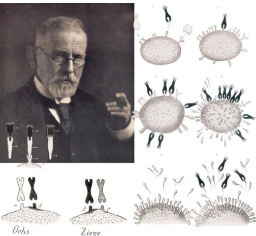
Figure 1 Ehlrlich’s side-chain theory
Paul Ehlrich (1854 -1915), drawings of the formation and effector functions of antibodies according to the side-chain theory. Reproduced from (Kaufmann, 2008).
24
The immune system:
The system that our body has evolved to fight against microbial components is called immunity. History of immunology (the study of immunity) goes back to the 15th century
when the first inoculation of smallpox was performed in China by Wan Quan, the paediatrician of Ming's family. Since then several factual observations show that exposure to a pathogen could lead to immunization. However, major advances were only made in the 19th century with Elie Metchnikoff and Paul Ehrlich explaining the
mechanisms leading to efficient immune responses. Metchnikoff first demonstrated the process of phagocytosis by punching the small thorns in the starfish larvae and finding unusual white cells surrounding the thorns. He proposed the hypothesis that this could be the process by which bacteria were attacked and killed by white blood cells. At the same time, Paul Ehrlich was developing side chain theory, where he postulated that cell protoplasm contains special structures with chemical side chains to which toxins bind (Figure 1). New side chains replace old side chains in case the organism survives injury caused by toxins. Elie Metchnikoff and Paul Ehrlich shared the Nobel Prize in Physiology for their “work on immunity" in 1908 (Kaufmann, 2008). They had basically discovered the innate and adaptive immune systems.
I.
Innate immune system:
The innate immune system refers to nonspecific defence mechanisms that come into play immediately or within hours of microbial infection. These mechanisms include barriers such as skin and mucosa, chemical compounds circulating in the blood such as complement system molecules and phagocytes and innate lymphocytes that are recognized microbes, a process known as innate sensing. The innate immune response is triggered by the recognition of the chemical properties of antigens. Neutrophils (phagocytes) are a good example of innate immune system cells. Whereas, macrophages and dendritic cells work at the frontier between innate and adaptive immunity.25
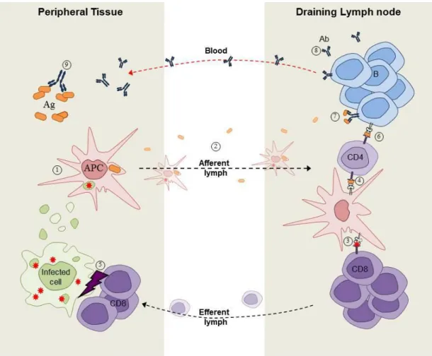
Figure 2. Overview of immune responses.
Antigen-presenting cells (APCs) that have internalized pathogens and/or infected cells ① leave peripheral tissues and reach, via the lymph, secondary lymphoid organs such as the spleen and lymph nodes ②. There, depending on its origin, the antigen will be presented to either CD8+ T cells ③ or CD4+ T cells ④. While CD8+ T cell activation results in the cytotoxicity-mediated killing of target cells ⑤, CD4+ T cells provide help ⑥ to antigen-stimulated B cells ⑦. B cell activation induces the production and secretion of antigen-specific antibodies ⑧ that recirculate through the blood to fight against pathogen within peripheral tissues ⑨. Ag=antigen, Ab=antibody [Picture credit: Dorian Obino]
26
II.
Adaptive immune system:
The adaptive immune system allows generating antigen-specific immune responses. It is more complex than the innate immune system as antigens must be specifically recognized and processed. First step of adaptive immune response is antigen uptake by dendritic cells in the peripheral system, soon after the uptake dendritic cells leave the periphery and travel through the lymph to reach secondary lymphoid organs such as lymph nodes, where they present processed antigen to antigen specific CD4+ or CD8+ T cells. While CD8+ T cell activation results in the cytotoxicity-mediated killing of target cells, CD4+ T cells provide help to launches the adaptive immune response that creates an army of immune cells specifically designed to attack the antigen (Figure 2). Adaptive immunity also includes a "memory" that makes future responses against a specific antigen more efficient. Development, structure and function of B lymphocytes are detailed in next chapter.28
B lymphocytes
B lymphocytes (B cells) are important cells of adaptive immune system and are essential for antibody production. For efficient antibody production, B cell needs to acquire antigens that are presented to them by neighbouring antigen presenting cells. B cells acquire antigen through their specific membrane receptors called B cell receptor (BCR). Following antigenic stimulation, B cells can process and present antigens in association with MHC class II molecules, thereby recruiting specific CD4+ T-cell help and stimulating B-T-cell proliferation and differentiation (Rock, Haber, Liano, Benacerraf, & Abbas, 1986). B cells can differentiate along two distinct pathways: they can either differentiate into extra follicular short lived plasma blasts, that are important for rapid antibody production and early protective immune responses, or they can enter into germinal centres (GC). There, they differentiate into long live plasma cells, which secrete high-affinity antibodies following affinity maturation*, or memory B cells, which confer long-lasting protection from secondary challenges (MacLennan, 1994)(Rajewsky, 1996). The process of B cell development, BCR signalling, antigen processing, presentation and internalization are detailed in following subparts.
*Affinity maturation: Affinity maturation occurs on surface immunoglobulin of GC B cells. It is the
process of high affinity antibody production by B cells which are activated by T follicular helper cells during the course of an immune response. With repeated exposures to the same antigen, B cells will produce antibodies with several log-fold higher affinities than in a primary response.
29
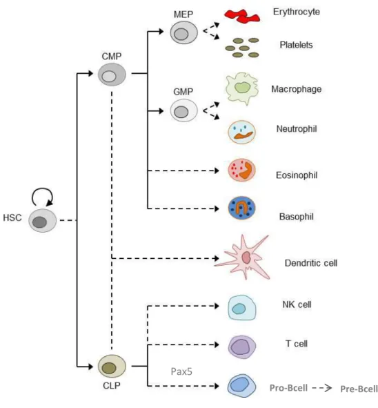
Figure 3. Overview of hematopoiesis.
HSC, hematopoietic stem cell; CMP, common myeloid progenitor; CLP, common lymphoid progenitor; MEP, megakaryocyte/erythroid progenitor; GMP, granulocyte-macrophage progenitor. Adapted from (Larsson & Karlsson, 2005)
Pax5
30
I. B cell development
Development of B cells begins in the foetal liver and continues in the bone marrow. B cells differentiate from hematopoietic stem cells (Sub capsular sinus). Sub capsular sinus give rise to common lymphoid progenitors (CLPs) that can differentiate into either T or B lymphocytes depending on the upregulation of unique transcription factors such as PAX5 (Larsson & Karlsson, 2005). Upon upregulation of PAX5, CLPs irreversibly differentiate into Pro-B cell, which express Immunoglobulin (Ig)α and Igβ on their surface (Nutt et al., 1999). Pro-B cells become pre-B cells when they express membrane µ chains with surrogate light chains in the pre-BCR. Checkpoint at pre-B cell stage only selects cells with functional pre-BCRs. Further gene rearrangement in Light (L) chain leads to differentiation to immature B cells, which express mature-BCRs at their surface. Another round of selection eliminates self-reactive B cells. Selected B cells at this stage exit the bone marrow and enter the spleen and complete their maturation process and develop into mature B cells residing within B cell follicles (follicular B cells) or the spleen marginal zone (marginal zone B cells). At that stage, a pool of mature B cells egress from the spleen, gains access to the blood circulation and colonize lymph nodes surface (Nagasawa, 2006) (Nutt et al., 1999) (Figure 3). Pro-B cells become pre-B cells when they express membrane µ chains with surrogate light chains in the pre-BCR. Checkpoint at pre-B cell stage only selects cells with functional pre-BCRs. Further gene rearrangement in Light (L) chain leads to differentiation to immature B cells, which express mature-BCRs at their surface. Another round of selection eliminates self-reactive B cells. Selected B cells at this stage exit the bone marrow and enter the spleen and complete their maturation process and develop into mature B cells residing within B cell follicles (follicular B cells) or the spleen marginal zone (marginal zone B cells). At that stage, a pool of mature B cells egress from the spleen, gains access to the blood circulation and colonize lymph nodes (Nagasawa, 2006).
31
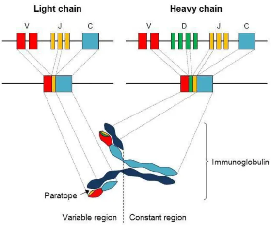
Figure 4. Structure and assembly of immunoglobulins
Immunoglobulins are composed of two light chains associated with two heavy chains. These chains are generated by an unique combination of rearranged coding sequences, divided in variable (V), diversity (D) and joining (J) segments through a process called V(D)J recombination. [Picture credit: Dorian Obino]
32
II. Structure of the B cell receptor
The BCR consists of an antigen binding transmembrane immunoglobulin (mIg) facing the extracellular environment. The BCR is composed of two parts: (1) a membrane-bound antigen-binding immunoglobulin (mIg) molecule of one isotype (IgD, IgM, IgA, IgG, or IgE), (2) two transmembrane polypeptides, Igα and Igβ, containing immunoreceptor tyrosine activation motifs (ITAMs), which enable the transmission of intracellular signalling. As all immunoglobulins (Igs), the BCR can be subdivided into two main regions: at the N-terminus, the variable region is formed of two paratopes involved in antigen binding, whereas the C-terminus part of the protein corresponds to the constant region and defines the isotype (IgM, IgD, IgA, IgE or IgGs) and function of Igs (Figure 4).
Immunoglobulins are large Y shaped proteins composed of two light chains associated with two heavy chains. These chains are generated by an unique combination of rearranged coding sequences, divided in variable (V), diversity (D) and joining (J) segments through a process called V(D)J recombination. During the lifetime of a B cell, immunoglobulin loci undergo several genetic rearrangements to generate mature BCR. The first recombination event occurs between one D and one J gene segment of the heavy chain locus that deletes any DNA between these two gene segments. This D-J recombination is followed by the joining of one V gene segment, from a region upstream of the newly formed DJ complex, forming a rearranged VDJ gene segment. All other gene segments between V and D segments are now deleted from the cell’s genome. This rearranged VDJ segment then associates with the constant region of IgH (Immunoglobulin heavy chain) to form the Pre-BCR. Undergoing allelic exclusion, Pre-B cell rearranges VL and JL segment of IgL (Immunoglobulin light chain) gene locus to
form a mature BCR with a unique variable region. At this stage, autoreactive B cells are subsequently eliminated, and the remaining B cells start to express both IgM and IgD (Metzger, Metzger, Ling, Hurst, & Van Cleave, 1992)
33
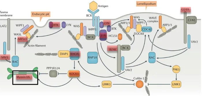
Figure 5 . B cell receptor signalling to the cytoskeleton
34
III. BCR signalling
As mentioned above, BCR is composed of membrane immunoglobulin (mIg) molecules and associated with Igα and Igβ heterodimers. The mIg subunits bind antigen, resulting in receptor aggregation, while the α/β subunits transduce signals to the cell interior. BCR activation induces activation of Lyn, a Src family kinase which phosphorylates the ITAMs and this in turn leads to phosphorylation of Syk, Btk and Vav. This initiates intracellular calcium release and the generation of secondary messengers e.g inositol-1.4.5-triphosphate (IP3) and diacylglycerol (DAG) (Tolar, Sohn, & Pierce, 2008)(Cambier, Pleiman, & Clark, 1994).
Although receptor aggregation is required for BCR signalling, it is not sufficient to elicit B cell activation. B cells needs to amplify these aggregation by spreading the membrane around the antigen presenting cell (Fleire et al., 2006) leading to actin remodelling. Spreading of B cell has been directly linked to BCR signalling, which depends on the BCR-associated tyrosine kinases Lyn and Syk, phosphorylation of the adaptor CD19, and recruitment of cytoplasmic proteins BLNK, PLCγ2, and Vav (Weber et al., 2008), (Depoil et al., 2009) (Figure 5). These molecules form a protein complex associated with the BCR that activates signalling pathways responsible for actin remodelling. Plasma membrane changes shape upon activation via dephosphorylation of Ezrin-Radixin-Moesin (ERM), these proteins influence the organization and integrity of BCR microclusters (Treanor, Depoil, Bruckbauer, & Batista, 2011). Activation of Vav stimulates actin polymerization through activation of Rac and the Arp 2/3 complex. Actin polymerization drives extension of the membrane edge and generation of lamellipodia (Arana et al., 2008)(Depoil et al., 2009)(Weber et al., 2008) and endocytic pits. RhoA activates actin polymerization and stimulates myosin II contractility through Rock1 activity (Saci & Carpenter, 2005).
35
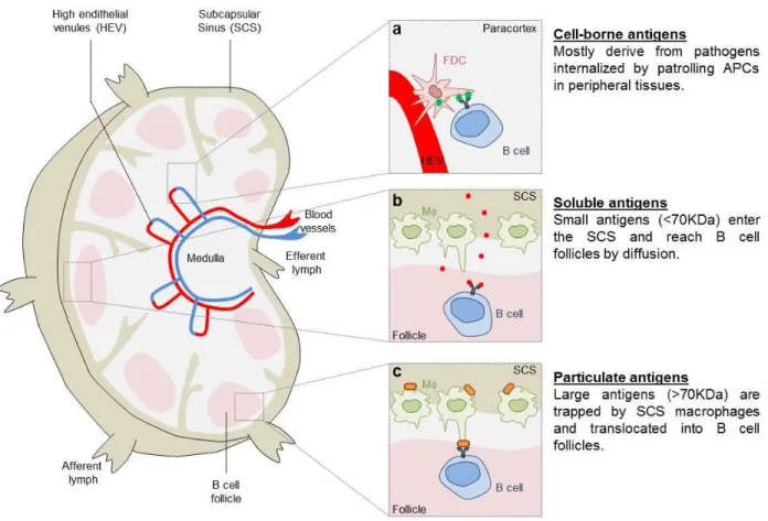
Figure 6. Antigen encounter by B cells.
Depending on their nature and origin, antigens follow different routes to be presented to B cells. Pathogen-derived antigens generated within antigen-presenting cells (APCs) are presented to B cells in areas close to high endothelial venules (a). Small soluble antigens reach B cell follicles by diffusion where they encounter antigen-specific B cells (b). In contrast, large/particulate antigens reaching the sub-capsular sinus of lymph nodes are trapped at the surface of specialized macrophages (SCS CD169+ macrophages), translocated within B cell follicles and presented to antigen-specific B cells in their native form (unprocessed antigens) (c). Adapted from (Batista & Harwood, 2009) [Picture Credit Dorian Obino]
36
IV. Antigen encounter by B cell
B cells are not migratory cells that patrol the body in search of antigen like T cells or dendritic cells. They reside in the follicles of secondary lymphoid organs where they encounter specific antigens. Antigens residing in the peripheral tissues can be taken up by sub-capsular macrophage or dendritic cells that transport them towards lymph node or directly present them to B cell (Qi et al., 2006). Antigens may also reach draining lymph nodes passively, through the lymph or the blood circulation, where B cells encounter them by different means that are detailed below (Figure 6).
i.
Encounter via macrophage or dendritic cells
Follicular dendritic cells have been described to acquire and present antigens to B cells (Suzuki, Grigorova, Phan, Kelly, & Cyster, 2009) as well as medullary macrophages, which acquire opsonized antigens. However, these cells were not required to initiate B cell responses in a model of influenza virus infection, which rather relies on dendritic cells presenting in the medullary sinus (Gonzalez et al., 2010)(Figure 6a).
ii.
Encounter with soluble antigen
Antigen smaller than 70 KDa are considered as soluble antigen. Afferent lymph vessels supply antigen to the lymph node and soluble antigen can be detected within minutes of subcutaneous administration (Nossal, Abbot, Mitchell, & Lummus, 1968). In1980, Farr. A. et al. identified small pores (0.1-1 µm) in the region of sub-capsular sinuses by electron microscopy (Figure 6b). These pores might allow small soluble antigens to enter the lymph node through the afferent vessels and get direct access to B cells.
iii.
Antigens immobilized on cell membranes
Large antigens (greater than 70 kDa) remain trapped at the sub capsular sinus (SCS) floor site where CD169+ macrophages have been shown to capture and transfer them to follicular B cells (Carrasco & Batista, 2006)(Junt et al., 2007)(Figure 6c). How antigens are transferred from the SCS to B cell follicles is unclear. Two routes have been suggested: first, the sub-capsular CD169+ macrophages that display a poor phagocytic capacity recycle antigens and expose them in their native form at the cell surface. Second, antigens that are immobilized at the surface of CD169+ macrophages could
38
be translocated from the SCS to B cell follicles through macrophage protrusions (Martinez-Pomares & Gordon, 2007). In all stated cases, antigens are presented to B cells triggering the internalization of antigen-BCR complexes into endo-lysosomal compartments. This leads to the processing of the antigen and the generation of antigenic peptides that will be loaded onto MHC-II molecules for further presentation to primed CD4+ T cells.
39
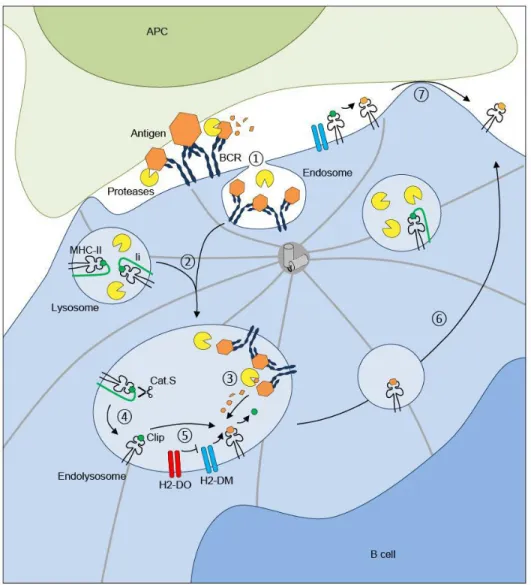
Figure 7. Antigen processing by B cells:
BCR cross-linking by antigens induces the internalization of BCR antigen complexes within B cell– endosomes ①. Endolysosomes that are formed by the fusion ② of antigen containing endosomes with lysosomes that carry MHC-II molecules allow the efficient processing of antigens ③. In the same compartment, Cathepsin S (Cat.S) cleaves the invariant chain (Ii) resulting in MHC-II–CLIP complex formation ④. Finally, H2-DM molecules promote the exchange between CLIP and antigenic peptides for them to be loaded onto MHC-II molecules ⑤. The catalysis of CLIP released by H2-DM is regulated by another non-classical MHC-II molecule, H2-DO. Peptide– MHC-II complexes are exported to the B-cell surface ⑥. Of note antigenic peptides might also be generated within the synaptic space where they are directly loaded onto MHC-II molecules at the cell surface ⑦. Adapted from (Obino & Lennon-Duménil, 2014)
40
V. Antigen processing by B cells
For the production of high affinity antibodies by B cells, it is essential that internalized antigen is processed and loaded on to MHC-II molecules to eventually present to primed CD4+ T cells. This step is known as T-cell/B-cell cooperation and is pivotal for the ultimate formation of GC and production of high-affinity antibodies by B cells (Mitchison, 2004).
BCR cross-linking by antigens induces the internalization of BCR-antigen complexes and polarization of MTOC to the BCR–antigen interaction zone, allowing the local recruitment of lysosomes. Antigens undergo limited proteolysis in order to preserve T-cell epitopes from excessive degradation (Delamarre, Pack, Chang, Mellman, & Trombetta, 2005) that was also shown to facilitate the arrival of antigen–BCR complexes into MHC-II endo lysosomes through its interaction with the cytosolic tail of invariant chain (Ii) (Vascotto et al., 2007). To prevent the premature binding of endogenous peptides and MHC-II molecules, Ii gets associates to MHC-II molecules during biogenesis in the endoplasmic reticulum. Once in endo-lysosomes Cathepsin S cleaves the Ii (Bakke & Dobberstein, 1990), (Lotteau et al., 1990), (Roche & Cresswell, 1991) (Figure 7) .This ultimately leads to the generation of the Ii CLIP fragment that occupies the MHC class II peptide-binding groove. Non-classical MHC-II molecule “H2-DM” catalyses the exchange between antigen-peptide complex and CLIP, this catalysis is regulated by another non-classical MHC-II molecule called “H2-DO” (Driessen et al., 1999)(Riese et al., 1996). H2-DO knockout B cells were found to exhibit increased amount of MHC-II peptide complexes and compete wild-type B cells for the entry to GCs. Finally, Peptide–MHC-II complexes are exported to the B-cell surface for further presentation to primed CD4+ T cells. Remarkably, antigen processing and peptide loading onto MHC class II molecules can also directly take place at the B-cell surface, where H2-DM molecules are also found(Lisa K. Denzin & Cresswell, 1995),(L K Denzin, Sant’Angelo, Hammond, Surman, & Cresswell, 1997).
42
VI. Antibody production
After antigen internalization and processing, a pool of B cells differentiates into short-lived plasmablasts producing antibodies with relatively low affinity (Cunningham et al., 2007), another pool migrates towards T cell boundary and receive signals from helper T cells to differentiate into T follicular helper cells (Reif et al., 2002)(Okada et al., 2005). This T-B cooperation is required for B cells to be fully activated, proliferate, and form GCs (Mitchison, 2004).
During the GC reaction, while proliferating, antigen-activated B cells undergo affinity maturation through a process named somatic hyper mutation (SHM). This results in the introduction of point-mutations within the V (D) J segment of immunoglobulin variable regions leading to changes in the affinity of the BCR for its cognate antigen. Here, affinity refers to the strength with which the epitope binds to an individual antigen-binding site on the antibody. High affinity antibodies bind quickly to the antigen with a stronger bond whereas low affinity antibodies bind weakly to the antigen and often do not detect efficiently the antigen. Following selection of B cells with higher affinity for the antigen, they go through immunoglobulin class switch recombination , leading to the generation of different classes of high-affinity Igs (De Silva & Klein, 2015). Following successive rounds of the GC reaction, selected class switched B cells* differentiate into either plasma cells, which produce antigen-specific high affinity antibodies, or memory B cells (Allen et al., 2007), (MacLennan, 1994).
* Class Switch: the mechanism that changes the antibody production of B cells from one type to
another, e.g. B cells switch from the isotype IgM to the isotype IgG. During class switch recombination the constant region portion of the antibody-heavy chain is changed, but the variable region of the heavy chain remains the same; thus, class switching does not affect antigen specificity. Since variable regions do not change, class switching does not affect antigen specificity. Instead, the antibody retains affinity for the same antigens, but can interact with different effector molecules.
43
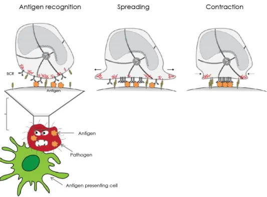
Figure 8. B lymphocytes form an immunological synapse when acquiring antigen in vivo:
Upon antigen recognition , B cells starts to spread around antigen presenting cell , soon after reaching the maximum spreading, it starts to contract and gather antigen in the center to eventually internalize it.
44
B cell immune synapse
Immune synapse is an interface between the APC and lymphocytes similar to the neurological synapse. It was discovered by Abraham Kupfer at the National Jewish Medical and Research Centre in Denver. The term immune synapse was coined by Michael Dustin at NYU. Where he coined the term in context of T and B cell interaction, where B cell act as an APC. In this thesis, immune synapse between APC and B cell is detailed.
Formation of B cell immune synapse is essential for antigen recognition. In order to efficiently acquire antigen, B cell first spread around the APC to gather maximum amount of antigen and then starts to contract to eventually pull off the antigen from the surface of APC (Figure 8). The acto-myosin cytoskeleton plays a central role in each of these steps that are detailed in subsequent sections. In the first section I will describe role of actin and myosin in antigen and the different extraction pathways, B cell spreading and contractions. I will also describe most common biophysical techniques to measure forces: in particular traction force microscopy that will be used through this entire thesis.
45
Figure 9. Actin cytosketon structure: showing lamellipodium , filopodium, microvilli, podosomes [http://jonlieffmd.com/blog/virus-tricks-manipulate-the-cytoskeleton].
46
Actin and Myosin:
Two core component of B cell cytoskeleton
The cytoskeleton is a highly interconnected structure, which regulates cell shape, cell division and movement. In eukaryotic cells, the cytoskeleton is mainly composed of three structures: microfilaments made of actin, intermediate filaments and microtubules (Wickstead & Gull, 2011). Of note, it is now well admitted that proteins belonging to the septin family form the fourth component of the cytoskeleton (reviewed in (Mostowy & Cossart, 2012)).
I.
Actin Cytoskeleton
Actin is the major cytoskeletal protein, which polymerizes to form actin filaments. Actin filaments are made up of thin, flexible fibers approximately 7 nm in diameter that can be cross-linked together to form different actin structures. In1942 , actin was first isolated from muscle cells (Needham, 1942) in which it constitutes approximately 20% of total cell protein. Although actin was initially thought to be uniquely involved in muscle contraction, it is now known to be an extremely abundant protein (typically 5 to 10% of total protein) in all types of eukaryotic cells.
Actin monomers are called globules. These higher-order structures form bundles or three-dimensional networks with the properties of semisolid gels. The assembly and disassembly of actin filaments, their crosslinking into bundles and networks, and their association with other cell structures (such as the plasma membrane) are regulated by a variety of actin-binding proteins, which are critical components of the actin cytoskeleton. Branched networks form lamellipodia (Figure 9) that helps the cell to propel itself on a surface. Parallel actin filament networks helps in membrane protrusions, called filipodia. Myosin II can connect two parallel actin fibres to make stress fibres. Actin not only provides mechanical support, determines cell shape, and allows movement of the cell surface but also help in several endocytic internalization processes described in subsequent sections.
47
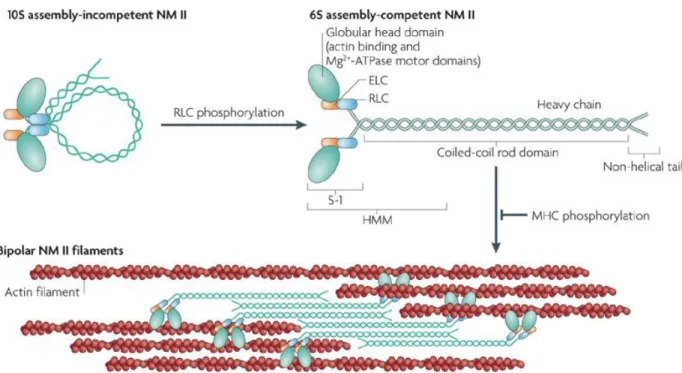
Figure 10. Domain structure of Myosin II
(a)The subunit and domain structure of non-muscle myosin II (NM II), which forms a dimer through interactions between the alpha-helical coiled-coil rod domains. The globular head domain contains the actin-binding regions and the enzymatic Mg2+-ATPase motor domains. The essential light chains (ELCs) and the regulatory light chains (RLCs) bind to the heavy chains at the lever arms that link the head and rod domains. In the absence of RLC phosphorylation, NM II forms a compact molecule through a head to tail interaction. This results in an assembly-incompetent form (10S; left) that is unable to associate with other NM II dimers. On RLC phosphorylation, the 10S structure unfolds and becomes an assembly-competent form (6S). S-1 is a fragment of NM II that contains the motor domain and neck but lacks the rod domain and is unable to dimerize. Heavy meromyosin (HMM) is a fragment that contains the motor domain, neck and enough of the rod to effect dimerization. (b) NM II molecules assemble into bipolar filaments through interactions between their rod domains. These filaments bind to actin through their head domains and the ATPase activity of the head enables a conformational change that moves actin filaments in an anti-parallel manner(Vicente-Manzanares, Ma, Adelstein, & Horwitz, 2009)
48
II.
Myosin II cytoskeleton
Myosins constitute a superfamily of motor proteins that play important parts in several cellular processes that require force and translocation. Most myosins belong to class II and, together with actin, make up the major contractile proteins. Thus, myosin II is the conventional motor protein involved in cardiac, skeletal and smooth muscle contraction. It is made up of two heavy chains. The N terminus of the heavy chain has a globular head domain, that binds to actin head domain and have the catalytic sites where energy is generated by ATP hydrolysis. The C-terminus tail domain has a coiled coil morphology that holds the two heavy chains together. The intermediate regulatory neck domain makes the angle between the neck and tail domain described in (Figure 10). Myosin II molecules that resemble their muscle counterparts, with respect to both structure and function, are also present in all non-muscle eukaryotic cells (Clark, Langeslag, Figdor, & van Leeuwen, 2007)and (Conti & Adelstein, 2008). Like muscle myosin II, non-muscle myosin IIA (NM II) molecules are comprised of three pairs of peptides: two heavy chains of 230 KDa, two 20 KDa regulatory light chains (RLCs) that regulate NM II activity and two 17 KDa essential light chains (ELCs) that stabilize the heavy chain structure. Although these myosins are referred to as ‘non-muscle’ myosin II to distinguish them from their muscle counterparts, they are also present in muscle cells, where they have distinct functions during skeletal muscle development and differentiation, as well as in the maintenance of tension in smooth muscle (Vicente-Manzanares et al., 2009).
49
Figure 11. The dynamic polymerization of actin filaments:
Actin filament (red) is involved in different processes that reshape or move cellular membranes. These processes include different forms of endocytic uptake at the plasma membrane — that is, clathrin-mediated, macropinocytosis and phagocytosis in mammalian cells. The protrusion of lamellipodia and filopodia in migrating mammalian cells is dependent on actin polymerization (Kaksonen, Toret, & Drubin, 2006), arrows indicating actin cytoskeleton organization in phagoc ytosis, macropinocytosis and clathrin mediated endocytosis .
50
Antigen extraction by B cells
I. Endocytosis
Internalization of extracellular fluid/particles represents an important path of entry within cells and particularly applies to cells of the immune system. Actin remodelling underneath the plasma membrane leads to the formation of membrane protrusions that are essential for the initiation of the internalization process. As most of the important chemical substances are large polar molecules that cannot pass through plasma and cell membrane by itself, they need to be transported , endocytosis is a form of active transport in which a cell transports molecules (such as proteins) into the cell by engulfing them. Reshaping of plasma membrane and actin polymerization plays a central role in different forms of endocytic internalization — for example phagocytosis, micro and macro-pinocytosis, and clathrin-mediated endocytosis.
Phagocytosis: Phagocytosis was discovered by Elie Metchnikoff. It is an internalization
process of harmful foreign particles such as bacteria, dead or dying cells by phagocytes. Dendritic cells, macrophages, mast cells are example of professional phagocytes. In general phagocytosis is induced by specific surface receptors within the cell, named pattern recognition receptors (PRRs), which recognize pathogen-associated molecular patterns (PAMPs), a class of molecules that are broadly shared by pathogens but distinguishable from host molecules (Figure 11).The size of a phagocytic particle goes up to several micrometers (Champion, Walker, & Mitragotri, 2008)
Micropinocytosis/Macropinocytosis: Pinocytosis is defined as the drinking up by cells in
greek language. We can define pinocytosis as the taking up of fluid into a cell of by invagination of the plasma membrane, which is then pinched off, resulting in small vesicles in the cytoplasm. When cells uptake molecules of less than 0.1 µm diameter, the process is named micropinocytosis and for the particles greater than 0.5 µm it is termed macropinocytosis. Macropinocytosis is the primary form of internalization in dendritic cells. No receptor is required for the initiation of micro and micropinocytosis (Figure 11).
52
Clathrin-mediated endocytosis: is a type of receptor mediated endocytosis involving
production of small (approx. 100 nm in diameter) vesicles that have a morphologically characteristic coat made up of the cytosolic protein clathrin. Clathrin-coated vesicles (CCVs) are found in virtually all cells and form domains of the plasma membrane termed clathrin-coated pits (Marsh & McMahon, 1999)(Figure 11).
i. B cell antigen internalization
Clathrin mediated endocytosis is the principle type of antigen internalization in B cells (Natkanski et al., 2013)(Stoddart et al., 2005). Natkanski et al., showed that knocking down clathrin leads to substantial reduction of internalization of both soluble and membrane presented antigens. Clathrin colocalize with most of internalized antigen clusters. They also showed that although actin polymerization and clathrin coated pits (CCPs) formation are essential for antigen internalization, they are not sufficient for extraction of membrane antigens in B cell synapse. They addressed the role of myosin II contractility in the invagination of BCR microclusters, which occurs before the formation of the CCPs. Formation of CCPs alone does not generate the forces required for endocytosis of membrane-attached antigens. However, DNA-based tension sensor measurements (detailed in next section) showed that B cell exert force of 10 pN to extract one antigen molecule and with the help of repetitive pulling on the BCR-antigen bond with the help of myosin II, B cells are able to extract BCR-antigens that have a stronger bond (Wan et al., 2015). Apart from mechanical extraction, there’s also enzymatic extraction through lysosomal proteases that polarize towards the synapse and cleaves the bond between antigen and APC (Yuseff et al., 2011). However recently it has been shown enzymatic extraction is used by B cell only on the substrate that cannot be mechanically deformed. Interestingly, once the mechanical way is activated the secretion of proteases is impaired (Spillane & Tolar, 2016). Even after all the recent progress in the field of B cell antigen internalization, a global picture of what types of forces are involved in antigen internalization is missing.
53
Figure 12. Atomic force microscopy :
(AFM) to measure bond strength as a function of applied force. (Basu & Huse, 2016)
Figure 13.DNA-based tension gauge:
DNA based tension gauge tethers remain attached to the surface unless a sufficient force is applied to rupture Watson–Crick base pairing. Right, a DNA hairpin coupled to a fluorophore (F) and quencher (Q) is used to generate a tension probe that fluoresces upon application of a threshold force. (Basu & Huse, 2016).
54
II. Techniques used in measurement of antigen extraction forces
Immune-synapse undergoes continuous architectural changes, these changes are the result of forces produced inside the cells and is transmitted through receptors, membrane, or integrins to the neighbouring cells. Immune synapse is not only a platform for chemical dialogues but also for mechanical ones. Our understanding of immune synapse mechanics relies on methods designed to measure synaptic forces between single cells such as AFM (Atomic force microscopy), DNA based force sensors, Traction force microscopy (TFM) and Micro-pillars that are described in the following sections (Basu & Huse, 2016).
i. Atomic Force Microscopy:
In this approach, a cell or molecule of interest is attached to a flexible cantilever and then brought into contact with a glass surface coated with target cells or cognate ligands (Figure 12). With atomic force microscopy one can measure adhesive forces up to molecular level between cells. Forces are measured by monitoring the negative deflection of the cantilever as it is withdrawn away from the surface (Basu & Huse, 2016). Although this technique is reliable to measure intermolecular forces, for inter cellular forces it is not always easy to interpret data and get rid of experimental artefacts.
ii. DNA based force sensors
This approach relies on molecular tension gauges containing DNA duplexes linked to stimulatory ligands (Wang & Ha, 2013). Suitable force exertion on the ligand will rupture the duplex, detaching the ligand from the surface. The strength of the duplex depends on its length, degree of base pairing, GC content, and junction point with the protein ligand (Figure 13) (e.g., the center or the end of the strand). Because mechanosensitive receptors, like integrins, only signal effectively under tension, they will be activated only by tension gauge tethers that are strong enough to withstand the associated pulling forces. Hence, by measuring signalling responses on a panel of different TGT surfaces, one can establish the force threshold required for receptor activation (Wan et al., 2015)and (Wang & Ha, 2013).
55
Figure 14. Gel-based TFM:
Force exertion is determined from the movements of beads embedded in the gel matrix. (Basu et al., 2016)
Figure 15. Micropillars:
Cells are imaged on an array of flexible micropillars, allowing applied forces to be calculated from pillar deflection (Basu & Huse, 2016).
56
iii. Traction Force Microscopy:
TFM is a technique for measurement of force based on the deformation of the elastic substrate on which the force is exerted (Harris, Wild, & Stopak, 1980). In this approach, small fluorescent beads are embedded on the top surface of a polyacrylamide hydrogel substrate bearing ligands (Dembo & Wang, 1999). This technique is generally used to measure forces developed during adhesion. Displacement observed in TFM is always measured by comparing each image to a non-stressed reference image. Cells form a contact structure with the deformable substrate (hydrogel), they distort the hydrogel, thereby moving the beads. The displacement field obtained by bead movement is used to obtain the force (inversion of the problem)(Butler, Tolić-Nørrelykke, Fabry, & Fredberg, 2002),(Mandal, Wang, Vitiello, Orellana, & Balland, 2014). The inversion of the problem is done in Fourier space by interpolating the data. Stress maps obtained reveal not only the magnitude and direction of applied forces but also their spatial distribution within the contact structure (Figure 14) (Basu & Huse, 2016). This technique has been generally used for bigger cells (≈100-120 µm) then lymphocytes with many beads underneath the cells to have precise force measurement (down to a single focal adhesion). We adapted this technique to use on B cell (≈5-7 µm) force measurement by optimizing the rigidity of the gel, number of beads and algorithm to measure forces, which are explained in the material and method section.
iv. Micropillars
This approach is a version of TFM in which the hydrogel substrate is replaced by a hexagonal array of PDMS micro pillars coated with stimulatory ligands (Bashour et al., 2014). Lymphocytes form immune synapse like contacts with the pillar tops and move them as they exert force against the surface. Each pillar deflection can be converted into a force measurement based on the height, width, and composition of the pillars. The micro pillar method provides enhanced spatial resolution relative to gel- based TFM because pillars (i) can be spaced within 1 µm of one another and (ii) they move independently of their neighbours (Basu & Huse, 2016) (Figure 15). Limitation of this would arise when using small cells like B cells (5-7 µm), which can easily leak into the pillar gaps.
58
III. Biomechanics of B cell antigen extraction
Immune cells probe their environment and their function replies on extraction and sensing the tissue that makes them constantly influenced by cellular mechanics. B cells also have been shown to have varying degree of activation based on the stiffness of the antigen presenting substrates (Wan et al., 2013). Antigens presented on stiff substrates, promote BCR signalling more efficiently with the generation of higher forces on the BCR, than to the antigens presented on softer substrates, which limit tension. Conversely, precise measurements using DNA tension sensors have revealed that the BCR-antigen bond is poorly responsive when the tension on the BCR is below 20 pN, but responds better to tensions between 20 pN and 40 pN, and reaches a maximum responsiveness to tensions above 40 pN (Wan et al., 2015). The low forces are probably generated in a myosin independent manner, whereas the high forces require myosin contractility. Even in the absence of the mechanosensitive activation of signalling, mechanical forces have strong effects on membrane receptor–ligand complexes by changing the rate of their dissociation.
Most protein–protein bonds are slip bonds, for which stability decreases when pulling forces are applied. In special cases, pulling can induce conformational changes within the proteins and somehow stabilize the bond. These types of bond, termed catch bond, have been observed in various adhesion receptors, such as integrins, and also in the T cell receptors (TCR) (B. Liu, Chen, Evavold, & Zhu, 2014) but this has not yet been proved in B cells. In B cells, it has been proposed that pulling on the antigen-bound BCR against resistance from the APC contributes to affinity discrimination by B cells because bonds with low-affinity antigens, but not high-affinity antigens, rupture before inducing a biological response(Natkanski et al., 2013). This is particularly important for affinity-dependent extraction of antigens, in which bonds need to sustain extraction forces before antigens are endocytosed by the B cell. The mechanical extraction of antigen provides an advantage for high-affinity B cells during antibody responses (Tolar, 2017).
60
Thus, mechanical aspects of B cell synapse emerges as a pivotal feature regulating the B cell immune synapse formation, activation, and eventually antigen extraction. More on cellular mechanics and its role in different cells and systems are mentioned in discussion.
61
Figure 16. The coordination of BCR and actin dynamics during signalling activation:
Stage 1: In resting B cells, surface BCRs and BCR nanoclusters have a relatively limited lateral mobility, which is constrained by the cortical actin network tethered to the plasma membrane by the interaction of ERM proteins with membrane anchored proteins. (B) Stage 2: In response to the binding of membrane-associated antigen, the lateral mobility of BCRs is transiently increased, which is concurrent with the detachment of the cortical actin from the plasma membrane and a transient actin disassembly. This is followed by receptor immobilization and the formation of small clusters, triggering signalling activation. (C) Stage 3: B cell spreading on antigen-associated membrane increases the number and sizes of BCR clusters as the signalling level rises. Actin polymerizes and is accumulated at the leading edge of the spreading membrane and at BCR microclusters. (D) Stage 4: BCR microclusters merge into a central cluster at one pole of the cell while the B cell contracts. The actin cytoskeleton reorganizes away from BCR clusters to the outer rim of the BCR central cluster. The signalling activity of BCRs in the central cluster is reduced. (Song, Liu, & Upadhyaya, 2014)
62
B cell spreading and BCR clustering
Dynamic changes in the actin cytoskeleton upon immune synapse formation induces B cell spreading, which helps to promote the gathering and extraction of membrane-tethered antigens. BCR activation is triggered by receptor clustering that induces receptor phosphorylation and signalling cascades (Tolar, Sohn, & Pierce, 2005). During signalling activation, occurrence of BCR clustering can be temporally described in four stages on rigid but fluid surface like supported lipid bilayer:
1. In the first stage BCRs exist in nano-clusters that have limited lateral mobility, an average diffusion coefficient of IgM-based BCR in naïve primary B cells of ~0.03 μm2/s (Tolar, Hanna, Krueger, & Pierce, 2009),(Treanor et al., 2010) (Figure 16A).
2. In the second stage in response to the binding of antigen, the lateral mobility of BCRs transiently increases (~0.05 μm2/s for membrane-associated antigen),
enabling interactions between BCRs and their nano-clusters to lead to the formation of microclusters (Fleire et al., 2006) (Treanor et al., 2010), (Treanor et al., 2011)(Figure 16B).
3. In the third stage, the B cell spreads on antigen-associated membrane (Fleire et al., 2006), thereby driving the formation of more BCR microclusters. Newly formed BCR microclusters move centripetally to one pole of the cells or to the center of the cell surface contact zone, recruiting more BCRs and apparently colliding with each other along the way (Tolar et al., 2009)(Figure 16C)
4. In the final stage, BCR microclusters merge together, resulting in the formation of a central cluster either at one pole of the cell (for soluble antigen) or in the center of the B cell contact zone (Carrasco, Fleire, Cameron, Dustin, & Batista, 2004) and (Tolar et al., 2009)(Figure 16D).
64
Actin polymerization at the membrane edge generates lamellipodia, the contact of lamellipodia with the antigen presenting cell is followed by integrin (LFA-1 and VLA-4) mediated adhesion, which stabilizes the synapse and contributes to the synaptic architecture (Carrasco & Batista, 2006)(Carrasco et al., 2004). Soon after actin reorganization, lamellipodial extensions proceed through cycle of protrusion and retraction (Tolar et al., 2009). It has been shown in fibroblasts that these protrusions are driven by actin polymerization at the leading edge of the cell, while the retractions are mediated by the non-muscle Myosin IIa pulling from the back of the lamellipodia (Giannone et al., 2017). Actin polymerization at the edges generates forces perpendicular to the edge of the synapse that initiate the centripetal BCR movements (Fleire et al., 2006)(Song et al., 2014). During the last stage of BCR clustering, the levels of F-actin diminish in the centre of the B cell contact zone as B cells contract and BCR microclusters merge into a central cluster (Song et al., 2014). To conclude, B cells spread around antigen-associated membranes and start the formation of microclusters. These microclusters move centripetally and fuse into a big central cluster.
65
Figure 17. Mechanics of BCR binding at the B-cell synapse:
Schematic depiction of the B-cell synapse (top) and the amount of local antigen binding (bottom). (1) BCR signalling in lamellipodia stimulates actin polymerization, protrusion of the leading edge, and pushing of the B cell membrane into contact with the APC. Binding is limited by the incomplete alignment of the B-cell membrane with the APC. (2) BCR microclusters are transported by actin flow and by myosin IIa contractions toward the center of the contact. Binding is promoted by alignment of the synaptic membranes and by BCR clustering. (3) Myosin IIa pulls on the microclusters and invaginates the B-cell membrane. The forces trigger accelerated dissociation. (4) Formation of clathrin-coated pits (CCPs) generates additional actin polymerization and eventual endocytosis of the antigen. (5) Forces are terminated and endocytosis reads the final number of antigens bound to the BCR . (Tolar & Spillane, 2014)
![Figure 9. Actin cytosketon structure: showing lamellipodium , filopodium, microvilli, podosomes [http://jonlieffmd.com/blog/virus-tricks-manipulate-the-cytoskeleton].](https://thumb-eu.123doks.com/thumbv2/123doknet/2332066.31933/46.918.141.690.128.605/cytosketon-structure-lamellipodium-filopodium-microvilli-jonlieffmd-manipulate-cytoskeleton.webp)