THÈSE
En vue de l’obtention du
DOCTORAT DE L’UNIVERSITÉ DE TOULOUSE
Délivré par l’Université Toulouse 3 Paul Sabatier (UT3 Paul Sabatier)Présentée et soutenue par
Céline Mathieu-Demazière
le Vendredi 31 janvier 2014Titre :
Cyclic-AMP signalling in Sinorhizobium meliloti and its role in the
control of infection during symbiosis with Medicago
ED SEVAB: Interactions Plantes-Microorganismes
Unité de recherche:
Laboratoire des Interactions Plantes-Microorganismes (LIPM) UMR CNRS-INRA 2594/441
Directeur de Thèse :
Jacques Batut, Directeur de recherche INRA
Rapporteurs :
Anne Galinier, Directeur de recherche CNRS Pascal Ratet, Directeur de recherche CNRS
Autres membres du jury :
Matthieu Arlat, Professeur UT3 Paul Sabatier, Président Christophe Guilhot, Directeur de recherche CNRS, Invité
2
4
Table of contents
REMERCIEMENTS ... 10 ABSTRACT ... 14 RESUME ... 16 LIST OF FIGURES ... 18 ABBREVIATIONS ... 20 INTRODUCTION ... 28A. THE SINORHIZOBIUM MELILOTI-MEDICAGO SYMBIOSIS ... 28
I. Introduction ... 28
1. Background and agronomic stakes ... 28
2. The nodule, a N2 fixing plant organ ... 30
3. Medicago truncatula and Lotus japonicus as model legumes ... 32
4. Sinorhizobium meliloti, the Medicago symbiont ... 34
II. Basis of Medicago colonization by S. meliloti ... 36
1. Infection of plant epidermis ... 36
2. Cortical events: nodule organogenesis and bacteria endocytosis ... 38
III. Signalling in the rhizobium-legume symbiosis ... 40
1. Early symbiotic signals... 40
2. Early signal transduction ... 46
3. Late symbiotic signal: oxygen-controlled regulatory system ... 46
IV. Negative control of symbiosis ... 50
1. Nodule number is controlled by phytohormones and CLE peptides ... 50
a) Hormonal control ... 50
b) AON ... 52
2. NCR peptides control bacteroid differentiation ... 54
3. Evidence for a negative regulation of infection ... 58
V. Bacterial escape of plant immunity ... 60
1. Rhizobia reduce stimulation of the host immune system ... 60
2. Rhizobia supress host defence responses ... 62
3. Plant immunity is key to symbiosis ... 64
B. SIGNAL TRANSDUCTION BY ADENYLATE CYCLASES ... 68
I. Signal transduction in bacteria is dominated by two-component systems and cyclic nucleotide signalling ... 68
1. Two-component phosphorelay systems ... 68
2. An overview of secondary messengers ... 70
II. Role of cAMP in bacteria... 78
1. Role of cAMP in bacterial metabolism ... 78
5
3. Role of cAMP in stress adaptation ... 84
4. Role of cAMP in biotic interactions ... 86
III. Adenylate cyclases, a case of convergent evolution ... 92
1. ACs are divided in six main classes ... 92
2. The universal class III of ACs ... 96
a) Distribution of class III ACs ... 96
b) Classification of class III ACs ... 98
c) The class III catalytic mechanism ... 100
d) Regulatory-domain composition of class III ACs ... 100
IV. The Cyclic-AMP receptor protein CRP ... 108
1. The CRP family ... 108
2. Allosteric activation of CRP ... 108
3. Transcription activation at CRP-dependent promoters ... 112
V. Switching off cAMP signalling: role of phosphodiesterases... 116
1. Phosphodiesterases are divided in three main classes ... 116
2. The class III phosphodiesterases ... 120
C. PHD WORK PRESENTATION ... 126
RESULTS AND DISCUSSION ... 132
A. CHAPTER 1: RHIZOBIA PLAY AN ACTIVE ROLE IN THE CONTROL OF INFECTION DURING SYMBIOSIS WITH MEDICAGO ... 136
I. Introduction ... 136
II. Article 1: Plant-activated bacterial receptor adenylate cyclases modulate epidermal infection in the Sinorhizobium meliloti-Medicago symbiosis ... 140
III. CyaK perceives the nodule signal ... 156
1. CyaK senses the Medicago nodule signal ex planta ... 156
2. CyaD1D2 and CyaK have complementary functions in symbiosis ... 156
3. Signal(s) perception ... 158
B. CHAPTER 2: BIOCHEMICAL AND FUNCTIONAL CHARACTERIZATION OF SPDA, A 2', 3'CYCLIC NUCLEOTIDE PHOSPHODIESTERASE FROM SINORHIZOBIUM MELILOTI ... 162
I. Introduction ... 162
II. Article 2: Biochemical and functional characterization of SpdA, a 2', 3'Cyclic Nucleotide Phosphodiesterase from Sinorhizobium meliloti ... 166
... 191
III. Complementary results ... 192
1. Biological characterization of a S. meliloti spdA overexpressing mutant ... 192
2. Study of CpdB, a putative 2'3'cAMP PDE from S. meliloti ... 192
a) Construction of a S. meliloti cpdB null mutant ... 192
b) Growth characteristics of a S. meliloti cpdB null mutant... 194
c) Expression of smc02178 in a cpdB mutant and ΔSpdAcpdB mutant background ... 194
6
GENERAL DISCUSSION AND PERSPECTIVES ... 198
I. cAMP signalling in Sinorhizobium meliloti ... 200
1. Biological roles of ACs/GCs in S. meliloti ... 200
2. A complex cAMP signalling network in S. meliloti ... 202
3. Spatial and temporal distribution of cAMP ... 204
4. Additional complexity in cNMP signalling pathways ... 206
II. cAMP signalling in symbiosis ... 208
1. Is cAMP signalling part of Autoregulation of Infection? ... 208
2. Molecular mechanism for the negative control of infection ... 212
3. Medicago signal identification ... 214
MATERIAL AND METHODS ... 218
8
« Un seul être vous manque et tout
est dépeuplé »
10
Remerciements
« Par l’esprit et par le marteau », devise des géologues.
Et oui, mes premiers remerciements s’adresse à M. Haroun Tazieff pour ses merveilleux récits qui m’ont donné envie d’être Volcanologue tout comme lui. J’ai ainsi suivi cet objectif dès l’école primaire jusqu’à l’université où j’ai abandonné les Sciences de la Terre et de l’Univers pour celles de la vie. Etant effrayée par l’infiniment grand, j’ai préféré étudier l’infiniment petit.
Et parce que j’ai commencé ma scolarité avec eux, je voudrai remercier Bruno et Nadine, ainsi que Martine, Sophie, Gilles et Cathy. Si je n’avais pas eu des instits’ comme vous, je n’aurais certainement pas aimé l’école…
Je voudrai également remercier tous mes professeurs du secondaire et plus particulièrement M. François Griffoul, mon professeur de SVT de Terminale S qui m’a conforté dans l’idée d’étudier les SVT et pour m’avoir accueilli comme stagiaire quand j’étais à l’IUFM.
Je remercie M. Claude Gutierrez et M. Philippe Rousseau de m’avoir fait confiance en m’acceptant en M2R Microbiologie sachant que je n’avais jamais fait de microbio.
Merci à M. Guillaume Bécard car même s’il l’ignore, c’est grâce à lui et ses superbes cours que j’ai choisi de faire mon stage de master sur les interactions plantes micro-organismes.
Il est donc temps de remercier Jacques et Catherine de m’avoir accepté dans leur équipe et de m’avoir ainsi donné une chance de devenir docteur. Merci Jacques de m’avoir si bien encadré tout au long de ma thèse.
Merci à Anne-Marie qui m’a tout appris et m’a supporté toutes ces années.
Merci à Delphine qui m’a beaucoup aidé durant mon stage de master afin que je puisse avoir une chance de réussir le concours de l’école doctorale.
Merci à Marta et Carine pour leur bonne humeur quotidienne, leur gentillesse et pour leur aide précieuse.
Merci aux jeun’s de l’équipe Suhua, Amandine, Camille et Dorian. Merci à toi Philippe pour nos nombreuses pauses café pendant lesquelles tu as toujours su m’écouter.
11
Merci à Cécile et Martina qui ont bien voulu de moi comme monitrice.
Merci à Peter Mergaert et Julie Cullimore d’avoir bien voulu faire partie de mon comité de thèse.
Merci à tous les membres du laboratoire qui ont participé de près ou de loin à ma vie de thésarde.
Merci à mes amis non-perm et perm Endrick, David, Alice, Brice, Joanne, Marie C, Marie T, Rémi, Violaine, Mehdi, et (les meilleures pour la fin) merci à Justine et Marie-Anne. Vous êtes toutes les deux de merveilleuses personnes et je suis sûr qu’une grande carrière vous attend. Justine, heureusement que tu étais là tous ces derniers mois. Tu as su rendre la thèse plus facile. Dommage qu’il ait fallu attendre si longtemps avant de se connaître. Il faudrait remercier qui tu sais, finalement c’est grâce à lui si on est amie. Marie-Anne, merci de m’avoir si souvent changé les idées, d’avoir été là pour me remonter le moral quand ça n’allait pas. Merci à vous tous, vous êtes géniaux.
Je voudrai également remercier mes amis de longue date tout simplement pour ce qu’ils sont et parce que sans eux je ne serais pas la personne que je suis aujourd’hui. Merci à Coralie, Nicolas, Gaby, Thomas, Marion, Julie et Louise.
Je remercie toute ma famille pour m’avoir encouragé à aller jusqu’au bout sans jamais me forcer. Merci à mes parents d’avoir cru en moi. Merci à mes grands-parents ainsi qu’à mon oncle Jean-Luc. Merci à ma sœur Mélanie qui par son caractère m’a forcé à forger le mien. Merci à Nolann et Morganne, les deux merveilleux anges que tu as mis au monde. Merci à Cédric puisqu’il y a participé un peu.
Je remercie ma cousine Noémie pour toutes les bêtises qu’on a pu faire et les fous rires qu’on a eu ensemble. Noémie, tu me manques, tu nous as quitté beaucoup trop tôt. J’aurais aimé connaître la femme que tu serais aujourd’hui.
Et merci à mon mari Adrien qui partage ma vie depuis 10 ans maintenant. Merci d’avoir été là depuis le début, de m’avoir accompagné toutes ces années. Mon Damour, merci pour tout, je n’y serais certainement jamais arrivé sans toi car il faut le dire, on ne peut réussir dans la vie que si on est bien accompagné. Merci d’être ce que tu es. Je t’aime.
12
Et pour être sûr de n’oublier personne, merci à tous ceux qui ont croisé mon chemin et qui d’une manière ou d’une autre m’ont tendu la main.
14
Abstract
The leguminous plant Medicago sativa (Alfalfa) can enter a symbiosis with a nitrogen-fixing bacterium called Sinorhizobium meliloti. S. meliloti elicits on M. sativa roots the formation of specialized organs called nodules that behave as miniature nitrogen-fixing factories in which fixed nitrogen is provided to the plant, in proportion of its needs. Extensive work on the model
Medicago truncatula/S. meliloti has revealed a great deal of the genetic pathway involved in
Nod Factor (NF) perception and signal transduction pathway leading to coordinated nodule organogenesis and controlled bacterial infection. The negative control of nodulation and infection is nowadays a very active area of research. Such a negative control is physiologically essential as excessive nodulation and/or bacterial infection would be detrimental to the symbiosis and to plant health. So far only plant (Medicago and Lotus) mutants have been
identified that are affected for the control of infection. As part of my PhD thesis, we have
shown that the bacterium S. meliloti is also involved in this process. Indeed, we have shown that three receptor-type bacterial adenylate cyclases (ACs) CyaD1, CyaD2 and CyaK, participate in the control of the infectious process in response to an unknown signal. In response to this signal, the three ACs synthesize cAMP which, via the regulator Clr (CRP -like regulator), activates a target gene, the gene smc02178 whose role is still unknown. The mutation of cascade components leads to a hyper-infectious phenotype on M. sativa roots. My PhD work focused on the characterization of signal perception mechanisms by the cyclases, its biosynthesis and the study of its perception by CyaK. We have shown that the signal perceived by the three cyclases is a plant signal. This signal is present in shoots and in nodules of M. sativa, and in a large number of plant species including non-legumes. In a second step, we were interested in a phosphodiesterase (PDE), the protein SpdA (SMc02179), potentially involved in the regulation of cAMP levels in S. meliloti. The study of the smc02179 regulation showed that the gene is expressed in symbiosis from the early stage of infection. We have thus functionally characterized the SpdA protein in vivo and in vitro and studied its role in the signalling cascade.
16
Résumé
La légumineuse Medicago sativa établit une symbiose fixatrice d’azote avec la bactérie
Sinorhizobium meliloti impliquant la formation d’un organe spécialisé, le nodule, au niveau de
la racine de la plante hôte. La formation de nodules fait intervenir un programme complexe de développement impliquant le processus d’organogenèse du nodule, l’infection intracellulaire des cellules de ce dernier, ainsi que des mécanismes de différenciation croisée des deux partenaires. Afin que la symbiose garde son caractère mutualiste, il est nécessaire qu’elle soit régulée négativement. Ainsi la nodulation est contrôlée de manière négative par
Medicago via en particulier une boucle de régulation appelée AON. Pour éviter une
surinfection racinaire, l’infection est également contrôlée de manière négative. Jusqu’à maintenant, on pensait que seule la plante jouait un rôle dans le contrôle négatif de l’infection. Dans le cadre de ma thèse, nous avons mis en évidence que la bactérie S. meliloti intervenait également dans ce processus. En effet, nous avons montré que trois adénylate cyclases (ACs) bactériennes de type récepteur, CyaD1, CyaD2, et CyaK, participent au contrôle du processus infectieux en réponse à un signal de nature inconnue. En réponse à ce signal, les trois ACs synthétisent de l’AMPc qui, via le régulateur Clr (CRP-like regulator), active un gène cible, le gène smc02178 dont le rôle est encore inconnu. La mutation des composants de la cascade conduit à un phénotype hyper-infectieux sur les racines de M. sativa. Mes travaux de thèse ont porté sur la caractérisation du signal perçu par les cyclases, sa biosynthèse et l’étude de son mode de perception par CyaK. Nous avons montré que le signal perçu par les trois cyclases est de nature végétale. Ce signal est présent dans les parties aériennes et dans les nodules de
M. sativa, ainsi que chez un grand nombre d’espèces végétales y compris non légumineuses.
Dans un deuxième temps, nous nous sommes intéressés à une phosphodiesterase (PDE), la proteine SpdA (SMc02179), susceptible d’être impliqué dans la régulation du taux d’AMPc présent dans la bactérie. L’étude de la régulation du gène smc02179 codant la PDE a montré que le gène smc02179 est exprimé en symbiose dès le stade précoce de l’infection. Nous avons également caractérisé de manière fonctionnelle la protéine SpdA in vivo et in vitro et étudié son rôle dans la cascade de signalisation.
18
List of Figures
Figure 1: Development of determinate and indeterminate root nodules. ... 29
Figure 2 : Medicago truncatula (A) and Medicago sativa (B) ... 31
Figure 3: The three components of the S. meliloti genome: a chromosome and two megaplasmids. 33 Figure 4 : Legume infection by S. meliloti. ... 35
Figure 5 : Structure of Nod factors. ... 39
Figure 6: Schematic illustration of genetic components involved in legume-rhizobia symbiotic signaling. ... 45
Figure 7: Role and distribution of oxygen in symbiotic nitrogen-fixing nodules. ... 47
Figure 8: Model proposed for long-distance autoregulation signaling in legume-rhizobia symbiosis. 51 Figure 9 : Model for the modulation of host immunity in the Rhizobium-legume symbiosis. ... 59
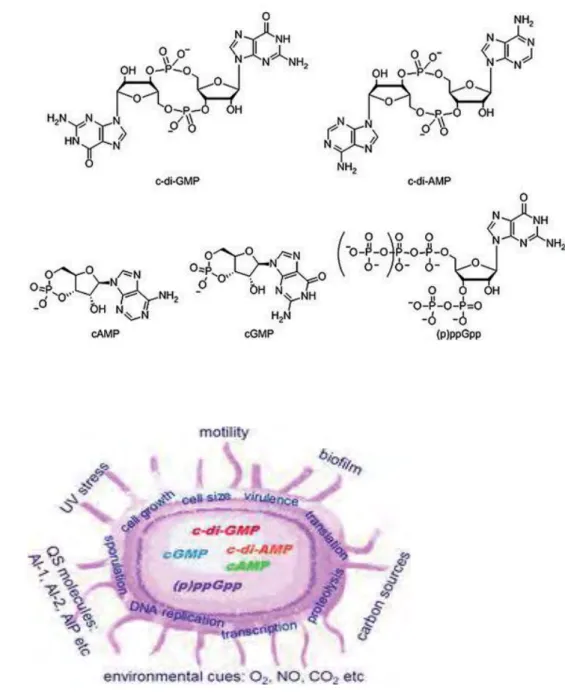
Figure 10 : Nucleotide second messengers. ... 71
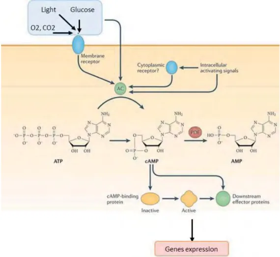
Figure 11: cAMP translates environmental signals into regulatory outcomes. ... 77
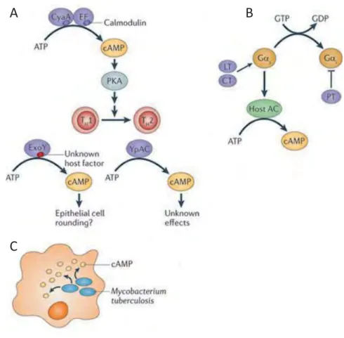
Figure 12 : Bacterial manipulation of host cAMP levels. ... 87
Figure 13 : Classification of class III adenylate cyclases (ACs). ... 97
Figure 14 : Dimeric structure of class III adenylate cyclase catalytic domains. ... 99
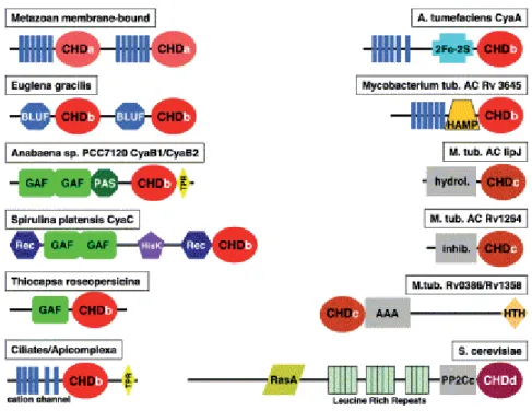
Figure 15 : Schematic representation of examples for variant domain organizations of class III adenylyl cyclases. ... 101
Figure 16 : Domain organization of the 26 ACs /GCs of S. meliloti ... 103
Figure 17 : The CRP-like proteins of S. meliloti ... 107
Figure 18 : Mechanism of allosteric control of CRP. ... 109
Figure 19 : Activation of transcription by the cyclic AMP receptor protein (CRP). ... 111
Figure 20: CyaK senses the plant nodule signal. ... 155
Figure 21: Expression of the smc02178-lacZ reporter gene fusion in M. sativa nodules. ... 155
Figure 22 : SpdA overexpressing mutant symbiotic phenotype. ... 191
Figure 23 : Growth characteristics of S. meliloti mutants. ... 193
Figure 24 : 2', 3'cAMP effect on smc02178 expression... 193
20
Abbreviations
A Alanine
A. hydrophila Aeromonas hydrophila
ABA Abscisic acid
ABC ATP binding cassette
AC Adenylate cyclase
AM Arbuscular mycorrhizal
5’AMP 5'-adenosine monophosphate
AnCrp Anabaena cAMP receptor protein
AOI Autoregulation of infection
AON Autoregulation of nodulation
AR1 Activating region 1
AR2 Activating region 2
AR3 Activating region 3
Asp Aspartic acid
ATP Adenosine tri-phosphate
B. anthracis Bacillus anthracis
B. japonicum Bradyrhizobium japonicum B. pertussis Bordetella pertussis B. subtilis Bacillus subtilis
BLUF Blue light using FAD
Bp base pair
C Carbon
C. albicans Candida albicans C. elegans Caenorhabditis elegans
C1 C2 Cyclase homology domain 1 of mammalian AC
Ca2+ Calcium
CaM Calmodulin
cAMP 3', 5'-cyclic adenosine monophosphate
2’3’cAMP 2', 3'-cyclic adenosine monophosphate
3’5’cAMP 3', 5'-cyclic adenosine monophosphate
CAP Catabolite gene activator protein
CBD cAMP-binding domain
CCR carbon catabolite repression
CCRH Colonized curled root hair
c-di-AMP 3', 5'-cyclic diadenylate monophosphate c-di-GMP 3', 5'-cyclic diguanylate monophosphate
cGMP 3', 5'-cyclic guanosine monophosphate
CHASE-2 Cyclase/histidine kinase associated sensory extracellular receptor domain 2
CHD Cyclase homology domain
cNMPs Cyclic nucleotide monophosphate
2’3’cNMP 2', 3'-cyclic nucleotide monophosphate 3’5’cNMPs 3', 5'-cyclic nucleotide monophosphate
CLE CLAVATA3/endosperm-surrending region
21
CNGCs Cyclic nucleotide gated channels
CO2 Carbon dioxyde
CpdA E. coli phosphodiesterase A
CREB cAMP-response element binding protein
CRP cAMP receptor protein
CSP Common symbiotic pathway
CT Cholera toxin
αCTD RNAP subunit α-C-terminal domain
CyaA AC of E. coli
CyaF5 AC CyaF5
D Aspartic acid
3D Three dimension
D. discoideum Dictyostelium discoideum D. melanogaster Drosophila melanogaster
DAC Dianenylate cyclase
DBD DNA-binding domain
DGC Diguanylate cyclase
DisA c-di-AMP synthase protein
DNA Deoxyribonucleic acid
E Glutamic acid
E. coli Escherichia coli
EF Oedema factor
EPACs Exchange proteins activated by cAMP
EPS Exopolysaccharide
F Phenylalanine
Fe Iron
Fe3+ IronIII
Flg22 Flagellin 22
FNR Fumarate and nitrate reductase
G Glycine
GAF cGMP specific phosphodiesterase, adenylyl cyclase and Fh1A
GdpP c-di-AMP hydrolase enzyme
Glc Glucose
GPCR G protein couple receptor
GTP Guanosine triphosphate
H Histidine
H. influenza Haemophilus influenza
HAMP tandemly arranged amphoteric α-helices present in histidine kinases, adenylyl cyclases, methyl-accepting chemotaxis proteins and phosphatases
HAR1 Hypernodulation aberrant root formation
His Histidine
Hisk Histidine kinase domain
HK Sensor kinase
ISR Induced systemic resistance
IT Infection thread
22
K. pneumoniae Klebsiella pneumoniae
KDa Kilodalton
L Leucine
L. japonicus Lotus japonicus
L. monocytogenes Listeria monocytogenes
LCO Lipochitooligosaccharide
LPS Lipopolysaccharide
LRR-RLK Leucine-rich repeat receptor-like kinase
LT E. coli heat-labile enterotoxin
LTA Lipoteichoic acid
LYK LysM receptor-like kinase
M. loti Mesorhizobium loti M. sativa Medicago sativa M. truncatula Medicago truncatula M. tuberculosis Mycobacterium tuberculosis M. xanthus Myxococcus xanthus
M1 M2 Cyclase homology domain 2 of mammalian AC
MA Mega annum
MAMPs Microorganism-associated molecular patterns
MASE-1 Membrane associated sensor 1
MASE-2 Membrane associated sensor 2
Mb Mega base Mg2+ Magnesium Mn2+ Manganese MoFe Molybdenum-iron MPEs Metallophosphoesterases Mt Medicago truncatula
MTI MAMP-triggered immunity
N Nitrogen
N2 di-Nitrogen
NARK Nodule autoregulation receptor kinase
NCR Nodule-specific cysteine-rich peptides
NF Nod factor
Ni2+ Nickel
nM Nanomolaire
NMP Nucleotide monophosphate
3’NMP 3'-nucleotide monophosphate
αNTD RNAP subunit α-N-terminal domain
P Phosphate
P. aeruginosa Pseudomonas aeruginosa P. sojae Phytophtora sojae
PAS Period clock protein, aryl hydrocarbon receptor, single-minded protein
PDE Phosphodiesterase
PdeA Phosphodiesterase A of M. xanthus
PdeB Phosphodiesterase B of M. xanthus
23
PF00149 pfam domain 00149
PGPR Plant-growth-promoting-rhizobacteria
PKA Protein kinase A
ppGpp 3', 5' guanosine bispyrophosphate
PPi Pyrophosphate
pppGpp guanosine 3'-diphosphate, 5'-triphosphate
PT B. pertussis toxin
PTS Carbonhydrate phosphotransferase system
PUB1 Plant U-box protein 1
R. etli Rhizobium etli
R. leguminosarum Rhizobium leguminosarum
Rdn1 Root determinated nodulation 1
REC Phosphorylation receiver domain
Rec Receiver domain
RNA Ribonucleic acid
RNAP RNA polymerase
ROS Reactive oxygen species
RR Response regulator
Rv0386 AC of M. tuberculosis, open reading frame number 386 Rv0805 PDE of M. tuberculosis, open reading frame number 3645 Rv1264 AC of M. tuberculosis, open reading frame number 1264 Rv3645 CRP of M. tuberculosis, open reading frame number 3645
S. aureus Staphylococcus aureus S. cerevisae Saccharomyces cerevisiae S. marcescens Serratia marcescens S. meliloti Sinorhizobium meliloti S. platensis Spirulina platensis
S. pombe Schizosaccharomyces pombe S. typhimurium Salmonella typhimurium
sAC Soluble adenylate cyclase
SAR Systemic aquired resistance
SKL Sickle
SMBDs Small molecule-binding domains
SMCR Succinate-mediated catabolite repression
SUNN Supernumerary nodules
T. roseopersicina Thiocapsa roseopersicina
TCA Tricarboxylic acid
TCS Two-component system
TM Transmembrane helix
TTSS Type III secretion system
V. cholerae Vibrio cholerae V. fischeri Vibrio fischeri V. vulnificus Vibrio vulnificus
Vfr Virulence factor regulator
Y. enterocolitica Yersinia enterocolitica Y. pestis Yersinia pestis
24
YybT c-di-AMP hydrolase enzyme
Zn2+ Zinc
26
« La vie a plus d’imagination que n’en
portent nos rêves »
28
Introduction
A. The Sinorhizobium meliloti-Medicago symbiosis
I. Introduction
1. Background and agronomic stakes
The settlement emergence during Neolithic permitted agriculture development. From that moment on, man was no more hunter but became producer. Agriculture continued to develop
for thousand years until industrialization. Agriculture modernization started during the 19th
century and Justus von Liebig is considered as its funder. In 1850, Liebig generalized the “Law of the Minimum” (Gorban et al., 2011) formulated for the first time in 1828 by Carl Sprengel. The Law of the Minimum says that the crop yield is limited by the first missing fertilizer element and thus it has to be complemented with an external amount of mineral fertilizer. The first limiting factor after water for crop yield is nitrogen. Although nitrogen represents 79% of the atmosphere, it is present in the form of gas that plants and animals are not able to use. Indeed, plants usually capture nitrogen in the form of nitrates and ammonia, from the soil (Oldroyd, 2013; Oldroyd et al., 2011). Nitrogen is used by plants to synthesise important molecules such as proteins, nucleotides, nucleic acids and chlorophyll. Thus, most cultivated plants heavily depend on applied nitrogen fertilizers for growth. Nitrogen fertilizers synthesis is extremely costly in fossil energy as N2 is very stable chemically. 1 ton of N-fertilizer requires
several tons of fuel for synthesis. Transport and spreading of N-fertilizers on fields by farmers further increase fossil energy waste. Last but no least a significant part of applied N-fertilizers are actually not taken up by growing plants but are leached in soil, hence contributing to water pollution and eutrophication in lands of intensive agriculture.
Leguminous plants (Fabaceae) are an exception since they are able to fix atmospheric nitrogen (N2) and thus to grow in the absence of nitrogen-containing chemical fertilizers. Indeed, even
if most of plants, as all eukaryotic organisms, are unable to fix atmospheric nitrogen by themselves, leguminous plants can do it thanks to their interaction with diazotroph microorganisms.
29
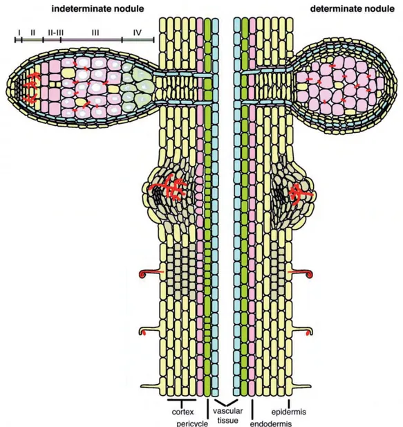
Figure 1: Development of determinate and indeterminate root nodules.
In legumes that develop indeterminate nodules, rhizobial infection of the plant roots induces periclinal cell divisions in the pericycle followed by inner cortical cell proliferation. Development of a nodule primordium is accompanied by the presence of a persistent meristem leading to a zonation of an indeterminate nodule with the meristem (zone I), the infection zone (II), an interzone (II-III), the fixation zone (III) and the senescence zone (IV). By contrast, determinate nodules derive from cell divisions in the outer root cortex where the meristematic activity is lost in mature nodules. Adapted from Popp and Ott, 2011
30
This interaction is mutualistic as it is beneficial for both partners. Indeed, legume plants receive fixed nitrogen from bacteria whereas bacteria receive carbon compounds from the plant. This symbiosis involves around 18.000 legume species (encompassing Caesalpinioideae, Mimosoideae, Papilionoideae) and a set of soil-borne bacteria collectively called rhizobia (Masson-Boivin et al., 2009). The process of biological nitrogen fixation is important to the environment and to world agriculture. The rhizobium-legume symbiosis as a whole is responsible every year for 50 million tons of nitrogen injected into agriculture, without greenhouse effects, as compared to 90 million tons of N-fertilizers (Fields, 2004). Legumes are thus plants of extreme ecological and agricultural importance. In addition legumes are a major feed (soybean) and food resource in South and Central America. Legume seeds are very rich in proteins and thus should become in future decades, a valuable alternative to animal proteins. Furthermore, largely thanks to their N2-fixing capacity, legumes are often pioneering
plant for (re)colonization of poor and degraded soils (Pérez-Montaño et al., 2013). Because of those agronomic stakes, legume-rhizobia symbiosis is subjected to important scientific researches worldwide.
2. The nodule, a N2 fixing plant organ
Nodules are specialized organs devoted to N2 fixation. They are mixed organs composed of
plant tissues and bacteria that are part of the plant microbiome. Legumes develop two types of root nodules (Figure 1). Indeterminate nodules of legumes such as Medicago truncatula and Pisum sativum have a persistent meristem and are continuously infected. These nodules can be divided into four major zones (Vasse et al., 1990). By contrast, determinate nodules (e.g in legumes like Lotus japonicus and Glycine max) have a defined lifespan and loose their central meristem as well as the ability to be continuously infected upon maturation (Popp and Ott, 2011). Within the developing root indeterminate nodule rhizobia differentiate into bacteroids that are able to fix atmospheric nitrogen.
The process of biological reduction of nitrogen to ammonia is catalysed by a metalloenzyme called nitrogenase. This enzyme contains two components that are named according to their metal composition. The smaller dimeric component, known as the iron (Fe) protein, functions as an ATP-dependent electron donor to the largest heterotetrameric component, known as the molybdenum-iron (MoFe) protein, which contains the enzyme catalytic site (Dixon and Kahn, 2004).
31
32
The process of nitrogen fixation is very energy costly since for one molecule of nitrogen fixed, 16 ATP molecules are used. There is also a metabolic cost to host plants that supply bacteria with photosynthates.
Nitrogen fixation is directed by two sets of genes, nif genes and fix genes (see page 34). nif genes encode structural proteins of the nitrogenase enzyme (nifHDK), enzymes involved in biosynthesis of the nitrogenase Fe-Mo cofactor (nifENB), the regulatory protein NifA, and proteins of unknown functions that are required for full nitrogenase activity (nifSWX). The
fixABCX genes might, based on their sequence, code for an electron transport chain to
nitrogenase. fixGHIS is involved in Cu2+ transport, while fixNOPQ encode a membrane-bound
cytochrome oxidase (Delgado et al., 1998). The fixL, fixJ, and fixK genes encode regulatory proteins (see page 48).
3. Medicago truncatula and Lotus japonicus as model legumes
Legumes are the third-largest family of angiosperms, the second most-important crop family, and a key source of biological nitrogen in agriculture (Young and Bharti, 2012). Legumes are usually defined by their typical flower structure and the ability of many of them to form root nodules in presence of rhizobia. The ability of about 70% of legumes to obtain nitrogen from the air through root nodules (Faria et al., 1989) was probably a major determinant in their evolutionary, ecological, and economical success. Interestingly, the study of symbiotic associations with rhizobia drove the development of model legumes such as M. truncatula (Ané et al., 2008; Barker et al., 1990) or L. japonicus (Sato and Tabata, 2006). L. japonicus is a model for determinate nodules while M. truncatula is used to study indeterminate nodule development. Studies on each nodule model are very efficient ways to compare common and different processes used by legumes to generate the N2-fixing organ.
M. truncatula (Figure 2A) is native of the Mediterranean basin and is found in a wide range of
habitats (Ané et al., 2008). Natural attributes of M.truncatula that make it a valuable genetic model compared to its close relative M. sativa (alfalfa) (Figure 2B) include its annual habit and rapid life cycle, its diploid (2n = 16) and autogamous nature, its prolific seed production, and a relatively small genome of about 550Mb. Jemalong A17 has been selected by the research community as a reference line for most genetic and genomic approaches (Ané et al., 2008; Barker et al., 1990).
33
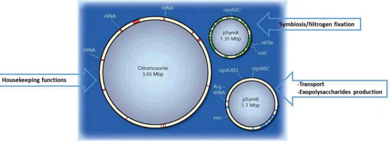
Figure 3: The three components of the S. meliloti genome: a chromosome and two megaplasmids.
Red, green and blue regions have a guanosine + cytidine (G + C) content of less than 60% (averaged over 10-kilobase windows). The positions of some genes are shown, including those needed for the synthesis of ribosomal RNA (rRNA) and for plasmid replication (rep genes), as well as the gene encoding the essential transfer RNA (Arg-tRNA) that recognizes the nucleotide triplet CCG. Also shown are the gene regions required for the bacterium to form nodules on the roots of legumes (nod genes), for the formation of external polysaccharides (exo genes), and for symbiotic nitrogen fixation (nif/fix genes). Adapted from Downie and Young 2001
34
M. truncatula is a member of Papilionoideae, a subfamily that diverged from the two other
legume subfamilies Mimosoideae and Caesalpinoideae approximately 60 MA (Young and Bharti, 2012). Research efforts on M. truncatula encompass a broad range of fields in plant biology from population biology and plant development to plant pathology, insect resistance
and biotechnology production (http://www.medicago.org).
However, most of times, researches on M. truncatula are associated with its capacity to develop two major plant root endosymbiosis, the arbuscular mycorrhizal (AM) and the rhizobial-legume symbiosis and links between those mutualistic interactions and pathogenic interactions. AM is a root endosymbiosis between fungi of the ancient phylum Glomeromycota and about 80% of land plant species, improving the uptake of water and mineral nutrient (Gough and Cullimore, 2011). During rhizobial-legume symbiosis, the microsymbiont allows the plant to gain access to ammonium, a product of the prokaryote-exclusive enzyme nitrogenase (Markmann and Parniske, 2009).
4. Sinorhizobium meliloti, the Medicago symbiont
Rhizobia are soil born bacteria that can fix atmospheric nitrogen to ammonia under ambient conditions, a reaction only mimicked on an industrial scale by a very costly chemical process (Dixon and Kahn, 2004). Rhizobia currently belong to 12 genera and more than 70 species of α- and β-proteobacteria (Masson-Boivin et al., 2009). Sinorhizobium meliloti, the symbiont of Medicago, is a α-proteobacterium, a phylogenetically disparate group of bacteria. It presents a tripartite 6.7-megabase (Mb) genome comprising a 3.65-Mb chromosome, and 1.35-Mb pSymA and 1.68-Mb pSymB megaplasmids (Figure 3) (Galibert et al., 2001). Genome sequence analysis indicates that all three elements contribute, in varying degrees, to symbiosis (Galibert et al., 2001). The chromosome is known to carry a wide variety of genes involved in general chemotaxis, (Figure 3). The psymA megaplasmid contains genes essential for nodulation, nitrogen fixation and catabolism of plant compounds (Figure 3). Interestingly, S. meliloti, together with Rhizobium leguminosarum have a restricted set of 9 and 8 nif genes
respectively, making them the N2-fixing organisms with the least nif genes described so far
35 Figure 4 : Legume infection by S. meliloti.
Bacteria are entrapped in a curled root hair, and from this site, infection threads are initiated. Infection threads progress into the inner cortex where the nodule primordium has formed through a series of cell divisions. Bacteria then differentiate into bacteroids and fix nitrogen in zone III of nodules.
36
The psymB megaplasmid carries genes involved in transport, exopolysaccharides synthesis, and genes required for thiamine biosynthesis and utilization of novel carbon sources (Figure 3) (Barloy-Hubler et al., 2000; Capela et al., 2001; Galibert et al., 2001). The three components of the S. meliloti genome may enable this bacterium to survive in soil, ready to jump at the chance of infecting a legume root.
II. Basis of Medicago colonization by S. meliloti
1. Infection of plant epidermis
The root epidermis represents the first barrier that must be breached by bacteria. Because the epidermis is the first point of contact with rhizobia it must influence where, when, and how many nodules will be formed. In some legumes species, particularly the more basal legumes, this epidermal barrier to bacterial infection can be overcome by rhizobial infection at points of epidermal damage, generally caused by the emergence of lateral roots. In these cases the bacteria infect these cracks and gain access to cortical cells from the infected cracks. However, bacterial infection generally occurs through root hair cells that differentiate into Infection Thread (IT). In most legume/rhizobial interactions, the root hair cells play a central role in facilitating bacterial infection.
Rhizobial infection via IT is accompanied by morphological alterations such as root hair curling and cortical cell divisions that initiate nodule organogenesis. In all but the most primitive rhizobial-host symbiosis, the bacteria must be internalized by plant cells in the root cortex before they begin to fix nitrogen. The bacteria penetrate these deeper plant tissues through the production of infection threads (IT) (Figure 4). To form those structures, the tip of a root hair to which rhizobia are bound, curls back on itself, trapping the bacteria within a pocket called CCRH for colonized curled root hair (Oldroyd, 2013). The bacteria divide at the growing IT tip, forming a column of bacteria. Thus bacteria induce the progressive ingrowth of the root hair cell membrane, resulting in bacterial invasion of interior plant tissue (Oldroyd, 2013). The tip of the developing IT is a site of new membrane synthesis, and is proposed to involve inversion of the tip growth that is normally exhibited by the root hair and to be the result of reorganization of cellular polarity (Gage, 2004).
38
Microscopic analysis of fluorescently tagged bacteria within IT indicates that only the bacteria at the tip of the ingrowing IT are actively dividing (Gage, 2002). Invasion appears to progress by continued bacterial proliferation at the tip and sustained induction of IT membrane synthesis (Jones et al., 2007). Rearrangement of the cytoskeleton and changes in microtubule organization is essential for IT formation (Oldroyd et al., 2011). However, the cytoskeletal reorganizations involved in IT formation have not yet been thoroughly dissected. Inward movement of the root hair cell nucleus, in advance of the ingrowing IT, has been observed (Timmers et al., 1999). In these cells, a network of microtubules connects the nucleus to the tip of the IT and surrounds the IT itself (Timmers et al., 1999). As the IT develops, the inside of this tubule is topologically outside the root hair cell and possesses a plant cell wall (Brewin, 2004). Interactions between plant cell-wall material and other components of the IT matrix might play an important part in IT growth (Brewin, 2004). Plant cell wall remodelling during root hair colonization and IT formation is another process that is not well understood, but is proposed to involve plant proteins that are exported to the cell wall and the IT matrix (Brewin, 2004).
2. Cortical events: nodule organogenesis and bacteria endocytosis
The epidermis regulates bacterial infection, whereas the root cortex controls the formation of the nodule. Intracellular infection and nodule organogenesis are genetically separable processes, thus genes involved in spatially coordinating and synchronizing nodule organogenesis with bacterial infection are essential for an efficient root nodulation symbiosis. However, whereas epidermal and cortical coordination events are essential, the coordinating process is still not clear.
The activation of the mitotic cell cycle in cortical cells is an important step during the interaction with the bacteria, and regulators of the cell cycle play an important role during the formation of a nodule primordium (Cebolla et al., 1999). Once an IT has penetrated to the base of a root hair cell the bacteria must induce new rounds of IT formation in each successive cell layer. The earliest IT that penetrates the growing M. truncatula or M. sativa nodule must grow past the actively dividing cells in the developing nodule primordium (Foucher and Kondorosi, 2000).
39 Figure 5 : Structure of Nod factors.
Nodulation (Nod) factors from Sinorhizobium meliloti and Rhizobium leguminosarum biovar viciae nodulate Medicago spp. or Pisum and Vicia spp., respectively. The backbone of beta1– 4-linked N-acetyl glucosamine residues can carry many different substituents depending on the rhizobial species. a) S. meliloti Nod factors carry a sulphate group (blue), which requires the bacterial proteins NodH, NodP and NodQ. b) At the equivalent location, a R.
leguminosarum biovar viciae Nod factor carries an acetyl group (red), which requires NodX.
This modification is specifically required for the nodulation of some types of P. sativum, which are homozygous for the Ps SYM2A locus. The NodF- and NodE-dependent acyl chains (green) can vary in their length and degree of saturation. Although the acetyl groups that are attached to the same residue by NodL are seen in Nod factors from both strains, they are absent from Nod factors of other rhizobial species. Other substitutions, which are not shown here, can be present in Nod factors from other rhizobia (Oldroyd and Downie, 2004).
40
Ultimately, cells adjacent to the initial primordium will give rise to a persistent nodule meristem that maintains a population of actively dividing cells and will continue to grow outward from the root for the life of the nodule (Figure 4) (Foucher and Kondorosi, 2000; Timmers et al., 1999).
When bacteria reach the target tissue layer, i.e. the inner plant cortex, they must be internalized by a cortical cell and establish a niche within that cell. Each bacterial cell is endocytosed by a target cell in an individual, unwalled membrane compartment that originates from the infection thread (Brewin, 2004). The entire unit, consisting of an individual bacterium and the surrounding endocytic membrane, is known as the symbiosome.In indeterminate nodules, a bacterial cell and its surrounding membrane divides synchronously before the bacteria differentiate into nitrogen-fixing bacteroids (Figure 4) (Robertson and Lyttleton, 1984).
III. Signalling in the rhizobium-legume symbiosis
Signal exchange is key to nodule establishment and maintenance. Here I will review best known signals.
1. Early symbiotic signals
Flavonoid compounds (2-phenyl-1,4-benzopyrone derivatives) produced by leguminous plants are the first signals exchanged by legumes-rhizobia symbionts (Jones et al., 2007). These compounds once perceived by bacteria activate the nodulation (nod) genes to produce and secrete the lipo-chitooligosaccharidic (LCO) Nod factors (NF) (Dénarié et al., 1996). NF induce multiple responses required for nodulation of appropriate host plants, and are the best characterized signals exchanged between plant host and rhizobial symbionts. NF consist of a
β-1,4-linked N-acetyl-D-glucosamine backbone which can differ in number not only between
bacterial species but also within a single species (e.g. NGR234). NF are N-acylated at the non-reducing terminal residue with acyl chains that can also vary between rhizobial species (Dénarié et al., 1996) (Figure 5). The nodABC operon encodes proteins that are required to make the core NF structure.
42
The products of other nod genes (and noe and nol genes) make modifications to NF that impart host specificity, including the addition of fucosyl, sulphuryl, acetyl, methyl, carbamoyl and arabinosyl residues, as well as introducing differences to the acyl chain (Dénarié et al., 1996). Many rhizobial species produce more than one type of NF, but it is not yet possible to predict the range of possible host plants from the NF structure.
AM fungi also secrete LCO compounds termed Myc factors almost identical to NF (Maillet et al., 2011). Myc Factors, although they have a similar to NF structure, are not able to induce nodulation on legume plant roots (Gough and Cullimore, 2011; Maillet et al., 2011). How plants discriminate between NF and Myc factors is a fascinating question.
Some EPS have also been postulated as early symbiotic signals. Binding of rhizobia to plant surfaces is essential for establishing a long-term interaction of the bacteria with their hosts (Brencic and Winans, 2005). Rhizobial surface polysaccharides clearly play a critical role during infection and the best-understood interaction is the one between S. meliloti and M.
truncatula. Most strains of S. meliloti produce two structurally distinct exopolysaccharides
called succinoglycan (also known as EPSI) and galactoglucan (EPSII). Succinoglycan is a polymer of an octasacharidde repeating unit modified with acetyl, succinyl, and pyruvyl substituents (Reinhold et al., 1994), is more efficient than EPSII in mediating infection thread formation on
M. sativa and is the only exopolysaccharide produced by S. meliloti that can mediate the
formation of infection threads on M. truncatula (Glazebrook and Walker, 1989; Pellock et al., 2000). A S. meliloti exoY mutant, which cannot produce any succinoglycan, can form CCRHs but initiates almost no infection threads, these mutants remain trapped in a microcolony at the tip of the root hair (Cheng and Walker, 1998). An S. meliloti exoH mutant produces succinoglycan that lacks the succinyl substituent, and this mutant forms abortive infection threads (Cheng and Walker, 1998).
Even if the role of EPSs in the success of infection processes is well established, the mechanisms by which they act remain to be elucidated. One possible role for these surface polysaccharide signals could be to affect plant cell defence responses (see IV.4) (D'Haeze and Holsters, 2004), although a positive signalling is also possible (González et al., 1996). Receptors for EPS are still unknown.
The active component of EPSI and EPSII are low-molecular-weight forms, because S. meliloti mutants unable to produce these forms are defective for infection (González et al., 1996; Leigh
44
et al., 1985; Urzainqui and Walker, 1992). These findings point toward the possibility of polysaccharide fragments acting as signals, which fits with reports that addition of polysaccharide fragments can complement exopolysaccharide mutants (Djordjevic et al., 1987; Leigh et al., 1985)
EPS production depends on the concentration of available phosphate, which might be sensed by the bacteria during the process of nodulation (Brencic and Winans, 2005). Phosphate concentration is very low in the soil (1 to 10 μM) and considerably higher within plant tissues (10 to 20 mM) (Brencic and Winans, 2005). EPS II is produced preferentially under low-phosphate conditions, whereas succinoglycan synthesis is stimulated at high concentrations of phosphate (Mendrygal and González, 2000). This suggests that inside the plant, bacteria produce EPS I rather than EPS II, which is consistent with the observation that although both EPS can mediate nodule invasion, EPS I is much more efficient in this process (Pellock et al., 2000).
45 Figure 6 : Sch ema tic ill us tr a tion of g en e tic c omponents i n v olv ed in l egume -r hiz o b ia s ymbiot ic signali n g. Fla v on oid s secr e te d by the pla nt induce NF and E P S s y n
thesis and secr
e ti o n by rhi zo b ia. Mec h anis ms by which EPS I act is s til l unknown. NF a re per ceiv e d by a re ce pt or co mple x c ont ai ning NF P. P e rc e p tion o f NF trig g e rs early respon ses, lik e ion fl ux es, de polari za tio n of p lasma mem br a n e, pr od uc tion of r e ac tiv e o x y g en species (R
OS), and cyt
o p lasmic alk a lin iz a ti o n . The signals ar e tr a n sduced downs tr e am to a ctiv a te LRR-rece pt or kinase, DMI2 a n d DMI1. Th e nuclear i on channel DM I1 is r e qui re d f o r NF -in duce d nuclear c a lcium spik ing. La ter these c alcium signals a re per ceiv e d an d dec o de d b y c a lcium/ ca lmod ulin -de penden t pr ot ein kinase, DMI3, wh ich acts as a ce ntr a l pla y er i n t he NF sign aling a n d c o or di na tes the e x pr ession o f s ymbi ot ic g e n es, i n cludi ng ear ly nod u li n g e nes. In par a llel, a mobil e signal, pos sibly cyt okinin, is activ a te d/ pr oduc e d by th e ep ide rmis downs tr e a m o f DMI3 and is tr a n sloc a te d t o the c o rte x wher e it i s sens e d by the cyt okinin r ece ptor CRE1. NF sig n al ing culmi n a tes with the c o or di na te d and s y n chr on ou s pr ogr essio n of inf e ct ion a nd c o rt ic al cell division , r esulting in the f o rma tion o f ma tur e, in fe ct ed a nd nitr og en -fixing nodules. Adapted fro m R o se e t al 201 2.
46
2. Early signal transduction
Flavonoids exudates are perceived in S. meliloti by a variety of NodD proteins, which are members of the LysR family of transcriptional regulators (Brencic and Winans, 2005). NodD proteins then bind to a “nod-box” promoter, which activates transcription of the downstream
nod/noe/nol genes resulting in NF synthesis (Figure 6) (Oldroyd, 2013).
NF exuded by rhizobia are sensed by receptor-like kinases with N-acetyl-glucosamine-binding lysine motif (LysM) in the extracellular domain (Gough and Jacquet, 2013). The LysM receptors NFP, LYK3 and LYK4 lead to activation of leucine-rich-repeat receptor-like kinases DMI2 and the nuclear potassium channel DMI1. DMI2 and DMI1 are active early in the epidermal
pathway and are at the origin of calcium spiking. Decoding this calcium signature by the Ca2+
calmodulin-binding protein DMI3 and its interacting partner IPD3 triggers the transcription factors NSP1, NSP2, ERN1 and NIN. Together, these transcription factors are involved in the activation of ENOD genes to initiate the infection process and induce cortical cell divisions. However, root hair deformation occurs via a signalling pathway independent of calcium spiking (Oldroyd and Downie, 2008). Indeed, Nod factor-induced cytokinin signalling, that is calcium spiking-independent, might influence epidermal susceptibility to rhizobial infection by participating in the mechanisms that regulate early plant symbiotic genes (nodulins) (Frugier et al., 2008).The phytohormone cytokinin might be produced by the epidermis downstream of DMI3 and translocated to the cortex where it is sensed by the cytokinin receptor CRE1. Downstream of CRE1, local inhibition of polar auxin transport contributes to nodule primordium development (Mortier et al., 2012). Hence, NF perception by root hair cells leads to at least two signalling events: one that involves calcium spiking and gives rise to gene expression changes and one that is associated with root hair deformation (Oldroyd and Downie, 2008). Some of the genes involved in NF signalling also control development of the AM symbiosis, as part of the so-called common symbiotic pathway (CSP) (Gough and Cullimore, 2011).
3. Late symbiotic signal: oxygen-controlled regulatory system
Bacteroids need oxygen to generate ATP, which is required in large amounts for the energy-costly nitrogen fixation.
47 Figure 7 : R o le and dis tri butio n o f o x y g e n i n s ymbi ot ic ni tr og en -f ixing no dul es. (A) Pr otectio n ag ains t o xy g en de fe ct on nitr og e n a se activity is pr ovide d by the nodule e n vir o n m e n t thr ou g h a co rtic al dif fusi o n bar rier so tha t t h e main r out e of o x y g e n dif fusion is thr ou g h the no dule ape x , whic h g en er a te s a lon gi tudinal o x y g en gr adien t. ( B ) R egula tory c asc a de c o n tr o lling nif tr anscri pti on in S. meli loti . In the a b sence of o x y g en , FixL autophosp horyla tes and tr a n sf er s the phos phoryl gr oup t o FixJ res u lt ing i n FixJ acti v a tio n.
FixJ in turn activ
a tes tr a n scriptio n of the r eg u la tory fix K a nd ni fA g e nes, whose pr o duc ts r egula te tr a n scripti on o f th e r e st o f t h e nitr og e n fix a tio n g en es. In S. meliloti, FixK e x pr ession is n e g a tiv e ly regula te d by F ixT , w h ich i nhibi ts the activi ty of Fix L. Adapted f rom D ix on and K ahn 20 04
48
However nitrogenase is extremely oxygen sensitive, thus some considerable physiological constraints on diazotrophy have to be imposed in order to protect the enzyme activity from oxygen damage (Dixon and Kahn, 2004). Therefore, a physiological paradox arises from the aerobic requirement of bacteroid metabolism compared with the extreme oxygen sensitivity of nitrogenase. Protection against oxygen is provided by the nodule environment through a cortical diffusion barrier so that the main route of oxygen diffusion is through the nodule apex, which generates a longitudinal oxygen gradient (Figure 7A). Oxygen diffusion is facilitated in the central zone by a high concentration of leghemoglobin, which accumulates in plant cells cytoplasms and binds oxygen with an extremely high affinity (Appleby, 1992). Its role is to buffer the oxygen concentration and control the diffusion of oxygen to the actively respiring bacteroids. The combination of leghemoglobin and the oxygen diffusion barrier leads to an extremely low concentration of free oxygen around the bacteroids. As a result, the free oxygen concentration drops to less than 50 nM in the central nitrogen-fixing zone containing bacteroids. The actual signal, therefore, that triggers transcription of nitrogen fixation genes is the drop in oxygen level that bacteria experience within the nodule. A gradient of oxygen was shown to exist inside nodules, and the expression pattern of the nitrogen-fixation genes corresponded to the distribution of oxygen along the nodule (Soupène et al., 1995). Oxygen concentrations are sensed by the bacteria through at least two proteins, FixL and NifA. At low oxygen concentrations, these proteins are active and are responsible for induction of genes involved in the fixation of nitrogen. Indeed, in the absence of oxygen, FixL autophosphorylates and transfers the phosphoryl group to FixJ (David et al., 1988) resulting in FixJ activation. FixJ in turn activates transcription of the regulatory fixK and nifA genes, whose products regulate transcription of the rest of the nitrogen fixation genes (Figure 7B) (Bobik et al., 2006).
Oxygen concentration is the major signal controlling the expression of nif and fix genes responsible for full nitrogenase activity (Soupène et al., 1995). Effect of oxygen on plant gene expression is not known.
50
IV. Negative control of symbiosis
1. Nodule number is controlled by phytohormones and CLE peptides
The nitrogen fixation process and nodulation itself are very costly for both plants and bacteria, thus the symbiosis has to be tightly controlled to avoid detrimental interactions and to ensure mutualism. Two well characterized ways that control the interaction are the control of nodule number by negative regulators such as phytohormones and a mechanism called autoregulation of nodulation (AON).
a) Hormonal control
The phytohormones ethylene, abscisic acid (ABA) and jasmonic acid (JA), which are also known to act during plant defence reactions, negatively regulate nodulation by modifying epidermal responses possibly because of their impact on Ca2+ spiking (Mortier et al., 2012). NF-induced
ethylene production inhibits nodulation, suggesting that a local negative feedback controls nodule number (van Spronsen et al., 1995). Indeed, by eliminating the NF-signalling pathway
either at or during the NF-induced Ca2+ spiking, ethylene inhibits root hair deformation,
shortens the Ca2+ spike period, blocks bacterial infection and supresses nodulin gene
expression (Oldroyd et al., 2001). In absence of ethylene actions, nodule formation is enhanced, whereas exogenous ethylene decreases nodule number (Nukui et al., 2000). These findings have been confirmed by the identification of sickle (skl), an ethylene-insensitive hypernodulation mutant of M. truncatula (Penmetsa and Cook, 1997; Penmetsa et al., 2003; Penmetsa et al., 2008). skl mutants display an exaggerated nodule number in the specific nodulation zone and uncontrolled growth of infection threads (Penmetsa and Cook, 1997; Penmetsa et al., 2003; Penmetsa et al., 2008).
ABA is believed to negatively influence nodulation at two different levels. On the one hand, at the nodule initiation level, ABA would interfere with NF signalling and affect the nature of the
NF-induced Ca2+ spiking, reducing the nodule number; on the other hand, at the nodule
development level, ABA would suppress cytokinin-dependent organogenesis (Mortier et al., 2012).
JA decreases NF-induced Ca2+ spiking and nodulation, with a similar effect as ethylene,
influencing the number of cells able to induce Ca2+ spiking (Oldroyd and Downie, 2008).
51
Figure 8: Model proposed for long-distance autoregulation signaling in legume-rhizobia symbiosis.
(1) Perception of the bacterial Nod factor is the start of the autoregulation of nodulation. (2) CLE peptides are proposed to arise in developing nodules in the roots and is further transmitted through the shoot. (3) CLE peptides are perceived in shoots by CLAVATA-like receptor kinases. (4) A shoot-derived inhibitor (SDI) signal is generated in shoots and translocated to the roots where it inhibits further nodulation. The SDI could act by indirectly downregulate cytokinin signalling. Adapted from Betsuyaku et al 2011.
52
Therefore, JA modulates the nature of NF-induced Ca2+ spiking (Oldroyd and Downie, 2008),
indirectly via its own concentration.
Altogether, these findings would suggest that hormone imbalance can indeed affect nodule number.
b) AON
In addition to local control mechanisms, split-root experiments have revealed that long-distance mechanisms exist, which control nodule numbers (Kosslak and Bohlool, 1984). In addition to skl, screens for supernodulation mutants of legumes revealed a class of mutants,
hypernodulation aberrant root formation 1 (har1) in Lotus japonicus, nodule autoregulation receptor kinase (nark) in soybean, and supernumerary nodules (sunn) in M. truncatula, that
are defective in AON (Oldroyd and Downie, 2008). These mutants all show excessive nodulation. Grafting revealed that this mode of regulation is shoot specific, because a mutant shoot grafted onto a wild-type root displays a mutant phenotype in the root (Delves et al., 1986). HAR1, NARK, and SUNN proteins are leucine-rich repeat receptor-like kinases (LRR-RLK) whose inactivation results in hypernodulation. These LRR-RLK belongs to a large family of receptor kinases similar to CLAVATA1 (CLV1), a receptor-like kinase involved in shoot meristem identity in Arabidopsis (Batut et al., 2011; Williams and Fletcher, 2005). CLV1-like LRR-RLK receptor kinases become activated upon binding of peptides belonging to the CLAVATA3/endosperm-surrounding region (CLE) family. CLE peptides are small secreted peptides composed of 12-13 conserved amino acids that are cleaved from the C-terminal end of CLE preproteins (Wang and Fiers, 2010). In L. japonicus, autoregulation is mediated by
CLE-RS genes that are specifically expressed in the developing root nodule, and the receptor kinase
HAR1 that functions in the shoot. Recently, Okamoto and coworkers have shown that arabinosylated CLE-RS peptides indeed directly bind to HAR1. In addition, CLE-RS2 glycopeptide specifically produced in the root is found in xylem sap collected from the shoot. Thus Okamoto and colleagues propose that CLE-RS glycopeptides are mobile signals responsible for the initial step of AON (Figure 8) (Okamoto et al., 2013). Thus, CLE peptides are a long-range signal translocated to the shoots where it could induce the formation/activation of an unknown shoot-controlled inhibition (“shoot-derived inhibitor”,
54
that could be cytokinin, see above) mechanism and prevent the roots from excessive nodule formation (Figure 8) (Mortier et al., 2012).
In addition to the shoot-regulated hypernodulation mutants, other mutants have been identified, such as too much love (tml) from L. japonicus and root determinated nodulation1 (rdn1) from M. truncatula that systemically inhibit nodulation and of which the hypernodulation phenotype is controlled by the root (Mortier et al., 2012).
Interestingly, Saur and coworkers have recently demonstrated a crosstalk between the NF signalling pathway and AON. Indeed, they have shown that NIN induces expression of
MtCLE12 which via SUNN inhibits cytokinin signalling indirectly in up-regulating the
shoot-derived-inhibitor (Saur et al., 2011).
Another signal controlling nodule number is the level of nitrate. High concentrations of nitrogen compounds inhibit nodule formation at different stages of the process, regardless of plant age, nodule size or former inoculation events (Mortier et al., 2012). Via split-root experiments with M. truncatula, nitrate limitation has been demonstrated to result in both local and systemic regulation of nodulation that partially depended on SUNN (Jeudy et al., 2010). Interestingly, Okamoto et al reported that LjCLE-RS2 transcripts are strongly up-regulated in response to nitrate, which indicates that nitrate responsiveness of nodulation is mediated by LjCLE-RS2 (Okamoto et al., 2009). Thus, LjCLE-RS2 is responsible for AON via HAR1 but is also involved in nitrate-dependent regulation of nodulation in a HAR1-dependent manner (Okamoto et al., 2009).
As the molecular network governing nodule organogenesis is linked with the one that control infection process in legumes, it is tempting to speculate that there is probably a mechanism similar to AON responsible for the negative control of infection during Sinorhizobium
meliloti-Medicago symbiosis.
2. NCR peptides control bacteroid differentiation
The host plant exerts control over the survival of bacteria within the symbiosome, and provide nutritional support and the correct micoaerobic environment required for nitrogen fixation. Bacteroids of S. meliloti from M. truncatula nodules undergo endoreduplication to yield a chromosome count of 24 compared to one to two for free-living bacteria (Mergaert et al., 2006).
56
M. truncatula bacteroids, but not free-living bacteria, are permeable to propidium iodide,
indicating enhanced membrane permeability (Mergaert et al., 2006), and these bacteroids are terminally differentiated and so cannot be cultured from nodules.
More than 400 nodule specific cysteine-rich peptides (NCR) are induced in M. truncatula that control bacteroid differentiation. These peptides induce membrane permeabilization, endoredupication, and loss of viability in free-living S. meliloti, all features characteristic of the bacteroid form (Van de Velde et al., 2010). NCR peptides are similar to defensine-like antimicrobial peptides that are part of the innate immune system in animals and plants (Alunni et al., 2007; Mergaert et al., 2003). They colocalize with bacteroids (Van de Velde et al., 2010) and targeted secretion across the peribacteroid membrane appears to be the function of a plant signal peptidase encoded by DNF1 (Wang et al., 2010a). DNF1 is required to target these cysteine-rich peptides to symbiosomes where they are incorporated into bacteroid development (Wang et al., 2010a). Mutation of Medicago dnf1 blocked bacteroid differentiation (Wang et al., 2010a), and the NCR peptides became trapped in the endoplasmic reticulum rather than in the bacteroid (Van de Velde et al., 2010).
Interestingly, the bacteria have evolved in order to survive in the host. Indeed, the BacA protein from S. meliloti is involved in protecting rhizobia against legume NCR peptides by reducing their antimicrobial activity. How BacA proteins achieve this protection is not yet fully understood. BacA is essential for bacteroid development and survival (Glazebrook et al., 1993) and may promote uptake or export of the NCR peptides (Marlow et al., 2009). BacA is an integral membrane protein in bacteria that belongs to the ATP binding cassette (ABC) superfamily of membrane transporters (Kereszt et al., 2011). S. meliloti bacA mutant is unable to support nitrogen-fixing symbiosis although it is not impaired in nodule formation, infection thread development or host cell entry. Instead of differentiating into nitrogen-fixing bacteroid, the bacteria are rapidly killed after the release from infection threads (Glazebrook et al., 1993). The absence of bacteroid development in bacA mutants would result from the lack of peptides transported to the symbiosomes, similarly to what happens in the dnf1 mutant, or a modification of cell envelope, that makes it more sensitive to plant peptides These indications suggest that the bacteroid state may be forced upon rhizobia by the host plant and may also reflect a level of control that ensures that bacteria remain beneficial to the plant.
58
3. Evidence for a negative regulation of infection
Little is known about mechanisms involved in the negative regulation of infection. So far, only plant proteins have been identified to be responsible for a putative feedback mechanism. NF signalling is essential during bacterial infection and specific NF receptors control NF recognition in this process. In M. truncatula, an entry receptor is encoded by LysM receptor-like kinase 3 (LYK3) and this LysM receptor-receptor-like kinase is involved in recognition of specific NF structures made by S. meliloti (Oldroyd et al., 2011). The regulation of these receptors and possibly their specific location within the plasma membrane is important during the infection process. An E3 ubiquitin ligase, Plant U-box protein 1 (PUB1), interacts with and is phosphorylated by LYK3 (Mbengue et al., 2010). RNAi knockdown of PUB1 in a lyk3 mutant (which normally forms aborted ITs) resulted in partial restoration of infection, demonstrating that PUB1 negatively regulates bacterial infection and that it may be regulated by LYK3. In both L. japonicus and M. truncatula, cytokinin regulation of NIN depends on LHK1/MtCRE1. In L. japonicus, lack of LHK1 function leads to hyperinfection (Murray et al., 2007), suggesting that cytokinin signalling has a role in limiting infection events. Although the role of cytokinin is likely to be complex, one mechanism might involve LHK1/MtCRE1-mediated nodule primordium inception in the root cortex, which in turn stimulates feedback loops that restrict root susceptibility to rhizobial infection (Frugier et al., 2008).
It is difficult to uncouple nodule organogenesis and IT formation. Indeed, most of the time, mutants leading to hyperinfection phenotypes are also impaired in AON and thus display hypernodulation phenotype or on the contrary, present less or no nodules on the plant root. In Medicago for example, the EFD transcription factor is a negative regulator of nodulation and infection in a way that remains to be elucidated but that may also involve cytokinin signalling (Vernié et al., 2008).
59
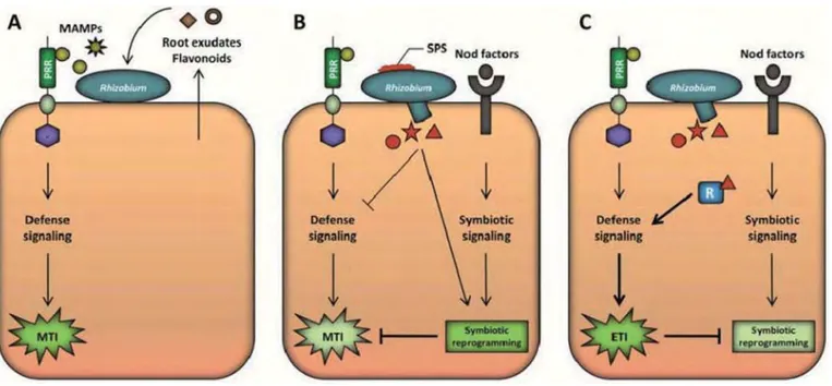
Figure 9 : Model for the modulation of host immunity in the Rhizobium-legume symbiosis.
A, Roots exudates recruit Rhizobium bacteria and secreted flavonoids prime the microsymbionts for the interaction. Host plants initially recognize rhizobia as potential invaders; pattern-recognition receptors (PRR) in the host perceive microbe-associated molecular patterns (MAMPs, yellow-colored shapes) and a signalling cascade is initiated that results in MAMP-triggered immunity (MTI). B, Surface polysaccharides (SPS) function early during the interaction, most likely as extracellular effectors to facilitate immune evasion. At later stages, the establishment of the symbiotic program in the plant cells, which is activated upon perception of the rhizobial nodulation (Nod) factors, counteracts the MTI with mechanisms yet to be defined. Rhizobial effectors that are secreted through the type III secretion system (brown-colored shapes) may assist in the suppression of the MTI response or act as symbiotic determinants. C, In the case that host resistance (R) protein recognizes a cognate rhizobial effector, effector-triggered immunity (ETI) is activated that, in turn, terminates the interaction as incompatible. Zamioudis and Pieterse 2012