The sexist behaviour of immune checkpoint inhibitors in cancer
therapy?
Andrea Botticelli1,2, Concetta Elisa Onesti1,2, Ilaria Zizzari3, Bruna Cerbelli4, Paolo Sciattella5, Mario Occhipinti1, Michela Roberto1,2, Francesca Di Pietro1,2, Adriana Bonifacino6, Michele Ghidini7, Patrizia Vici8, Laura Pizzuti8, Chiara Napoletano3, Lidia Strigari9, Giulia D’Amati4, Federica Mazzuca1,2, Marianna Nuti3 and Paolo Marchetti1,2
1Medical Oncology Department, Sant’Andrea Hospital, Rome, Italy
2Department of Clinical and Molecular Medicine, “Sapienza” University of Rome, Rome, Italy 3Department of Experimental Medicine, “Sapienza” University of Rome, Rome, Italy
4Department of Radiological Oncological and Pathological Sciences, “Sapienza” University of Rome, Rome, Italy 5Statistical Department, “Sapienza” University of Rome, Rome, Italy
6Breast Diagnosis and Treatment Unit, Sant’Andrea Hospital, “Sapienza” University of Rome, Rome, Italy 7Oncology Unit, ASST Cremona, Cremona, Italy
8Division of Medical Oncology 2, IRCCS Regina Elena National Cancer Institute, Rome, Italy
9Laboratory of Medical Physics and Expert Systems, IRCCS Regina Elena National Cancer Institute, Rome, Italy Correspondence to: Concetta Elisa Onesti, email: elisaonesti@gmail.com
Keywords: immune checkpoint inhibitors; anti-CTLA-4; anti-PD-1; sex differences; gender differences Received: July 28, 2017 Accepted: October 10, 2017 Published: November 01, 2017
Copyright: Botticelli et al. This is an open-access article distributed under the terms of the Creative Commons Attribution License
3.0 (CC BY 3.0), which permits unrestricted use, distribution, and reproduction in any medium, provided the original author and source are credited.
ABSTRACT
Background: Immune checkpoint inhibitors, targeting the molecules CTLA-4, PD-1 and PD-L1, showed efficacy against several type of cancers and are currently used in clinical practice. An important biological variable that influences innate and adaptive immunity is the sex, acting through genetic, hormonal and environmental factors. The overall differences between sexes could be crucial to evaluate the response to ICIs.
Materials and methods: We performed a meta-analysis of Phase II-III Clinical Trials published up to June 2017 in which anti-CTLA-4, anti-PD-1 and anti-PD-L1 were studied. We extracted the OS and PFS HR differentiated by sex from subgroups analysis of each trial. We analyzed the three classes of drugs separately.
Results: We selected 36 Phase II-III Clinical Trials, 9 of which reported results for OS and 6 for PFS. We analyzed 2 Clinical Trials for OS with anti-CTLA-4, including 1178 patients, observing a benefit for males vs females (HR 0.65, 95% CI 0.55-0.77
vs HR 0.79, 95% CI 0.65-0.96, p 0.078).
Not statistically significant results were observed with anti-PD-1 neither for OS (males vs females: HR 0.72, 95% CI 0.64-0.83 vs HR 0.81, 95% CI 0.70-0.94, p 0.285) neither for PFS (males vs females: HR 0.66, 95% CI 0.52-0.82 vs HR 0.85, 95% CI 0.66-1.09, p 0.158). We cannot perform a meta-analysis for anti-PD-L1 due to the lack of data.
Conclusions: Different mechanisms could be involved in sex differences with regard to immunotherapy. These differences could be relevant to identify immunological targets in order to draw studies exploring novel combinations of immunotherapy agents.
INTRODUCTION
Several immune checkpoint inhibitors (ICIs), mostly against the molecules CTLA4, PD-1 and PD-L1, are approved in clinical practice, given the promising results in several types of solid tumors. [1] Interestingly, the target of these drugs is not the tumor, as conventional chemotherapy and targeted therapies, but critical checkpoints of the patient’s immune system. In fact CTLA-4 is predominantly expressed on CD4+ “helper” T lymphocytes and its physiological engagement by CD80 and CD86 molecules down-regulates the T cell response. [2] PD-1 is mainly expressed by activated T cells and binds PD-L1, expressed on the surface of antigen presenting cells (APCs) and tumour cells, inducing a negative control damping of the immune response. [2]. These drugs are able to unleash existing T cell, permitting the expansion of effector cytotoxic T cells, that could recognize and destroy the tumor.
Currently, despite the remarkable success also in the metastatic setting of several approved monoclonal antibodies, such as anti-CTLA4 (Ipilimumab, Tremelimumab) and anti-PD1/PD-L1 (Nivolumab, Pembrolizumab, Atezolizumab, Avelumab, Durvalumab), a good percentage of patients do not respond to treatment and may develop different pattern of toxicities, known as immune related adverse events (irAE).
Various biomarkers (e.g. PD-L1 expression, intratumoral immune infiltrate, mutational burden etc...) have been supposed to reflect the ICIs pharmacodynamics
and to predict ICIs efficacy and/or toxicity, but their relevance is still unclear. [3].
It was shown that the sex, defined by the differential organization of chromosomes, reproductive organs, and sex steroid levels, represents an important biological variable that influences innate and adaptive immunity. The sex influence on the immune system is well documented in the pathogenesis and prognosis of autoimmune diseases, infections and malignancies [4].
Generally, as showed in various immune-modulated disease, adult females make a stronger innate and adaptive immune responses than males. In particular, females have a higher incidence of autoimmune diseases than males and the onset and the severity is related with the reproductive status, suggesting that their pathogenesis could be influenced by sex hormones. [5, 6].
The different innate immune responses among mammals suggests that some sex differences may be germline encoded. Indeed, in preclinical models of human cells or rats tissue, females show higher expression than males of genes along TLR pathways, like Toll-like receptor 7 (TLR7) gene, encoded on the X chromosome, as well as a better induction of type I interferon (IFN) responses. [4, 7-10] Murine models were recently studied to evaluate the different sexual response to ICIs. The treatment with anti-PD-L1 Abs appeared to be more efficacious in female than in male murine model of melanoma. [11]. Interestingly, a recent research reports that PD-1 expression and function correlate with a better response to hormone treatment. [12] Despite of great interest in the improved clinical outcomes
of cancer patients with immunological approach, only few data are published regarding potential sex-based differences in responses and toxicity to immunotherapy.
In this context, the overall differences between males and females could be crucial to evaluate the response to ICIs and the toxicity profile.
On these bases, we performed a phase II and III trials’ meta-analysis to determine sex-differential effects of ICIs in cancer patients.
RESULTS
We selected 36 Phase II and Phase III Clinical Trials published up to June 2017 in which immunotherapy was tested (anti-CTLA-4, anti-PD-1 and anti-PD-L1) in patients affected by solid cancers. Of these studies, 17 were Phase III Trials subdivided as following: 8 on melanoma, 6 on NSCLC, 1 on RCC, 1 on head and neck tumors and 1 on urothelial carcinoma. [13-30] Nineteen were Phase II Trials: 10 on melanoma, 5 on NSCLC, 1 on RCC and 3 on urothelial cancer. [31-50] From this selection, 2 studies were excluded because results were
published only as abstracts, 21 because the subgroup analysis differentiated by sex was not reported and 2 because the patients in the control arm received ICIs. (Figure 1).
In the final statistical analysis for OS 9 studies were selected as reported in Table 1, including the three classes of drugs. [13, 16, 20, 25-30] The patients enrolled in the standard arm received a platinum doublet chemotherapy in first line or Docetaxel in second line for NSCLC, Everolimus for RCC, Dacarbazine or Gp100 for melanoma, investigator’s choice chemotherapy (Paclitaxel, Docetaxel or Vinflunine) for urothelial cancers. The patients were treated in first or second line, according to the protocol. Only two trials, CheckMate-025 on RCC and Keynote-045 or urothelial carcinoma, enrolled patients in third line of treatment. [29, 30].
We conducted a meta-analysis dividing the selected trials according to the target of the studied drug. We cannot perform a meta-analysis for anti-PD-L1, because only one study was available, showing a HR in females of 0.64 (95% CI 0.49-0.84) vs HR 0.79 (95% CI 0.64-0.97) in males. [25].
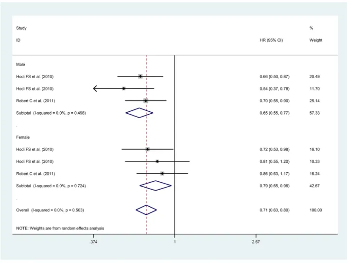
We performed a meta-analysis for OS with anti-CTLA-4 including 2 studies enrolling patients with melanoma treated with ipilimumab. (Figure 2) These trials enrolled 1,178 patients, 702 males (59.6%) and 476 females (40.4%). Overall, 480 males and 310 females received experimental treatment. [13, 16] The heterogeneity test between the two groups showed a benefit for males vs females (HR 0.65, 95% CI 0.55-0.77 vs HR 0.79, 95% CI 0.65-0.96, p 0.078).
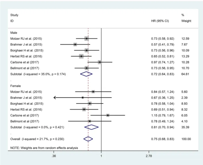
We considered 6 Clinical Trials with anti-PD-1 for OS. Overall we analyzed 3,792 patients, 2,514 males (66.3%) and 1,278 females (33.7%), 55.1% (1386) and 53.4% (683) of which received immunotherapy, respectively. [22, 26-30] In this analysis we observed a not statistically significant lower HR for males than females (HR 0.72, 95% CI 0.64-0.83 vs HR 0.81, 95% CI 0.70-0.94, p 0.285), as reported in Figure 3.
Finally, we performed a meta-analysis for PFS, considering one Phase II and 5 Phase III Trials, as reported in Table 2. All the 6 studies randomized patients to receive anti-PD-1 (Pembrolizumab in 3 studies or Nivolumab in 3 studies). [22, 26-29, 32] The patients
in the control arms received chemotherapy in first or second line, according to the trial. Only one study enrolled patients after failure of two lines of treatment. [32] Overall, we analyzed 3,274 patients, 2,007 males (61.3%) and 1,267 females (38.7%), 1,176 and 728 of which received experimental treatment, respectively. The heterogeneity test between sexes was not statistically significant, despite the HR lower in males group (HR 0.66, 95% CI 0.52-0.82 vs HR 0.85, 95% CI 0.66-1.09, p 0.158).(Figure 4) No PFS data differentiated by sex were available for anti-CTLA-4 or anti-PD-L1.
DISCUSSION
Males and females differ in the immune response to infections and vaccines and in the predisposition to develop autoimmune diseases. [4].
The observation that not only hormonal factors but also genetic, and environmental mediators are involved in sex-based immunological differences lead to hypothesize that different outcome during immunotherapy could depend on the patient’s sex.
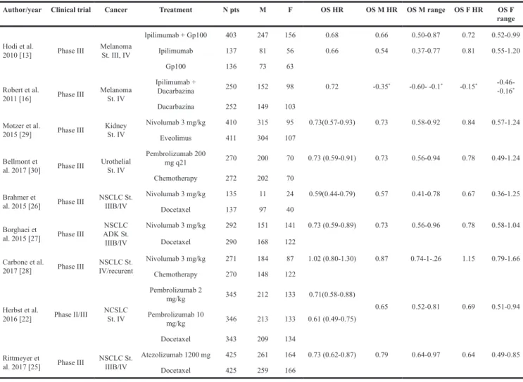
Table 1: Clinical trials selected for OS
Author/year Clinical trial Cancer Treatment N pts M F OS HR OS M HR OS M range OS F HR OS F range
Hodi et al.
2010 [13] Phase III Melanoma St. III, IV
Ipilimumab + Gp100 403 247 156 0.68 0.66 0.50-0.87 0.72 0.52-0.99 Ipilimumab 137 81 56 0.66 0.54 0.37-0.77 0.81 0.55-1.20
Gp100 136 73 63
Robert et al.
2011 [16] Phase III Melanoma St. IV
Ipilimumab +
Dacarbazina 250 152 98 0.72 -0.35* -0.60- -0.1* -0.15* -0.46- -0.16*
Dacarbazina 252 149 103 Motzer et al.
2015 [29] Phase III Kidney St. IV
Nivolumab 3 mg/kg 410 315 95 0.73(0.57-0.93) 0.73 0.58-0.92 0.84 0.57-1.24 Eveolimus 411 304 107
Bellmont et
al. 2017 [30] Phase III Urothelial St. IV
Pembrolizumab 200
mg q21 270 200 70 0.73 (0.59-0.91) 0.73 0.56-0.94 0.78 0.49-1.24 Chemotherapy 272 202 70
Brahmer et
al. 2015 [26] Phase III NSCLC St. IIIB/IV
Nivolumab 3 mg/kg 135 11 24 0.59(0.44-0.79) 0.57 0.41-0.78 0.67 0.36-1.25 Docetaxel 137 97 40
Borghaei et
al. 2015 [27] Phase III
NSCLC ADK St. IIIB/IV Nivolumab 3 mg/kg 292 151 141 0.73 (0.59-0.89) 0.73 0.56-0.96 0.78 0.58-1.04 Docetaxel 290 168 122 Carbone et al.
2017 [28] Phase III IV/recurentNSCLC St.
Nivolumab 3 mg/kg 271 184 87 1.02 (0.80-1.30) 0.87 0.74-1-.26 1.15 0.79-1.66 Chemotherapy 270 148 122 Herbst et al. 2016 [22] Phase II/III NCSLC St. IV Pembrolizumab 2 mg/kg 345 212 133 0.71(0.58-0.88) 0.65 0.52-0.81 0.69 0.51-0.94 Pembrolizumab 10 mg/kg 346 213 133 0.61 (0.49-0.75) Docetaxel 343 209 134 Rittmeyer et
al. 2017 [25] Phase III NSCLC St. IIIB/IV
Atezolizumab 1200 mg 425 261 164 0.73 (0.62-0.87) 0.79 0.64-0.97 0.64 0.49-0.85 Docetaxel 425 259 166
The aim of our study is to analyze differences in the response to ICIs treatment in both sexes.
Interestingly in our study we observed a better OS associated to the anti-CTLA4 treatment in males compared to females. As far as anti-PD-1 treatment is concerned, despite the sample size and the number of clinical trials evaluated considerably wider than those for the anti CTLA-4 analysis, statistical significance has not been achieved.
Moreover we observed lower HR in males compared to women both for OS and PFS for anti-PD-1 treatments. Instead for anti PD-L1 antibodies it was not possible to perform a meta-analysis since in literature only one study presented HR for females and males.
It has been demonstrated that females have an immune system that acts predominantly by T helper (CD4+) response, in particular with a humoral response in various species and in cell culture systems in presence of high levels of estrogen and progesterone. [4] Hormone receptors are present in many cells of immune system; especially the
estrogen receptor is expressed in lymphocytes, macrophages and dendritic cells, while progesterone receptor is present, also in the natural killer cells. [4, 51, 52] Conversely, males’ immune system mainly acts through a cytotoxic action, having at baseline a higher number of T CD8 + lymphocytes and a lower CD4 + / CD8 + ratio than females. [4] This last observation could results in a stronger CD8+ response against tumor and in a better sensibility to ICIs.
Furthermore it is important to underline that many genes involved in the immune response are located on the X chromosome and therefore they showed a higher expression in females. These genes encode for receptors belonging to the class of PRRs, as TLR4 and TLR7, involved in a humoral response, some receptors for interleukins and some transcription factors such as FOXP3 that acts as negative regulator. [5, 53].
Interestingly Griesbeck demonstrated, in a murine model, that the production of IFN-alfa after TRL7 stimulations was higher in female dendritic cells than males, suggesting a crucial role of IRF5 gene. [54].
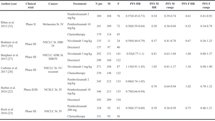
Table 2: Clinical trials selected for PFS
Author/year Clinical
trial Cancer Treatment N pts M F PFS HR PFS M HR PFS M range PFS F HR PFS F range
Ribas et al. 2015 [32] Phase II Melanoma St. IV Pembrolizumab 2 mg/kg 180 104 76 0.57(0.45-0.73) 0.54 0.39-0.74 0.61 0.41-0.92 Pembrolizumab 10 mg/kg 181 109 72 0.50(0.39-0.64) 0.50 0.36-0.68 0.52 0.34-0.78 Chemotherapy 179 114 65 Brahmer et al.
2015 [26] Phase III NSCLC St. IIIB/IV
Nivolumab 3 mg/kg 135 11 24 0.59(0.44-0.79) 0.57 0.41-0.78 0.67 0.36-1.25 Docetaxel 137 97 40
Borghaei et al.
2015 [27] Phase III NSCLC ADK St. IIIB/IV
Nivolumab 3 mg/kg 292 151 141 0.92(0.77-1.1) 0.81 0.63-1.04 1.04 0.80-1.37 Docetaxel 290 168 122
Carbone et al.
2017 [28] Phase III NSCLC St. IV/recurent
Nivolumab 3 mg/kg 271 184 87 1.15(0.91-1.45) 1.05 0.81-1.37 1.36 0.98-1.90 Chemotherapy 270 148 122 Herbst et al. 2016 [22] Phase II/III NCSLC St. IV Pembrolizumab 2 mg/kg 345 212 133 0.88(0.74-1.05) 0.78 0.64-0.94 1.02 0.78-1.32 Pembrolizumab 10 mg/kg 346 213 133 0.79(0.66-0.94) Docetaxel 343 209 134 Reck et al. 2016 [23] Phase III NSCLC St. IV Pembrolizumab 200 mg 154 92 62 0.50(0.37-0.68) 0.39 0.26-0.58 0.75 0.46-1.21 Chemotherapy 151 95 56
Indeed, several genes, whose function is implicated in immune system activity, have in their promoter an estrogen response element (ERE), such the IRF5 gene, which encodes for the IFN regulatory factor 5. [54, 55] IFNγ gene presents also an ERE on its promoter, with a transcription directly regulated by estrogens. [4] IFNγ consequently promotes, through a IRF1 mediated mechanism, the expression of CD-274 gene, coding for the PD-L1, and of IDO gene, encoding for indoleamine-2,3-dioxygenase, an enzyme that transform tryptophan into kynurenine, leading to an immunosuppressive tumor microenvironment in which the T cells’ proliferation and activity are inhibited. [56-58].
Several studies demonstrated that Treg cells express high immune inhibitory receptors, such as CTLA-4 and PD-1. Paradoxically, on Treg cells the binding of these receptors with the ligand leads to an increase in the activation of the Treg cells themselves, with consequent inhibitory activity on T lymphocytes. [59] As it has been observed in different preclinical and clinical studies, the use of anti-CTLA-4 antibodies could induce the depletion of Treg cells. [60, 61] As Treg cells are present at higher levels in males, this mechanism could affect the different response to anti-CTLA-4 in the two sexes. [4] As far as PD-1, despite having a Treg cell activation function similar to that described for CTLA-4, an inhibitory action on Treg cells has not been reported in literature.
The inhibitory activity of anti CTLA-4 on Treg cells also occurs at the gastrointestinal tract, with dysregulation of their immunomodulatory function and consequent widespread inflammation, particularly at the colon level. [62, 63] This is certainly the basis of the gastrointestinal toxicity mechanism but also responsible for the modification of the intestinal flora. The function of microbiota is relevant for the modulation of the immune system; moreover it’s affected by gender and diet differences, and it is also involved in immune-related toxicity mechanisms during treatement. [64] The effect of the microbiota may also affect the different efficacy observed according to the sex, and among different drugs with different mechanism of action.
Furthermore the differences of PFS and OS HR between anti-CTLA-4 and anti-PD-1 could depend on their different action’s mechanism. Anti-CTLA-4 and anti PD-1 act in different point of cancer- immune cycle (in priming phase and killing cancer cells phase respectively): while anti CTLA4 enhances early T cell activation, anti-PD-1 reverse the exhausted state of existing effector T cells. Thus the different activation status of antigen-specific T cells in males and females could influences the response not only to monotherapy but mostly to combination of ICIs.
Moreover, we have to consider also the differences in gender and not only in sex. The environmental estrogens
could have an impact on immune response such as xenoestrogens added in cosmetic or hormone replacement therapy and oral contraceptives.
Our work presents some weakness and limitations depending on the trials’ heterogeneity, on the different cancer type considered that could have different immunogenicity and on the absence of information about hormonal status and on PD-L1 status according to sex.
Furthermore, some studies enrolled patients according to biomarkers expression at different cut-off, for example PD-L1 for Pembrolizumab trials or for first-line Nivolumab, while for anti-CTLA-4 trials none biomarker was used as inclusion criteria. Considering the previously discussed sex related implications on PD-L1 expression, this heterogeneity between trials could be considered as a bias of our meta-analysis.
Moreover, this is not an individual patients’ data meta-analysis, leading to exclusion of many literature data. We also excluded trials with control arm treated with ICIs and trials studying combination immunotherapy, even if it could be interesting to analyze the impact of the double block of immune target in the two sexes. However, we decided to include only Phase II-III trials and to exclude trials with combination of ICIs to reduce the possible bias.
These observations need to be confirmed by further studies, in particular by direct comparison of males and females on a homogeneous and numerically larger sample, with an analysis of lymphocyte subpopulations, of expression of different markers on the surface of immune cells, of mutational burden, with the dosage of cytokines and antibodies during therapy, with an accurate study of the microbiota and studying the possible associations with immune related adverse events (irAE).
In conclusion, the differences in immune response between males and females could be relevant to determine the response and the resistance to ICIs and to identify new immunological targets in order to draw studies exploring novels combinations of immunotherapy agents.
MATERIALS AND METHODS
Data retrievalWe conducted a meta-analysis of Clinical Trials including treatment with immunotherapy (anti-CTLA-4, anti-PD1, anti-PD-L1), which data were published up to June 2017. The databases of PubMed, www.clinicaltrials. gov and the American Society of Clinical Oncology (ASCO) University Meeting was searched for relevant publications about lung (NSCLC), renal cells (RCC), head and neck, urothelial cancers and melanoma. Furthermore, we completed the data retrieval with a manual search between the references of the previously selected articles. The selection of items was restricted to phase II and phase III trials.
Data extraction
Three independent reviewers selected and analyzed all the articles. The following data were extracted: type of study; type of cancer; stage of disease; arms of treatment; number and sex of patients for each arm of treatment; PFS and OS expressed as HR for the whole population and differentiated by sex. The abstracts were excluded for lack of subgroups’ analysis. Similarly, the articles where subgroup analysis differentiated by was not reported were excluded from the final statistical analysis. The studies in which the control arm received ICIs where excluded too. Statistical analysis
Hazard Ratios (HR) with 95% confidence intervals (CIs) for PFS and OS were extracted from papers. Subgroups random-effects meta-analysis (inverse-variance weighted method) were performed to evaluate differences in treatment effects according to patients’ sex. In all figures the derived results are reported as conventional meta-analysis forest plots, with a HR<1.0 indicating better outcome in the experimental arm. All the analysis were accomplished using Stata/SE14.0 (Stata Corp, College Station, TX, USA).
Author contributions
Andrea Botticelli: Design of the project, data collection and manuscript writing
Onesti CE: Data collection, manuscript writing Zizzari I, Roberto M, Di Pietro FR, Bonifacino A, Ghidini M, Vici P, Pizzuti L, Napoletano C, D’Amati C: Manuscript review
Cerbelli B: Manuscript writing
Sciattella P, Strigari L: Statistical analysis Occhipinti M: Data collection, manuscript writing Mazzuca F, Nuti M, Marchetti P: Design of the project, manuscript review.
CONFLICTS OF INTEREST
Nothing to declare.FUNDING
None.REFERENCES
1. Topalian SL, Drake CG, Pardoll DM. Immune checkpoint blockade: A common denominator approach to cancer therapy. Cancer Cell. 2015;27:450-461.
2. Ribas A. Tumor immunotherapy directed at PD-1. N Engl J Med. 2012; 366: 2517-2519.
3. Manson G, Norwood J, Kohrt H, Houot R. Biomarkers associated with checkpoint inhibitors. Ann Oncol. 2016; 27: 1199-1206.
4. Klein SL, Flanagan KL. Sex differences in immune responses. Nat Rev Immunol 2016; 16:626-638.
5. Quintero OL, Amador-Patarroyo MJ, Montoya-Ortiz G, Rojas-Villarraga A, Anaya JM. Autoimmune disease and gender: plausible mechanisms for the female predominance of autoimmunity. J Autoimmun. 2012; 38: J109-J119. 6. Borchers AT, Naguwa SM, Keen CL, Gershwin ME. The
implications of autoimmunity and pregnancy. J Autoimmun. 2010; 34: J287-J299.
7. Libert C, Dejager L, Pinheiro I. The X chromosome in immune functions: when a chromosome makes the difference. Nat Rev Immunol. 2010; 10: 594-604.
8. Pisitkun P, Deane JA, Difilippantonio MJ, Tarasenko T, Satterthwaite AB, Bolland S. Autoreactive B cell responses to RNA-related antigens due to TLR7 gene duplication. Science. 2006; 312: 1669-1672.
9. Berghofer B, Frommer T, Haley G, Fink L, Bein G, Hackstein H. TLR7 ligands induces higer IFNα production in females. J Immunol. 2006; 177: 2088-2096.
10. Roved J, Westerdahl H, Hasselquist D. Sex differences in immune response: hormonal effects, antagonistic selection, and evolutionary consequences. Horm Behav. 2017; 88: 95-105.
11. Lin PY, Sun L, Thibodeaux SR, Ludwig SM, Vadlamudi RK, Hurez VJ, Bahar R, Kious MJ, Livi CB, Wall SR, Chen L, Zhang B, Shin T, et al. B7-H1-Dependent sex realted differences in tumor immunity and immunotherapy responses. J Immunol. 2010; 185: 2747-2753.
12. Dinesh RK, Hahn BH, Singh RP. PD-1, gender and autoimmunity. Autoimmun Rev. 2010; 9: 583-587.
13. Hodi FS, O’Day SJ, McDermott DF, Weber RW, Sosman JA, Haanen JB, Gonzalez R, Robert C, Schadendorf D, Hassel JC, Akerley W, van den Eertwegh AJ, Lutzky J, et al. Improved survival with ipilimumab in patients with matastatic melanoma. N Engl J Med. 2010; 363: 11-23. 14. Weber JS, D’Angelo SP, Minor D, Hodi FS, Gutzmer R,
Neyns B, Hoeller C, Khushalani N, Miller WH Jr, Lao CD, Linette GP, Thomas L, Lorigan P, et al. Nivolumab versus chemotherapy in patients with advanced melanoma who progressed after anti-CTLA-4 treatment (CheckMate 037): a randomised, controlled, open-label, phase 3 trial. Lancet. 2015; 16: 375-384.
15. Robert C, Long GV, Brady B, Dutriaux C, Maio M, Mortier L, Hassel JC, Rutkowski P, McNeil C, Kalinka-Warzocha E, Savage KJ, Hernberg MM, Lebbé C, et al. Nivolumab in previously untreated melanoma without BRAF mutation. N Engl J Med. 2015; 372: 320-330.
16. Robert C, Thomas L, Bondarenko I, O’Day S, Weber J, Garbe C, Lebbe C, Baurain JF, Testori A, Grob JJ, Davidson N, Richards J, Maio M, et al. Ipilimumab plus dacarbazine
for previously untreated metastatic melanoma. N Engl J Med. 2011; 364: 2517-2526.
17. Eggermont AM, Chiaron-Sileni V, Grob JJ, Dummer R, Wolchok JD, Schmidt H, Hamid O, Robert C, Ascierto PA, Richards JM, Lebbé C, Ferraresi V, Smylie M, et al. Adjuvant ipilimumab versus placebo after complete resection of high-risk stage III melanoma (EORTC 10071): a randomized, double-blind, phase 3 trial. Lancet Oncol. 2015; 16: 522-530.
18. Eggermont AM, Chiarion-Sileni V, Grob JJ, Dummer R, Wolchok JD, Schmidt H, Hamid O, Robert C, Ascierto PA, Richards JM, Lebbé C, Ferraresi V, Smylie M, et al. Prolonged Survival in Stage III Melanoma with Ipilimumab Adjuvant Therapy. N Engl J Med. 2016; 375: 1845-1855. 19. Ribas A, Kefford R, Marshall MA, Punt CJ, Haanen JB,
Marmol M, Garbe C, Gogas H, Schachter J, Linette G, Lorigan P, Kendra KL, Maio M, et al. Phase III randomized clinical trial comparing tremelimumab with standard-of-care chemotherapy in patients with advanced melanoma. J Clin Oncol. 2013; 31: 616-622.
20. Larkin J, Chiaron-Sileni V, Gonzalez R, Grob JJ, Cowey CL, Lao CD, Schadendorf D, Dummer R, Smylie M, Rutkowski P, Ferrucci PF, Hill A, Wagstaff J, et al. Combined nivolumab and ipilimumab or monotherapy in untreated melanoma. N Engl J Med. 2015; 373: 23-34. 21. Robert C, Schachter J, Long GV, Arance A, Grob JJ,
Mortier L, Daud A, Carlino MS, McNeil C, Lotem M, Larkin J, Lorigan P, Neyns B, et al. Pembrolizumab versus ipilimumab in advanced melanoma. N Engl J Med.2015; 372: 2521-2532.
22. Herbst RS, Baas P, Kim DW, Felip E, Pérez-Gracia JL, Han JY, Molina J, Kim JH, Arvis CD, Ahn MJ, Majem M, Fidler MJ, de Castro G Jr, et al. Pembrolizumab versus docetaxel for previously treated, PD-L1-positive, advanced non-small-cell lung cancer: a randomised controlled trial. Lancet.2016; 387:1540-1550.
23. Reck M, Rodriguez-Abreu D, Robinson AG, Hui R, Csőszi T, Fülöp A, Gottfried M, Peled N, Tafreshi A, Cuffe S, O’Brien M, Rao S, Hotta K, et al. Pembrolizumab versus Chemotherapy for PD-L1–Positive Non–Small-Cell Lung Cancer. N Engl J Med.2016; 375: 1823-1833.
24. Ferris RL, Blumenschein G Jr, Fayette J, Guigay J, Colevas AD, Licitra L, Harrington K, Kasper S, Vokes EE, Even C, Worden F, Saba NF, Iglesias Docampo LC, et al. Nivolumab for Recurrent Squamous-Cell Carcinoma of the Head and Neck. N Engl J Med.2016; 375: 1856-1867.
25. Rittmeyer A, Barlesi F, Waterkamp D, Park K, Ciardiello F, von Pawel J, Gadgeel SM, Hida T, Kowalski DM, Dols MC, Cortinovis DL, Leach J, Polikoff J, et al. Atezolizumab versus docetaxel in patients with previously treated non-small-cell lung cancer (OAK): a phase 3, open-label, multicentre randomised controlled trial. Lancet. 2017; 389: 255–265.
26. Brahmer J, Reckamp KL, Baas P, Crinò L, Eberhardt WE, Poddubskaya E, Antonia S, Pluzanski A, Vokes
EE, Holgado E, Waterhouse D, Ready N, Gainor J, et al. Nivolumab versus Docetaxel in Advanced Squamous-Cell Non-Small-Cell Lung Cancer. N Engl J Med. 2015; 373: 123-135.
27. Borghaei H, Paz-Ares L, Horn L, Spigel DR, Steins M, Ready NE, Chow LQ, Vokes EE, Felip E, Holgado E, Barlesi F, Kohlhäufl M, Arrieta O, et al. Nivolumab versus Docetaxel in Advanced Non-squamous Non-Small-Cell Lung Cancer. N Engl J Med. 2015; 373: 1627-1639. 28. Carbone DP, Reck M, Paz-Ares L, Creelan B, Horn L,
Steins M, Felip E, van den Heuvel MM, Ciuleanu TE, Badin F, Ready N, Hiltermann TJN, Nair S, et al. First-Line Nivolumab in Stage IV or Recurrent Non-Small-Cell Lung Cancer. N Engl J Med.2017; 376: 2415-2426.
29. Motzer RJ, Escudier B, McDermott DF, George S, Hammers HJ, Srinivas S, Tykodi SS, Sosman JA, Procopio G, Plimack ER, Castellano D, Choueiri TK, Gurney H, et al. Nivolumab versus Everolimus in Advanced Renal-Cell Carcinoma. N Engl J Med. 2015;373:1803-1813.
30. Bellmunt J, de Wit R, Vaughn DJ, Fradet Y, Lee JL, Fong L, Vogelzang NJ, Climent MA, Petrylak DP, Choueiri TK, Necchi A, Gerritsen W, Gurney H, et al. Pembrolizumab as Second-Line Therapy for Advanced Urothelial Carcinoma. N Engl J Med. 2017; 376: 1015-1026.
31. Wolchok J, Neyns B, Linette G, Negrier S, Lutzky J, Thomas L, Waterfield W, Schadendorf D, Smylie M, Guthrie T Jr, Grob JJ, Chesney J, Chin K, et al. Ipilimumab monotherapy in patients with previously treated, advanced melanoma:a randomized, double. blind, multicenter, phase 2, dose-ranging study. Lancet Oncol. 2010; 11: 155-164. 32. Ribas A, Puzanov I, Dummer R, Schadendorf D, Hamid O,
Robert C, Hodi FS, Schachter J, Pavlick AC, Lewis KD, Cranmer LD, Blank CU, O’Day SJ, et al. Pembrolizumab versus investigator-choise chemotherapy for ipilimumab-refractory melanoma: a phase 2 trial. Lancet Oncol.2015; 16: 908-918.
33. Postow MA, Chesney J, Pavlick AC, Robert C, Grossmann K, McDermott D, Linette GP, Meyer N, Giguere JK, Agarwala SS, Shaheen M, Ernstoff MS, Minor D, et al. Nivolumab and ipilimumab versus ipilimumab in untreated melanoma. N Engl J Med. 2015; 372: 2006-17.
34. Hodi FS, Chesney J, Pavlick AC, Robert C, Grossmann KF, McDermott DF, Linette GP, Meyer N, Giguere JK, Agarwala SS, Shaheen M, Ernstoff MS, Minor DR, et al. Combined nivolumab and ipilimumab versus ipilimumab alone in patients with advanced melanoma: 2-year overall survival outcomes in a multicentre, randomised, controlled, phase 2 trial. Lancet Oncol. 2016; 17: 1558-1568.
35. Hersh EM, O’Day SJ, Powderly J, Khan KD, Pavlick AC, Cranmer LD, Samlowski WE, Nichol GM, Yellin MJ, Weber JS. A phase II multicenter study of ipilimumab with or without dacarbazine in chemotherapy-naïve patients with advanced melanoma. Invest New Drugs. 2011; 29: 489-498. 36. Zimmer L, Eigentler TK, Kiecker F, Simon J, Utikal J, Mohr
A, Fluck M., Terheyden P, et al. Open-label, multicenter, single-arm phase II DeCOG-study of ipilimumab in pretreated patients with different subtypes of metastatic melanoma. J Transl Med. 2015; 13: 351.
37. Hodi FS, Lee S, McDermott DF, Rao UN, Butterfield LH, Tarhini AA, Leming P, Puzanov I, Shin D, Kirkwood JM. Ipilimumab plus sargramostim vs ipilimumab alone for treatment of metastatic melanoma: a randomized clinical trial. JAMA. 2014; 312: 1744-1753.
38. Weber J, Thompson JA, Hamid O, Minor D, Amin A, Ron I, Ridolfi R, Assi H, Maraveyas A, Berman D, Siegel J, O’Day SJ.. A randomized, double-blind, placebo-controlled, phase II study comparing the tolerability and efficacy of ipilimumab administered with or without prophylactic budesonide in patients with unresectable stage III or IV melanoma. Clin Cancer Res. 2009; 15: 5591-5598.
39. O’Day SJ, Maio M, Chiarion-Sileni V, Gajewski TF, Pehamberger H, Bondarenko IN, Queirolo P, Lundgren L, Mikhailov S, Roman L, Verschraegen C, Humphrey R, Ibrahim R, et al. Efficacy and safety of ipilimumab monotherapy in patients with pretreated advanced melanoma: a multicenter single-arm phase II study. Ann Oncol. 2010; 21: 1712-1717.
40. Di Giacomo AM, Ascierto PA, Pilla L, Santinami M, Ferrucci PF, Giannarelli D, Marasco A, Rivoltini L, Simeone E, Nicoletti SV, Fonsatti E, Annesi D, Queirolo P, et al. Ipilimumab and fotemustine in patients with advanced melanoma (NIBIT-M1): an open-label, single-arm phase 2 trial. Lancet Oncol. 2012; 13: 879-886.
41. Margolin K, Ernstoff MS, Hamid O, Lawrence D, McDermott D, Puzanov I, Wolchok JD, Clark JI, Sznol M, Logan TF, Richards J, Michener T, Balogh A, et al. Ipilimumab in patients with melanoma and brain metastases: an open-label, phase 2 trial. Lancet Oncol. 2012; 13: 459-465.
42. Motzer RJ, Rini BI, McDermott DF, Redman BG, Kuzel TM, Harrison MR, Vaishampayan UN, Drabkin HA, George S, Logan TF, Margolin KA, Plimack ER, Lambert AM, et al. Nivolumab for Metastatic Renal Cell Carcinoma: Results of a Randomized Phase II Trial. J Clin Oncol. 2015; 33: 1430-1437.
43. Rizvi NA, Mazières J, Planchard D, Stinchcombe TE, Dy GK, Antonia SJ, Horn L, Lena H, Minenza E, Mennecier B, Otterson GA, Campos LT, Gandara DR, et al. Activity and safety of nivolumab, an anti-PD-1 immune checkpoint inhibitor, for patients with advanced, refractory squamous non-small-cell lung cancer (CheckMate 063): a phase 2, single-arm trial. Lancet Oncol. 2015; 16: 257-265.
44. Felip E, Van Meerbeeck J, Wolf J, Ardizzoni A, Li A, Srinivasan S, Popat S.193TiP: CheckMate 171: A multicenter phase 2 trial of nivolumab (nivo) in patients (pts) with stage IIIB/IV squamous cell (SQ) NSCLC who have received ≥1 prior systemic treatment. J Thorac Oncol. 2016; 11: S141.
45. Langer CJ, Gadgeel SM, Borghaei H, Papadimitrakopoulou VA, Patnaik A, Powell SF, Gentzler RD, Martins RG, Stevenson JP, Jalal SI, Panwalkar A, Yang JC, Gubens M, et al. Carboplatin and pemetrexed with or without pembrolizumab for advanced, non-squamous non-small-cell lung cancer: a randomised, phase 2 cohort of the open-label KEYNOTE-021 study. Lancet Oncol. 2016; 17: 1497-1508. 46. Fehrenbacher L, Spira A, Ballinger M, Kowanetz M,
Vansteenkiste J, Mazieres J, Park K, Smith D, Artal-Cortes A, Lewanski C, Braiteh F, Waterkamp D, He P, et al. Atezolizumab versus docetaxel for patients with previously treated non-small-cell lung cancer (POPLAR): a multicentre, open-label, phase 2 randomised controlled trial. Lancet. 2016;387:1837-1846.
47. Lynch TJ, Bondarenko I, Luft A, Serwatowski P, Barlesi F, Chacko R, Sebastian M, Neal J, Lu H, Cuillerot JM, Reck M. Ipilimumab in combination with paclitaxel and carboplatin as first-line treatment in stage IIIB/IV non-small-cell lung cancer: results from a randomized, double-blind, multicenter phase II study. J Clin Oncol. 2012; 30: 2046-2054.
48. Galsky M, Retz AO, Siefker-Radtke A, BVaena D, Grimm MO, Bracarda S, Arranz-Arija J, Pal SK, Ohyama C, et al. Efficacy and safety of nivolumab monotherapy in patients with metastatic urothelial cancer (mUC) who have received prior treatment: Results from the phase II CheckMate 275 study. Ann Oncol. 2016; 27: LBA31_PR.
49. Bajorin DF, Plimack ER, Siefker-Radtke AO, Choueiri TK, De Wit R, Sonpavde G, Gipson A, Holly Brown H, Mai Y, Pang L, Perini RF, Bellmunt J. KEYNOTE-052: Phase 2 study of pembrolizumab (MK-3475) as first-line therapy for patients (pts) with unresectable or metastatic urothelial cancer ineligible for cisplatin-based therapy. J Clin Oncol. 2015; 33: TPS4572.
50. Rosenberg JE, Hoffman-Censits J, Powles T, van der Heijden MS, Balar AV, Necchi A, Dawson N, O’Donnell PH, Balmanoukian A, Loriot Y, Srinivas S, Retz MM, Grivas P, et al. Atezolizumab in patients with locally advanced and metastatic urothelial carcinoma who have progressed following treatment with platinum-based chemotherapy: a single-arm, multicentre, phase 2 trial. Lancet. 2016; 387: 1909-1920.
51. Phiel KL, Henderson RA, Adelman SJ, Elloso MM. Differential estrogen receptor gene expression in human peripheral blood mononuclear cell populations. Immunol Lett. 2005; 97: 107–113.
52. Teilmann SC, Clement CA, Thorup J, Byskov AG, Christensen ST. Expression and localization of the progesterone receptor in mouse and human reproductive organs. J Endocrinol. 2006; 191: 525–535.
53. Kim CH. FOXP3 and its role in the immune system. Adv Exp Med Biol. 2009; 665: 17-29.
54. Griesbeck M, Ziegler S, Laffont S, Smith N, Chauveau L, Tomezsko P, Sharei A, Kourjian G, Porichis F, Hart M,
Palmer CD, Sirignano M, Beisel C, et al. Sex differences in plasmacytoid dendritic cell levels of IRF5 drive higher IFNα production in women. J Immunol. 2015; 195: 5327-5336.
55. Hawegama A, Patel D, Yariagadda S, Strickland FM, Richardson BC. Stronger inflammatory/cytotoxic T cell response in women identified by microarray analysis. Genes Immun. 2009; 10:509-516.
56. Lu C, Redd PS, Lee JR, Savage N, Liu K. The expression profiles and regulation of PD-L1 in tumor-induced myeloid-derived suppressor cells. Oncoimmunology. 2016; 5: e1247135.
57. Liang C, Jiang E, Yao J, Wang M, Chen S, Zhou Z, Zhai W, Ma Q, Feng S, Han M. Interferon-γ mediates the immunosuppression of bone marrow mesenchymal stem cells on T-lymphocytes in vitro. Hematology. 2017. https:// doi.org/10.1080/10245332.2017.1333245.
58. Löb S, Königsrainer A, Rammensee HG, Opelz G, Terness P. Inhibitors of indoleamine-2,3-dioxygenase for cancer therapy: can we see the wood for the trees? Nat Rev Cancer. 2009; 9: 445-452.
59. Miller JF, Sedalain M. The journey for discoveries in fundamental immunology to cancer immunotherapy. Cancer Cell. 2015; 27: 439-449.
60. Yang ZZ, Novak AJ, Stenson MJ, Witzig TE, Ansell SM. Intratumoral CD4+CD25+ regulatory T-cell-mediated suppression of infiltrating CD4+ T cells in B-cell non-Hodgkin lymphoma. Blood. 2006; 107: 3639–3646. 61. Riella LV, Liu T, Yang J, Chock S, Shimizu T, Mfarrej B,
Batal I, Xiao X, Sayegh MH, Chandraker A. Deleterious effect of CTLA4-Ig on a Treg-dependent transplant model. Am J Transpl. 2012; 12: 846-855.
62. Carbonnel F, Soularue E, Coutzac C, Chaput N, Mateus C, Lepage P, Robert C. Inflammatory bowel disease and cancer response due to anti-CTLA-4: is it in the flora? Semin Immunopathol. 2017; 39: 327-331.
63. Read S, Greenwald R, Izcue A, Robinson N, Mandelbrot D, Francisco L, Sharpe AH, Powrie F. Blockade of CTLA-4 on CD4+CD25+ regulatory T cells abrogates their function in vivo. J Immunol. 2006; 177: 4376–4383.
64. Andrea Botticelli, Zizzari I, Mazzuca F, Ascierto PA, Putignani L, Marchetti L, Napoletano C, Nuti M, Marchetti P. Cross-talk between microbiota and immune fitness to steer and control response to anti PD-1/PDL-1 treatment. Oncotarget 2017; 8: 8890-8899.