Combining an original method for preserving RNA expression in situ with an effective RNA extraction method makes it possible to study gene expression in any banana fruit tissue.
Abstract –– Introduction. RNA isolation is a prerequisite to studying gene expression in
banana and to understanding changes occurring in response to the environment. Standard extraction methods do not efficiently extract RNA from plants such as banana, with high levels of phenolics, carbohydrates, or other compounds that bind to and/or coprecipitate with RNA.
Materials and methods. Five to seven RNA extraction methods were compared. Four
crown-tissue storage methods were also compared. cDNA-AFLP was used to ensure that the obtained RNA was of sufficient quality for molecular applications and that RNA expression was unaltered by in
situ storage. Results and discussion. The modified hot-borate method proved to be the best
RNA extraction method, allowing high yields of good quality, undegraded RNA from the crown, fruit peel and pulp at all stages of ripening. The RNA obtained by this method was of sufficient quality for molecular applications such as cDNA-AFLP that give highly reproducible results. Freeze-drying of fresh tissues and tissue conservation in hot-borate buffer, two original storage methods, appear appropriate for preserving RNA in situ without ultra-low temperature. The RNA obtained was of high quality, undegraded, and useful for all downstream applications. The genome expression profile obtained by cDNA-AFLP analysis was unaltered by these methods for storing collected tissues. Conclusion. By applying all the suggested procedures in this work, it is possible to store and study gene expression in any banana fruit tissue, whatever the maturity stage, without affecting the RNA expression level.
Belgium / Musa sp. / bananas / freeze-drying / RNA / storage / extraction
La combinaison d'une méthode originale permettant de maintenir l'expression de l’ARN in situ et d’une méthode efficace pour extraire l’ARN permet d’étudier l'expression de gènes dans n'importe quel tissu de banane.
Résumé –– Introduction. L'extraction de l’ARN est un préalable pour étudier l'expression des
gènes dans la banane et pour comprendre les changements intervenant en réponse à l'envi-ronnement. Les méthodes standard d'extraction de l’ARN ne sont pas efficaces avec des plantes comme le bananier, qui présentent des niveaux élevés de composés phénoliques, d’hydrates de carbone, ou autres composés qui se lient à et/ou coprécipitent l'ARN. Matériel et méthodes. Cinq à sept méthodes d'extraction de l’ARN ont été comparées. Quatre méthodes de stockage de tissus de couronne ont été également comparées. La technique de cDNA-AFLP a été utilisée pour s'assurer que l'ARN extrait était de qualité suffisante pour des applications moléculaires et que l'expression de l'ARN était inchangée après un stockage in situ. Résultats et discussion. La méthode du borate chaud modifiée s’est avérée être la meilleure méthode d'extraction de l’ARN, permettant des rendements élevés de bonne qualité, et donnant de l'ARN non dégradé à partir des tissus de la couronne, de la peau des fruits et de la pulpe à toutes les étapes de la maturation. L'ARN obtenu par cette méthode a été de qualité suffisante pour les applications moléculaires telles que le cDNA-AFLP qui donne des résultats fortement reproductibles. La lyo-philisation des tissus frais et la conservation de tissu dans la solution tampon de borate chaud, deux méthodes originales de stockage, semblent appropriées à la conservation de l'ARN in situ en absence d’ultra basse température. L'ARN obtenu a été de qualité, non dégradé, et utilisable pour toutes les applications. Le profil d'expression du génome obtenu par la technique de cDNA-AFLP est resté inchangé par ces méthodes de stockage des tissus collectés. Conclusion. En appliquant toutes les procédures suggérées dans ce travail, il est possible de stocker et d’étudier l'expression de gènes dans n'importe quel tissu de banane, quel que soit le stade de maturité, sans affecter le niveau d'expression de l'ARN.
Belgique / Musa sp. / banane / lyophilisation / ARN / stockage / extraction 1Gembloux Agric. Univ., Plant
Pathol. Unit, Passage des Déportés 2, B-5030 Gembloux, Belgium
jijakli.h@fsagx.ac.be 2CIRAD, Persyst, UPR Syst. Banan. Ananas, TA B-26 / PS4, Blvd. de la Lironde,
34398 Montpellier Cedex 5, France
Combining an original method for preserving RNA expression
in situ with an effective RNA extraction method makes it possible
to study gene expression in any banana fruit tissue
Ludivine LASSOIS1, Luc dE LAPEYRE dE BELLAIRE2, Haïssam JIJAKLI1*
* Correspondence and reprints
Received 24 October 2008 Accepted 5 December 2008
Fruits, 2009, vol. 64, p. 127–137 © 2009 Cirad/EDP Sciences All rights reserved
DOI: 10.1051/fruits/2009008 www.fruits-journal.org
1. Introduction
Banana is cultivated in more than 120 coun-tries and holds the second place in world fruit production, just behind orange and in front of grape. Exported dessert bananas belong to the Cavendish subgroup. Gene expression studies are essential to under-standing the physiological processes of this plant species [1, 2]. Expression profiling, however, requires adequate sample conser-vation and high-quality RNA isolation for cDNA library construction and molecular analysis. The total RNA and mRNA obtained must notably be free of protein, genomic DNA and secondary metabolites. RNA extraction is thus a crucial step, but it can be problematic because of the relative insta-bility of RNA, largely due to RNA degrada-tion by ribonucleases [3]. Conservadegrada-tion and isolation of RNA from plant material, partic-ularly from fruit tissues, presents special challenges [4, 5]. First, most plant materials contain relatively high levels of RNase activ-ity, mostly located in the vacuoles [6]. They also contain various plant metabolites, such as polysaccharides and polyphenols. They bind to and/or co-precipitate with RNA, affecting the yield and quality of RNA [7] and rendering it unsuitable for cDNA synthesis. Thus, qualitative and quantitative differ-ences in polysaccharide and polyphenol content among different plant tissues signif-icantly influence the efficiency of nucleic acid extraction and purification procedures [3, 8]. Many procedures [4, 5, 8–14] have been developed to cope with endogenous polysaccharides, phenolics and RNases dur-ing RNA isolation from various plants, but the efficiency of an extraction method depends largely on the kind of plant or tissue.
Another potential problem with RNA is its rapid degradation or alteration during tissue storage. In some cases, plant tissues must be preserved as collected, especially when a genome-wide expression profile is to be studied. The most commonly used method to store tissues for preparation of RNA is to remove fresh tissues and to maintain them at a strictly ultra-low temperature [(–50 to –70) °C] [15]. However, other efficient stor-age methods without ultra-low temperature
could be of interest but have not been exten-sively documented. Freeze-drying is pro-posed in some cases to conserve animal tis-sues [16–20], human tistis-sues [21, 22] and bacteria [23] in order to further RNA extrac-tion. However, in plant tissues, freeze-dry-ing has not been used extensively to pre-serve RNA [4, 24] and contradictory results have been reported [25, 26].
The first aim of our study was to evaluate various techniques of RNA extraction from fresh banana tissues for quantity, quality and integrity, but also its adequacy for downstream molecular analyses such as cDNA-Amplified Fragment Length Polymor-phism analysis (cDNA-AFLP). This RNA fin-gerprinting technique derives from AFLP (Amplified Fragment Length Polymor-phism), a method described by Vos et al. [27] for genomic DNA fingerprinting, which was applied for the first time like cDNA-AFLP to plants by Bachem et al. [28].
Classical RNA isolation techniques were compared with methods specifically elabo-rated for banana or other plants rich in polyphenols and polysaccharides.
As collected materials sometimes have to be stored prior to RNA extraction, the sec-ond aim of our study was to assess different methods for preserving RNA in situ. This means both avoiding RNA degradation between sample collection and RNA extrac-tion and ensuring that nothing happens, during storage, to alter the expression pro-file obtained. Checking that this latter crite-rion is met requires obtaining pre- and post-storage differential expression profiles by cDNA-AFLP.
2. Materials and methods
2.1. Plant materials
Ripe yellow bananas of the Cavendish sub-group were bought on the market. Precli-macteric green bananas of the Cavendish subgroup were obtained from a commercial source in Belgium (Van Damme, Brussels) just before the ethylene treatment that trig-gers ripening.
2.2. RNA extraction methods
RNA extraction methods were tested on var-ious fruit tissues from green and ripe bananas.
Samples of peel, pulp and crown tissue were sliced, immediately frozen in liquid nitrogen, and ground to a fine powder with a pre-cooled mortar and pestle before extraction. Five extractions were performed for each tested method. All materials and reagents were treated so as to be RNase-free. Glassware was oven-baked at 240 °C over-night. Sterile disposable plastic wares were used when it was possible and non-dispos-able plastic wares were rinsed with 0.1 M NaOH, 1 mM EDTA, followed by RNase-free water. Solutions (water and other solu-tions) were treated with 0.1% DEPC (diethyl pyrocarbonate), incubated overnight at 37 °C, then autoclaved to eliminate residual DEPC.
Five RNA extraction methods were applied to peel and pulp from green bananas. Meth-ods based on commercial products were carried out according to the supplied instructions. Methods taken from the litera-ture were carried out according to the authors’ protocols. These methods were: 1. The commercial ready-to-use RNA isola-tion product TRIZOL® Reagent (Invitrogen, Carlsbad, CA, USA), according to the proto-col proposed for extraction from plant tis-sues: this is an improved version of the sin-gle-step RNA isolation method developed by Chomczynski and Sacchi [29]. During sam-ple homogenization or lysis, TRIZOL Rea-gent maintains RNA integrity while disrupt-ing cells and dissolvdisrupt-ing cell components. 2. The ‘SV Total RNA Isolation System’ developed by Promega (Madison, WI, USA): this is a quick and simple technique for pre-paring purified, intact total RNA. The system combines the disruptive and protective properties of guanidine thiocyanate and β-mercaptoethanol to inactivate the ribonu-cleases present in cell extract. The system also incorporates a DNase treatment step, and purification is achieved without any phenol/chloroform extraction.
3. The method elaborated by Liu et al. [30] to isolate total RNA from banana fruit tis-sues: RNA was extracted at room
tempera-ture with a high-ionic-strength buffer. Proteins, genomic DNA and secondary metabolites were then removed from the extract by precipitation with pre-cooled potassium acetate and repeated phenol/ chloroform/isoamyl alcohol extractions. RNA was recovered by ethanol precipitation without LiCl. This procedure can be com-pleted in less than 4 h.
4. The hot-borate method [31], adapted from that described for cotton [32]: RNA extraction was performed in a basic hot-borate/proteinase K buffer. A RNA-enriched pellet is precipitated from the supernatant with lithium chloride and dissolved in DEPC water. Two phenol/chloroform/isoamyl alco-hol (25/24/1) extractions and one chloro-form/isoamyl alcohol (24/1) extraction are incorporated into the RNA purification pro-tocol to remove contaminant proteins. Total RNA is precipitated with ethanol.
5. The simple procedure developed by Asif et al. [33] for RNA isolation from banana tis-sue: these authors have modified the CTAB/ NaCl method [11] by removing PVP from the extraction buffer and including a simple polysaccharide precipitation step. A RNA-enriched pellet is precipitated from the supernatant with lithium chloride and dis-solved in DEPC water before phenol/chlo-roform extraction. Total RNA is precipitated with ethanol.
On peel and pulp from ripe bananas, in addition to these five protocols, two addi-tional ones among those proposed by Gehrig et al. [7] were tested. Their tech-niques based on GITC (guanidium isothio-cyanate) (for technique 6) and GHCL (gua-nidium hydrochloride) (for technique 7) buffers were selected. Both methods use a high-molecular-weight polyethylene glycol (HMW-PEG). The hot-borate method [31] was also tested on crown tissues. All extrac-tions were performed on a small scale in a 1.5- or 2-mL tube, by using the protocols proportionally.
2.3. Evaluation of RNA quality, quantity and integrity
After extraction, RNA was recovered by cen-trifugation for 20 min at 16 000 g and 4 °C.
The pellet was washed once in 1 mL of 70% cold ethanol, vortexed, and centrifuged for 5 min at 16 000 g and 4 °C. The pellet was air-dried at 37 °C, and then resuspended in RNase-free water. The purified RNA was quantified by measuring the absorbance at 260 nm (A260) with a Nanodrop (ND-1000 spectrophotometer, Isogen Life Sciences, Maarssen, Netherlands). An A260 of 1 is equivalent to 40 ng RNA·µL–1. The absorb-ance at 280 nm was also measured, and sample purity was estimated by the [A260/ A280] absorbance ratio. RNA integrity was evaluated by the sharpness of the bands cor-responding to 28S and 18S ribosomal RNA (rRNA) visualized by ethidium bromide staining on a 1% agarose gel.
2.4. Evaluation of the RNA adequacy for downstream molecular analyses RNA extracts were treated for 30 min at 37 °C with 20 units of RNase-free DNase (Roche, Basel, Switzerland) in the presence of 40 units of RNase inhibitor (Roche Diag-nostics, Mannheim, Germany). The cDNA-AFLP technique involves three steps: (1) restriction of cDNA and ligation of oligonu-cleotide adapters; (2) selective amplification of sets of restriction fragments using PCR primers bearing selective nucleotides at the 3’ end; (3) gel analysis of the amplified frag-ments. Double-stranded cDNA was synthe-sized from total RNA according to the instructions for use of the Superscript Dou-ble-Stranded cDNA Synthesis kit (Invitro-gen, Carlsbad, CA, USA). The double-stranded cDNA was digested with EcoRI and
MseI, ligated with EcoRI and MseI adapters, and pre-amplified with the Eco (5’-GACT-GCGTACCAATTC-3’) and Mse (5’-GAT-GAGTCCTGAGTAA-3’) primers, following the instructions of the AFLP Analysis System kit for microorganisms (Invitrogen, Carlsbad, CA, USA). After twenty-fold dilutions of the PCR fragments, specific amplifications were carried out with combinations of an Eco primer and a Mse primer containing two additional bases at their 3’ ends. The Eco primers were labeled with γ33P dATP. Amplification products were separated by electrophoresis at 50 W on a vertical dena-turing polyacrylamide gel (6%) containing 7 M urea. Gels were dried on a Whatman paper before screening with a phosphoim-ager (BioRad, Richmond, CA, USA). To eval-uate the reproducibility of the cDNA-AFLP, a single banana crown was ground in liquid nitrogen to a fine powder and divided into two pools. RNA was extracted according the hot-borate method [31]. After DNase treat-ment, two different pre-amplifications were performed per sample and two different selective amplifications were performed per pre-amplified sample (figure 1). Two repli-cates were performed.
2.5. In situ RNA preservation methods
Four tissue storage methods were compared on crown tissue material.
The first two methods involved placing the tissues on RNAlater (Ambion, Austin, TX, USA) according to the manufacturer’s instructions. Here, crown tissue was first either sliced into tiny lamellas before storage in RNAlater at 4 °C or ground in liquid nitro-gen before storage in RNAlater at 4 °C.
The third conservation method (buffer storage) consisted of storing crown tissues (ground to a fine powder in liquid nitrogen) in the hot-borate buffer [31] (200 mM BORAX, pH 9.0; 30 mM EGTA; 1% SDS; 10 mM DTT; 2% PVP 40; 2% DIECA; 0.5% Igepal) to which 0.5 mg proteinase K·mL–1 buffer was added. This was stored at room temperature.
The last method consisted of freeze-dry-ing the crown sample at –80 °C for 24 h (Tel-star, Cryodos, Barcelona, Spain) and storing
Figure 1.
Protocol applied to test the reproducibility of the cDNA-AFLP analyses based on RNA extracted from banana fruit tissues.
it at room temperature before RNA extrac-tion (freeze-drying storage). The freeze-dried crown samples were stored with silica gel to avoid an aqueous environment.
In each case, RNA extraction was per-formed 21 d later by the hot-borate method [31]. The extraction efficiency was assessed by analysis of RNA quality, quantity and integrity as described in section 2.2.
The adequacy of the RNA, obtained after a buffer or a freeze-drying storage, for downstream molecular application was evaluated by cDNA-AFLP as described in section 2.4. Furthermore, this technique allows evaluating the effect of the storage conditions on gene expression by compar-ing pre- and post-storage differential expression profiles. To do this, three sam-ples were collected from a single crown. One of these served as control and RNA extraction was performed immediately; both of the others were stored either by freeze-drying or in hot-borate storage buffer [31] as described above prior to RNA extrac-tion and cDNA-AFLP. This experiment was repeated twice.
3. Results and discussion
3.1. Compared performances of the RNA extraction methods Among the RNA extraction methods tested, the hot-borate method [31] emerges as
par-ticularly adequate for banana pulp and peel, whatever the maturity stage, and for the crown, which is particularly fibrous. Total RNA isolated by this method was of high purity and integrity and of sufficient yield, making it suitable for molecular analysis. This was confirmed in several ways.
First, spectrophotometric analysis revealed an [A260/ A280] ratio ranging consistently from 2.0 to 2.2, indicating that the RNA iso-lated by this approach was largely free of contaminating proteins. This was true what-ever the tissue type and maturity stage (table I). Pure RNA has an [A260/ A280] ratio of 2.0, but, for most applications, a lower or higher ratio probably will not affect the results [3]. As a result of variations between individual starting materials and in perform-ing the procedure, the expected range of the [A260/ A280] ratio for RNA is 1.7–2.2. Three other methods gave this ratio: Liu et al. [30] and both methods described by Gehrig et al. [7]. Both commercial kits failed to extract RNA from banana fruit tissues. The TRIZOL reagent was developed for animal tissues, especially those rich in RNase, but it seems unsuitable for recalcitrant plant tissues such as banana. This is in agreement with the results obtained by other authors [12]. The method based on CTAB [33] was also unsuc-cessful. The same conclusion was reached with different recalcitrant plant species [13]. Secondly, a comparison of RNA yields based on spectrophotometric determina-tions showed a significantly lower yield (as Table I.
[A260/ A280] ratio values according to different tissue and RNA extraction methods. The best methods give ratios between 1.7 and 2.2.
Methods Peel Pulp Crown
Green Mature Green Mature Trizol® Reagent (Invitrogen) 0.9–1.1 0.9–1.2 0.1–3.4 1.0–1.3 – SV Total RNA Isolation System (Promega) 0.2–0.8 0.4–1.0 0.06–0.20 0.6–0.9 –
Liu et al. [30] 1.7 1.7–2.0 1.7–1.8 1.8–2.1 –
Mbéguié-A-Mbéguié et al. [31] 2.0–2.2 2.0–2.2 2.0–2.2 2.0–2.1 2.00–2.14
Asif et al. [33] 1.2–1.4 1.2–2.3 1.1–1.5 0.8–1.5 –
Gehrig et al. (GITC) [7] – 1.7–2.0 – 1.0–2.3 –
Gehrig et al. (GHCL) [7] – 1.7–2.0 – 1.7–2.0 –
much as 15 times lower) for the method of Liu et al. [30] than for the hot-borate method [31] (table II). The yield obtained in this study by this latter method is comparable with those reported for other methods designed for high-phenol fruit tissues [4, 7]. The yield obtained in this study by the hot-borate [31] protocol depended heavily on the tissue type and, to a lesser extent, on the maturity stage. By decreasing yield (table II), the different tissues can be ranked as fol-lows: green pulp (67.8–79.5 µg·g–1FW) > mature pulp (61.8–65.9 µg·g–1FW) > green
peel (45.2–60.2 µg·g–1FW) > mature peel (43.4–50.5 µg·g–1FW) > crown (10.2– 17.4 µg·g–1FW).
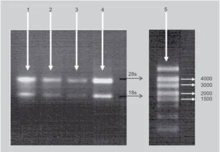
Thirdly, denaturing agarose gel electro-phoresis yielded clear rRNA bands with a 28s rRNA band equal to or more abundant than the 18s rRNA band (figure 2), indicat-ing that little or no degradation had occurred during extraction with the hot-borate [31] and Liu et al.'s [30] methods. In high-quality RNA, the 28s band should be approximately twice as intense as the 18s band [3]. In degraded RNA samples, the 18s band will be enhanced, since the 28s ribosomal RNA is typically degraded to an 18s-like species. No rRNA was observed with any other tech-nique, and notably not with either of those described by Gehrig et al. [7]. The latter methods did give, however, a good [A260/ A280] ratio (table I) and thus a high esti-mated yield (table II). An explanation might be that A260 could be falsely boosted by con-taminants such as salt, organic solvent or protein, or by one or more of the many chemicals used in nucleic acid purification which absorb at 260 nm [34]. Thus, the [A260/ A280] ratio can accurately describe nucleic acid purity, but it can also be mis-leading [35]. For this reason, it is essential to have both the [A260/ A280] ratio and an agarose gel result to evaluate a RNA extrac-tion method.
Table II.
Yield values (µg RNA·g–1 fresh material), based on A260, according to different tissue and RNA extraction methods. (x) Means that the yield could not be calculated because the [A260/ A280] ratio was not between 1.7 and 2.2.
Methods Peel Pulp Crown
Green Mature Green Mature
Trizol® Reagent (Invitrogen) (×) (×) (×) (×) –
SV Total RNA Isolation System (Promega)
(×) (×) (×) (×) –
Liu et al. [30] 3.9–5.7 7.6–8.9 3.6–9.9 50.3–56.8 –
Mbéguié-A-Mbéguié et al. [31] 45.2–60.2 43.4–50.5 67.8–79.5 61.8–65.9 10.2–17.4
Asif et al. [33] (×) (×) (×) (×) –
Gehrig et al. (GITC) [7] – 72.0–75.8 – (×) –
Gehrig et al. (GHCL) [7] – 102.4–109.7 – 117.5–125.2 –
GHCL: guanidium hydrochloride; GITC: guanidium hydrochloride. Figure 2.
Band patterns obtained by ethidium bromide staining after electrophoresis, through a 1% agarose gel, of total RNA extracted from green banana peel (1 and 3) or pulp (2 and 4). The extraction protocols used were that of Liu et al., [30] (1 and 2) and the hot-borate method [31] (3 and 4). (5) is a RNA ladder and the positions of the 28s and 18s rRNAs are shown. Similar results were obtained for both protocols with peel and pulp from ripe banana. No rRNAs were observed with any other method tested in this work (results not shown).
Finally, the adequacy of the hot-borate method [31] for extracting total RNA from banana tissues in order to generate cDNA for downstream molecular analyses was successfully performed by cDNA-AFLP after DNase treatment (figures 3, 4).
For these reasons, although the hot-borate method [31] is labor-intensive and time-consuming, it seems to be the most suitable for extracting RNA from banana tis-sues in order to carry out genome-wide expression analyses. This RNA extraction technique is applicable to large samples of up to 25 g fresh material [36], but also to small samples of 0.24 g of material in a 2-mL tube, as in this work. It could be more con-venient to extract RNA from a small sample, because many samples can be extracted at the same time, and the yield obtained is still sufficient for subsequent molecular applica-tions.
3.2. In situ RNA preservation methods
On the basis of quality, quantity and integ-rity, it appears that all four methods tested can preserve RNA (table III). The choice of a method must thus be based on the exper-imental constraints and the equipment avail-able. Yet, although all four techniques appeared to preserve RNA quality and integ-rity, their yields were different. RNAlater is
an aqueous tissue storage reagent that sta-bilizes and protects cellular RNA in intact, unfrozen tissue samples. According to Table III.
[A260/ A280] ratio and yield values (based on A260) according to four RNA in situ preservation methods. Extractions were performed by the hot-borate method [31].
Methods [A260/ A280] ratio Yield (µg RNA·g–1 fresh material) 28s:18s RNA bands Crown sliced into tiny lamellas
and stored in RNAlater at 4 °C for 21 d
1.9–2.0 3.2–5.7 Yes
Crown ground in liquid nitrogen and stored in RNAlater at 4 °C for 21 d
1.8 1.5–3.7 Yes
Crown ground in liquid nitrogen and stored in the hot-borate buffer [31] + proteinase K at room temperature for 21 d
1.9 10.2–14.0 Yes
Crown freeze-dried
and stored for 21 d at room temperature before extraction
2.2 12.0–16.2 Yes
Figure 3.
Reproducibility of cDNA-AFLP based on total RNA. cDNA-AFLP was applied to various pools of total RNA from a single crown according to figure 1. (1) to (8) represent the various cDNA-AFLP pools and (9) is a blank.
Ambion (Austin, TX, USA), it eliminates the need to process tissue samples immediately or to freeze them in liquid nitrogen for later processing. Tissue pieces can be harvested and submerged in RNAlater for storage with-out jeopardizing the quality or quantity of RNA obtained by subsequent RNA isolation. In this study, RNAlater has the advantage of being a simple and fast technique, but its yield is two to five times lower than those
obtained by tissue storage in extraction buffer or freeze-drying. For the latter two methods, the yields are similar. These two techniques present the advantages of stor-age at room temperature and a similar RNA extraction quality and yield to those obtained after immediate extraction of RNA from fresh crowns (tables II, III). Freeze-dry-ing has not been used extensively in plant tissues [4, 24]. Contradictory results, ranging from good RNA preservation in leaf tea [25] to complete degradation in cotton [26], have been reported. In these previous studies, only the RNA quantity and quality on agar-ose gel were analyzed [24]. In some cases, authors have tested the expression conser-vation of a single or two particular genes [19–21, 23, 25]. In this original study, the entire pre- and post-storage genome-wide expression profile were compared, in order to evaluate the eventual global modification of RNA expression that could happen dur-ing the freeze-drydur-ing process or storage. The results obtained by cDNA-AFLP clearly indicate that the RNA obtained by both buffer storage and freeze-drying was of suf-ficient quality for molecular application and that cDNA-AFLP pre- and post-storage dif-ferential expression profiles were identical (figure 4). Thus, these two storage methods do not affect either the RNA quantity and quality, or the genome expression. For this study, cDNA-AFLP was selected because it is a non-biased technique based on PCR amplification, offering the advantage of almost no false positives [37]. It provides a straightforward way to check band identity and homogeneity [37] and it is more sensi-tive than hybridization-based techniques and highly specific. It was also confirmed in this study that cDNA-AFLP is a highly repro-ducible method for genome-wide expres-sion profile analysis (figure 3). In both rep-licates, the steps comprising DNase treatment, amplification and selective amplification led to the same AFLP profile. Reproducibility of cDNA-AFLP has already been reported by many authors [28, 38–40]. Khun [37] even evaluates the reproducibility of cDNA-AFLP at 100%.
Both storage techniques are thus per-fectly suitable for cDNA-AFLP applied to the study of differential gene expression in different cell populations. However,
Figure 4.
Reproducibility of the cDNA-AFLP gene expression profile whatever the in situ
preservation method. (10–13) = replicate 1; (14–17) = replicate 2. (10) and (14) are blanks; (11) and (15) are profiles obtained after immediate RNA extraction from fresh material; (12) and (16) are profiles obtained with RNA from freeze-dried tissues; (13) and (17) are profiles obtained with RNA from tissue samples stored in the hot-borate buffer [31].
freeze-drying, which preserves enzymatic activity, offers further advantages high-lighted in this study; they can be summa-rized: (1) the extraction yield, quality and integrity equal those obtained by fresh tis-sue RNA extraction or by conventional stor-age techniques; (2) the freeze-dried RNA can be stored even at room temperature, without any alteration of the expression pro-file, for further RNA extraction; (3) freeze-drying is an easy way to obtain a fine dry power without using liquid nitrogen, which is not always available or economically fea-sible in all banana-producing countries; (4) freeze-dried tissues can be easily trans-ported from one laboratory to another and even between countries; (5) a final advan-tage of freeze-drying is the large amount of RNA that can be obtained in a single extrac-tion carried out in a 2-mL tube. Such a tube can hold only about 0.25 g fresh sample material, but if the material is freeze-dried, a mere 0.2 g is equivalent to about 2.4 g fresh weight. Assuming the same yield from freeze-dried material as from fresh material, the amount of RNA that can be obtained per tube is ten to twelve times higher when the material is freeze-dried. In applications requiring a lot of RNA, it could be very con-venient to meet this requirement while working on a small scale with a 2-mL tube. These advantages make freeze-drying an attractive alternative to conventional storage. Moreover, the two efficient storage meth-ods proposed in this study to conserve fruit banana RNA in situ without altering the quality, quantity or expression profile could probably be applied to other plant tissues in particular fruits which are particularly recalcitrant.
4. Conclusion
We thus evaluated and made choices among various techniques spanning all steps required to study differential gene expres-sion in banana fruit. Our results highlight two good, reliable and practical storage methods that preserve RNA in situ before extraction without requiring ultra-low tem-perature. We also identified a RNA extrac-tion method yielding high-quality RNA
suit-able for gene expression profiling. To illustrate this, we used the extracted RNA successfully to generate cDNA-AFLP differ-ential expression profiles. Thus, it is possi-ble to study gene expression in any banana fruit tissue, whatever the maturity stage, by applying all the procedures selected in our work.
Acknowledgements
The authors wish to thank Van Damme (Brus-sels), an independent banana importer for Benelux, for its interest in this project. Thanks are also due to Didier Mbéguié-A-Mbéguié of CIRAD, UMR 1270 QUALITROP, Guadeloupe, France, for his assistance with our extraction protocol research and to Frettinger Patrick from the Plant Biology Unit of Agricultural University of Gembloux, Belgium, for his advice.
References
[1] Xu B.Y., Su W., Liu J.H., Wang J.B., Jin Z.Q., Differentially expressed cDNAs at the early stage of banana ripening identified by sup-pression subtractive hybridization and cDNA microarray, Planta 226 (2007) 529–539. [2] Choudhury S.R., Roy S., Sengupta D.N.,
Characterization of transcriptional profiles of MA-ACS1 and MA-ACO1 genes in response to ethylene, auxin, wounding, cold and dif-ferent photoperiods during ripening in banana fruit, J. Plant Physiol. 165 (2008) 1865–1878.
[3] Martin L.A., Smith T.J., Obermoeller D., Bruner B., Kracklauer M., Dharmaraj S., RNA purification, in: Gerstein A.S. (Ed.), Molecular biology problem solver, Wiley-Liss, NY, USA, 2001.
[4] Hu C., Honda C., Kita M., Zhang Z., Tsuda T., Moriguchi T., A simple protocol for RNA isola-tion from fruit trees containing high levels of polysaccharides and polyphenol compounds, Plant Mol. Biol. Rep. 20 (2002) 69a–69g. [5] Ding L., Sun Q., Wang Z., Sun Y., Xu Z., Using
silica particles to isolate total RNA from plant tissues recalcitrant to extraction in guanidine thiocyanate, Anal. Biochem. 374 (2008) 426– 428.
[6] Claros M., Canovas F., RNA isolation from plant tissues: a practical experience for bio-logical undergraduates, Biochem. Educ. 27 (1999) 110–113.
[7] Gehrig H., Winter K., Cushman J., Borland A., Taybi T., An improved RNA isolation method for succulent plant species rich in polyphenols and polysaccharides, Plant Mol. Biol. Rep. 18 (2000) 369–376.
[8] Liu W., Wang B., Duan C., Li B., A method for isolating functional RNA from callus of Den-drobium candidum contented rich polysac-charides, Colloid Surf. B-Biointerfaces 42 (2005) 259–262.
[9] Venugopalan C., Kapoor H., Single step iso-lation of plant RNA, Phytochem. 46 (1997) 1303–1305.
[10] Wu Y., Llewellyn D., Dennis E., A quick and easy method for isolating good-quality RNA from cotton (Gossypium hirsutum L.) tissues, Plant Mol. Biol. Rep. 20 (2002) 213–218. [11] Chang S., Puryear J., Cairney J., A simple
and efficient method for isolating RNA from pine trees, Plant Mol. Biol. Rep. 11 (1993) 113–116.
[12] Gao J., Liu J., Li B., Li Z., Isolation and puri-fication of functional total RNA from blue-grained wheat endosperm tissues contain-ing high levels of starches and flavonoids, Plant Mol. Biol. Rep. 19 (2001) 185a–185i. [13] Salzman R.A., Fujita T., Zhu-Salzman K.,
Hasegawa P.M., Bressan R.A., An improved RNA isolation method for plant tissues con-taining high levels of phenolic compounds or carbohydrates, Plant Mol. Biol. Rep. 17 (1999) 11–17.
[14] Zeng Y., Yang T., RNA isolation from highly viscous samples rich in polyphenols and polysaccharides, Plant Mol. Biol. Rep. 20 (2002) 417a–417e.
[15] Maniatis T., Fritsch E., Sambrook J., (Eds.). Molecular Cloning: A laboratory Manual. Cold Spring Harbor Laboratory Press, NY, USA,1982.
[16] Chen T., Gagliardo R., Walker B., Zhou M., Shaw C., Partial structure of the phylloxin gene from the giant monkey frog, Phyl-lomedusa bicolor: Parallel cloning of precur-sor cDNA and genomic DNA from lyophilized skin secretion, Pept. 26 (2005) 2624–2628. [17] Chen T., Xue Y., Zhou M., Shaw C.,
Molecu-lar cloning of mRNA from toad granuMolecu-lar gland secretion and lyophilized skin: identifi-cation of Bo8 a novel prokineticin from Bom-bina orientalis, Pept. 26 (2005) 377–383.
[18] Gadbois D., Salo W., Ann D., Downing S., Carlson D., The preparation of poly(A)+mRNA from the hagfish slime gland, Prep. Biochem. Biotechnol. 18 (1988) 67–76. [19] Huang Z., Ortmeyer H., Hansen B., Shuldiner R., Preparation of RNA from lyophilized tissue: a stable and reliable method for long term storage, Biotech. 17 (1994) 4.
[20] Matsuo S., Sugiyama T., Okuyama T., Yoshikawa K., Honda K., Takahashi R., Maeda S., Preservation of pathological tis-sue specimens by freeze-drying for immuno-histochemical staining and various molecular biological analyses, Pathol. Int. 49 (1999) 383–390.
[21] Tsuka H., Mori H., Okada K., Matsukawa S., Utilization of the freeze-drying method in the preparation of biologically active, intact RNA from hard frozen tissues of human prostate, Anal. Biochem. (1997) 458–461.
[22] Vaughan H., Chalker V., Mee Z., Rossouw A., James V., Stability of lyophilised specimens for the molecular detection of viral DNA/ RNA, J. Clin. Virol. 35 (2006) 135–140. [23] Sessitsch A., Gyamfi S., Stralis-Pavese N.,
Weilharter A., Pfeifer U., RNA isolation from soil for bacterial community and functional analysis: evaluation of different extraction and soil conservation protocols, J. Microbiol. Methods 51 (2002) 171–179.
[24] Drouet A., Hartmann C., Polyribosomes from pear fruit, Plant Physiol. 69 (1982) 885–887. [25] Jaiprakash M., Pillai B., Venkatesh P.,
Subra-manian N., Sinkar V., Sadhale P., RNA isola-tion from high-phenolic freeze-dried tea (Camelia sinensis) leaves, Plant Mol. Biol. Rep. 21 (2003) 465a–465g.
[26] Saha S., Callahan F., Dollar D., Creech J., Effect of lyophilisation of cotton tissue on quality of extractable DNA, RNA, and pro-tein, J. Cotton Sci. 1 (1997) 10–14.
[27] Vos P., Hogers R., Bleeker M., Reijans M., van de Lee T., Hornes M., Frijters A., AFLP: a new technique for DNA fingerprinting, Nucleic Acids Res. 23 (1995) 4407–4414. [28] Bachem C., Visualization of differential gene
expression using a novel method of RNA fin-gerprinting based on AFLP: analysis of gene expression during potato tuber develop-ment, Plant J. 9 (1996) 745–753.
[29] Chomczynski P., Sacchi N., Single step method of RNA isolation by acid guanidin-ium thiocyanate-phenol-chloroform extrac-tion, Anal. Biochem. 162 (1987) 156–159.
[30] Liu J., Goh C., Loh C., Liu P., Pua E., A method for isolation of total RNA from fruit tissues of banana, Plant Mol. Biol. Rep. 16 (1998) 1–6.
[31] Mbéguié-A-Mbéguié D., Fils-Lycaon B., Chillet M., Hubert O., Galas C., Gomez R.-M., Extraction and purification of total RNA from banana tissues (small scale), Fruits 63 (4) (2008) 255–261.
[32] Wan C., Wilkins T., A modified hot borate method significantly enhances the yield of high-quality RNA from cotton (Gossypium hirsutum L.), Anal. Biochem. 233 (1994) 7–12. [33] Asif M.H., Dhawan P., Nath P., A simple pro-cedure for isolation of high quality RNA from ripening banana fruit, Plant Mol. Biol. Rep. 18 (2000) 109–115.
[34] Huberman J.A., Importance of measuring nucleic acid absorbance at 240 nm as well as at 260 and 280 nm, Biotech. 18 (1995) 636. [35] Troutman T., Prasauckas K., Kennedy M.,
Stevens J., Davies M., Dadd A., How to prop-erly use and maintain laboratory equipment, in: Gerstein A.S. (Ed.), Molecular biology problem solver, Wiley-Liss, NY, USA, 2001.
[36] Mbeguie-A-Mbeguie D., Hubert O., Sabau X., Chillet M., Fils-Lycaon B., Baurens F.C., Use of suppression subtractive hybridization approach to identify genes differentially expressed during early banana fruit develop-ment undergoing changes in ethylene respon-siveness, Plant Sci. 172 (2007) 1025–1036. [37] Kuhn E., From library screening to
microar-ray technology: strategies to determine gene expression profiles and to identify differen-tially regulated genes in plants, Ann. Bot. 87 (2001) 139–155.
[38] Matz M., Lukyanov S.A., Different strategies of differential display: areas of application, Nucleic Acids Res. 26 (1998) 5537–5543. [39] Campalans A., Pagès M., Messeguer R.,
Identification of differentially expressed genes by the cDNA-AFLP technique during dehydration of almond, Tree Physiol. 21 (2001) 633–643.
[40] Brugmans B., Fernadez del Carmen A., Bachem C., van Hos H., van Eck H.J., Visser R., A novel method for the construction of genome wide transcriptome maps, Plant J. 31 (2002) 1–15.
La combinación de un método original que permita mantener la expresión del ARN in situ junto con un método eficaz para extraer el ARN permite estudiar la expresión de genes en cualquier tejido de banano.
Resumen –– Introducción. La extracción del ARN es un paso previo para estudiar la expresión
de los genes en la banana y para comprender los cambios que intervienen en respuesta al entorno. Los métodos estándar de extracción del ARN no son eficaces con plantas como el banano, que presentan elevados niveles de compuestos fenólicos, de hidratos de carbono u otros compuestos ligados con y/o coprecipitan el ARN. Material y métodos. Se compararon entre cinco y siete métodos de extracción del ARN. Asimismo se compararon otros cuatro méto-dos de almacenamiento de tejiméto-dos de corona. Se empleó la técnica de cDNA-AFLP para ase-gurarse de que el ARN extraído poseía la calidad suficiente para aplicaciones moleculares y de que la expresión del ARN no sufría alteraciones tras almacenamiento in situ. Resultados y
dis-cusión. El método del borato caliente modificado resultó ser el mejor método de extracción
del ARN mostrando unos elevados rendimientos de gran calidad; y, ofreciendo a la vez ARN no degradado a partir de los tejidos de la corona, la cáscara de los frutos así como de la pulpa en todas las etapas de maduración. El ARN obtenido mediante este método presentó suficiente calidad para las aplicaciones moleculares tales como el cDNA-AFLP que ofrece resultados alta-mente reproducibles. La liofilización de los tejidos frescos así como la conservación de tejido en la solución tampón de borato caliente, dos originales métodos de almacenamiento, parecen ser apropiados para la conservación del ARN in situ en ausencia de temperatura ultra baja. El ARN obtenido fue de calidad, no degradado, y utilizable para todas las aplicaciones. El perfil de expresión del genoma obtenido por la técnica de cDNA-AFLP permaneció intacto por estos métodos de almacenamiento des los tejidos colectados. Conclusión. Mediante la aplicación de todos los procedimientos sugeridos en este estudio es posible almacenar y estudiar la expresión de genes en cualquier tejido de banano, independientemente de su estado de madurez, sin afec-tar el nivel de expresión del ARN.