Molecular characterization of the contribution of autophagy to
antigen presentation using quantitative proteomics
par Christina Bell
Département de Chimie Faculté des Arts et des Sciences
Thèse présentée à la Faculté des études supérieures et postdoctorales en vue de l’obtention du grade de
Philsosophiæ Doctor (Ph.D.) en chimie
July 2014 © Christina Bell, 2014
Résumé
L’autophagie est une voie hautement conservée de dégradation lysosomale des constituants cellulaires qui est essentiel à l’homéostasie cellulaire et contribue à l’apprêtement et à la présentation des antigènes. Les rôles relativement récents de l'autophagie dans l'immunité innée et acquise sous-tendent de nouveaux paradigmes immunologiques pouvant faciliter le développement de nouvelles thérapies où la dérégulation de l’autophagie est associée à des maladies auto-immunes. Cependant, l'étude in vivo de la réponse autophagique est difficile en raison du nombre limité de méthodes d'analyse pouvant fournir une définition dynamique des protéines clés impliquées dans cette voie. En conséquence, nous avons développé un programme de recherche en protéomique intégrée afin d’identifier et de quantifier les proteines associées à l'autophagie et de déterminer les mécanismes moléculaires régissant les fonctions de l’autophagosome dans la présentation antigénique en utilisant une approche de biologie des systèmes. Pour étudier comment l'autophagie et la présentation antigénique sont activement régulés dans les macrophages, nous avons d'abord procédé à une étude protéomique à grande échelle sous différentes conditions connues pour stimuler l'autophagie, tels l’activation par les cytokines et l’infection virale.
La cytokine tumor necrosis factor-alpha (TNF-α) est l'une des principales cytokines pro-inflammatoires qui intervient dans les réactions locales et systémiques afin de développer une réponse immune adaptative. La protéomique quantitative d'extraits membranaires de macrophages contrôles et stimulés avec le TNF-α a révélé que l'activation des macrophages a entrainé la dégradation de protéines mitochondriales et des changements d’abondance de plusieurs protéines impliquées dans le trafic vésiculaire et la réponse immunitaire. Nous avons constaté que la dégradation des protéines mitochondriales était sous le contrôle de la voie ATG5, et était spécifique au TNF-α. En outre, l’utilisation d’un nouveau système de présentation antigènique, nous a permi de constater que l'induction de la mitophagie par le TNF-α, a entrainée l’apprêtement et la présentation d’antigènes mitochondriaux par des molécules du CMH de classe I, contribuant ainsi la variation du répertoire immunopeptidomique à la surface cellulaire. Ces résultats mettent en évidence un rôle insoupçonné du TNF-α dans la mitophagie et permet une meilleure compréhension des mécanismes responsables de la présentation d’auto-antigènes par les molécules du CMH de classe I.
Une interaction complexe existe également entre infection virale et l'autophagie. Récemment, notre laboratoire a fourni une première preuve suggérant que la macroautophagie peut contribuer à la présentation de protéines virales par les molécules du CMH de classe I lors de l’infection virale par l'herpès simplex virus de type 1 (HSV-1). Le virus HSV1 fait parti des virus humains les plus complexes et les plus répandues. Bien que la composition des particules virales a été étudiée précédemment, on connaît moins bien l'expression de l'ensemble du protéome viral lors de l’infection des cellules hôtes. Afin de caractériser les changements dynamiques de l’expression des protéines virales lors de l’infection, nous avons analysé par LC-MS/MS le protéome du HSV1 dans les macrophages infectés. Ces analyses nous ont permis d’identifier un total de 67 protéines virales structurales et non structurales (82% du protéome HSV1) en utilisant le spectromètre de masse LTQ-Orbitrap. Nous avons également identifié 90 nouveaux sites de phosphorylation et de dix nouveaux sites d’ubiquitylation sur différentes protéines virales. Suite à l’ubiquitylation, les protéines virales peuvent se localiser au noyau ou participer à des événements de fusion avec la membrane nucléaire, suggérant ainsi que cette modification pourrait influer le trafic vésiculaire des protéines virales. Le traitement avec des inhibiteurs de la réplication de l'ADN induit des changements sur l'abondance et la modification des protéines virales, mettant en évidence l'interdépendance des protéines virales au cours du cycle de vie du virus. Compte tenu de l'importance de la dynamique d'expression, de l’ubiquitylation et la phosphorylation sur la fonction des proteines virales, ces résultats ouvriront la voie vers de nouvelles études sur la biologie des virus de l'herpès.
Fait intéressant, l'infection HSV1 dans les macrophages déclenche une nouvelle forme d'autophagie qui diffère remarquablement de la macroautophagie. Ce processus, appelé autophagie associée à l’enveloppe nucléaire (nuclear envelope derived autophagy, NEDA), conduit à la formation de vésicules membranaires contenant 4 couches lipidiques provenant de l'enveloppe nucléaire où on retrouve une grande proportion de certaines protéines virales, telle la glycoprotéine B. Les mécanismes régissant NEDA et leur importance lors de l’infection virale sont encore méconnus. En utilisant un essai de présentation antigénique, nous avons pu montrer que la voie NEDA est indépendante d’ATG5 et participe à l’apprêtement et la présentation d’antigènes viraux par le CMH de classe I. Pour comprendre l'implication de NEDA dans la présentation des antigènes, il est essentiel de caractériser le protéome des autophagosomes isolés à partir de macrophages infectés par HSV1. Aussi, nous avons développé une nouvelle approche
de fractionnement basé sur l’isolation de lysosomes chargés de billes de latex, nous permettant ainsi d’obtenir des extraits cellulaires enrichis en autophagosomes. Le transfert des antigènes HSV1 dans les autophagosomes a été determine par protéomique quantitative. Les protéines provenant de l’enveloppe nucléaire ont été préférentiellement transférées dans les autophagosome lors de l'infection des macrophages par le HSV1. Les analyses protéomiques d’autophagosomes impliquant NEDA ou la macroautophagie ont permis de decouvrir des mécanismes jouant un rôle clé dans l’immunodominance de la glycoprotéine B lors de l'infection HSV1. Ces analyses ont également révélées que diverses voies autophagiques peuvent être induites pour favoriser la capture sélective de protéines virales, façonnant de façon dynamique la nature de la réponse immunitaire lors d'une infection.
En conclusion, l'application des méthodes de protéomique quantitative a joué un rôle clé dans l'identification et la quantification des protéines ayant des rôles importants dans la régulation de l'autophagie chez les macrophages, et nous a permis d'identifier les changements qui se produisent lors de la formation des autophagosomes lors de maladies inflammatoires ou d’infection virale. En outre, notre approche de biologie des systèmes, qui combine la protéomique quantitative basée sur la spectrométrie de masse avec des essais fonctionnels tels la présentation antigénique, nous a permis d’acquérir de nouvelles connaissances sur les mécanismes moléculaires régissant les fonctions de l'autophagie lors de la présentation antigénique. Une meilleure compréhension de ces mécanismes permettra de réduire les effets nuisibles de l'immunodominance suite à l'infection virale ou lors du développement du cancer en mettant en place une réponse immunitaire appropriée.
Mots-clés: Autophagie, présentation antigénique, protéomique quantitative, spectrométrie de masse, TNF-α, macrophages, Herpès Simplex Virus
Abstract
Autophagy is a highly conserved lysosomal-mediated protein degradation pathway that plays a crucial role in maintaining cellular homeostasis and contributes to antigen processing and presentation. The emerging roles of autophagy in both innate and adaptive immunity underpin novel immunological paradigms that may provide opportunities for the development of new therapies where impaired autophagy is associated with autoimmune diseases. However, the in vivo study of autophagic response is challenging in view of the limited number of analytical approaches that can provide a dynamic definition of the key proteins involved in this pathway. Accordingly, we developed an integrated proteomics research program to unravel the molecular machines associated with autophagy and to decipher the fine details of the molecular mechanisms governing the functions of the autophagosome in antigen presentation using a systems biology approach. To study how autophagy and antigen presentation are actively modulated in macrophages, we first conducted comprehensive, global proteomics studies under different conditions known to stimulate autophagy. Autophagy is modulated by cytokines as well as by viral infection in various ways.
TNF-α is one of the major proinflammatory cytokines that mediate local and systemic responses and direct the development of adaptive immunity. Label-free quantitative proteomics analysis of membrane extracts from TNF-α activated and resting macrophages revealed that TNF-α activation led to the downregulation of mitochondrial proteins and the differential regulation of several proteins involved in vesicle trafficking and immune response. Importantly, we found that the downregulation of mitochondria proteins occurred through Atg5-dependent mitophagy, and was specific to TNF-α. Furthermore, using a novel antigen presentation system, we observed that the induction of mitophagy by TNF-α enabled the processing and presentation of mitochondrial antigens at the cell surface by MHC class I molecules, suggesting that TNF-α induced mitophagy contributes to the modification of the MHC class I peptide repertoire. These findings highlight an unsuspected role of TNF-α in mitophagy and expanded our understanding of the mechanisms responsible for MHC class I presentation of self-antigens.
A complex interplay also exists between viral infection and autophagy. Recently, our lab provided the first evidence that macroautophagy can contribute to the presentation of viral proteins on MHC class I molecules during Herpes Simplex Virus type 1 (HSV1) infection. HSV1 are among the most complex and widespread human viruses. While the composition of viral particles has been studied, less is known about the expression of the whole viral proteome in infected cells. To comprehensively characterize the system, we analyzed the proteome of the prototypical HSV1 in infected macrophages by LC-MS/MS. We achieved a very high level of protein coverage and identified a total of 67 structural and non-structural viral proteins (82% of the HSV1 proteome) using LC-MS/MS on a LTQ-Orbitrap instrument. We also obtained a comprehensive map of 90 novel phosphorylation sites and ten novel ubiquitylation sites on different viral proteins. Interestingly all ubiquitylated proteins could either localize to the nucleus or participate in membrane fusion events, suggesting that ubiquitylation of viral proteins might affect their trafficking. Treatment with inhibitors of DNA replication induced changes of both viral protein abundance and modifications, highlighting the interdependence of viral proteins during the life cycle of the virus. Given the importance of expression dynamics, ubiquitylation and phosphorylation for protein function, these findings will serve as important tools for future studies on herpes virus biology.
Interestingly, HSV1 infection in macrophages triggers a novel form of autophagy which remarkably differs in many ways from macroautophagy. This process, referred to as nuclear envelope-derived autophagy (NEDA), leads to the formation of 4-membrane layered vesicles originating from the nuclear envelope where some viral protein such as glycoprotein B are highly enriched. To which extent this process differs from macroautophagy and participates in the pathogenesis of HSV infection is still largely unknown. Using a novel antigen presentation assay we could show that NEDA is an Atg5-independent pathway that participates in the capture of viral proteins, and their processing and presentation on MHC class I molecules. To understand the involvement of NEDA in antigen presentation it is crucial to characterize the autophagosomal proteome in HSV1 infected macrophages. We developed a novel isolation method based on the loading of the lysosomal compartment with latex beads, a unique tool to obtain very pure cell extracts, upon autophagy induction. The transfer of HSV1 antigens into autophagosomes was monitored using quantitative proteomics. Nuclear enveloped-derived proteins were preferentially transferred to the autophagosome during HSV1 infection. Detailed proteomics characterization of autophagosomes formed during NEDA
and macroautophagy led to the discovery of mechanisms that play a key role in glycoprotein B immunodominance during HSV1 infection. These analyses also revealed that various autophagic pathways can be induced to promote the capture of selective sets of viral proteins, thus actively shaping the nature of the immune response during infection.
In conclusion, the application of quantitative proteomics methods played a key role in identifying and quantifying important regulators of autophagy in macrophages and allowed us to identify changes occurring during the remodeling of autophagosomes in response to disease and inflammatory conditions such as viral infections. Furthermore, our systems biology approach that combined mass spectrometry-based quantitative proteomics with functional screens such as antigen presentation assays revealed novel biological insights on the molecular mechanisms governing the functions of autophagy in antigen presentation. Harnessing the contribution of autophagy in antigen presentation has the potential to minimize the deleterious effects of immunodominance in viral infection and cancer by shaping an appropriate immune response.
Keywords: Autophagy, antigen presentation, quantitative proteomics, mass spectrometry, TNF-α, macrophages, Herpes Simplex Virus
Table of contents
Résumé ... i
Abstract ... v
Table of contents ... ix
List of tables ... xvii
List of figures ... xix
List of abbreviations ... xxi
Acknowledgements ... xxv
Chapter 1 : Introduction ... 1
1.1. The immune system... 2
1.1.1 Innate immunity... 2
1.1.2. Adaptive immunity... 3
1.1.2.1. T cells – important effector cells of adaptive immunity ... 3
1.2. Macrophages – key players in innate and adaptive immunity ... 4
1.2.1. Macrophage activation ... 6
1.3. Antigen processing and presentation ... 9
1.4. Autophagy ... 12
1.4.1. Molecular regulation of autophagy and the autophagy pathway ... 14
1.4.2. Signaling pathways regulating autophagy ... 15
1.4.3. Origin of the autophagosomal membrane ... 17
1.4.5. Mitophagy ... 20
1.4.6. Autophagy in infection and immunity ... 22
1.4.6.1. Xenophagy ... 22
1.4.6.2. Autophagy and antigen presentation ... 23
1.4.6.3. Cross-talk between autophagy and cytokines ... 25
1.4.6.4. Autophagy and viral infection ... 26
1.5. Characteristics of Herpes simplex virus type 1 ... 28
1.5.1. Virion structure ... 29
1.5.2. Genome organization ... 30
1.5.3. Viral life cycle ... 30
1.5.4. Viral gene expression ... 31
1.5.5. Latent infection ... 32
1.5.6. Treatment of HSV infections ... 32
1.5.7. Viral infection control- Viral evasion ... 33
1.5.8. HSV-1 and autophagy: a complex interplay ... 34
1.5.8.1. Nuclear envelope derived autophagy (NEDA) ... 34
1.6. Mass spectrometry-based proteomics ... 35
1.6.1. Sample preparation ... 37
1.6.2. Separation of proteins and peptides ... 37
1.6.3. Mass spectrometry ... 40
1.6.3.1. Ionization techniques – Electrospray Ionization ... 40
1.6.3.2. Tandem mass spectrometry (MS/MS)... 42
1.6.3.2.1. Dissociation methods for MS/MS ... 43
1.6.3.2.2. Nomenclature of fragment ions ... 44
1.6.3.3. Mass analyzers ... 45
1.6.3.3.1. Linear quadrupole ion traps (LIT) ... 46
1.6.3.3.2. Orbitrap Mass Analyzer ... 47
1.6.3.3.3. LTQ Orbitrap ... 48
1.6.3.3.4. Obitrap Elite ... 49
1.6.4. Quantification in mass spectrometry-based proteomics ... 50
1.6.4.1. Stable-isotope based methods ... 52
1.6.4.1.1. Metabolic labeling ... 52
1.6.4.2. Label-free quantitation ... 53
1.6.4.2.1. Spectral count based approaches ... 53
1.6.4.2.2. MS1 intensity based approaches ... 54
1.6.5. Bioinformatics and data analysis ... 54
1.6.6. Proteomics of post-translational modifications ... 57
1.6.7. Contribution of proteomics to the understanding of HSV-1 infection ... 58
1.6.8. Characterization of autophagy by quantitative proteomics ... 59
1.7. Research objectives ... 61
1.8. Thesis overview ... 63
1.9. References ... 65
Chapter 2 : Quantitative proteomics reveals the induction of mitophagy in
TNF-
α activated macrophages ... 93
2.1. Author contributions ... 96 2.2. Summary ... 97 2.3. Introduction ... 98 2.4. Experimental Procedures ... 101 2.4.1. Cell lines ... 101 2.4.2. Raw-Kb construction ... 1012.4.3. pIRES-gB30-694-Mito vector ... 101
2.4.4. RAW-Kb-Mito gB30-694 cell line ... 102
2.4.5. Crude membrane preparation ... 102
2.4.6. Protein and peptide fractionation ... 102
2.4.7. Proteolytic digestion ... 103
2.4.8. Mass spectrometry ... 104
2.4.9. Protein identification and quantitative analysis ... 104
2.4.10. Bioinformatics analysis ... 105
2.4.12. Flow Cytometry Analysis ... 106
2.4.13. Immunofluorescence ... 106
2.4.14. Electron microscopy ... 107
2.4.15. Antigen presentation Assay ... 107
2.5. Results ... 108
2.5.1. Quantitative proteomics of TNF-α activated macrophages ... 109
2.5.2. Protein interaction network analysis of TNF-α activated macrophages uncovers the regulation of mitochondrial proteins ... 113
2.5.3. TNF-α induces specific autophagic elimination of mitochondria in macrophages ... 116
2.5.4. Macrophage activation by TNF-α increases MHC Class I presentation of mitochondrial antigens ... 121 2.6. Discussion ... 124 2.7. Acknowledgements ... 127 2.8. Supplemental data ... 128 2.8.1. Supplementary figures ... 128 2.8.2. Supplemental tables ... 137 2.9. References ... 138
Chapter 3 : Proteomics analysis of Herpes Simplex Virus type 1-infected
cells reveals dynamic changes of viral protein expression, ubiquitylation
and phosphorylation ... 145
3.1. Author contributions ... 146
3.2. Abstract ... 147
3.3. Introduction ... 148
3.4.1. Cells & viruses ... 150
3.4.2. Infection & drug treatment ... 150
3.4.3. SDS-PAGE and Mass Spectrometry ... 150
3.4.4. Protein identification and data analysis ... 151
3.4.5. Identification of ubiquitylated and phosphorylated residues ... 151
3.4.6. Immunoblot ... 152
3.5. Results & Discussion ... 153
3.5.1. Coverage of HSV1 proteins ... 153
3.5.2. Dynamic changes in the HSV1 proteome after inhibition of DNA replication ... 154
3.5.3. Ubiquitylation might regulate protein trafficking during the viral life cycle .... ... 157
3.5.4. Phosphorylation of HSV1 regulatory proteins ... 160
3.5.5. Phosphorylated protein motifs display great variability ... 163
3.6. Conclusions... 165 3.7. Acknowledgement ... 166 3.8. Abstract figure... 167 3.9. Supporting Information ... 168 3.9.1. Supporting figures ... 168 3.9.2. Supporting tables ... 169 3.10. References ... 170
Chapter 4 : Nuclear envelope-derived autophagy contributes to MHC I
antigen presentation of a viral protein that resides in the nuclear envelope
... 175
4.1. Author contributions ... 176
4.3. Introduction ... 178
4.4. Results ... 180
4.4.1. Contribution of NEDA to viral antigen presentation ... 180
4.4.2. Quantitative proteomics analyses of viral proteins in autophagolysosomes ... 186
4.5. Discussion ... 196
4.6. Experimental Procedures ... 200
4.6.1. Cells, viruses, and antibodies ... 200
4.6.2. Generation of pIRES-gB1-2715 Nuclear Envelope and pIRES-gB1-2094 ER lumen vectors ... 201
4.6.3. Generation of stable RAW gB-nuclear envelope & RAW gB-ER lumen cell lines ... 201
4.6.4. Infection and drug treatment ... 201
4.6.5. Antigen presentation assays ... 202
4.6.6. Autophagolysosome isolation ... 202
4.6.7. Mass spectrometry analysis of APL ... 202
4.6.8. Immunofluorescence ... 203
4.6.9. SDS-PAGE & immunoblots ... 204
4.6.10. Electron microscopy ... 204 4.7. Acknowledgements ... 205 4.8. Supplemental data ... 206 4.8.1. Supplementary figures ... 206 4.8.2. Supplemental tables ... 207 4.9. References ... 208
Chapter 5 : Conclusion ... 213
5.1. Conclusion and Discussion ... 214
5.1.2. Potential roles of TNF-α induced mitophagy... 216
5.1.3. TNF-α induced mitophagy contributes to antigen presentation: ‘Shaping an efficient immune response’ ... 218
5.1.4. HSV1 infection and autophagy ... 219
5.1.5. A comprehensive characterization of viral protein expression in macrophages ... 220
5.1.6. NEDA is Atg5-independent and contributes to MHC class I antigen presentation ... 221
5.1.7. A novel autophagosome isolation method ... 221
5.1.8. Selectivity of NEDA: beneficial for host or virus? ... 222
5.1.9. Autophagy can shape the immune response by selectively targeting different protein subsets: a novel concept ... 223
5.2. Future perspectives ... 225
5.2.1. Iterative proteomics: Autophagy and antigen presentation - ’network of influence’ ... 225
5.2.2. Quantitative proteomics approaches to investigate selectivity in autophagy pathways ... 226
5.2.3. Modulation of the Immunopeptidome by selective autophagy ... 227
5.3. References ... 229
List of tables
Table II-1. Changes in abundance of selected macrophage membrane proteins upon
TNF-α stimulation. ... 111
Table II-S1. Protein identification for different separation platforms: mRP-C18, GELFREE and SCX (CD-ROM). ... 137
Table II-S2. List of peptides identified in each separation platforms (mRP-C18, GELFREE and SCX) (CD-ROM). ... 137
Table II-S3. Quantification of protein abundance changes upon TNF-alpha stimulation (CD-ROM). ... 137
Table II-S4. Ubiquitinated peptides upon TNF-alpha stimulation (CD-ROM)... 137
Table III-1. Ubiquitylation sites in HSV1 proteins. ... 158
Table III-2. Phosphorylation sites identified in HSV1 proteins. . ... 161
Table III-S1. Summary of all HSV1 proteins that were identified in this study (under all experimental conditions) (CD-ROM). ... 169
Table III-S2. Summary of detailed peptide identifications in HSV1 infected cells treated with DMSO, acyclovir or PAA (CD-ROM). ... 169
Table III-S3. Statistics on peptides identified after DMSO, acyclovir and PAA treatment in replicate experiments (CD-ROM). ... 169
Table III-S4. Organisation of HSV1 proteins in clusters according to sensitivity to acyclovir and PAA (CD-ROM). ... 169
Table III-S5. Description of ubiquitylated peptides including links to spectra (CD-ROM). ... 169
Table III-S6. Statistics on ubiquitylated residues detected in replicate experiments (CD-ROM). ... 169
Table III-S7. Description of phosphorylated peptides including links to spectra (CD-ROM). ... 169
Table III-S8. Statistics on phosphorylated residues detected in replicate experiments (CD-ROM).. ... 169
Table IV-1. Statistics of quantitative proteomics analysis of APL and TCL from HSV1 WT and HSV1 ΔPP1α infected macrophages. ... 189
Table IV-S1. HSV1 protein identifications (under all experimental conditions) (CD-ROM). ... 207
List of figures
Figure 1.1. Cytokine mediated macrophage activation. ... 8
Figure 1.2. MHC class I and MHC class II antigen processing and presentation pathways.. ... 10
Figure 1.3. Autophagy pathway. ... 13
Figure 1.4. Signaling pathways regulating autophagy. ... 16
Figure 1.5. Autophagy mechanisms and potential sources of membrane. ... 18
Figure 1.6. Cross-talk between autophagy and cytokines. ... 25
Figure 1.7. HSV virion structure. ... 29
Figure 1.8. General shotgun proteomics workflow. ... 36
Figure 1.9. Nomenclature for fragment ions. ... 44
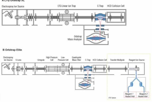
Figure 1.10. Schematics of the LTQ Orbitrap XL and Orbitrap Elite hybrid mass spectrometers. ... 49
Figure 1.11. Overview of quantitative proteomics workflows. ... 51
Figure 1.12. Analytical outcome of a proteomics experiment. ... 56
Figure 2.1. Workflow for large-scale quantitative proteomics analyses of RAW264.7 macrophages. ... 109
Figure 2.2. Large Scale membrane proteome analysis of resting and TNF-α activated macrophages. ... 110
Figure 2.3. Quantitative proteomics analysis of membrane proteins in TNF-α stimulated macrophages identified the overexpression of cPLA2. ... 112
Figure 2.4. Bioinformatics analyses of the membrane proteome from TNF-α activated macrophages reveals the downregulation of mitochondria proteins. ... 115
Figure 2.5. Changes in mitochondrial functions associated with TNF-α activated macrophages. ... 118
Figure 2.6. TNF-α induces mitophagy. ... 120
Figure 2.7. Influence of TNF-α on antigen presentation. ... 123
Figure 2.S1. Flow cytometry analysis and clone showing the highest fluorescence levels selected, amplified and used... 128
Figure 2.S2. Fractionation efficiency using immunoblots for several cellular markers. ... 129
Figure 2.S3. Reproducibility of peptide intensities across replicates. ... 130
Figure 2.S4. Comparison of three different fractionation techniques for quantitative membrane proteomics of RAW264.7 macrophages. ... 131
Figure 2.S5. MS/MS spectra of phosphopeptides. (CD-ROM). ... 131
Figure 2.S6. Scatter plots of abundance measurements for peptide ions identified in control and TNF-α stimulated extracts. ... 132
Figure 2.S7. Fold change measurements for citrate synthase and ATP synthase subunit b. ... 133
Figure 2.S9. Abundance of fluorescently-labeled Annexin A5 at the plasma membrane and 7-amino actinomycin D using flow cytometry. ... 134 Figure 2.S10. Lysosomal degradation activities when stained with Lysosensor. ... 135 Figure 2.S11. Integrated model of the TNF-α modulated functions favoring antigen MHC class I presentation. ... 136
Figure 3.1 Effect of DNA replication inhibitors on HSV1 protein expression. ... 156 Figure 3.2. Ubiquitylation of HSV1 proteins. ... 159 Figure 3.3. Phosphorylation of HSV1 proteins. ... 162 Figure 3.4. Phosphorylation motifs. ... 164 Figure 3.5. Abstract figure. ... 167 Figure 3.S1. Coverage of HSV1 structural proteins and proteins of different kinetic classes. ... 168
Figure 4.1. NEDA is regulated differently than macroautophagy and is
Atg5-independent. ... 181 Figure 4.2. NEDA contributes to the presentation of gB to MHC class I molecules. .. 183 Figure 4.3. NEDA contributes to MHC class I antigen presentation of a nuclear
envelope-resident viral antigen. ... 185 Figure 4.4. A novel autophagolysosome isolation method. ... 188 Figure 4.5. Quantitative proteomics analysis of APL extracts. ... 191 Figure 4.6. NEDA leads to the enrichment of gB on the APL. ... 193 Figure 4.7. Atg5-dependent transfer of a specific subset of proteins to the APL during ΔPP1α infection. ... 195 Figure 4.S1. Atg5 knockdown efficiency. ... 206
List of abbreviations
1D One dimensional
2D Two dimensional
3-MA 3-methyladenine APC Antigen presenting cell APL Autophagolysosome ATG Autophagy related genes AtgX Autophagy-related protein X
Baf Bafilomycin A1
Bcl B-cell lymphoma
CCCP Carbonyl cyanide m-chlorophenylhydrazone CD Cluster of differentiation
CH Cycloheximide
CID Collision-induced dissociation cPLA2 Cytosolic phospholipase A2 DDA Data-dependent acquisition DMSO Dimethylsulfoxide
DNA Deoxyribonucleic acid ER Endoplasmic reticulum
ERK Extracellular signal-regulated kinase ESI Electrospray ionization
ETD Electron transfer dissociation FDR False Discovery Rate
FP False positive FPR False positive rate
FT-ICR Fourier transform ion cyclotrone resonance GABARAP γ-amino butyric acid receptor-associated protein GELFREE Gel-eluted liquid fraction entrapment electrophoresis
gX Glycoprotein X HBV Hepatitis B virus
HCD Higher energy collisional induced dissociation HSV1 Herpes Simplex Virus type 1
ICP Infected cell protein IFN-γ Interferon-γ
IL Interleukin
IRGM Human immunity-related GTPase
JC-1 5,5’,6,6’-tetrachloro 1,1’,3,3’-teramethyl benzimidazoyl carbo cyanine iodide
LB-C Latexbead-compartment LC Liquid chromatography
LC3 Microtubule-associated protein 1A/1B-light chain 3 LIR LC3-interacting motif
LIT Linear ion trap
LTQ Linear trap quadrupole m/z Mass-to-charge ratio
MALDI Matrix-assisted laser desorption/ionization MAMP Microbial-associated pattern
MEF Mouse embryonic fibroblasts MHC Major histocompatibility complex MIIC MHC class II compartment MOI Multiplicity of infection
mRP-C18 Macroporous reversed phase-C18
MS Mass Spectrometry
MS/MS Tandem mass spectrometry mTORC1 Mammalian TOR complex 1
MudPIT Multidimensional protein identification technology NEDA Nuclear envelope derived autophagy
PAA Phosphonoacetic acid
PAGE Polyacrylamide gel electrophoresis PAS Phagophore assembly site
pi Post infection
PINK1 PTEN (phosphatase and tensin homolog)-induced putative protein kinase 1
PP1α Protein Phosphatase α ppm Parts per million
PSM Peptide-to-spectrum match PtdIns3K Phosphatidylinositol 3-kinase PTM Posttranslational modification pXY Protein encoded by HSV1 gene XY
rf Radio frequency
ROS Reactive oxygen species
RP Reversed phase
RSD Relative standard deviation SCX Strong cation exchange SD Standard deviation
SILAC Stable isotope labeling with amino acids in cell culture SQSTM1 Sequestosome
TAP Transporter associated with antigen processing TCL Total cell lysate
THX T helper type X TLR Toll-like receptor
TM Total membrane
TMD Transmembrane domain TNF-α Tumor necrosis factor-α ToF Time-of-flight
TOR Target of rapamycin
TRAP1 TNF receptor-associated protein 1 TSC Tuberous sclerosis complex UBL Ubiquitin like
UL Unique region long ULK Unc-51-like kinase
UPS Ubiquitin dependent proteasome system US Unique region short
Vps Vacuolar protein sorting
WT Wild Type
Acknowledgements
This thesis would never have been possible without the generous advice and support of many people.
I would like to express my deepest gratitude to Prof. Pierre Thibault, my PhD research director at the Institute for Research in Immunology and Cancer at Université de Montréal, who believed in my abilities and offered me this research position in his lab. I am very grateful for the challenge he presented to me in the shape of this project and for the opportunity to perform graduate studies in mass spectrometry-based proteomics. Pierre was always ready and patient to answer my questions concerning my work and listen to my problems. I thankfully acknowledge all his scientific support and advice on science and academic life in general. Thank you for being a great mentor and for making my stay at the IRIC very pleasant. I am very glad that I could be a member of your group. I would like also like to express my sincere gratitude to Prof. Michel Desjardins, my PhD research co-director, from the Department of Pathology and Cell Biology at Université de Montréal for the opportunity to be a part of his lab and to learn from his expertise. I am thankful for not only his willingness to share his knowledge, but also for always motivating me to explore and develop my research further and thus get better.
Thank you to both of you for encouraging my research and for allowing me to grow as a scientist, while working on a very exciting, interdisciplinary project. Your advice on both research as well as on my career have been precious. I also really appreciate that you made it possible for me to realize a research stay during my PhD in the group of Prof. Markus Wenk at the National University of Singapore, to whom I would also like to express my gratitude. This research stay was a great experience, which not only taught me a lot about lipidomics, but it also significantly expanded my horizons.
I am very much obliged to the past and present lab members of the Thibault and Desjardins labs for sharing their expertise, for their support and for a very friendly atmosphere throughout my thesis work. The privilege to work closely with them, was of great importance to my work. I want to thank Dr. Kerstin Radtke for all her expertise with the viral infection system and for all her advice and help in general. I would also like to thank Dr. Luc English for introducing me to all the concepts of autophagy and antigen presentation and for teaching me several techniques crucial for my PhD project. Also, I
am very grateful to Dr. Jonathan Boulais for sharing his knowledge and teaching me useful bioinformatics tools. He was always ready to answer all my questions, help me with the data analysis and discuss about science. Thank you very much also to Dr. Matthias Trost for sharing his knowledge and experimental skills and for introducing me to my PhD project at first. Thank you to Dr. Diana Matheoud, Angélique Bellemare-Pelletier, Christiane Rondeau, Dr. Francis McManus and Prof. Étienne Gagnon for help and support. I am also very grateful to Dr. Mathieu Courcelles as well as to Olivier Caron-Lizotte for all their help and patience with the analysis of my large proteomics datasets. I would like to especially thank Dr. Eric Bonneil for all his help and expertise with the mass spectrometry equipment as well as all his scientific advice and guidance, and particularly for making every day in the lab much more fun. I really enjoyed working with you. I would also like to thank Prof. Roger Lippé and his team, especially Johanne Duron, for all their expertise with the viral infection system and for making their equipment available to us.
I would like to express my special appreciation and thanks to Dr. Sonja Hess from California Institute of Technology, who always supported me and gave me valuable advice and who first introduced me to the field of proteomics during my diploma thesis research, which continued to fascinate me ever since.
Finally, I would like to thank the Chemistry department as well the FESP for supporting my application to the Vanier Canada Graduate Scholarship and of course the Natural Sciences and Engineering Research Council (NSERC) for selecting me as a Vanier scholar. It was an honor and a privilege to benefit society through my research as a Vanier scholar. This award not only opened up opportunities for my research, but it also was a constant motivation to fulfill my goals.
Above all, I´m very grateful to my parents Werner and Brunhilde, and my loving family for always being there for me and for accompanying me during this journey from afar. Thank you for always supporting my academic endeavors and for always encouraging me to strive towards my goals and make my dreams come true.
Most of all, I would like to thank Vincent for his relentless support, love, encouragement and understanding. Words cannot express how grateful I am. Thank you for always being there for me and for always staying on my side, especially in hard times.
You miss 100% of the shots you don’t take.
1.1. The immune system
The immune system is composed of structures and mechanisms that allow an organism to discriminate between ”self” and ”non-self” 1. These defense mechanisms provide the body with protection against foreign pathogens such as bacteria, viruses, parasites or fungi and it also prevents autoreactive damage causing autoimmune disease, an immunosurveillance mechanisms referred to as self tolerance2. Furthermore, the immune system is also able to recognize an “altered- self" as in the case of cancer cells. The immune system is broadly divided into two main systems composed of innate immunity (or nonspecific immunity) and acquired/adaptive immunity (or specific immunity). Although these two subsystems were often seen as completely separate, the current opinion is that innate immunity and adaptive immunity systems are functionally interconnected and complement each other to contain infectious diseases3.
1.1.1 Innate immunity
The innate immune system represents an excellent and rapid first line of defense against microbes. It is composed of physical barriers, antimicrobial substances, inflammatory mechanisms, complement system, leukocytes and fever, which are very effective against a variety of pathogens. The innate immune system recognizes molecular patterns common to various pathogens via a restricted number of receptors on phagocytic cells such as Toll-like receptors and is hence not specific2. This initial response is very rapid (in a time frame of hours) as these pathogen receptors are widely expressed on many cells. However, due to the lack of specificity the system rapidly fails when attacked by a pathogen that has evolved to evade the defense mechanisms. The presence of a constant synergizing interaction between innate and adaptive immunity allows the rapid establishment of specific defense mechanisms targeted at the pathogen. For example, phagocytosis and autophagy, two non-specific defense mechanisms for degradation of pathogens that are intra-or extracellular, are now recognized as part of the specific immune response1, 4.
1.1.2. Adaptive immunity
Adaptive immunity, which has been acquired later in evolution, comprises a limited number of cells with very specific recognition capabilities for any pathogen. The adaptive immune system uses two types of responses, the cell-mediated response and the humoral response. For each of these responses, specialized cells have evolved that express antigen-specific receptors on their surface to exclusively recognize antigens associated with different pathogens2. The antigen receptor, if linked with a specific antigen, generates a clonal expansion of the latter, necessary to obtain enough immune cells to elucidate an efficient immune response. The adaptive immune response is hence slower (in a timeframe of days). In the case of cell-mediated immunity, the main actors are T lymphocytes, previously activated by professional antigen-presenting cells (APCs). To provide humoral immunity and perform the role of APCs, B cells actively produce antibodies after activation by antigen interaction. In addition to the development of effector cells, the adaptive response also enables the development of memory cells, thus providing a more rapid immune response if re-infected with the same pathogen4, a mechanism used in vaccination strategies3.
1.1.2.1. T cells – important effector cells of adaptive immunity
Adaptive immune cells that cause cell-mediated immunity originate from pluripotent hematopoietic stem cells in the bone marrow. These pluripotent cells generate multipotent stem cells that give rise to lymphoid and myeloid precursors. These precursors generate all cells comprising the immune system; T cells (derived from lymphoid precursors) and monocytes/macrophages and dendritic cells (formed from myeloid precursors)5.
Bone marrow derived T cells undergo a ''selection'' in the thymus. Thymic selection comprises positive and negative selection4. During positive selection only T cells able to bind major histocompatibility (MHC) molecules expressed by the epithelial cells of the thymus cortex with sufficient affinity survive, while others are eliminated by apoptosis6. Thereafter, the cells having passed the positive selection, migrate to the thymus to undergo negative selection (or clonal deletion). Here the lack of affinity with peptide-MHC complexes ensures the survival of lymphocytes, since T cells that interact too strongly with “self”-peptide-MHC complexes are eliminated7. This last step is crucial to avoid auto-reactive lymphocytes causing autoimmune diseases and plays an important role in
central tolerance8. After the thymic selection, “naïve” T cells enter the blood and lymphatic circulation, where they can get activated as “effector” T lymphocytes and / or "memory" T lymphocytes following recognition of specific antigens. The differentiation of naive T cells into effector T lymphocytes is executed by professional antigen presenting cells, including dendritic cells and macrophages, regulated by co-stimulatory molecules and cytokines, and allows T effector cells to eliminate infected cells4.
There are different subgroups of effector T cells. Cluster of differentiation (CD) 4+ T lymphocytes play a central role in the development of the humoral response and cell mediated immune response. CD4+ T cells can differentiate into type I T helper (TH1) cells or type II T helper (TH2) cells, following binding of a peptide antigen on MHC class II complex from macrophages or dendritic cells and T cell receptor (TCR). TH1 and TH2 cells differ based on their subsequent production of cytokines. TH1 cells modulate cell-mediated immunity by secreting cytokines such as interferon and interleukin-2 (IL-2), whereas TH2 cells modulate humoral immunity by secreting IL-4, IL-10 and IL-139. Naïve CD8 + T cells differentiate into cytotoxic T lymphocytes (CTL) following activation through the interaction of their TCR with peptide-MHC class I and strong co-stimulatory activity on the surface of an APC. The co-stimulatory activity is promoted by the cytokine production of CD4+ T cells, resulting in the increased expression of co-stimulatory molecules. CD8+ T lymphocytes are particularly effective in eliminating virus-infected cells10. CD8+ T cells can destroy infected cells directly via the release of serine proteases called granzymes and the protein perforin11, which allows granzymes to enter into infected cells.
1.2. Macrophages – key players in innate and adaptive
immunity
Macrophages are innate immune cells with well-established roles in the primary response to pathogens, but also in the coordination of the adaptive immune response, inflammation, tissue homeostasis, and repair. Macrophages were initially recognized by Elli Metchnikoff as phagocytic cells responsible for pathogen elimination and housekeeping functions in a wide range of organisms, from invertebrates to vertebrates.
Ellie Metchnikoff, who won the Nobel Prize in Physiology or Medicine in 1908 for his description of phagocytosis, proposed that the key to immunity was to “stimulate the phagocytes”12.
Macrophages evolved in simple multicellular organisms to phagocyte and clear dying cells during development, and to protect the host through innate immunity, both as resident tissue macrophages and monocyte-derived cells mobilized during inflammation. Macrophages are very plastic and can change their functional phenotype depending on the environmental stimuli they receive. These cells have a vital role in protecting the host through their ability to kill pathogens and instruct other immune cells, but also contribute to the pathogenesis of inflammatory and degenerative diseases.
Macrophages are present in essentially all tissues. They differentiate from circulating peripheral-blood mononuclear cells (PBMCs), which migrate into tissue in the steady state or in response to inflammation13. PBMCs develop from a common myeloid progenitor cell in the bone marrow. This myeloid progenitor cell is the precursor of many different cell types, including, eosinophils, basophils, neutrophils, macrophages, dendritic cells and mast cells. During monocyte development, myeloid progenitor cells (termed granulocyte/macrophage colony-forming units) successively generate monoblasts, pro-monocytes and finally pro-monocytes, which are sent from the bone marrow into the bloodstream13. Monocytes travel from the blood into tissue to form long-lived tissue- specific macrophages of the bone (osteoclasts), alveoli, central nervous system (microglial cells), gastrointestinal tract, liver (Kupffer cells), connective tissue (histiocytes), skin (Langerhans cells), peritoneum and spleen. Resident macrophage populations in different organs adjust to their local microenvironment. The stimuli that regulate tissue-specific phenotypes of macrophages include surface and secretory products of neighboring cells and extracellular matrix. Macrophages can respond to endogenous cues that are quickly generated following infection or injury. These early stimuli are typically produced by innate immune cells and can result in a distinct, though usually transient, effect on the physiology of macrophages14. Macrophages can also respond to stimuli produced by antigen-specific immune cells. These signals are more specific and prolonged than innate immune stimuli and generally exert longer term alterations in macrophages15.
Macrophages are professional phagocytes that internalize large particles like dead cells or pathogens, and play crucial roles in immunity by the initiation of microbicidal
mechanisms as a part of innate immunity14. In mammals, the internalization of microorganisms at sites of infection by macrophages proceeds via the internalization of pathogens in phagosomes, where they are killed and degraded by hydrolytic enzymes. Phagosomes obtained these functional properties relatively recently during the evolution of multicellular organisms through the acquisition of molecular machineries that transformed phagosomes from a lytic vacuole into an organelle fully competent for antigen presentation16. Indeed, the processing of proteins from internalized pathogens to derive antigens for presentation at the cell surface on major histocompatibility complex (MHC) class I and class II molecules is a key mechanism of adaptive immunity to initiate specific defense mechanisms17. Importantly, the initiation of an efficient immune response depends on the capability of APCs such as macrophages to display peptide MHC complexes on their cell surface.
1.2.1. Macrophage activation
Cytokines produced by immune cells can activate macrophages to convey different responses. Classically macrophages are activated in response to interferon-γ (IFN-γ), which can be produced during an adaptive immune response by T helper 1 (TH1) cells or CD8+ T cells or during an innate immune response by natural killer cells, and tumor necrosis factor (TNF), which is produced by antigen presenting cells (Figure 1.1). The term classically activated has been used to refer to the effector macrophages that are produced during cell-mediated immune responses14, 15, 18.
IFN-γ activation results in the transcriptional regulation of several genes including nitric oxide synthase-2 and phagocyte oxidase that are associated with reactive oxygen species (ROS) production. ROS are an established feature of the macrophage’s microbicidal activity and improve the killing abilities of macrophages19. IFN-γ transforms resting macrophages into potent cells with increased antigen presenting capacity, increased synthesis of proinflammatory cytokines and toxic mediators, and enhanced complement mediated phagocytosis. Thus, macrophages obtain the capacity to kill bacteria, especially intracellular pathogens. IFN-γ also mediates phagosome maturation and antigen loading on MHC class I and class II molecules20-22 and regulates the upregulation of MHC class I as well as co-stimulatory molecules on macrophages14.
Classically activated macrophages secrete pro-inflammatory cytokines such as 1, IL-6 and IL-23, a hallmark of the macrophage’s microbicidal activity14. Proinflammatory cytokines produced by classically activated macrophages are an important feature of the host defense, but they can also cause extensive damage to the host. The expansion of TH17 cells has been linked to IL-1, IL-6 and IL-2323, 24. These cells produce IL-17, a cytokine that can contribute to inflammatory autoimmune diseases14. Interestingly, macrophages activated through Toll-like receptor (TLR) ligand stimulation produce tumor necrosis factor α (TNF-α), another important cytokine that synergizes with INF-γ to enhance macrophage activation. Macrophages can also be stimulated exogenously from the secretion of this cytokine by antigen presenting cells. TNF-α stimulation is particularly important in Leishmania infections as macrophages stimulated with IFN-γ alone are less efficient at killing this parasite due to lack of TLR ligands expression. TNF-α also has a central role in inflammatory cell activation and recruitment, and is associated with the development of various chronic inflammatory diseases such as Crohn’s disease25. Taken together, classically activated macrophages are products of a cell-mediated immune response. They can also be transiently generated in response to innate stimuli following viral infections or stress. Some pathogens have acquired the ability to interfere with IFN-γ signaling and inhibit efficient macrophage activation. Classically activated macrophages are vital components of the host defense, but their activation must be tightly controlled, because the cytokines and mediators they produce can lead to host tissue damage. Indeed classically activated macrophages are key mediators of the immunopathology that occurs during several autoimmune diseases, including rheumatoid arthritis26 and inflammatory bowel disease27.
Similarly to classically activated macrophages, wound healing or alternatively activated macrophages can arise in response to innate or adaptive signals (Figure 1.1). Macrophages are alternatively activated by IL-4 and IL-1328, which trigger a distinct phenotype that accounts for allergic, cellular and humoral responses to parasitic and extracellular pathogens28. Basophils and mast cells are important early sources of innate IL-4 production, while other granulocytes might also contribute. It is well established that IL-4 and IL-13 are associated with TH2 type responses. IL-4- and IL-13 can promote the development of wound-healing macrophages, though this activation yields poor antigen-presenting cells that are less efficient at producing ROS or at clearing intra cellular pathogens than classically activated macrophages29.
Figure 1.1. Cytokine mediated macrophage activation.
Classically activated macrophages are generated from IFN-γ and TNF-α stimulation. IL-4 stimulation results in wound-healing (alternatively) activated macrophages. Regulatory macrophages are formed in response to various stimuli including glucocorticoids and IL-10. From reference14.
Due to the role of TH1-derived interferon-γ in cell-mediated immunity to intracellular infection and of IL-4 (TH2) in extracellular parasitic infection, macrophages were designated analogously as M1 (classic) and M2 (alternative) macrophages. This concept was recently extended to a wider range of immune modulatory mediators and functions30.
In addition to classically and alternatively activated macrophages, regulatory macrophages develop in response to various stimuli, including immune complexes, glucocorticoids, apoptotic cells, prostaglandins, G-protein coupled receptor ligands or IL-10 (Figure 1.1). This innate or acquired deactivation of macrophages causes the production of IL-10, which suppresses immune responses and is the most important and reliable characteristic of regulatory macrophages14.
Taken together, both innate and adaptive signals can influence macrophage physiology and these alteration allow macrophages to participate in homeostatic processes such as tissue remodeling and wound healing as well as in host defense. However, each of these alterations can have potentially dangerous consequences and must be appropriately regulated.
1.3. Antigen processing and presentation
The efficiency of T cell-mediated adaptive immune responses depends on the ability of antigen presenting cells, such as macrophages, to display peptide and MHC complexes on their surface31. MHC molecules are divided into MHC class I and MHC class II, where the former presents peptides at the cell surface to CD8+ and the latter to CD4+ T cells. MHC class I and II also differ by the structure of the groove where antigenic peptides are bound. The antigen-binding groove of MHC class I molecules is closed at each end, resulting in the binding of peptides with a defined length (8-10 amino acids)32. In contrast, the antigen binding groove of MHC class II molecules has open ends, permitting the loading of peptides of a more variable length (13-25 amino acids).33.
While MHC I complexes are expressed ubiquitously by all nucleated cells and recognized by CD8+ T cells, MHC II molecules are limited to professional APCs such as B cells, macrophages and dendritic cells, and are then recognized by CD4+ T cells.34 However, MHC class II expression can be induced by IFN-γ and other stimuli in non-APCs. Non-APCs can express MHC class II molecules in the absence of co-stimulatory molecules to maintain peripheral tolerance34.
Membrane trafficking plays a key role in both endogenous and exogenous antigen processing and presentation (see Figure 1.2). Initially, two segregated pathways of antigen presentation were proposed. Endogenous antigens, including viral proteins
synthesized by infected cells, are degraded in the cytoplasm by the proteasome. The resulting peptides are translocated into the endoplasmic reticulum (ER) lumen by the MHC locus-encoded peptide transporter associated with antigen processing (TAP), where they can be further trimmed and are loaded onto nascent MHC class I molecules35. The constitutive 26S proteasome is composed of a 20S core barrel that has protease activity36 and two 19S caps. It generates the bulk peptides for MHC class I molecules. Two alternative proteasomes exist: the immunoproteasome, which is expressed by many immune cells and the thymus specific proteasome, which is expressed in thymic epithelial cells37. Immune cell-specific variants of the proteolytic core are incorporated into the 20S barrel altering the degradation pattern of the proteasome38.
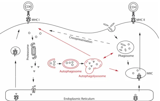
Figure 1.2. MHC class I and MHC class II antigen processing and presentation pathways. Endogenous antigens are degraded in the cytoplasm by the proteasome. The resulting peptides are translocated into the endoplasmic reticulum (ER), where they are loaded on MHC class I molecules and transported to the cell surface to be presented to CD8+ T cells. Exogenous antigens are internalized by endocytosis or phagocytosis, processed by hydrolases, and then loaded onto MHC class II molecules before translocation to the cell surface and presentation to CD4+ T cells. Exogenous antigens can also be presented on MHC class I molecules, a process referred to as cross-presentation. Autophagy contributes to endogenous and exogenous MHC class II antigen processing and presentation by delivering endogenous antigens to MHC class II-containing compartments (MIICs) for lysosomal antigen processing and MHC class II loading of antigens. Autophagy can also contribute to the presentation of endogenous antigens by MHC class I molecules. Endogenous antigens may be transported from the autophagosome to the cytosol to enter the classical pathway, or peptides may be loaded onto MHC class I molecules in the autophagolysosome compartment. Adapted from reference39.
In the ER, the MHC class I heterodimer is assembled from a polymorphic heavy chain and a light chain called β2-microglobulin (β2m). In order to be stable a peptide in the MHC I binding groove is required. Without peptides, MHC class I molecules are stabilized by ER chaperone proteins such as calreticulin, ERp57, protein disulfide isomerase and the chaperone tapasin. Tapasin interacts with TAP, thus coupling peptide translocation into the ER with peptide delivery to MHC class I molecules. The complex of TAP, tapasin, MHC class I, ERp57 and calreticulin is known as the peptide loading complex (PLC)34. Peptides may bind to MHC class I molecules directly or may need further trimming by ER aminopeptidase associated with antigen processing (ERAAP). When peptides bind to MHC class I molecules, the chaperones are released and fully assembled peptide-MHC class I complexes leave the ER and are transported to the cell surface through the secretory pathway (Figure 1.2) for presentation to CD8+ T cells35, 40. Peptides and MHC class I molecules that fail to bind are transported back into the cytosol and degraded by the ER-associated protein degradation system (ERAD)41.
In contrast, exogenous antigens are internalized by endocytosis or phagocytosis, processed by hydrolases in lytic endovacuolar compartments and the resulting peptides loaded onto MHC class II molecules. The transmembrane α – and β chains of MHC class II are assembled in the ER and associate with the invariant chain (Ii). The resulting Ii-MHC class II complex is transported to a late endosomal compartment termed the Ii-MHC class II compartment (MIIC). Here Ii is cleaved, leaving a residual class II associated Ii peptide (CLIP) in the peptide binding groove of the MHC class II heterodimer. In the MIIC, MHC class II molecules require HLA DM (H2-M in mice) to enable the exchange of the CLIP fragment for a specific peptide derived from the degradation of exogenous proteins in the endosomal pathway. MHC class II molecules are then translocated to the cell surface for presentation of their cargo to CD4+ T cells (Figure 1.2).34
Although initially thought to be strictly segregated, these two pathways were revisited to account for the cells’ ability to present exogenous antigens on MHC class I molecules, a process referred to as cross-presentation (Figure 1.2)42. Different models have been proposed for the cross-presentation pathway. In the phagosome to cytosol model, antigens internalized by phagocytosis are transported across the phagosome membrane into the cytosol, where they can be further processed by the proteasome and enter the classical processing pathway for MHC class I presentation43. In the phagosome to cytosol to phagosome model, peptides are loaded on MHC class I molecules in the phagosome
itself. This model involves two transmembrane transport steps of the phagocytosed antigens44, 45. The first step transports peptides into the cytoplasm where antigens are further trimmed by the proteasome. The second step enables the re-entry of the processed antigens in the phagosome lumen through TAP44-46. It was recently shown that antigenic peptides that bind to MHC class I molecules can also be generated by proteases in the lumen of vacuolar organelles hence avoiding the detour to the proteasome in the cytosol. These peptides are directly loaded on the MHC class I complexes in the ”vacuolar pathway”47. While the contribution of the endo-phagocytic pathway to the processing of exogenous antigen for MHC class I and II antigen presentation has been recognized for several years, it has been shown only very recently that endogenous and exogenous antigens can also be taken up by autophagosomes and presented on both MHC class I and MHC class II molecules, thus making autophagy an emerging immunological paradigm (see chapter 1.4.6.2 for more details)48.
1.4.
Autophagy
A constant balance between biosynthetic and catabolic processes is crucial for cellular homeostasis. Eukaryotic cells primarily use two distinct mechanisms for large-scale degradation, the ubiquitin dependent proteasome system (UPS) and autophagy. Proteasome-mediated degradation requires previous ubiquitylation of the cargo, which is then recognized by ubiquitin receptors directing it to the 26S proteasome. The UPS rapidly eliminates proteins to regulate many cellular processes, including signal transduction, cell division and gene expression and is considered to be highly selective. Conversely, autophagy, a highly conserved lysosomal degradation pathway, can degrade almost any cargo including whole organelles. Half a century ago, Christian de Duve coined the term “autophagy” (literally, “self-eating”) to describe a process whereby cells digest their cytoplasmic materials within lysosomes49, 50. This was based on his discovery of lysosomes in 195551, for which he won the Nobel Prize in Physiology or Medicine in 197452.
Half a decade of research has helped to understand that there is not one autophagic pathway, but at least three primary types of autophagy: macroautophagy, microautophagy and chaperone-mediated autophagy (CMA), which share a common
destiny of lysosomal degradation, but are mechanistically different from one another53, 54. Both micro and macroautophagy can be selective or non-selective. During microautophagy, lysosomes engulf cytoplasmic materials by inward invagination of the lysosomal membrane. Chaperone-mediated autophagy, mediated by the chaperone hsc70, co-chaperones, and the lysosomal-associated membrane protein type 2A (LAMP2A), transports proteins across the lysosomal membrane without membrane rearrangements55, 56. During classical macroautophagy a phagophore is formed at the phagophore assembly site (PAS). The phagophore subsequently engulfes cytosolic components such as organelles and packages them in a two-membrane–bound compartment called the autophagosome. The autophagosome subsequently fuses with the lysosome to form the autophagolysosome in which the content is completely degraded and the resulting macromolecules are released back into the cytosol for reuse (Figure 1.3)56.
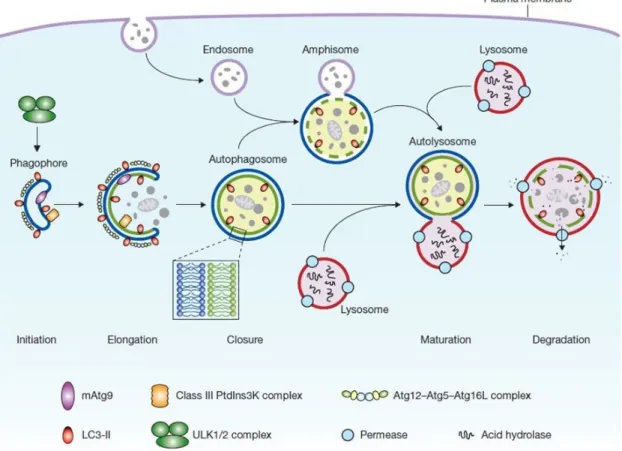
Figure 1.3. Autophagy pathway.
Mammalian autophagy proceeds through a series of steps. Upon several, yet unknown signals the phagophore is formed. The phagophore subsequently engulfes cytosolic components such as organelles and packages them in a double-membrane autophagosome. Autophagosome maturation proceeds via fusion with an endosome and/or lysosome, followed by breakdown and degradation of the autophagosome inner membrane and cargo, and recycling of the resulting macromolecules. The core molecular machinery is also shown. Adapted from reference56.
1.4.1. Molecular regulation of autophagy and the autophagy pathway
Although autophagy was first identified in mammalian cells approximately half a century ago, the transition from morphology to molecular mechanisms only started in the last decade mainly based on the discovery of autophagy related (ATG) genes initially identified in yeast57. Mammalian homologs were found soon after58. Among these ATG genes, one subgroup comprised of approximately 18 genes is common to the various types of autophagy including non-selective macroautophagy and mitophagy. The corresponding gene products of this subgroup are crucial for autophagosome formation and are thus referred to as the core autophagy machinery59, 60. There are four different functional subgroups: (1) the Atg1/unc-51-like kinase (ULK) complex (Atg1, Atg11, Atg13, Atg13, Atg17, Atg29 and Atg31), which plays a role in the initial induction of autophagosome formation; (2) the transmembrane protein Atg9 and associated proteins (Atg2, Atg9 and Atg18), which regulates membrane recruitment to the elongating phagophore after the association of the Atg1 complex at the PAS; (3) the class II phosphatidylinositol 3-kinase (PtdIns3K) complex (Vps (vacuolar protein sorting) 34, Vps15, Vps30/Atg6, and Atg14), which plays a role during vesicle nucleation and is implicated in the targeting of phosphatidylinositol-3-phosphate (PtdIns3P)-binding proteins to the PAS; and (4) two ubiquitin like (UBL) conjugation systems: the Atg12 (Atg5, Atg7, Atg10, Atg12 and Atg16) and Atg8/LC3 (Atg3, Atg4,Atg7 and Atg8) conjugation system, which function in vesicle expansion61, 62.
Mammalian autophagy is regulated by the serine/threonine protein kinases ULK1 and ULK2 (mammalian homologues of the yeast autophagy-related protein (Atg1)) and the lipid kinase activity of PtIns3K Vps34 (Vps-34 in yeast) associated to Beclin 1 (mammalian orthologue of yeast Atg6) and Atg14-like protein (Atg14L). ULK1, ULK2 and the Vps34-Beclin 1-ATG14L complex integrate upstream signaling pathways (see chapter 1.4.4.) and activate the downstream Atg conjugation cascade62, 63. Two ubiquitin-like systems regulate autophagosome formation and closure, conjugating three UBL proteins (Atg12, Atg5 and Atg8) to target proteins or membranes64. Atg7 and Atg10 exert the conjugation of Atg12 to Atg5 respectively, which forms a complex with Atg16. A complex of Atg12-Atg5-Atg16 together with Atg3 controls conjugation of Atg8 (processed by Atg4) to phosphatidylethanolamine (PE) on the isolation membrane64. Although these two UBL systems are dependent on each other, they have distinct functions. The Atg12-Atg5-Atg16 complex localizes transiently to the outer membrane of the PAS. However,