Université de Montréal
Perception des visages auprès des adolescents et des adultes autistes
par Karine Morin
Département de psychoéducation, Université de Montréal Faculté des arts et des sciences
Mémoire présenté à la Faculté des arts et des sciences en vue de l’obtention du grade de Maitrise es science
En psychoéducation Option mémoire et stages
Juin, 2012
Faculté des arts et des sciences
Ce mémoire intitulé:
Perception des visages auprès des adolescents et des adultes autistes
Présentée par: Karine Morin
a été évalué par un jury composé des personnes suivantes: Marc Lanovaz président-rapporteur Linda S. Pagani Directeur de recherche Armando Bertone Codirecteur ………. membre du jury Dave Saint-Amour examinateur externe ………. représentant du doyen
Résumé
La perception faciale est la mesure visuelle la plus commune pour mesurer les habiletés sociales chez les personnes autistes. Lorsque cette habileté est atypique, la nature de son origine est souvent contentieuse. Une hypothèse suggère qu’une analyse visuelle orientée localement dans l’autisme affecterait leur performance sur les tâches faciales lorsque l’analyse configurale est nécessaire. Objectif. Évaluer cette hypothèse en mesurant la discrimination de l’identité faciale avec des visages synthétiques présentés avec ou sans changement de point de vue, afin de minimiser ou non l’accès aux attributs locaux. Méthodes. Cinquante-huit participants, autistes et neurotypiques, appariés selon leur quotient intellectuel, genre et âge, ont accompli une tâche de discrimination de l’identité faciale similaire à celle de Habak, Wilkinson et Wilson (2008). Les stimuli étaient des visages synthétiques, présentés de face ou de profil, simplifiés et écologiquement validés. Les seuils de discrimination de l’identité faciale, pourcentage minimum de changement dans la géométrie faciale à 75 % de réponses correctes, ont été obtenus en utilisant un système à deux choix alternatifs. Résultats. Les analyses montraient une interaction significative entre les groupes et conditions, avec une différence significative entre les groupes pour la condition avec changement de point de vue, où la performance du groupe autiste était inférieure comparativement au groupe neurotypique. Discussion. La performance inférieure pour la condition avec changement de point de vue suggère que la discrimination de l’identité des visages est plus difficile chez les individus autistes lorsque l’accès aux éléments locaux est minimisé et lorsqu’une analyse globale des informations est nécessaire.
Abstract
Face perception is the most commonly used visual metric of social abilities in autism. When found to be atypical, the nature of its origin is often contentious. One hypothesis proposes that locally-oriented visual analysis, which characterizes persons with autism, influences performance on most face tasks where configural analysis is optimal. Objective. We evaluate this hypothesis by assessing face identity discrimination with synthetic faces presented with and without changes in viewpoint, with the former condition minimizing access to local face attributes used for identity discrimination. Methods. Fifty eight participants, with and without autism, matched for global intellectual quotient, age, and gender, were asked to perform a face identity discrimination task similar to that of Habak, Wilkinson, and Wilson (2008). Stimuli were frontal and side viewpoint of simplified and ecologically validated synthetic faces. Face identity discrimination thresholds, defined by the minimum percentage of change in face geometry at 75% correct performance, were obtained using a two-alternative, temporal forced choice match-to-sample paradigm. Results. Analyses revealed a significant interaction effect between groups and conditions, with significant group differences found only for the viewpoint change condition, where performance of the autism group was significantly decreased compared to that of neurotypical participants. Discussion. The selective decrease in autism performance for the viewpoint change condition suggests that face identity discrimination in autism is more difficult when (i) access to local cues are minimized, and (ii) an increased dependence on integrative analysis is introduced to the face task used.
Table des matières
Introduction générale………..1
Position du problème……….2
Relevé de littérature………...3
L’autisme………..3
La perception des visages……….4
Développement de la perception des visages ………..………5
Perception visuelle locale et globale……….6
Importance de la recherche dans l’avancement des connaissances dans l’autisme..8
Spécification sur la méthode………....10
Instruments de mesure utilisé dans cette étude pour le diagnostic d’autisme…….10
Instruments de mesure pour les variables contrôles………10
Quotient intellectuel………...10
Acuité visuelle………....11
Paradigme………..……..11
Méthode des stimuli constants………12
Article: Atypical face perception in autism: A point of view?...……….14
Abstract………15
Introduction………..17
Methods………...22
Participants……….….22
Apparatus, & Stimuli………...23
Results……….25
Discussion………27
Conclusion………..30
Références de l’article..………32
Conclusion générale……….42
Interventions au niveau du développement de la perception visuelle globale……….45
Impact sur la vie des personnes autistes………..47
Adapter l’environnement pour miser sur les forces de personnes autistes…………..48
Forces et limites de la présente étude………..50
La liste des figures
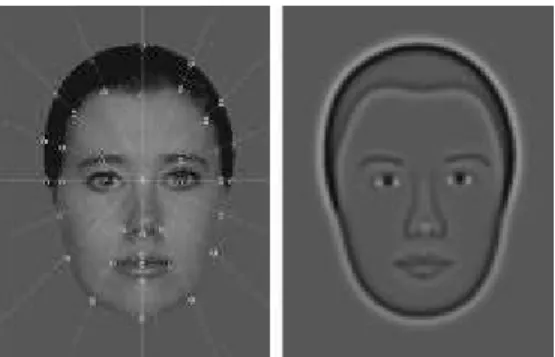
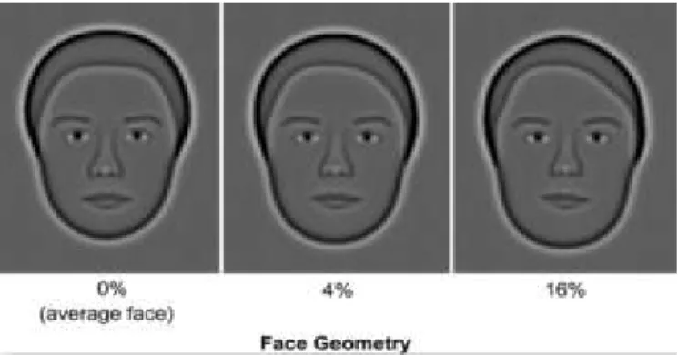
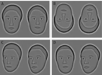
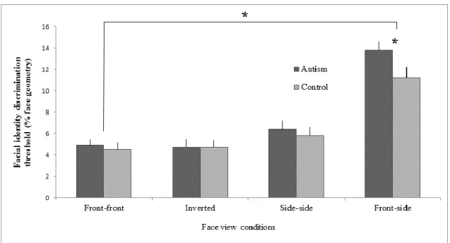
Figure 1: An original digital photo using 37 measurements and its synthetic face.………...35 Figure 2: Faces with increasing geometrical change (in % face geometry change) from the average face. ………..36 Figure 3: The four faceview conditions assessed………...37 Figure 4: Mean identity discrimination thresholds (% change face geometry) as a function of face view condition for autism and neurotypical groups. Error bars depict the standard error of the mean.…...….38
La liste des tableaux
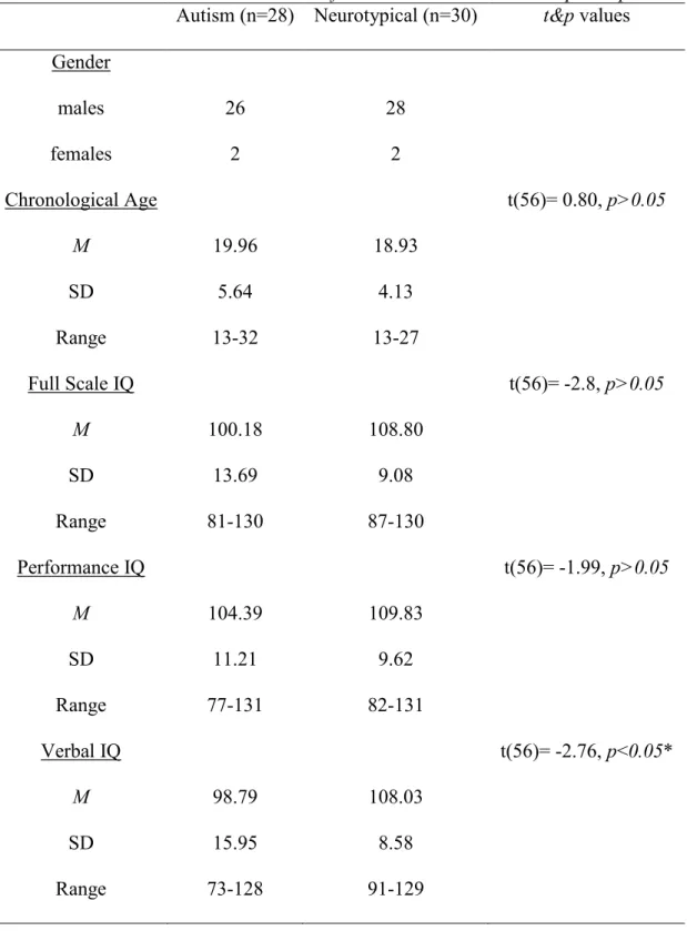
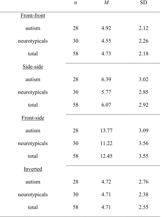
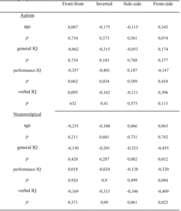
Tableau 1: Means and Standard Deviations for Variables Used to Match participants………..39 Tableau 2: Means and standard deviations for the facial identity discrimination threshold for the four conditions………....40 Tableau 3: Correlation between age, intellectual quotient and performance on condition tasks for both group………...41
ANNEXES
ANNEXE 1: Éthique………xi ANNEXE 2: Formulaires de consentement………..xiv ANNEXE 3: Curriculum Vitae, cheminement scolaire et professionnel………..xxi
La liste des sigles et la liste des abréviations ADI-R - Autism Diagnosis Interview-Revised
ADOS - Autism Diagnosis Observation Schedule AFF - Aire fusiforme faciale
ASD - Autism spectrum disorder
CIE - Commission Internationnale de l’Éclairage QI/IQ -Quotient intellectuel
WAIS-III - Wechsler Adult Intelligence Scale III WISC-IV - Wechsler Intelligence Scale for Children IV
Position du problème
En général, les personnes autistes présentent une perception atypique des visages. Un tel déficit dans l’habileté à accéder à l’information faciale serait une manifestation possible de leurs problèmes de socialisation et de communication (Schultz, 2005). Par contre, l’origine de cette difficulté est contentieuse (Jemel, Mottron & Dawson, 2006). Parmi les diverses recherches sur le domaine, une hypothèse énonce que la complexité des visages plutôt que leurs caractéristiques sociales serait à l’origine du problème (origine non-sociale), car un manque au niveau de l’intégration des informations chez les personnes autistes, relié ou non à des indicateurs sociaux, pourrait se manifester lors du traitement des visages (Davies, Bishop, Manstead & Tantam, 1994; Frith, 1989). Ceci pourrait possiblement affecter l'architecture neuronale impliquée dans la perception d’objets complexes, dont l’aire fusiforme faciale (AFF), une aire particulièrement impliquée dans la perception des visages. Par contre, cette hypothèse demeure incertaine. Afin de tester l’hypothèse d’une origine non sociale, notre étude a utilisé un paradigme de discrimination de l’identité faciale qui impliquait des stimuli variant en fonction de leur niveau d’intégration visuelle, c’est-à-dire en fonction de leur complexité, afin d’être identifiés efficacement. En effet, les divers stimuli créaient un biais vers l’analyse locale ou vers l’analyse globale des informations disponibles. Il était attendu qu’une performance intergroupe différentielle, en fonction de cette manipulation biaisée vers une analyse locale ou globale, appuiera une origine non sociale en ce qui concerne le traitement des visages chez les personnes autistes.
Relevé de littérature L’autisme
L’autisme est un trouble du développement d’origine neurobiochimique qui influence la façon dont la personne interagit avec son milieu. Ce diagnostic a été décrit pour la première fois par Kanner (1943). Selon le DSM-IV-TR (American Psychological Association [APA], 2000), les symptômes se manifestent dès un jeune âge et ils se caractérisent par une perturbation au niveau de la communication, verbale et non verbale, au niveau des interactions sociales et par la présence de comportements stéréotypés. Les perturbations sociales se traduisent par des comportements atypiques variant considérablement d’une personne à une autre. En effet, elles peuvent se manifester par la présence de pleurs parfois incontrôlables, une indifférence aux contacts physiques, l’évitement de contacts physiques, la présence de réaction excessive et l’apparence d’indifférence chez la personne. Un autre trait de cette perturbation sociale est l’absence de comportements d’anticipation qui représente souvent le premier signe qui alerte les parents, car ces comportements font habituellement leurs apparitions dès un jeune âge. Par exemple, un enfant a tendance à tendre les bras lorsque son parent s’apprête à le prendre. Certains enfants autistes ne présentent pas cette caractéristique comportementale. En ce qui concerne les difficultés au niveau de la communication, elles apparaissent vers l’âge de 2 à 5 ans et elles touchent le registre verbal et non-verbal. Ces perturbations pourraient se traduire par une difficulté à comprendre et à utiliser les signes ainsi que les codes sociaux, une expression gestuelle déficitaire ou absente (ex. pointer du doigt pour s’exprimer ou demander quelque chose à quelqu’un), une absence de gestes de sollicitation aux jeux et une absence de jeux de mimes (ex. jeux de cache-cache ou faire semblant). Les personnes autistes peuvent aussi présenter
des difficultés au niveau de l’interprétation des émotions, au niveau de l’expression verbale et ils peuvent répéter mot à mot des longs passages verbaux entendus dans le passé. Par contre, il est intéressant de noter que la compréhension de gestes instrumentaux ne semble pas affectée. Un exemple de geste instrumental est de mettre le doigt devant la bouche pour demander le silence. Pour conclure, souvent les personnes autistes présentent des déficits chroniques en ce qui concerne le traitement de l’information sociale et émotionnelle. Ils peuvent aussi présenter des comportements stéréotypés tels que des fixations persistantes sur des parties d’objet, des comportements répétitifs prolongés et de l’auto-stimulation. Les difficultés sociales des personnes autistes ont fait l’objet de nombreuses études empiriques afin d’avoir une meilleure idée de l’origine de ces difficultés. Par contre, cette origine demeure incertaine.
La perception des visages
Le visage est un des stimuli visuels les plus importants, et ce, dès les premières heures de vie des humains (Golstein, 1983). Le visage fournit un contenu riche en information sociale et il se différencie des autres stimuli de l’environnement par sa fréquence d’apparition. Compte tenu de leurs difficultés sociales, il n’est pas étonnant d’entendre que la perception des visages serait un des processus sociaux affectés chez les personnes autistes (Hobson, Ouston & Lee, 1988; Klin, Jones, Schultz, Volkmar & Cohen, 2002). En effet, il a été avancé que les perturbations sociales dans l’autisme pourraient être en lien avec un déficit au niveau de la perception des visages (Schultz, 2005), ceci engendrant des troubles dans le développement des habiletés sociales et de communication. Plusieurs études ont investigué le manque au niveau de l’intérêt social chez les personnes autistes afin d’avoir une meilleure compréhension de son origine et
de l’impact possible de cette difficulté sur leur vie. Alors, puisque le stimulus par excellence pour évaluer la perception sociale est le visage, plusieurs études, dont la présente étude, ont utilisé ce stimulus auprès de cette population.
Développement de la perception des visages
Les humains typiques seraient des « experts » en ce qui a trait à la reconnaissance des visages puisqu’ils sont capables de discriminer précisément et rapidement des visages similaires entre eux (Carey, 1992). Cette discrimination est difficile, car les visages humains présentent tous les mêmes caractéristiques et similarités. Cette capacité serait en partie innée (Bowlby, 1969), car les nouveau-nés montrent déjà des signes de reconnaissance des visages malgré leur manque d’expérience (Sasson, 2006). Avec l’expérience, cette capacité à percevoir les visages continue à se développer et l’acquisition d’un niveau d’expertise similaire à celui de l’adulte est atteinte vers l’adolescence. Même si les mécanismes sous-jacents ne sont pas bien compris, divers aspects de la perception des visages s’améliorent entre l’âge de 6 et 14 ans, une amélioration qui ne peut pas être expliquée par le développement des processus mnésiques (Carey, Diamond & Woods, 1980; Sasson, 2006), mais proviendrait d’une combinaison du développement et des apprentissages. Il n’est alors pas surprenant qu’un déficit au niveau de l’habileté à acquérir l’information faciale d’autrui serait l’un des premiers indicateurs d’un développement atypique du cerveau chez les personnes autistes, car le système neuronal impliqué dans la reconnaissance faciale se développe en bas âge (Dawson, Webb & McPartland, 2005; Tantam, Monaghan, Nicholson & Stirling, 1989).
En ce qui concerne les personnes autistes plus spécifiquement, il a été démontré que certaines caractéristiques que l’on retrouve chez les individus typiques, en ce qui a trait à la perception des visages, seraient atypiques dans l’autisme. Tout d’abord, il a été démontré que les personnes autistes présentent des contacts visuels plus pauvres, et ce, dès l’âge de un an (Osterling & Dawson, 1994) et ils accordent peu d'importance à la région des yeux (Hobson et al., 1988; Klin, Sparrow, de Bilt, Cicchetti, Cohen & Volkmar, 1999; Langdell, 1978 ; Spezio, Adolphs, Hurley & Piven, 2007). Il est à noter que les yeux sont des éléments essentiels au bon déroulement des interactions sociales et de la communication (Kleinke, 1986). En ce sens, Klin et al. (2002) ont démontré que les personnes autistes focalisent davantage leur regard sur la bouche contrairement aux individus typiques qui présentent un intérêt pour la région des yeux. De plus, ils ont démontré un facteur prédictif entre le temps passé à fixer la bouche et les compétences sociales. Spécifiquement, plus le temps de fixation au niveau de la bouche est long, plus les interactions sociales sont altérées. D’autre part, Klin et al. (1999) ont démontré que les difficultés au niveau de la perception des visages dans l’autisme ne semblent pas fortement associées avec les habiletés cognitives générales. Finalement, plusieurs auteurs suggèrent que des stratégies d’encodage visuel partie par partie ou perception locale seraient privilégiées chez les personnes autistes et pourraient expliquer la présence de difficultés sociales (Boucher & Lewis, 1992; Davies et al., 1994; Tantam et al., 1989).
Perception visuelle locale et globale
En général, diverses hypothèses suggèrent que l’habileté à reconnaître et à identifier les visages se développe au travers des habiletés perceptives basées sur
l’accumulation d’expériences. En effet, divers modèles ont tenté d’expliquer comment l’exposition à des visages peut améliorer les capacités de reconnaissance faciale pendant le développement (Ellis, 1992). Appuyant le tout, une étude de Schwarzer (2000) a démontré que les enfants, en général, montrent une préférence pour l’analyse locale des informations, tel que l’on retrouve chez les personnes autistes. Avec le temps, l’expérience avec la perception des visages amène les enfants grandissants à analyser les visages en utilisant des moyens d’analyse plus intégrative ou global (Sasson, 2006). Le traitement intégratif découle donc de l’expertise et n’est pas propre à la perception des visages (Gauthier & Tarr, 1997). Contrairement aux enfants ayant un développement typique, les enfants autistes ne passeraient pas d’une analyse locale des informations du visage à une analyse intégrative (Sasson, 2006). Ceci représente une caractéristique perceptive visuelle générale des personnes autistes. Ils ont tendance à présenter un intérêt pour les éléments locaux des stimuli sociaux ou non. En ce sens, les personnes autistes sont excellentes dans la réalisation de tâches nécessitant une perception locale, c’est-à dire distinguer les éléments locaux d’un contexte global, telles que les casse-têtes, les cubes de Khos et dans les tâches de détection de figures imbriquées (Shah & Frith, 1993). D’autres travaux, dont celui de Mottron, Peretz et Ménard (2000), appuient cette hypothèse comme quoi les individus autistes utilisent de façon préférentielle l’analyse des propriétés locales des stimuli de leur environnement. Par contre, ceci ne veut pas dire qu’ils ne sont pas capables de percevoir les éléments dans leur ensemble.
Chez les adultes autistes, les études montrent qu’ils sont moins affectés que les enfants autistes en ce qui concerne leur capacité à bien percevoir les visages, ceci pouvant être expliqué par le développement de stratégies d’adaptation avec le temps (Sasson, 2006). Par contre, alors que les enfants en général ne présentent pas de « face
inversion effect » (Carey, 1996), seulement les personnes autistes ne présentent toujours pas cet effet à l’âge adulte (Hobson et al., 1988; Langdell, 1978). Puisque ces tâches utilisant des visages inversées perturbent l’analyse intégrative, mais pas l’analyse locale (Yin, 1969), le fait que les personnes autistes sont moins affectées appuie l’hypothèse d’un manque au niveau de l’intégration des informations ou l’utilisation de stratégies d’analyse visuelle locale (Sasson, 2006). Ceci peut alors suggérer qu’ils ne développent pas une expertise pour les visages comme cela est le cas pour les individus ayant un développement typique.
Comme il a été mentionné, la perception des visages serait partie intégrante du fonctionnement et des interactions sociales (Schultz, Gauthier, Klin, Fullbright, Anderson, Volkmar et al., 2000). Il est alors pertinent de croire qu’un déficit au niveau de l’habileté d’accès à l’information faciale chez les personnes autistes, influencé par les stratégies perceptives visuelles employées, pourrait être une cause possible de leurs problèmes de socialisation et de communication (Dawson et al., 2005; Schultz, 2005; Schultz et al., 2000). Les difficultés sociales pourraient découler de l’absence de réciprocité sociale qui est liée au manque d’intérêt envers les visages (Osterling & Dawson, 1994) ainsi qu’aux difficultés au niveau de la perception des visages.
Importance de la recherche dans l’avancement des connaissances dans l’autisme Jusqu’à présent, beaucoup d’études ont été menées sur la perception des visages chez les personnes autistes, et ce, sur un large éventail d’âges et de moyens. Toutefois, cette variété rend difficile l’élaboration de conclusion sur l’origine des difficultés au niveau de la perception des visages chez les individus autistes (Sasson, 2006). De plus, peu d’études ont évalué cette habileté en utilisant des stimuli pouvant créer un biais vers
une analyse globale ou locale des informations faciales afin de spécifier l’origine du problème. Puisque le visage humain est une source importante d’informations sociales et que les implications d’un déficit à ce niveau engendrent plusieurs difficultés, il s’avère pertinent d’étudier comment les personnes autistes le traitent afin de bien comprendre les processus sous-jacents. Effectivement, une meilleure connaissance de l’origine de la perception des visages atypique chez les individus autistes pourrait éventuellement permettre une meilleure compréhension des comportements sociaux et émotifs spécifiques à cette population. Comme il a été mentionné plus tôt, l’autisme représente un trouble neurodéveloppemental où certaines capacités comme la communication et la socialisation ne se présentent pas ou sont retardées. En ce sens, les connaissances générées par ce type d’études nous permettront peut-être de mieux comprendre comment communiquer efficacement avec les personnes autistes ainsi qu’élaborer des interventions qui maximiseront leurs capacités à interagir et à communiquer dans des situations sociales et éducatives. De plus, de telles données pourront nous servir dans un contexte clinique afin de soutenir le diagnostic précoce de ce trouble. Alors, notre étude évaluera des adolescents et des adultes, autistes et neurotypiques, sur une tâche de discrimination de l’identité faciale pouvant créer un biais vers l’analyse locale ou globale des informations afin d’avoir une meilleure vision du fonctionnement de cette habileté chez les personnes autistes. La prochaine section détaillera la méthode en ce qui concerne les instruments de mesures utilisés ainsi que le paradigme de l’étude.
Spécification sur la méthode
Instruments de mesure utilisés dans cette étude pour le diagnostic d’autisme
Deux instruments de mesure ont été utilisés pour établir le diagnostic d’autisme, l’« Autism Diagnosis Interview-Revised » (ADI-R) et l’« Autism Diagnosis Observation Schedule » (ADOS). Dans un premier temps, l’ADI-R constitue une entrevue semi-structurée valide qui couvre toutes les informations nécessaires pour l’établissement d’un diagnostic d’autisme selon le DSM-IV-TR (questions générales sur le développement de l’individu, sur son langage, sur ses comportements, sur ses intérêts et sur ses jeux) (Rutter, Le Couteur & Lord, 2005). Dans un deuxième temps, l’ADOS constitue une évaluation semi-structurée valide où un observateur expérimenté évalue le niveau de communication, d’interaction sociale et de jeu d’un individu afin d’établir un diagnostic (Lord, Rutter, DiLavore & Risi, 2007). Cette évaluation se fait dans le cadre d’activités déterminées en fonction de l’âge et des capacités de la personne évaluée. Les diagnostics ont été établis par un neuropsychologue expérimenté.
Instruments de mesure pour les variables contrôles.
Quotient intellectuel. Le quotient intellectuel est contrôlé afin de s’assurer que les résultats ne sont pas expliqués par une divergence au niveau des quotients intellectuels. Deux instruments de mesure ont été utilisés en fonction de l’âge des participants. Pour les enfants et adolescents de 6 ans et 10 mois à 16 ans et 11 mois, le « Wechsler Intelligence Scale for Children – IV (WISC-IV) » a été utilisé pour l’évaluation de leurs aptitudes intellectuelles au niveau des domaines cognitifs spécifiques et des aptitudes intellectuelles générales (Wechsler, 2007). Pour les adultes, le « Wechsler Adult Intelligence Scale III (WAIS-III) » a été utilisé pour évaluer leurs aptitudes
intellectuelles au niveau des domaines cognitifs spécifiques et des aptitudes intellectuelles générales (Wechsler, 1997). Ces évaluations ont été effectuées par un neuropsychologue expérimenté.
Acuité visuelle. L’acuité visuelle était évaluée à l’aide d’une charte Snellen afin de s’assurer que les résultats ne sont pas expliqués par une vision non corrigée. La Charte Snellen correspond à une planche où figurent des rangées de lettres dont la taille est standard et diminue progressivement. Le participant doit nommer les lettres jusqu’à ce qu’il ne puisse plus les identifier correctement. L’acuité visuelle est évaluée à 10 pieds et à 40 cm. Ce test a une très forte corrélation avec d’autres types de tests de vision, donc représente une bonne validité (Lovie-Kitchin, 1988). L’acuité visuelle était évaluée le jour même, soit avant la passation de la tâche.
Paradigme
Dans le cadre de cette étude, une approche psychophysique était utilisée afin d’évaluer les capacités perceptives des participants autistes de haut niveau ainsi que celles des participants du groupe témoin appariés. Cette étude reproduisait la procédure utilisée lors de l’étude de Habak, Wilkinson et Wilson (2008). Les participants devaient faire une tâche de discrimination de l’identité faciale entre des visages synthétiques. Cette étude utilisait un paradigme d’appariement qui se définit comme suit: identifier lequel des deux visages présentés dans un deuxième temps est identique au premier visage présenté. Chaque essai débutait suite à une pression sur la barre espace du clavier d’ordinateur. Voici un exemple typique d’un essai: dans un premier temps, un visage cible était présenté à l’écran (visage moyen) pour 1000 ms. Dans un deuxième temps, un
masque visuel était présenté pendant 200 ms, suivi par la présentation de deux visages synthétiques (visages de choix) présentés côte à côte. Le sujet devait identifier et nommer lequel des deux visages de choix était apparié au visage moyen présenté dans le premier temps. Les deux visages de choix étaient présentés sur l’écran jusqu’à ce que l’expérimentateur enregistre la réponse. L’expérimentateur devait enregistrer la réponse du participant en appuyant sur la touche du clavier d’ordinateur qui correspond à cette réponse. Pour chaque condition testée, 80 représentations comme celle-ci avaient lieu. Une session était divisée en quatre conditions: 1. le visage moyen et les visages de choix étaient présentés de profil (vue de profil), 2. le visage moyen et les visages de choix étaient présentés de face (vue de face), 3. le visage moyen étaient présenté de face et les visages de choix étaient présentés de profil ou vice versa (vue avec changement de point de vue) et 4. les visages étaient présentés de façon inversée, soit le menton vers le haut et le front vers le bas (vue inversée). Il est à noter qu'il y avait variation dans l'ordre de passation des conditions entre les participants, afin de contrôler l’effet de séquence possible. La période de test était d’une durée approximative d'une heure. Les réponses du participant étaient utilisées pour calculer les seuils différentiels de discrimination de l’identité faciale pour chaque condition afin de définir ses capacités. Un seuil différentiel est défini comme étant la limite à partir de laquelle une personne n’est plus apte à faire la distinction entre deux stimulations. Les seuils différentiels comportementaux étaient obtenus en utilisant une méthode des stimuli constants.
Méthode des stimuli constants
La méthode des stimuli constants a été développée pour étudier les seuils sensoriels absolus et différentiels. Cette méthode collecte la performance d’un individu à
une tâche donnée à divers niveaux de la propriété d’un stimulus (pour la présente étude, ce sera le pourcentage de changement géométrique à l’aide d’un paradigme d’appariement). Lorsque les visages de choix étaient présentés aux participants, entre chaque présentation, il y avait variation du pourcentage de changement géométrique entre ces visages. Par exemple, lors d’une première représentation, les choix alternatifs étaient présentés avec 2 % de différence dans leur géométrie faciale. Lors d’une seconde représentation, ils étaient présentés avec 8 % de différence dans leur géométrie faciale, etc. Pour chaque condition à l’étude, il y avait quatre niveaux de changement géométrique préétablis (ex. 2 %, 4 %, 6 % et 8 %). Pour chaque niveau, il y avait 20 représentations, d’où les 80 représentations par condition. Les différents niveaux étaient présentés de façon aléatoire, ce qui empêchait au participant de pouvoir prévoir le niveau de la prochaine représentation, réduisant les erreurs d’accoutumance ou d’espérance. Dans la présente étude, le seuil différentiel était établi au moment où le sujet était capable d'apparier correctement les visages 75 % du temps. En d’autres mots, la capacité de discriminer l’identité faciale était définie par le changement géométrique minimal nécessaire afin d’identifier correctement 75 % du temps lequel des deux visages était apparié au visage moyen présenté dans le premier temps.
ARTICLE
Morin, K., Guy, J., Habak, C., Wilson, H.R., Pagani, L.S., Mottron, L., Bertone, A., (in preparation). Atypical face perception in autism: A point of view?
Abstract
Background. Face perception is the most commonly used visual metric of social perception in autism. However, when found to be atypical, the nature of its origin is often contentious. One hypothesis proposes that locally-oriented visual analysis, which characterizes persons with autism, ultimately affects performance on most face tasks where global analysis is optimal. Objective. We evaluate this hypothesis by assessing face identity discrimination with synthetic faces presented with and without changes in viewpoint, with the former condition minimizing access to local face attributes used for identity discrimination. Methods. Twenty eight individuals with autism (13 to 32 years) and 30 neurotypical participants (13 to 27 years) were asked to perform a face identity discrimination task. Included in this task were synthetic face stimuli extracted from traditional face photographs in both frontal and 20° side viewpoints. The face photographs were then digitized from 37 points to provide a continuous measure of facial geometry. Face identity discrimination thresholds were obtained using a two-alternative, temporal forced choice match-to-sample paradigm consisting of a target face, followed by a mask, then by two choice faces presented side-by-side. Participants were asked to identify which choice face matched the target. Results. Analyses revealed a significant interaction effect between groups and conditions, with significant group differences found only for the viewpoint change condition, where performance in the autism group was significantly decreased compared to that of neurotypical participants. Conclusions. The selective decrease in autism performance for the viewpoint change condition suggests that face identity discrimination in autism is more difficult when (i) access to local cues are minimized, and/or (ii) an increased dependence on integrative
analysis is introduced to the face task used. These results lend support to a perceptual, rather than social, origin of atypical face perception in autism.
Introduction
Autism spectrum disorder (ASD) is a neurodevelopmental disorder characterized by impairment in social interaction and communication, co-occurring with restricted interests and repetitive behaviors (such as persistent fixations on parts of objects, repetitive behaviors and prolonged self-stimulation (American Psychological Association (APA), 2000). In the cognitive domain, visual perception in autism is best characterized by a superior ability to process non-social, or elementary visual information, with concurrent difficulties perceiving information that is laden with social content, such as that conveyed by faces (Weigelt et al., 2011). Influential theories have reconciled these differences by proposing that perception in autism is locally-oriented, resulting often in enhanced performance on tasks when a local visual analysis is advantageous, and inferior performance where an integrative or global approach is usually needed, as during face perception (Behrmann et al., 2006; Dakin & Frith, 2005; Mottron et al., 2006). Given that, in autism, behavioral phenotype is most often associated with difficulties related to communicative, social, and emotional functioning while interacting with their environment, much research has assessed social cognition in autism. These characteristic differences, exemplified by a preference for inanimate objects and a lack of social interest, are often detected early in childhood and therefore are present at an early age in diagnosed cases (Sasson, 2006). Large-scale models explain a general deficit in social cognition in autism by suggesting altered functioning of the social brain network (Baron-Cohen & Belmonte, 2005; Baron-Cohen et al., 2000; Baron-Cohen et al., 1999; Dziobek et al., 2010): a larger scale network comprised of several, inter-related brain regions specifically-sensitive to different types of social information that include the amygdala, superior-temporal sulcus and most importantly,
the fusiform face area (Pelphrey et al., 2005). Dysfunction in one or more of these regions, or the connectivity between them, would result in deficits in social cognition, with prominent hypotheses proposing an altered functioning of face-specific brain areas as a probable origin (Dawson et al., 2005; Schultz, 2005; Schultz et al., 2000).
The most common method of assessing social functioning in autism has involved measuring the ability of individuals with autism to detect, discriminate or recognize face stimuli. Since face perception is argued to be an integral part of reciprocal social interactions and functioning (Schultz et al., 2000), it is suggested that a deficit in processing facial information may be implicated in the socialization and communication difficulties seen in autism (Dawson et al., 2005; Schultz, 2005; Schultz et al., 2000). Although several studies investigating face perception abilities have found differences between individuals with and without autism (please see Weigelt et al., 2011 for a comprehensive review), no firm conclusion has been reached with respect to their origin (Jemel et al., 2006).
Two competing hypotheses have been proposed to reconcile the differences in how individuals with autism process facial information with one suggesting a social origin, while the other a perceptual origin. The first hypothesis suggests that differences in performance on face perception tasks reflect the abnormal development of brain mechanisms implicated in the «social brain network»: a network of inter-related brain areas involved in social perception, cognition and behaviour (Boucher & Lewis, 1992; Schultz, 2005). Particular attention has been drawn to the fusiform face area, located in the fusiform gyrus, which has been identified as a key region for the processing of facial information (Kanwisher et al., 1997). It has been repeatedly demonstrated that the fusiform face area is differentially activated (less activated) in autism exclusively for the
processing of face stimuli (Pierce et al., 2001; Schultz, 2005) and not for non-social stimuli, such as objects (Boucher & Lewis, 1992; Klin et al., 1999; Serra et al., 2003). The second hypothesis suggests that the differences in face processing abilities in autism stem from a perceptual origin. Specifically, researchers have argued that the detail-oriented processing style characteristically used by individuals with autism contributes to their differences in face perception, such that individuals with autism are biased towards detailed facial information and therefore preferentially process local facial features over their global form (Davies et al., 1994; Frith, 1989; Mottron et al., 2006). Accordingly, some studies have demonstrated that individuals with autism prefer to recognize individual parts of the face (Langdell, 1978), specifically the features located in the lower part of the face, for example the mouth, in comparison to typically-developing individuals (Hobson et al., 1988; Langdell, 1978). These results have been supported with eye-tracking studies (Klin et al., 2002), which have shown that individuals with autism fail to engage in the emotionally- or socially-relevant content of social scenes by devoting significantly more time fixating on areas such as the mouth and not the eyes.
Support for a more locally-driven analysis for face perception in autism stems from the well-known fact that faces are better recognized when presented upright than inverted. This is referred to as the face inversion effect (Yin, 1969). The viewing of faces in an inverted position limits configural (global) analysis and requires one to rely more heavily on a feature-based (local) analysis to correctly identify a face. Studies examining the face inversion effect have found that individuals with autism to be less affected by the inversion of faces (Hobson et al., 1988; Langdell, 1978; Tantam et al., 1989), supporting the notion that they may preferentially use a featural over a configural
analysis for facial identity discrimination. Additional support for a preferred local analysis in autism has been illustrated by the work of Deruelle et al. (2004). In this study, both children with autism and Asperger’s syndrome discriminated faces using high-spatial frequency cues, contrary to their typically-developing peers who primarily used low spatial frequency information. Since detailed facial information is better represented at high-spatial frequencies than low spatial frequencies, this result supports the idea that individuals with autism rely more on featural than configural information in face perception tasks. Similarly, Vlamings et al. (2010) demonstrated that a bias for processing high spatial frequency information, both for neutral and socially-relevant stimuli, such as faces, was present in autism from ages of 3 to 4. These findings, along with others (Leonard et al., 2011; Leonard et al., 2010), suggest that the abnormal processing of spatial frequency information, in addition to the detail-driven approach to facial analysis, may directly impact the development of neural mechanisms responsible for the processing of facial information; thus affecting certain aspects of social interaction and discourse.
Despite the large number of studies investigating face processing abilities in autism, few have addressed facial identity discrimination across viewpoints. A viewpoint-specific approach allows for the investigation of local and global processing strategies. Specifically, in conditions where faces are presented in the same direction, facial discrimination based on local attributes is facilitated because the observer can use a particular feature or set of features and/or symmetric information (Habak et al., 2008). The same is true for conditions using inverted faces, which also create a bias towards a local analysis, as previously mentioned. In contrast, the view-change condition creates a bias towards a more global analysis, because faces can no longer be matched according
to their specific or symmetric elements (Mondloch et al., 2002) or to specific features, which vary according to viewpoint. In order to successfully complete the view-change condition, the observer must have access to a global and integrative mental representation of the face identity in order to identify the face from several points of view (Habak et al., 2008). In the real world, the faces of persons we interact with are constantly moving (i.e. while speaking) and therefore, we must form a global representation of an individual’s identity so we can recognize them despite such movement. In this and other experiments, stationary faces are used making it easier for identity discriminations to be based on the comparison of local features, particularly in the same-view condition where such information is readily available. The use of a viewpoint change therefore offers an alternative to typical, same-view stationary faces in offering a presentation that more readily resembles that which is encountered in natural settings.
The aim of this study was therefore to assess the performance of adolescents and adults, with and without autism, on a facial identity discrimination task using synthetic faces across viewpoints in order to assess local and global processing strategies. We did this to evaluate whether manipulating the access to local facial information differentially affected the ability to identify faces in autism. The following view conditions were used: front-view (A), front-view inverted (B), side-view (C), and view-change (front-side) condition (D). Since the view-change condition requires a global analysis to be performed optimally, we predicted that the autism group’s ability to discriminate faces across viewpoint would be selectively decreased in comparison to the neurotypical group.
Methods Participants
Twenty-eight adolescents and adults with autism (aged 13 to 32) and thirty neurotypical participants (aged 13 to 27) were recruited from the Rivière-des-Prairies Hospital database. Autism was diagnosed using the Autism Diagnostic Interview – Revised (ADI-R) and Autistic Diagnostic Observation Schedule - General (ADOS-G), both of which were conducted by a trained clinician-researcher. The comparison group consisted of neurotypical adolescents and adults with no history of neurological or psychiatric conditions (including their first-degree relatives), as was screened with a questionnaire for personal or familial history of neurological or psychiatric disorders. Participants with and without autism were matched on age, gender, and Wechsler Full-Scale and Performance IQ.
[INSERT TABLE 1 HERE]
Participants could not be matched on verbal IQ scores. However, the significant difference between groups in terms verbal IQ was not viewed as a serious drawback in the interpretation of the results because the verbal demands of our face task were quite limited. If participants chose not to provide a verbal response, they were able to respond by pointing at the stimulus. All participants had Wechsler full-scale IQ scores of 80 or higher and normal or corrected-to-normal near and far vision, which was measured using both near and far visual acuity charts before the testing session (i.e. near point directional –E- and –C cards, Snellen letter sequence-A-new Logmar). All participants or participating families (if under 18 years) provided written informed consent. The study was carried out in accordance with the Declaration of Helsinki and was approved by the research ethics committee at Rivière-des-Prairies Hospital.
Apparatus, & Stimuli
Visual synthetic face stimuli were presented on a 17-inch LCD monitor with a screen resolution of 1600 X 1000 pixels using a MacBook Pro laptop testing station. Experiments (stimulus presentation and response recording) were run in MATLAB using custom routines and extensions from the Psychophysics and Video Toolbox (Brainard, 1997; Kleiner et al., 2007; Pelli, 1997). A Minolta Chroma Meter CS-100 was used for reading the color/ brightness and the gamma correction of the monitor. The average luminance of the monitor was 50.0 cd/m2 (x = 0.2783, y = 0.3210 in CIE (Commission Internationale de l'Eclairage) u' v' color space) where Lmin and Lmax were 0.5 and 99.50 cd/m2 respectively.
The synthetic face stimuli used in this study are the same as those described in Wilson et al. (2002). Briefly, individuals were photographed from a front view (0o rotation) and profile view (20o rotation). These photographs were then digitized with the facial geometry of each image defined by 37 measurements.
[INSERT FIGURE 1 HERE]
Each subset of 37 points defined the shape of the head, hairline, internal facial features (nose, mouth, eyes, etc.), and feature placement, so that features and the distances between them were varied. Photographs of 40 women and 40 men were digitized and averaged to create an average male face and an average female face for each view separately. The 37-point vector representing an individual face was normalized relative to the average face, and a face-space containing different individual identities could be defined. In addition, geometric difference from the average face could be controlled (% geometric change) to yield a face with varying “identity strength”, for example a same
individual can have facial geometry that is closer to the average face or further away and would look like a more exaggerated version of him/herself.
[INSERT FIGURE 2 HERE]
These synthetic faces have been simplified (hair and skin texture removed) in order to eliminate the visually distracting elements of real photographic images such as wrinkles, unwanted texture cues originating from color, and hair and skin characteristics to optimally control facial attributes without sacrificing their ecological validity (Wilson et al, 2002). Studies have substantiated the ecological validity of these stimuli: they have been shown to activate brain areas responsible for the processing of faces (Loffler et al., 2005), elicit the face-inversion effect and are comparable with their original black and white photographic counterparts (Wilson et al., 2002).
The testing procedure incorporated a psychophysical approach similar to that used by Habak et al. (2008), whereby participants were asked to perform a facial identity discrimination task. Specifically, a two-alternative temporal forced choice (2AFC) match-to-sample paradigm and the method of constant stimuli were used to establish facial identity discrimination thresholds. These thresholds were defined as the minimum percent change of face geometry, needed to match the faces correctly 75% of the time. Each trial was initiated with the press of the space bar, which was then followed by the presentation of a target face for 1000 milliseconds (ms). Subsequently, a visual mask (random noise) appeared for 200 ms after which two choice faces were presented side-by-side. The choice faces remained on the screen until participants identified which of the two faces matched the target face. All conditions (A. front-view, B. front-view inverted, C. side-view, D. view-change) included the presentation of faces at four
different levels of face geometry each shown twenty times, amounting to a total of eighty trials per condition.
[INSERT FIGURE 3 HERE]
For conditions A, B, and C, faces were presented in the same-view, which allowed for the matching of faces based on local attributes alone. In contrast, access to local information was minimized in condition D (view-change condition), which consequently required a greater reliance on global analysis to correctly match the choice and target faces. To limit any effects of learning, a different set of faces was used in each condition and the testing order of the four conditions was randomized across participants.
Results
Table 2 shows the means, standard deviations of the facial identity discrimination results obtained for the four faceview conditions measured in the study; sample sizes are also reported. Mean facial identity discrimination threshold as a function of faceview condition is shown for autism (black bars) and neurotypical (grey bars) groups in Figure 4.
INSERT TABLE 2 HERE
A two-way mixed factorial ANOVA with Group (autism vs control) as a between-subjects factor and faceview condition (front-front, inverted, side-side and front-side) as between-subjects factors was used to analyze the data. Overall, a significant main effect of condition, group combined, was found, (F(3, 56) = 120.24, p < .001; ƞ2partial = 0.682),
[INSERT FIGURE 4 HERE]
with the average facial identity discrimination thresholds higher for the view-change condition in comparison to all other conditions. In addition, a significant main effect of
group, all conditions combined, was also demonstrated, (F(1, 56) = 4.09, p = .048;
ƞ2partial = 0.068), with the average facial identity discrimination thresholds higher for the
autism group in comparison to the neurotypical group, indicating that the performance of the autism group was worse than that of neurotypical group across all conditions. Importantly, a significant group x faceview condition interaction effect was manifested, (F(3,56) = 2.81, p = .041; ƞ2partial = 0.048), suggesting that between-group performance
was not the same across faceview conditions. Specifically, post hoc simple effects analysis conducted between groups at each faceview condition, revealed a significant group-difference for the view-change condition only (F(1,56) = 8.44, p = .005; ƞ2partial =
0.131), suggesting that decreased performance in autism was contingent on accessibility to local facial information. Between-group differences were not manifested for front-front, inverted nor side-side faceview conditions. Simple effect tests conducted. As expected, this result is in line with the demands of the view-change condition, which unlike all other conditions, cannot be solved using a “local” matching strategy.
Age and IQ
To determine if age or IQ affected the results obtained on the face task, Pearson bivariate correlations were performed between age, IQ and each face condition, respectively. In order to adjust the p value, a Bonferroni correction was provided. No significant correlation was found between performance thresholds and age or IQ scores for both groups (p > 0.0016).
Discussion
The aim of the present study was to assess the performance of a large sample of adolescents and adults with autism on a facial identity discrimination task that created a bias toward local or global facial analysis. It should be noted that identifying faces in the viewpoint change condition necessitates a global representation of the identity and is very difficult to complete using local cues: a strategy that is possible for identity discrimination in other conditions. Our findings demonstrate that there is no between group difference on facial identity discrimination when synthetic faces are presented in the same view when a local analysis is possible (i.e. front-front, updown, and side-side conditions). The autism group, however, is more affected than the neurotypical group when faces are not presented in the same way (i.e. viewpoint change condition), or when a global analysis is need to perform the task optimally. These results suggest that facial identity discrimination is more difficult for participants with autism when (i) access to local cues are minimized, and (ii) an increased dependence on integrative analysis is introduced to the face task used.
These findings are consistent with those of a recent meta analysis examining the tasks traditionally used in facial perception studies in autism (same view condition and inverted condition) conducted by Weigelt et al. (2011). Specifically, our results demonstrate that overall, face identity perception is qualitatively similar between autism and typical participants for more classic face paradigms where access to local facial information is available, non viewpoint change conditions. This access was manipulated in our study, allowing us to dissociate between the aforementioned social versus perceptual origins of face deficits in autism. In doing so, we demonstrated that our autism group did not manifest a general face impairment per se; group differences were
not manifested for the front-front nor side-side view conditions where a local analysis can be used. A decreased performance of the autism group across all viewpoint conditions would be more consistent with a deficit in the social brain network (Baron-Cohen & Belmonte, 2005; Baron-(Baron-Cohen et al., 2000; Baron-(Baron-Cohen et al., 1999; Dziobek et al., 2010; Pelphrey et al., 2005). Instead, a selective decrease in facial identity performance was manifested for the viewpoint change condition alone, suggesting that less proficient identity discrimination in autism may be most often manifested when an integrative analysis is necessary, as is the case for the viewpoint change condition in the present study.
Interestingly, no differences were found between groups in the inverted face view condition, contrary to what is sometimes found in the face processing autism literature. Although speculative, this lack of local advantage may be absent due to the simplified nature of the synthetic stimuli used here where unwanted “local” cues (i.e., wrinkles, shadows) normally found in photographic face stimuli were eliminated.
Whereas arguably hundreds of studies have been published on face identification, discrimination and/or recognition in autism over the last few decades (Weigelt, et al., 2011), surprisingly few have assessed the effect of viewpoint change on face perception in autism. In one such study, Gepner et al. (1996) found that the performance of a small group of children with autism was significantly reduced for identity matching of unfamiliar faces when presented across viewpoint, but not for other face perception tasks assessed. Based on the non-generalizability of face impairments in their study, Gepner et al., (1996) suggested that the less efficient viewpoint change performance in their autism group might have been of a perceptual (rather than social) origin. Although these authors did not elaborate on the possible nature of the perceptual differences, we suggest
that discriminating face identity in autism is more difficult when access to local cues are minimized, and/or when an increased dependence on integrative analysis is introduced to the face task used. In support of this perceptual account, the ability to efficiently match facial identity across viewpoints for typically developing children matures at a later age compared to other matching tasks for faces not differing in viewpoint (Mondloch et al., 2003). In addition, selective impairments for facial discrimination across viewpoint has been previously demonstrated in both normal aging (Habak et al., 2008) and prosopagnosia (Lee et al., 2010), populations who unlike autism, do not manifest socially-related core symptomology, but whose perceptual abilities have been demonstrated to be, at least in part, defined by less efficient visuo-integrative abilities (Habak & Faubert, 2000).
Influential cognitive hypotheses concerned with visual information processing in autism have reconciled atypical elementary and social perception in autism by suggesting that perception in autism is locally-oriented, resulting often in enhanced performance on tasks where a local or detailed visual analysis is advantageous. Concurrently, such detailed analysis also results in inferior performance where an integrative or global approach is usually needed, such as exemplified by the viewpoint change condition in our study. This predominantly detailed analysis has been attributed to either a bottom-up processing style dominated by the altered activity of local visual mechanisms (Bertone et al., 2010; Mottron & Burack, 2001; Mottron et al., 2006), and a reduced influence, or dysfunction of large-scale neuro-integrative mechanisms resulting in a reduced global representation of non-social or social information (Frith & Happé, 1994; Happé & Frith, 2006). Such a concurrent bias towards a local, and away from an integrative approach would predict a selective decrease in the ability to discriminate the
identity between faces that are oriented across view point, where access to local facial information is minimized. This argument is supported by recent findings from Vlamings et al. (2010), who demonstrated a processing bias for high-spatial frequency gratings subserving local, detailed information in a group of 3- to 4-year-old children with ASD. These and other results (Leonard et al., 2010) indicate the possibility that an atypical early bias for local spatial information in autism may directly affect the development of neural mechanisms involved in face processing, with consequences regarding emotion processing and/or social interaction.
Conclusion
The goal of this study was to assess face identity discrimination with and without changes in viewpoint, with the former condition minimizing access to local face attributes used for identity discrimination. Although hundreds of studies evaluating face perception in autism have been published, our study represents one of few investigating face identify discrimination across viewpoint conditions. In addition, this study represents one of the most important in terms of number of participants, with 28 adolescents and adults with an autism diagnosis. The result of a selective decrease in performance in the autism group for the viewpoint change condition suggests that face identity discrimination in autism is more difficult when access to local cues is minimized, and/or when an increased dependence on integrative analysis is introduced to the face task used. The autism group performed as well as the controls for no view-change condition (i.e., front-front or side-side). This suggests that the autism group does not have a general impairment for discriminating facial identity per se. We interpret the selective decrease in autism performance for the view-change condition as support for a
perceptual, rather than social, origin of atypical face perception in autism. Future autism studies should address this question within a developmental framework in order to assess whether a similar pattern of response is manifested in childhood. In addition, it would be important to reproduce these findings while measuring eye-movements using eye-tracking technology in order to confirm that participants with autism use local information during task completion. If the perceptual hypothesis is indeed supported, behavioral interventions can target perceptual integration abilities in autism rather than focusing uniquely on improving on social information.
Références de l’article
American Psychological Association (2000). Manuel diagnostique et statistique de
troubles mentaux (4 ed. TR). Washington, DC: Masson.
Baron-Cohen, S., & Belmonte, M. (2005). Autism: A window onto the development of the social and the analytic brain. Annual Review of Neuroscience 28, 109-126. Baron-Cohen, S., Ring, H., Bullmore, E., Wheelwright, S., Ashwin, C., & Williams, S.
(2000). The amygdala theory of autism. Neuroscience and Behavioural Reviews,
24, 355-364.
Baron-Cohen, S., Ring, H., Wheelwright, S., Bullmore, E., Brammer, M., Simmons, A., & Williams, S. (1999). Social intelligence in the normal and autistic brain: An fMRI study. European Journal of Neuroscience, 11, 1891-1898.
Behrmann, M., Avidan, G., Leonard, G. L., Kimchi, R., Luna, B., Humphreys, K., & Minshew, N. (2006). Configural processing in autism and its relationship to face processing. Neuropsychologia, 44, 110-129.
Bertone, A., Hanck, J., Kogan, C. S., Chaudhuri, A., & Cornish, K. M. (2010). Associating neural alterations and genotype in autism and fragile x syndrome: Incorporating perceptual phenotypes in causal modeling. Journal of Autism and
Developmental Disorders, 40, 1541-1548.
Boucher, J., & Lewis, V. (1992). Unfamiliar face recognition in relatively able autistic children. Journal of Child Psychology and Psychiatry, 33, 843-859.
Brainard, D. H. (1997). The psychophysics toolbox. Spatial Vision, 10, 433-436. Dakin, S., & Frith, U. (2005). Vagaries of visual perception in autism. Neuron, 48,
497–507.
Davies, S., Bishop, D., Manstead, A.S.R., & Tantam, D. (1994). Face perception in children with autism and asperger's syndrome. Journal of Child Psychology and
Psychiatry, 35, 1033-1057.
Dawson, G., Webb, S. J., & McPartland, J. (2005). Understanding the nature of face processing impairment in autism: Insights from behavioral and
electrophysiological studies. Developmental Neuropsycholoy, 27, 403-424. Deruelle, C., Rondan, C., Gepner, B., & Tradif, C. (2004). Spatial frequency and face
processing in children with autism and Asperger syndrome. Journal of Autism and
Developmental Disorders, 34, 199-210.
Dziobek, I., Bahnemann, M., Convit, A. & Heekeren, H. R. (2010). The role of
fusiform-amygdala system in the pathophysiology of autism. Archives of General
Psychiatry, 67, 397-405.
Frith, U. (1989). Autism: Explaining the enigma. Oxford: Basil Blackwell.
Frith, U., & Happé, F. (1994). Autism: Beyond “theory of mind”. Cognition, 50, 115-132.
Gepner, B., de Gelder, B., & de Schonen, S. (1996). Face processing in autistics: Evidence for a generalised deficit?. Child psychology, 2, 123-139.
Habak, C., & Faubert, J. (2000). Larger effect of aging on the perception of higher-order stimuli.Vision Research, 40, 943-950.
Habak, C., Wilkinson, F., & Wilson, H. R. (2008). Aging disrupts the neural
Happé, F., & Frith, U. (2006). The weak coherence account: Detail-focused cognitive style in autism spectrum disorders. Journal of Autism and Developmental
Disorders, 37, 5–25.
Hobson, R. P., Ouston, J., & Lee, A. (1988). What’s in face? The case of autism. British
Journal of Psychology, 79, 441-453.
Jemel, B., Mottron, L., & Dawson, M. (2006). Impaired face processing in autism: Fact or artifact? Journal of Autism and Developmental Disorders, 36, 91-106.
Kanwisher, N., McDermott, J., & Chun, M. M. (1997). The fusiform face area: A module in human extra striate cortex specialized for face perception. Journal of
Neuroscience, 17, 4302–4311.
Kleiner. M., Brainard, D., & Pelli. D. (2007). "What's new in Psychtoolbox-3?".
Perception, 36, 1-235.
Klin, A., Jones, W., Schultz, R. T., Volkmar, F., & Cohen, D. (2002). Visual fixation patterns during viewing of naturalistic social situations as predictors of social competence in individuals with autism. Archives of General Psychiatry, 59, 809-816.
Klin, A., Sparrow, S. S., de Bilt, A., Cicchetti, D. V., Cohen, D. J., & Volkmar, F. R. (1999). A normed study of face recognition in autism and related disorders.
Journal of Autism and Development Disorders, 29, 499-508.
Langdell, T. (1978). Recognition of faces: An approach to the study of autism, Journal
of Child Psychology and Psychiatry, 19, 255-268.
Lee, Y., Duchaine, B., Wilson, H. R., & Nakayama, K. (2010). Three cases of
developmental prosopagnosia from one family: Detailed neuropsychological and psychophysical investigation of face processing. Cortex, 46, 949-964.
Leonard, H. C., Annaz, D., Karmiloff-Smith, A., & Johnson, M. H. (2011). Brief report: Developing spatial frequency biases for face recognition in autism and williams syndrome. Journal of Autism and Developmental Disorders, 41, 968-973. Leonard, H. C., Karmiloff-Smith, A., & Johnson, M. H. (2010). The development of
spatial frequency biases in face recognition. Journal of Experimental Child
Psychology, 106, 193-207.
Loffler, G., Yourganov, G., Wilkinson, F., & Wilson, H. R. (2005). FMRI evidence for the neural representation of faces. Nature Neuroscience, 8, 1386–1390.
Mondloch, C. J., Geldart, S., Maurer, D., & Le Grand, R. (2003). Developmental changes in face processing skills. Journal of Experimental Child Psychology, 86, 67–84.
Mondloch, C. J., Le Grand, R., & Maurer, D. (2002). Configural face processing develops more slowly than featural face processing. Perception, 31, 553-566. Mottron, L., & Burack, J. (2001). Enhanced perceptual functioning in the development
of autism. In J.A. Burack, T. Charman, N. Yirmiya, & P.R. Zelazo (Ed). The
development of autism: Perspectives from theory and research (pp.131-148).
Erlbaum: Mahwah, New Jersey.
Mottron, L., Dawson, M., Soulières, I., Hubert, B., & Burack, J. (2006). Enhanced perceptual functioning in autism: An update, and eight principles of autistic perception. Journal of Autism and Develpmental Disorders, 36, 27-43.
Pelli, D. G. (1997) The VideoToolbox software for visual psychophysics: Transforming numbers into movies. Spatial Vision, 10, 437-442.
Pelphrey, K. A., Morris, J. P., & McCarthy, G. (2005). Neural Basis of eye gaze processing deficits in autism. Brain, 128, 1038-1048.
Pierce, K., Muller, R. A., Ambrose, J., Allen, G., & Courchesne, E. (2001). Face processing occurs outside the fusiform ‘face area’ in autism: Evidence from functional MRI. Brain, 124, 2059–2073.
Sasson, N. J. (2006). The development of face processing in autism. Journal of Autism
and Develpmental Disorders, 36, 381-393.
Schultz, R. T. (2005). Developmental deficits in social perception in autism: The role of the amygdale and fusiform face area. International Journal of Developmental
Neuroscience, 23, 125-141.
Schultz, R. T., Gauthier, I., Klin, A., Fullbright, R., Anderson, A. W., Volkmar, F. et al. (2000). Abnormal ventral temporal cortical activity during face discrimination among individuals with autism and Asperger syndrome. Archives of General
Psychiatry, 57, 331-340.
Serra, M., Althaus, M., de Sonneville, L. M. J., Stant, A. D., Jackson, A. E., & Minderaa, R. B. (2003). Face recognition in children with a pervasive developmental disorder not otherwise specified. Journal of Autism and
Developmental Disorders, 33, 303-315.
Tantam, D., Monaghan, L., Nicholson, H., & Stirling, J. (1989). Autistic children’s ability to interpret faces: A research note. Journal of Child Psychology and
Psychiatry, 30, 623-630.
Vlamings, P. H. J. M., Jonkman, L. M., van Daalen, E., van der Gaag, R. J. & Kemner, C. (2010). Basic abnormalities in visual processing affect face processing at an early age in autism spectrum disorder. Biological Psychiatry, 68, 1107-1113. Weigelt, S., Koldewyn, K., & Kanwisher, N. (2011). Face identity recognition in autism
spectrum disorders: A review of behavioral studies. Neuroscience and
Biobehavioral Reviews, 36, 1060-1084.
Wilson, H. R., Loffler, G., & Wilkinson, F. (2002). Synthetics faces, face cubes, and the geometry of face space. Vision Research, 42, 2909-2923.
Yin, R.K. (1969). Looking at upside-down faces. Journal of Experimental Psychology,
Figure 2. Faces with increasing geometrical change (in % face geometry
Figure 4. Mean identity discrimination thresholds (% change face geometry) as a
function of face view condition for autism and neurotypical groups. Error bars depict the standard error of the mean (* = p < .05)